Chapter II · CONSTITUENTS OF THE LICHEN THALLUS (continued 2)
Reading edition
Isolated algal cells have been observed within the cortex of various lichens. They are carried thither by the hyphae from the gonidial zone in the process of cortical formation, but they soon die off as in that position they are deprived of a sufficiency of air and of moisture. Forssell[299] found Xanthocapsa cells embedded in the hymenium of Omphalaria Heppii. They were similar to those of the thallus, but they were not associated with hyphae and had undergone less change than the thalline algae.
C. Constancy of Algal Constituents
Lichen hyphae of one family or genus, as a rule, combine with the same species of alga, and the continuity of genera and species is maintained. There are, however, related lichens that differ chiefly or only in the characters of the gonidia. Among such closely allied genera or sections of genera may be cited Sticta with bright-green algae and the section Stictina with blue-green; Peltidea similarly related to Peltigera and Nephroma to Nephromium. In the genus Solorina, some of the species possess bright-green, others blue-green algae, while in one, S. crocea[300], there is an upper layer of small bright-green gonidia that project in irregular pyramids into the upper cortex; while below these there stretches a more or less interrupted band of blue-green Nostoc cells. The two layers are usually separated by strands of hyphae, but occasionally they come into close contact, and the hyphal filaments pass from one zone to the other. In this genus cephalodia containing blue-green Nostoc are characteristic of all the “bright-green” species. Harmand[301] has recorded the presence of two different types of gonidia in Lecanora atra f. subgrumosa; one of them, the normal Protococcus alga of the species, the other, pale-blue-green cells of Nostoc affinity.
Forssell[302] states that in Lecanora (Psoroma) hypnorum, the normal bright-green gonidia of some of the squamules may be replaced by Nostoc. In that case they are regarded as cephalodia, though in structure they exactly resemble the squamules of Pannaria pezizoides, and Forssell considers that there is sufficient evidence of the identity of the hyphal constituent in these two lichens, the alga alone being different.
It may be that in Archilichens with a marked capacity to form a second symbiotic union with blue-green algae, a tendency to revert to a primitive condition is evident—a condition which has persisted wholly in Peltigera with its Nostoc zone, but is manifested only by cephalodia formation in the Peltidea section of the genus. In this connection, however, we must bear in mind Forssell’s view that it is the Archilichens that are the more primitive[303].
The alien blue-green algae with their gelatinous sheaths are adapted to the absorption and retention of moisture, and, in this way, they doubtless render important service to the lichens that harbour them in cephalodia.
D. Displacement of Algae within the Thallus
a. Normal displacement. Lindau[304] has contrasted the advancing apical growth of the creeping alga Trentepohlia with the stationary condition of the unicellular species that multiply by repeated division or by sporulation, and thus form more or less dense zones and groups of gonidia in most lichens. The fungus in the latter case pushes its way among the algae and breaks up the compact masses by a shoving movement, thus letting in light and air. The growing hypha usually applies itself closely round an algal cell, and secondary branches arise which in time encircle it in a network of short cells. In the thallus of Variolaria[305] the hyphae from the lower tissues, termed push-hyphae by Nienburg[306], push their way into the algal groups and filaments composed of short cells come to lie closely round the individual gonidia. Continued growth is centrifugal, and the algae are carried outward with the extension of the hyphae (Fig. 12). Cell-division is more active at the periphery, that being the area of vigorous growth, and the algal cells are, in consequence, generally smaller in that region than those further back, the latter having entered more or less into a resting condition, or, as is more probable, these smaller cells are aplanospores not fully mature.
b. Local displacement. Specimens of Parmelia physodes were found several times by Bitter, the grey-green surface of which was marbled with whitish lines, caused by the absence of gonidia under these lighter-coloured areas. The thallus was otherwise healthy as was manifested by the freely fruiting condition: no explanation of the phenomenon was forthcoming. Bitter compared the condition with the appearance of lighter areas on the thallus of Parmelia obscurata.
Something of the same nature was observed on the thallus of a Peltigera collected by F. T. Brooks near Cambridge. The marking took the form of a series of concentric circles, starting from several centres. The darker lines were found on examination to contain the normal blue-green algal zone, while the colour had faded from the lighter parts. The cause of the difference in colouration was not apparent.
E. Non-gonidial Organisms associated with Lichen Hyphae
Bonnier[307] made a series of cultures with lichen spores and green cells other than those that form lichen gonidia. In one instance he substituted Protococcus botryoides for the normal gonidia of Parmelia (Xanthoria) parietina; in another of his cultures he replaced Protococcus viridis by the filamentous alga Trentepohlia abietina. In both cases the hyphae attached themselves to the green cells and a certain stage of thallus formation was reached, though growth ceased fairly early. Another experiment made with the large filaments of Vaucheria sessilis met with the same amount of success (Fig. 34). The germinating hyphae attached themselves to the alga and grew all round it, but there was no advance to tissue formation.
Cultures were also made with the protonema of mosses. Either spores of mosses and lichens were germinated together, or lichen spores were sown in close proximity to fully formed protonemata. The developing hyphae seized on the moss cells and formed a network of branching anastomosing filaments along the whole length of the protonema without, however, penetrating the cells. If suitable algae were encountered, proper thallus formation commenced, and Bonnier considers that the hyphae receive stimulus and nourishment from the protonema sufficient to tide them over a considerable period, perhaps until the algal symbiont is met. An interesting variation was noted in connection with the cultures of Mnium hornum[308]. If the protonema were of the usual vigorous type, the whole length was encased by the hyphal network; but if it were delicate and slender, the protoplasm collected in the cell that was touched by hyphae and formed a sort of swollen thick-walled bud (Fig. 35). This new body persisted when the rest of the filament and the hyphae had disappeared, and, in favourable conditions, grew again to form a moss plant.
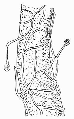
Fig. 34. Germinating hyphae of Lecanora subfusca Ach., growing over the alga Vaucheria sessilis DC., much magnified (after Bonnier).
F. Parasitism of Algae on Lichens
A curious instance of undoubted parasitism by an alga, not as in Strigula on one of the higher plants, but on a lichen thallus, is recorded by Forssell[309]. A group of Protococcus-like cells established on the thallus of Peltigera had found their way into the tissue, the underlying cortical cells having degenerated. The blue-green cells of the normal gonidial layer had died off before their advance but no zone was formed by the invading algae; they simply withdrew nourishment and gave seemingly no return. The phenomenon is somewhat isolated and accidental but illustrates the capacity of the alga to absorb food supply from lichen hyphae.
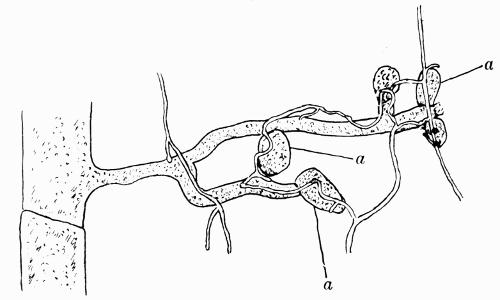
Fig. 35. Pure culture of protonema of Mnium hornum L. with spores and hyphae of Lecidea vernalis Ach. a,a,a, buds forming × 150 (after Bonnier).
An instance of epiphytic growth has also been recorded by Zahlbruckner[310]. He found an alga, Trentepohlia abietina, covering the thallus of a Brazilian lichen, Parmelia isidiophora, and growing so profusely as to obscure the isidiose character towards the centre of the thallus. There was no genetic connection of the alga with the lichen as the former was not that of the lichen gonidium. Lichen thalli are indeed very frequently the habitat of green algae, though their occurrence may be and probably is accidental.