Chapter IV · THE PATHOLOGY AND BACTERIOLOGY OF PNEUMONIA FOLLOWING INFLUENZA
Reading edition
E. L. Opie, M.D.; F. G. Blake, M.D.; and T.M. Rivers, M.D.
Many observers have described isolated phases of the recent epidemic and of past epidemics of influenza. Few have had an opportunity to follow the pathology of influenza from the onset of an epidemic through a period of several months and to observe the succession of acute and chronic changes which occur in the lungs. Our commission arrived on September 5, 1918, at Camp Pike two weeks before the outbreak of influenza. The commission had previously made a careful study of the clinical course, the bacteriology and to a limited extent the pathology of pneumonia occurring at Camp Funston where there was little if any influenza. Study of the records preserved at the base hospital at Camp Funston had convinced us that this camp had passed through an epidemic of influenza during the spring of 1918, this epidemic being followed by a very severe outbreak of pneumonia. Our investigation at Camp Funston had brought to our attention those phases of pneumonia which with the facilities of a base hospital laboratory could be most profitably studied with a view to determining the causation, the epidemiology and the prevention of the pneumonias prevalent in the American army.
Study of pneumonia after death offers the only opportunity of determining the relation of pulmonary lesions to the considerable variety of microorganism associated with them. Clinical diagnosis furnishes no certain criterion for distinguishing lobar and bronchopneumonia; suppurative 108pneumonia is rarely recognizable during life. The relation of pneumococci, streptococci, staphylococci or B. influenzæ to one or other type of pneumonia can be determined with accuracy only after death; for the demonstration of one or more of these microorganisms in material obtained from the upper respiratory passages in life, though of value, furnishes us no definite evidence that the organism which has been identified has entered the lung and passed from the bronchi to produce pneumonia.
Study of autopsies following examination of the sputum during life has shown that an individual primarily attacked by influenza may suffer with a succession of pneumonias, one microorganism having prepared the way for another. The complexity of the subject is much increased by the truth that pyogenic microorganisms, like the tubercle bacilli, are capable of producing a considerable variety of pulmonary lesions.
Examination of the lungs of a large number of individuals who have died as the result of pneumonia following influenza has disclosed a succession of acute and chronic diseases. Immediately succeeding the height of the epidemic of influenza, death occurred with acute lobar pneumonia or with diffusely distributed hemorrhagic bronchopneumonia caused in the majority of instances by Pneumococcus IV in association with B. influenzæ. Superimposed infection with hemolytic streptococci increased in frequency and in individuals who had occupied certain wards was almost invariable. At a later period, from one to two months following the maximum incidence of influenza chronic lesions, namely, bronchiectasis, unresolved pneumonia, and chronic empyema were common and often occurred as the result of influenza which had had its onset at the height of the epidemic.
When influenza attacked the encampment, about 50,000 troops were quartered in it, and for a considerable period no more troops were brought into the camp and none left 109it. All cases of pneumonia occurring among these troops were brought to the base hospital so that the autopsies which were studied were representative of all the pneumonias following influenza in this limited group of men. It is noteworthy that autopsy disclosed no instance of fatal influenza unaccompanied by pneumonia.
Pneumonia of Influenza.—Knowledge concerning the bacteriology of the pneumonia of influenza dates from the study of the epidemic of 1889–90. The frequency with which Diplococcus lanceolatus occurred in association with influenzal pneumonia was well recognized, although several observers, notably Finkler[57] and Ribbert,[58] found Streptococcus pyogenes so often that they attributed the pneumonia of influenza to this microorganism.
During a subsidiary outbreak of influenza occurring in 1891–92 Pfeiffer[59] discovered the microorganism which he believed was the cause of the disease. Pneumonia, he believed, was caused by the invasion of this microorganism into the lung, and the pneumonia of influenza, if death occurred at the height of the disease, was characterized not only by the presence of the bacillus of influenza, but was recognizable by its anatomic peculiarities. He described lobular patches of consolidation which were separated from one another by air containing tissue or were confluent, so that, although the lobular character was still recognizable, whole lobes might be affected. The consolidated tissue was dark red and within each lobular area were small, yellowish gray spots varying in size from that of a pinhead to a pea. In the mucus of the larynx and trachea were numerous microorganisms, including diplococci and streptococci, among which influenza bacilli were predominant; in the larger bronchi, bacteria other than influenza bacilli were less abundant, whereas in the finer bronchi filled with 110purulent fluid and in the lung tissue influenza bacilli had undivided sway. Pfeiffer stated that the changes described were found when death occurred at the height of the disease, whereas other pulmonary lesions might be sequelæ of this typical influenzal pneumonia.
Observations upon the pathology of influenzal pneumonia made during the epidemic of 1889–92 have been collected in the monograph of Leichtenstern[60] published in 1896. He combats the opinion held by some observers that pneumonia with influenza is always catarrhal and cites many writers to prove that lobar pneumonia not infrequently accompanies the disease. Indeed, some have found “croupous” pneumonia more often than “catarrhal.” Krannhals[61] at Riga found typical fibrinous pneumonia in 53 instances, doubtful forms in 22 and bronchopneumonia in 37. Cruickshank[62] in England found croupous forms predominant. Among 43 autopsies performed upon individuals dead with influenza Birch-Hirschfeld[63] found 11 instances of croupous lobar pneumonia, 8 instances of croupous lobular pneumonia and 24 instances of catarrhal pneumonia. Leichtenstern thinks that the atypical symptomatology of lobar pneumonia with influenza—for example, the purulent character of the sputum—has led many physicians to believe that lobar pneumonia rarely occurs. It is equally true that many instances of confluent lobular pneumonia are mistaken for lobar.
There appears to be no comprehensive description of the pneumonias of influenza based upon the epidemics of 1889–92. Descriptions dating from this period are much obscured by attempts to separate croupous or fibrinous from catarrhal pneumonias. Croupous lobular pneumonia has been recognized, for example, by Birch-Hirschfeld. Leichtenstern describes a form of pneumonia which he regards as 111neither lobar nor lobular although it implicates whole lobes; the consolidated tissue is homogeneous and varies in color from fleshy red to bluish red; it is tough and elastic in consistency. The author thinks that it is an error to regard this lesion as a confluent lobular pneumonia.
Kuskow[64], who has discussed the pathology of influenza in considerable detail, has seldom seen lobar pneumonia but has almost invariably found, even when there is lobar distribution of the lesion, lobular patches of consolidation involving groups of lobules, single lobules or only parts of lobules; the lung tissue has been hyperemic and in places edematous.
Opinions concerning the pathology and bacteriology of the pneumonias of influenza, published since the recent epidemic, have varied almost as much as those just cited. Few observers have had the opportunity of making a considerable number of observations under conditions which determine the relation of the pulmonary lesions to the primary disease.
Keegan[65] has found with influenza a massive and confluent bronchopneumonia, frequently resembling lobar pneumonia but distinguishable particularly in its early stages when the cut section of the consolidated lung has a finely granular character and each bronchiole stands out as a grayish area with intervening dark red alveolar tissue.
Symmers[66] states that the pneumonia of influenza is a confluent lobular exudative and hemorrhagic pneumonia which bears a close resemblance to the pneumonic variety of bubonic plague.
Our commission[67] published a preliminary report on pneumonia following influenza observed at Camp Pike. The occurrence of purulent bronchitis, distention of lungs, 112peribronchial hemorrhage and bronchiectasis were described. B. influenzæ was isolated from the bronchi in approximately 85 per cent of instances of influenzal pneumonia but from the consolidated lung in less than half this number of autopsies. Lobar pneumonia was present in a large proportion of autopsies but was less frequent than bronchopneumonia. Bronchopneumonic consolidation is in part red, lobular and confluent, in part nodular; pneumococci have a predominant part in the production of these lesions. Three types of suppurative pneumonia are described: (a) Abscess caused by hemolytic streptococci usually in contact with the pleura and accompanied by empyema; (b) Suppuration of interstitial tissue of the lung caused by hemolytic streptococci and accompanied by empyema; (c) multiple foci of suppuration clustered about a bronchus of medium size and caused by staphylococci. We have expressed the opinion that B. influenzæ descends into the bronchi; pneumococci, usually Type IV, may enter the lung and produce either lobar or bronchopneumonia. Hemolytic streptococci may descend and infect the pneumonic lung causing death often before suppuration has occurred. Epidemics of such superimposed streptococcus pneumonia in wards of the hospital were described.
LeCount[68] says: “The pneumonia of influenza is commonly referred to as bronchopneumonia. It may be so designated, but it differs from other bronchopneumonias in its predilection for the periphery of the lungs and in the extent to which the inflammation is hemorrhagic.”
MacCallum[69] states that the following types of pneumonia following influenza may be recognized. 1. Pneumococcus pneumonia. The lobular character of the consolidation is in these cases well marked, although it tends to lose its definiteness through the confluence of adjacent areas. The cut surface of the lung shows in the more acute cases a 113peculiar lobular or confluent consolidation which corresponds well with what is commonly written of the stage of engorgement in the description of lobar pneumonia. Later stages in the pneumonia show within these areas patches of rough gray consolidated tissue from which definite plugs of exudate project. 2. Staphylococcal pneumonia. 3. Streptococcal pneumonia. There is lobular pneumonia, the interlobular septa are edematous and, microscopically, bronchi and alveoli are loaded with streptococci. Whole areas of lung, though retaining their form, are entirely necrotic. Lymphatics are distended with exudate containing streptococci in great numbers, but in none of these cases is interstitial bronchopneumonia found. 4. Pneumonia produced by B. influenzæ of Pfeiffer. The lung is studded with palpable shot-like grayish yellow nodules; the bronchi exude thick yellow pus, which contains influenza bacilli. Microscopically, the walls of the bronchi are found to be thickened by mononuclear infiltration and new formation of connective tissue. The walls of the alveoli are thickened and indurated and the alveoli often contain fibrin in process of organization. Absence of conspicuous changes in lymphatics, absence of intense pleural infection and relatively scant numbers of polynuclear leucocytes in the exudate within the alveoli and bronchial walls are, MacCallum states, all that distinguish this pulmonary change from the interstitial bronchopneumonia caused by the hemolytic streptococcus and described by him in previous papers.
Lyon[70] designates the pulmonary lesion following influenza, hemorrhagic pneumonitis, the lung tissue containing serous fluid loaded with red blood corpuscles; in many instances there was such scant consolidation that the process could not be regarded as a true pneumonia. In 35 instances the lesion was lobular in distribution and in 16 114instances was sufficiently extensive to be designated lobar, but it was not typical lobar pneumonia, and, often associated with lobular patches of consolidation, appeared to be a confluent lobular pneumonia.
Goodpasture and Burnett[71] say: “The difficulties of analyzing the pulmonary lesions in any group of influenza pneumonias as they have appeared in this epidemic, are very apparent to anyone who has had an opportunity to observe the bacteriology and pathology of this accompaniment of the disease.” There is an acute outflow of the fluid elements of the blood and of hemorrhage into the lung tissue filling the alveoli in lobular areas and not infrequently in an entire lobe. By a special method of staining these authors have studied the distribution of Gram-negative bacilli with the morphology of B. influenzæ. The fact that in certain very early cases demonstrable bacteria of any kind are scarce or not found at all, has lead them to believe “notwithstanding the demonstration of influenza bacilli in pure culture in the lung in all but one instance, that at this stage organisms are comparatively few within the alveoli, and the primary injury is due to a very potent toxic agent elaborated in and disseminated through the larger air passages. In the later stages or from the beginning, if the injury be slight, the infection focalizes about the bronchi or their terminations, so that the bronchial and lobular distribution becomes very conspicuous.” Typical lobar pneumonia with “croupous” exudate within the alveoli occurs in cases complicated by secondary pneumococcus infection.
Wolbach[72] has found that two types of pneumonia are characteristic of influenza. In cases in which death has occurred within a few days after onset of pulmonary signs, the lung tissue is dark red and “meaty in consistency” and contains abundant blood-tinged serous fluid which drips 115from the cut surface. The other type of lesion is found in patients who have lived for ten days or more after onset of the disease; there is extensive bronchitis, bronchopneumonia, discrete or confluent, and peribronchitis. The lungs are voluminous and the smaller bronchi are distended. Microscopically, there is peribronchitis with extensive infiltration of the interlobular septa and organization in alveoli and bronchioles. This lesion is that designated by MacCallum as “interstitial bronchopneumonia.” Wolbach thinks that the two types of lesion represent different stages of the same process. He regards as distinctive of the pneumonias of influenza the presence of hyalin fibrin lining distended air spaces. With the two types of lesion which have been described, B. influenzæ was the only organism which could be cultivated, and the author associates these distinctive conditions with that microorganism.
Streptococcus Pneumonia.—Finkler emphasized the importance of streptococcus pneumonia as a complication of influenza. In 1888 he described instances of acute primary streptococcus pneumonia observed in 1887 and 1889. This form of pneumonia, Finkler thought, occurred in Bonn in epidemic form before the influenza epidemic of 1889–90 and, he states, exhibited an astonishing similarity to the pneumonia of influenza. He thought that later there was in Bonn a mixed infection of influenza and primary streptococcus pneumonia. In one type of streptococcus pneumonia, described by Finkler, there was lobular consolidation which in multiple patches produced “pseudolobar” consolidation; the consolidated lung was smooth and red, and similar to spleen, rather than hepatized. In another group of instances the lesion merited the name “erysipelas of the lung.” The lesion was an acute interstitial pneumonia in some places, a cellular or occasionally fibrinous pneumonia with involvement of the interstitial tissue in other places. There was edematous swelling and accumulation of leucocytes in the interstices between the alveoli and about 116the blood vessels and bronchi. Finkler stated that the similarity to erysipelas might suggest that the lymphatics contain streptococci, but this relation did not exist although large lymphatic channels were occasionally filled with coagulum. He asserted that the disease was contagious and cited cases which he believed had their origin in hospital wards.
The widespread occurrence of streptococcus pneumonia in the army camps in this country attracted attention during the first months of 1918. Cummings, Spruit and Lynch[73] at Fort Sam Houston, Texas, recognized the prevalence of streptococcus pneumonia, both as a complication of measles and in association with lobar pneumonia, and showed that the microorganism concerned was a hemolytic streptococcus. In 7 autopsies upon individuals with lobar pneumonia, they found pneumococcus alone in 2 instances and pneumococcus and hemolytic streptococcus or hemolytic streptococcus alone in 5 instances. Hemolytic streptococcus was found in all of 24 instances of bronchopneumonia, three-fourths of which followed measles. They recommend the bacteriologic examination of the throat of patients with measles and the segregation of those who harbor hemolytic streptococci.
Cole and MacCallum[74] have published almost simultaneously, with that just cited, a report upon the pneumonia which has occurred at Fort Sam Houston and have shown the importance of hemolytic streptococci in its causation. They have found two varieties of pneumonia, namely, acute lobar pneumonia which does not differ essentially from that which occurs elsewhere and bronchopneumonia which in most cases has followed measles and is caused by S. hemolyticus. They think this infection is usually acquired in the hospital. They believe that the pulmonary lesions are characteristic. In this publication and elsewhere MacCallum 117has designated the lesion “interstitial bronchopneumonia.”
The epidemic of streptococcus pneumonia and empyema occurring at Camp Dodge, Iowa, from March 20 to May 10 is described by Miller and Lusk[75]. During this period there were 400 cases of pneumonia, whereas from September 20, 1917, to March 20 there had been only 276 instances of lobar pneumonia. The type of pneumonia changed, there was more severe intoxication and empyema became very frequent; in 85 of 95 exudates streptococci were found. The outbreak of pneumonia bore no relation to measles. The authors state that a mild tracheitis was prevalent in the cantonment during March, and whenever a large group of soldiers congregated coughing was noticeable.
MacCallum[76] studied the pneumonia at Camp Dodge during May and found 17 instances of the lesion which he had designated interstitial bronchopneumonia; of these, 9 followed measles, although in the earlier part of the epidemic there appear to have been, he states, few such cases. Cultures made at autopsy, except in a few fatal cases of uncomplicated lobar pneumonia caused by the pneumococcus, showed the hemolytic streptococcus in every situation throughout the respiratory tract and pleura.
The pneumonia which occurred at Camp Funston is of special interest to our commission because we were for a time stationed at this camp and had the opportunity of following in the excellent records of the hospital the history of the occurrence of pneumonia during the year following the establishment of the camp in September, 1917. Stone, Phillips and Bliss[77] have described the outbreak of pneumonia which occurred in March, 1918. At this time there was, the authors state, severe pneumonia with frequent empyemas due to hemolytic streptococci. This condition 118which did not follow measles was responsible for the greatly increased death rate in March; 9 deaths occurred in February, 45 in March, 25 in April and 14 in May. They found during March 26 instances of multiple pulmonary abscess. In 29 autopsies they found a pleural lesion which they designate “subcostosternal pus pockets”; it occurs only in association with empyema caused by hemolytic streptococci.
Our commission[78] has shown that an epidemic of influenza, well characterized by its epidemiology and symptoms, preceded and accompanied the outbreak of pneumonia just described. Between March 4 and 29 1,127 men from Camp Funston, which then contained 29,000 men, were sent to the base hospital with influenza and many more were treated in the infirmaries of the camp; on March 11 107 patients with influenza were admitted to the hospital. The greatest incidence of pneumonia in the history of the encampment up to this time occurred between March 9 and 29, immediately following the outbreak of influenza, the maximum incidence of pneumonia occurring five days after the maximum for influenza.
The foregoing observations are cited to prove that streptococcus pneumonia, which occurred during the spring of 1918 at Camp Funston and doubtless at other camps, had its origin in influenza and did not differ in character from that which occurred on a much larger scale in the fall of 1918.
Table of Autopsies.—In order to present as briefly as possible the data upon which the present study has been based, autopsies have been assembled in tabular form in the order of their performance (Table XXVII). During the early period of the epidemic autopsies were performed on all who died with pneumonia, but later, with increase in the number of deaths, this became impossible and autopsies were performed on all those who died in certain wards.
119Comparison of charts representing incidence of influenza and of deaths from pneumonia furnishes evidence that fatal pneumonia during the period of investigation was with few exceptions referable to influenza. During two weeks, namely, from September 1 to 14, before the presence of the epidemic was evident, there were only 2 fatal cases of pneumonia. In most instances the relation of pneumonia to influenza is established by a definite history of influenza having its onset during the epidemic. Bronchopneumonia usually develops gradually as a sequence of influenza in which purulent bronchitis has occurred. Lobar pneumonia may develop in cases of influenza complicated by purulent bronchitis. In some instances there is apparent recovery from influenza indicated by return of temperature to normal; after from one to three days of normal temperature there is typical lobar pneumonia with rusty sputum. In many instances of pneumonia having their onset at the height of the epidemic of influenza, the history indicates that pneumonia was present immediately after the onset of symptoms, so that the onset resembled that of pneumonia rather than of influenza.
Cases of pneumonia following measles have been excluded from the table in order that they may be studied separately and compared with the pneumonias of influenza. It is noteworthy that the lesions of pneumonia following measles have shown a very close resemblance to the pneumonias of influenza, with regard both to pathologic characters and to bacteriology.
Five instances of pneumonia following typhoid fever (Autopsies 245 and 329), scarlet fever (Autopsy 311) or mumps (Autopsies 403 and 417) have been excluded from the table. These secondary pneumonias are grouped as an appendix to the section on pneumonia following measles. It is not improbable that individuals with the diseases named are just as susceptible as others to influenza. Included in the table is an instance (Autopsy 487) in which a definite attack of influenza preceded scarlet fever.
120
Table XXVII
NO. OF AUTOPSY RACE LENGTH OF MILITARY SERVICE DURATION OF ILLNESS DURATION OF PNEUMONIA CLINICAL DIAGNOSIS PURULENT BRONCHITIS LOBAR PNEUMONIA PERIBRONCHIOLAR CONSOLIDATION HEMORRHAGIC PERIBRONCHIOLAR CONSOLIDATION LOBULAR CONSOLIDATION PERIBRONCHIAL CONSOLIDATION ABSCESS INTERSTITIAL SUPPURATIVE PNEUMONIA MULTIPLE ABSCESSES IN CLUSTERS EMPYEMA BRONCHIECTASIS UNRESOLVED BRONCHOPNEUMONIA ORGANIZING BRONCHITIS BACTERIA IN SPUTUM BACTERIA IN BRONCHUS BACTERIA IN LUNG BACTERIA IN BLOOD OF HEART
229 W 1m 12+ 12+ L P + Pn. I. B. inf. 0
231 W 2m 13 4 B P +
232 W 14d 9 6? B + Pn. IIa. B. inf. Pn. IIa. Pn. IIa.
233 C 11m 5 2 L + Pn. IIa. Pn.
236 W 3w 8 7+ L + Pn. IV. B. inf. B. inf. Staph. 0
237 W 10d 8 2 L + + N E St. h. St. h.
238 W 2d 8 5 L + Pn. IV. B. inf. Pn. IV
239 W 11d 9 5 L + Pn. II. Pn. II. B. inf. Pn. II.
240 W 13d 8 4 L + M + Pn. IV. B. inf. Pn. IV.
241 W 14d 5 5 L + Pn. IV. B. inf. Pn. IV.
242 C 14d 7 4 L P + + Pn. IIa. B. inf. Pn. IIa.
243 W 15d 5 5 L + M Pn. IV.
244 C 1m 6 3 B P M + + + Pn. IV. B. inf. Pn. IV. B. inf. 0
121246 C 2m 6 3 L P + M Pn. IIa. B. inf. 0
247 W 10d 10 5 L P M + + Pn. IV. B. inf. Pn. IV.
248 W 1m 4 2+ I + E St. h. St. h. St. h.
249 W 15d 12 6+ L M + Pn. III.
250 W 14d 11 7 L M + E Pn. IIa. B. inf. Pn. IIa.
251 W 25d 7 1 B P + + + + St. h. St. h.
252 W 21d 14 12 B P M + Pn. II. B. inf. St. v. Pn. II. B. inf. 0
253 W 12d 19 12+ B M + + E Pn. Ia. Pn. II. B. inf. Staph. Pn. II. B. inf. Pn. II.
254 W 21d 7 6+ B P + M + Pn. I. B. inf. Pn. I. Staph 0
255 W 12d 5 4? B P M + + + E St. h. Pn. IIa. Staph. St. h. St. h.
256 W 17d 8 4 B P + M Pn. Staph. Pn. IIa. Pn. IIa.
257 C 21d 10 7+ L P + B. inf. Staph Pn. I.
258 W 1m 6 2+ B M + + E St. h. Pn. IV. B. inf. St. h.
259 W 3m 4 1 B + + + St. h. Pn. III. Staph. St. h.
260 W 1m 2 1 L + 0
261 W 2m 7 5+ B P + B. inf. Pn. IV. 0
262 W 12d 5 4? B + + E St. h. B. inf. St. h. St. h.
263 W 21d 7 5 L P + + E St. h. B. inf. Staph. St. h. Staph.
264 W 21d 10 7 L + Pn. IV. Staph. St. h. Staph. St. h. St. h.
265 W 14d 7 6+ L + St. h. Staph. inf. Pn. IV. B. inf. St. h.
122266 W 1m 7 2 L + + St. h. St. h. B. inf. St. h. St. h.
267 W 2m 22 10 B + Pn. IV. Pn. II. B. inf. Pn. II. Pn. II.
268 W 2m 6 4? L + Pn. III. B. Pn. III. B. inf. St. h. Pn. III. B. inf. St. h. 0
269 W 25d 8 2 L + Pn. IV. B. Pn. IV. Staph. Pn. IV. 0
270 W 17d 18 3 L + h St. h. B. inf. Staph. St. h. Staph. St. h.
271 W 2d 12 5 L P + St. h. Pn. IV. B. inf. St. h. St. h.
272 W 3m 7 2 L + St. h.
273 W 1m 8 4+ L + + N E St. h. B. inf. Staph. St. h. Pn. IV. Staph. St. h.
274 W 2w 9 5 B + N Pn. IV. St. h. Staph. St. h. St. h.
275 W 4m 9 4 L M + + N St. h. B. inf. Staph. St. h. B. inf. Staph. St. h. Pn. IV.
123276 W 1m 6 4+ L + St. h. Staph B. inf. St. h. Pn. IV. B. inf. St. h.
277 W 21d 10 3 L + St. h. Staph St. h. Staph St. h.
278 W 2m 16 6+ L + E St. h. B. inf. St. h. St. h.
279 W L + Pn. IV Pn. IV.
280 W 21d 8 8 L + + + St. h. B. inf. Staph. St. h. St. h.
281 W 21d 9 5 L + + h St. h. B. inf. St. h.
282 W 1m 10 ? B + + E Pn. IV. B. inf. St. h. B. inf. Pn. II Staph. St. h. Pn. II. St. h. Pn. II.
283 W 7d 19 8 B P + + + M + B. inf. Pn. IV. Staph. Staph. B. inf. Pn. IV.
284 W 21d 11 ? B P + + E St. h. B. inf. Staph. St. h. St. h.
285 W 11 9? B P M + E Pn. IIa. B. inf. St. h. B. inf. St. h. St. h.
286 W 20d 9 4+ L P M + Pn. IV. B. inf. Staph. 0 Pn. IV.
287 W 3m 12 4 L P + + + Pn. IV. B. inf. Staph. Pn. IV. B inf. Staph. Pn. IV.
288 W 1m 10 5 B P M + h + E + St. h. B. inf. St. h. B. inf. St. h. St. h.
289 W 19d 12 7 B P M + M + Pn. IV. B. inf. Staph. Pn. IV. Pn. IV.
290 W 1m 3+ 3+ B + St. h. B. inf. St. h. St. h.
291 W 2w 18 11 B P + + M + Pn. IV. B. inf. B. inf. Staph. 0 0
292 W 3m 5 5 L + E St. h. B. inf. St. h.
293 W 2m 3+ 3+ L P + Pn. III. B. inf. Pn. III. B. inf. Pn. III.
295 W 1m 12 5 L P + + E + Pn. IV. St. h. St. h. B. inf. St. h.
124296 W 16d 18 3 L P + + E + St. h. B. inf. St. h. B. inf. St. h.
297 W 1m 5 ? I P + B. inf. Pn. IV. St. h. Pn. IV. B. inf. 0
298 C 21d 13 10 B P + Pn. IIa. B. inf. Pn. IIa.
299 W 28d 9 3 L P + B. inf. Pn. IV. Staph. St. h.
300 C 22d 16 12+ L P + M h Pn. IIa. B. inf. Pn. IIa. B. inf. Pn. IIa.
301 W 1m 7 5 B + Pn. IV. B. inf. Staph. St. h. Pn. IV.
302 C 5d 6 3+ B + Pn. IV. B. inf. Pn. IV.
303 W 1m 7 3+ L + Staph. Pn. IV. B. inf. Pn. IV. B. inf.
304 W 4m 10 2? L + St. h. B. inf. St. h. B. inf.
125305 W 5m 3+ 3+ L + B. inf. St. h. Pn. IV. Staph. B. inf. St. h Pn. IV. Staph.
306 W 1y 6+ 3 B + B. inf. Pn. IV. Staph. 0 0
308 W 1m 6 6 L + + Pn. IV. B. inf. St. h. Staph. St. h. Pn. IV.
309 W 1m 4 2? L + E St. h. St. h.
310 W 21d ? 3? L + Pn. III. B. inf. Staph. Pn. III. Pn. IV. B. inf.
312 W 1m 17 7 L P M + h N + Pn. IV. B. inf. St. h. St. h. B. inf. Staph. St. h. B. inf. St.
313 W 1y 5 2 B + St. h. B. inf. Staph. St. h. St. h.
314 W 1m 3+ 3+ L P + + Pn. IV. B. inf. Staph. Pn. IV. Pn. IV.
315 W 1m 9 2 B + Pn. IV. B. inf. Staph. Pn. IV.
316 W 1m 11 4 L + B. inf. Pn. III. B. inf. Pn. III.
317 W 13d 9 2 L + Pn. IV. B. inf. Pn. IV.
318 W 2m 8 3 L + Pn. IV. B. inf. Pn. IV. B. inf.
319 W 1m 4+ 4+ B P + St. h. Staph. St. h. B. inf. Staph. St. h.
320 C 5m 1+ 1+ L + Pn. IIa. Pn. IIa. Pn. IIa.
321 W 28d 4+ 4+ L + E B. inf. Pn. IV. B. inf. Pn. IV.
322 W 10d 8 6 L P + + + Pn. IV. Pn. III. Staph. aur. 0
323 W 2m 12 4 B + Pn. IV. B. inf. Staph. 0
126324 W 22d 9 6 B + Pn. I. B. inf. 0 Pn. I.
325 W 1m 8 8 B P M N + + St. h. B. inf. Staph. St. h. St. h.
326 W 1m 5+ 2 B P + h St. h. B. inf. Staph. St. h. B. inf. St. h.
327 W 1m 4 ? B +
328 W 5m 13 3+ L P + Pn. III. Pn. IV. B. inf. Staph. Pn. III. Pn. IV. B. inf. Pn. III.
330 W 1m 10 3 L M N + E Pn. IV. Pn. IV.
331 W 1m 12 11+ B P + Pn. IV.
332 W 1m 17 3 L P + M h 0
333 W 19d 15 7 L P M + + + Staph. aur. B. inf. St. h. Staph. aur. Pn. IIa. St. h. Pn. IIa.
334 W 14d 16 5 B M N + E St. h. Staph St. h.
127335 W 1m 7 ? ? + St. v. B. inf. Staph. Pn. IV. Pn. IV.
336 W 2m 12 6 B P M + + 0
337 W 1m 9 2? L + Pn. IV.
338 W 1m 7 5+ B + Pn. IV.
339 W 2m 9 6? L + Pn. IV.
340 W 35d 8 3 L P + + 0
341 W 3m 6 4+ + 0
342 W 2m 9 3? L P + +
343 W 1m 11 1? L + E Pn. IV. Pn. IV.
344 W 4m 13 6 L + + E St. h.
345 W 1d ? 7 L P + + + E Pn. III. St. h Staph.
346 W 26d 16 10 B P + M + N B. inf. Pn. IV. St. h. B. inf. St. h. B. inf. St. h.
347 C 3d 10 3 L P + h St. h.
348 C 4d 8 8 B P + Pn. IIa.
349 W 2d 12 6 L P + Pn. III St. h Pn. I.
350 W 2m 6 2? B + St. h.
351 C 4m 4 3+ L P +
352 C 2m 8 4 L P + + + + Pn. IIa.
353 C 6m 18 18 L P + E Pn. I. B. inf. B. inf. Pn. Pn. IIa. Pn. IIa.
354 W 15d 2+ 2 L P M h + E St. h. B. inf. Staph. St. h. St. h.
355 W 21d 7 7 B P + + E St. h. B. inf. Pn. IV. Staph. St. h. B. inf. Staph. 0
356 C 5d 8 4 L + Pn. IV.
357 W 1m 10 6+ L + + + Pn. IV. B. inf. Pn. IV.
358 W 1m 15 6? L + E Pn. IIa.
128359 W 1m 7+ ? L M h + Pn. IV.
360 W 36d 10 3 L P + + + 0
361 W 3m 8 2 L + Pn. IV.
362 W 4m 15 13+ L + St. h.
363 W 3m 8 1+ L P + Pn. IV.
364 W 6d 9 5+ B + St. h.
365 W 2m 11 2 B + + + Pn. IV. Pn. IV.
366 W 6d 8 1+ B + M Pn. IV.
367 W 22d 15 8+ L + Pn. IV.
368 C 4d 15 11+ L P + M Pn. I.
369 W 68d 7+ 4 B P + + N + E St. h. St. h. St. h.
370 W 17d 17 14 L P + + + + + [No. St. h.] Staph. aur. Pn. IV. B. inf. Staph. aur 0
372 W 1m 17 5 L P + + Mh +
373 W 4d 11 1? B + St. h.
374 C 4d 10 5 L P + + + Pn. IV.
375 C 4d 12 6 L P + + + 0
129376 W 1m 10 7+ B P + + M + E + [No. St h.] St. h. B. inf. Staph. aur. St. h. St. h.
377 W 1m 7 4? B P + + Pn. IV.
378 W 1m 28 7 B P + + E + St. h. B. inf. Pn. IIa. St. h. B. inf. Pn. IIa. Pn. IIa.
379 W 11m 7 1 B + + E B. inf. + ? Pn. IIa.
380 W 3m 11 ? B P + + + Pn. III. Pn. III.
381 W 21d 13 9? L P + + E + St. h. Pn. II. Pn. IV. Staph. aur. St. h.
382 W 1m 9 6+ L P + 0
383 W 2m 9 2 L + + B. inf. Pn. III. St. h. Pn. III.
384 W 1m 13 5 L + Pn. IV.
385a W 3w 12 6? B P + + E St. h. St. h.
385b W 2m 11 4 L + Pn. IV.
385c W 24d 17 10 B P M + + + + St. h. B. inf. St. h.
386 W 1m ? 5 L P + + Pn. III. B. inf. Staph. aur. Pn. III.
387 W 3w 19 9 B P + + + + E + St. h. B. inf. Staph. aur. Pn. Staph. alb Pn. II. B. inf. St. h.
388 W 3m 11 7 L P + Pn. IV.
389 W 1m 15 15 L P + + E + St. h. St. h.
391 W 25d 13 13 L P + + + h + Pn. IV. Pn. IV.
392 W 1m 12 8+ L P + M + Pn. II.
393 W 1m 20 4 L + Pn. IV.
394 W 21d ? ? L P + + h + St. h.
395a W 1m 19 11? L P M + + E + St. h. B. inf. St. h.
395b W 3m 12 3? B P + h E Pn. IIa. Pn. IIa.
396 W 2m 7 1+ B + 0
130397 W 21d 22 14 B P M + + E St. h. B. inf. St. h.
398 W 1m 16 6? B P + M + + 0
399 W 1m 18 4 L + Pn. IIa.
400 W 1m 15 11+ L + Pn. IV.
401 W 1m 13+ 9 B P + M h + Pn. IV.
402 W 1m 14 8 B P M + + E + + + Pn. IV. St. h.
404 B P + +
405 W 21d 13 11+ B + M Pn.
406 C 2d 18 15? L P + + E Pn. IV.
408 C 1m 13 ? L P + + 0
409 C 6d 12 9+ L P + + M 0
410 W 35d 13+ 13+ L P + + M + E + St. h. B. inf. Staph. aur.
411 W 3m 16 2 B P + + Pn. IIa. B. inf. Pn. IIa.
412 W 1m 15 13? L + M + Pn. II. B. inf. Pn. II.
131413 C 2m 13 8+ L P + + Pn. III. B. inf. Pn. III.
414 C 7d 18 4 L P + Pn. IIa. B. inf. Pn. IIa.
415 C 16d 8 6+ L P + + + St. h. 0
416 C 7d 14 6? L P + Pn. IV. Pn. IV. Pn. IV.
418 W 2m 19 4 B + Pn. IIa. St. v. B. inf. Pn. IIa.
419 W 4m 20 ? L P + + M + Pn. II. B. inf. Pn. II. B. inf. 0
420 W 1m 11 3 B P + M + E + + St. h. B. inf. Staph. aur. St. h.
421 W 21d 19 15? B P + M + + + Pn. IV. St. h. St. h.
422 W 3m 11+ 11+ B P + + M + + Pn. IIa. B. inf. 0
423 W 1m 16 12? B P + M E + + St. h. B. inf. St. h.
424 W 5y 14 6 L P + + Pn. IV.
425 W 1m 29 14 B P + + M + + + St. h. B. inf. Staph. alb. St. h.
426 W 4m 20 13 ? + Pn. IIa. B. inf. Pn. IIa.
427 W 1m 16 ? L P + + St. h. St. h.
428 W 3w 25 21 L P M + + E + St. h. B. inf. St. h. B. inf. St. h.
429 C 2m 7+ 5 L P + + + B. inf. Staph. alb 0
430 W 2m 16+ 7 L P + + N St. h. B. inf. Staph. aur. St. h.
431 W 21d 23 18 L P + + + + 0 0
432 W 42d 19 12+? L P + E B. inf. Pn. IIa. Pn. IIa.
433 W 1m 19 17 B P M + M + E + St. h. B. inf. Staph. aur. 0
132434 W 4m 14+ 10 L + + E St. h. B. inf. Staph. aur. St. h. Staph. aur. St. h.
435 W 1m 16+ 2? B + St. h. B. inf. B. inf. + St. h.
436 C 11m 5 3+? B + Pn. IIa. B. inf. Staph. aur. Pn. IIa. Pn. IIa.
437 C 5m 11 7? L P + + 0
440 W 1m 19 12? L P + + + M + + B. inf. Staph. aur. B. inf. Staph. aur. 0
445 W 1m 27 16? P M h + + Staph. aur. Staph. aur. Pn. IV. St. h.
446 W 8d 13 ? P + + 0
447 W 8d 10 2 B + Staph. aur. B. coli. Staph. aur. 0
448 W 70d 17 14+ L P + + Mh + 0 0 0
449 W 2m 27 13+ B P + + + E + + St. h. B. coli. St. h. B. coli. St. h.
452 W 4m 14 9 B P M + E St. h. B. inf. St. h. B. inf. St. h.
133451 C 2m 7 3+ B + E B. coli. Staph. St. v. Pn. IIa. B. inf. Pn. IIa.
455 C 26 22+ B + + E St. h. B. inf. St. h. St. h.
456 W 1m 23+ 20+ L P + E B. coli. St. h.
457 W 17m 17+ 17+ B + + + + + Pn. IV. B. inf.
458 W 11m 10 8+? L + + Pn. IV. B. inf. St. v. 0
459 C 10d 6 3 B P + + Staph. aur. Pn. IV. 0
460 W 1m 17 17 L P + + M + E + St. h. B. inf. Staph. St. h. B. inf. St. h.
461 C 5d 14 8+ L P + M + Staph. aur. Pn. I. St. h. Pn. I.
462 C 5d 15+ 12 B + 0
463 W 3m 20 12 B P + + Mh + + B. inf. Staph. B. inf. Staph. 0
464 C 21d 24 17? L P + B. inf. Pn. I. Staph. B. inf. Pn. I. 0
465 W 1m 24+ 11 LB P M + E + St. h. B. inf. Staph. St. v. St. h. St. h.
466 W 2d 13 3 B + + Pn. IIa. Pn. IIa. Pn. IIa.
467 W 3m 30 25? B P + + + E + St. h. B. inf. 0 St. h.
468 W 1m 22+ 6 L P + + + Staph. aur. B. inf. St. v. B. inf. St. v. 0
469 W 1m 25 12 B P + + +
471 C 6+? 4 L P + Pn. IV. B. inf. Staph. aur. Pn. I. Pn. IV. B. inf. 0
472 W 1m 37 21 B P + + + + E + + B. coli. St. h. B. coli. St. h.
134473 W 2w 28+ 24 B P + + E + + + B. inf. St. v. Staph. M. cat. St. v. Pn. III.
474 W 1m 36 31+ L + + E St. v. B. inf. M. cat. St. h. St. h. B. inf.
476 W 7d 6 2 B P M + 0
477 W 5d 9 5 B + + N St. h. B. coli. St. h. B. coli. St. h.
478 W 2m 9 3 L + St. h. St. h.
479 C 8d 29 15 L P + M N B. inf. St. h. Staph. M. cat. St. h. Staph. aur. St. h.
480 W 4m 31+ 29 B + + E Staph. aur. St. v. Staph. aur. B. inf. St. v. B. coli. St.
482 W 2m 11 5+ B P + + + M + B. inf. Pn. IV. St. h. B. inf. Pn. IV. 0
485 W 3d 9+ 3+ B P M + E St. h. B. inf. Staph. aur. B. coli. St. h. St. h.
135487 W 21d 55 40 B P + + E + + B. inf. St. h. B. inf. St. h.
488 W 4d 16 8 L P M + E + St. h. Pn. IIa. St. h. St. h.
489 C 8d 11 4 B P + + M + B. inf. Pn. IV. Pn. IV. B. inf. 0
494 W 2m 11 3 L + h + E St. h. B. inf. St. h. B. inf. St. h.
498 W 1y 6 1? L + N St. v. St. v. Staph. aur. Staph. alb. St. v.
499 W 5m 36 5 B P M E + St. h. B. inf. 0 St. h.
504 W 3m 6 3 L M + E St. h. B. inf. Staph. St. h. St. h.
506 W 8m 7+ 2? B + Pn. IV. B. inf. Staph. aur. M. cat. Pn. IV. Staph. aur. Pn. IV.
136In successive columns the table gives the autopsy number, race, and length of military service. These factors have had an important influence upon the incidence of influenza and pneumonia and have been discussed in a preliminary report.[79] The duration of illness (4th column of table), counted from the date of onset of symptoms of influenza or in some instances, when the earliest symptoms were those of pneumonia, from onset of pneumonia, can usually be determined accurately. The duration of pneumonia (5th column of table) is much more uncertain, because its determination dates from the first recognition of the physical signs of pneumonia.
Clinical Diagnosis.—The clinical diagnosis recorded upon the clinical history of the patient is given in column 6. Many clinicians have been impressed with the difficulty of determining during life the type of pneumonia associated with influenza. The occurrence of purulent bronchitis, the frequent coexistence of lobar and bronchopneumonia and an atypical onset often make the recognition of lobar pneumonia more difficult than usual. The diffuse consolidation of confluent lobular pneumonia increases the difficulty of recognizing bronchopneumonia. In the table (column 6) lobar pneumonia is indicated by L., bronchopneumonia by B. Among 227 autopsies the clinical diagnosis agreed with the condition found at autopsy in 109 instances (48 per cent); in 35 instances (15.4 per cent) both lobar and bronchopneumonia were found at autopsy and a diagnosis of one or other was made during life. In 83 instances (36.6 per cent) the diagnosis made during life was incorrect. Cases admitted to the base hospital at Camp Pike were as carefully studied as the conditions in a base hospital during an epidemic permitted and diagnosis of pneumonia was doubtless as accurate as in other base hospitals. 137Statistics from military and other hospitals based upon clinical diagnosis of the pneumonias of influenza are probably subject to an error of at least 1 in 3 cases, and conclusions based upon them are almost valueless.
The inaccuracy of clinical diagnosis of the pneumonia of influenza is further illustrated by a consideration of lobar pneumonia. This diagnosis on the one hand was made 136 times and was correct 67 times and incorrect 69 times; on the other hand, lobar pneumonia was found at autopsy 98 times and had been diagnosed in only 67 of these cases (68.4 per cent).
Classification of the Pulmonary Lesions of Influenza.— Influenzal pneumonia exhibits the following noteworthy characters:
1. Acute bronchitis with injury or destruction of lining epithelium and accumulation of inflammatory exudate within the lumen.
2. Hemorrhagic pneumonia with accumulation of blood within the alveoli and within and about the bronchi.
3. Susceptibility of bronchi and pulmonary tissue to secondary pyogenic infection with necrosis and suppuration.
4. Bronchiectasis.
5. Tendency to the occurrence of chronic pneumonia following failure of pneumonia to undergo resolution.
All these changes are doubtless referable to the severity of the primary injury to the lower air passages.
In the presence of destructive changes in the bronchi many bacterial species, including B. influenzæ, pneumococci of various types, streptococci (notably hemolytic streptococci) and staphylococci may invade the lungs and produce acute inflammation. The anatomic characters of the pneumonic lesions following influenza are equally varied.
In order to obtain insight into the pathogenesis of these lesions, it is desirable to imitate the historical development of knowledge concerning the characters and causes of disease, namely, first to define accurately the lesions concerned 138and later to determine with what microorganisms these lesions are associated. The difficulties of this undertaking are increased by the multiplicity of the microorganisms concerned and by the well-known truth that the same microorganism, e. g., the tubercle bacillus, may produce widely different anatomic lesions.
In the table of autopsies the following lesions are listed:
Column 7. Purulent bronchitis.—“P” indicates that the small bronchi contain mucopurulent fluid.
Column 8. Lobar pneumonia.—The occurrence of the lesion is indicated by the plus sign (+).
Column 9. Peribronchiolar consolidation.—The presence of nodular patches of consolidation about respiratory bronchioles is indicated by the plus sign (+) when the lesion has been recognized at the time of autopsy. When the lesion has been first recognized by microscopic examination the letter “M” is used.
Column 10. Hemorrhagic peribronchiolar consolidation.—The occurrence of this lesion which represents the preceding on a background of hemorrhage is indicated by the plus sign (+).
Column 11. Lobular consolidation.—The presence of the lesion is indicated by the plus sign (+).
Column 12. Peribronchial consolidation.—Peribronchial pneumonia recognized at the time of autopsy is indicated by the plus sign (+). Peribronchial pneumonia recognized microscopically is indicated by “M.” The presence of peribronchial hemorrhage without consolidation is indicated by “h.”
Column 13. Abscess formation with pneumonia.—Suppuration with abscess formation almost invariably just below the pleura is indicated by the plus sign (+). Necrosis of lung tissue recognized microscopically and unaccompanied by suppuration is indicated by “N.”
Column 14. Suppurative interstitial pneumonia.—This lesion invariably associated with suppurative lymphangitis is indicated by the plus sign (+).
Column 15. Multiple abscess in clusters.—Abscesses in clusters about a bronchus of medium size are indicated by the plus sign (+).
Column 16. Empyema.—The presence of the lesion is indicated by “E.”
Column 17. Bronchiectasis.—“B” indicates the lesion.
Column 18. Unresolved bronchopneumonia.—Presence of the lesion is indicated by the plus sign (+).
Column 19. Organizing bronchitis and bronchiolitis.—“O” indicates the lesion.
The lesions of columns 7 to 12 are acute inflammatory processes, columns 9 to 12 represent different types 139of bronchopneumonia. Columns 13 to 15 represent suppurative lesions. Columns 17 to 19 represent chronic lesions. A survey of the table shows the predominance of acute lesions in the early period of the study and the gradual increase of chronic lesions.
The last four columns of the table of autopsies give the bacteriology of the sputum during life and the bacteria found in the bronchi, in the lungs, and in the blood of the heart after death.
Mortality of Pneumonia Following Influenza.—From September 6 to December 15, 250 autopsies were performed on patients who had died with pneumonia at the base hospital at Camp Pike, and with few exceptions bacteriologic cultures were made from them. Although it was not possible to perform autopsies on all who died, those which were performed afford a fair index of all deaths, for throughout the epidemic of influenza and its outbreak of pneumonia approximately one half of all who died were examined after death. The relation of autopsies to deaths is shown by a comparison by weeks of the number of deaths and number of autopsies during the months of September and October.
WEEK DEATHS AUTOPSIES
Sept. 1–7 1 1
Sept. 8–14 1 1
Sept. 15–21 4 3
Sept. 22–28 15 14
Sept. 29–Oct. 5 121 67
Oct. 6–12 191 78
Oct. 13–19 78 43
Oct. 20–27 22 15
Oct. 28–31 8 6
441 228
For most of these autopsies there is a record of the date of onset of the illness, namely, influenza, which finally resulted in pneumonia and death. Comparison of the number of cases of influenza which developed on any day with the number of fatal cases which had their onset on the same 140day will determine the mortality of influenza at different periods of the epidemic. Chart 1 shows the number of cases of influenza which had their onset on each day from September 1 to October 31 and the number of fatal cases with autopsy which had their origin on corresponding days. The comparison by weeks between autopsies and total number of deaths shows that the autopsies represent with considerable accuracy the deaths. If there is any error it occurs at the height of the outbreak of pneumonia from September 29th to October 5th and not at its beginning, or end. The chart shows that the highest mortality occurred among cases of influenza which had their origin at the beginning of the epidemic from September 21 to October 1, whereas after October 1, though the maximum number of cases of influenza occurred on October 3, very few developed fatal pneumonia.
Mortality from Pneumococcus and Streptococcus Pneumonias.—By referring fatal cases of streptococcus pneumonia back to their date of origin it is possible to determine what proportion of the cases of influenza, which developed on any day, died with infection by hemolytic streptococcus. The accompanying chart (Chart 1) shows that infection with hemolytic streptococci has been very frequent at the beginning of the epidemic of influenza (shown by area with double hatch in chart) that is, from September 20 to 30 and subsequently has gradually diminished so that few cases have had their onset in the second half of the epidemic from September 30 to October 15.
Pneumococcus pneumonia uncomplicated by streptococcus infection (shown by area with single hatch in chart) pursued a course which more closely conformed to the curve representing influenza. The cases of influenza which resulted fatally bore a fairly constant ratio to the total number of cases of influenza from the onset of the epidemic until October 1, but subsequently few cases of influenza developed fatal pneumococcus pneumonia.
141These charts arranged with reference to the onset of fatal pneumonias dissociate very clearly the outbreak of streptococcus pneumonia, which reached its height at the beginning of the influenza epidemic, from the uncomplicated pneumococcus pneumonia which reached its maximum at the midpart of the influenza epidemic and then abruptly abated.
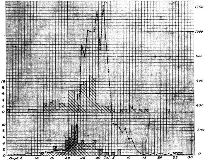
Chart 1—Showing the relation of (a) onset of cases of pneumonia shown by autopsy to be uncomplicated by secondary infection with hemolytic streptococcus, indicated by upper continuous line with single hatch, and of (b) onset of fatal cases of streptococcus pneumonia, indicated by the lower continuous line with double hatch, to (c) the occurrence of influenza, indicated by the broken line. The onset of each case of fatal pneumonia is represented by a single square.
Our study of ward infection in pneumonia furnishes an explanation of the outbreak of fatal streptococcus pneumonia coincident with the initial stage of the influenza epidemic. This outbreak is a true epidemic of streptococcus infection superimposed, in many instances at least, upon preexisting pneumococcus pneumonia, but in some instances, 142doubtless, a primary streptococcus pneumonia, following the bronchitis of influenza. In the absence of secondary streptococcus infection a very large proportion of these individuals would have recovered. This epidemic of streptococcus pneumonia, it has been shown, was the result of unfavorable conditions produced by great overcrowding of the hospital and in the early part of the epidemic by inadequate separation of those with streptococcus infection from those with none. With control of these conditions, streptococcus pneumonia rapidly diminished.
Greater susceptibility to pneumococcus pneumonia in the early than in the late period of the epidemic is perhaps explained by differences in the severity of influenza; the more susceptible individuals were attacked by influenza in the early period, whereas the less susceptible did not acquire the disease until they had been exposed to an immensely increased number of infected individuals. A better explanation is furnished by the greater opportunity at the beginning of the epidemic for the transmission of microorganisms causing pneumonia, for at this time patients with influenza were crowded together and methods to prevent the transmission of infection were little used.
Bronchitis
Clinical study has shown that purulent bronchitis (see Fig. 2) occurs in about one-third of the cases of influenza. In a large proportion of cases of bronchitis there is no clinical evidence of pneumonia. The bronchial lesions found in association with the pneumonia of influenza are an index of the ability of the agent, which causes influenza, to injure the bronchi.
In those who have died with pneumonia following influenza the large bronchi (with cartilage) are intensely injected, so that the mucosa has a deep red color which on cross section contrasts very sharply with the pearly white of the cartilage. Superficial injury to the bronchi is 143not infrequently evident in the larger bronchial branches; superficial loss of epithelium is indicated by erosion of the surface, whereas somewhat deeper destructive changes are occasionally evident. Microscopic examination accurately determines the degree of destructive change.
Purulent bronchitis was noted in 134 autopsies (55.6 per cent of autopsies). From the small bronchi, in many instances, purulent fluid welled up upon the cut surface of the lung, whereas in other instances tenacious mucopurulent fluid could be squeezed from small, cut bronchi by pressure upon lung tissue. The consistency of the material within the bronchi varied greatly, ranging from a viscid and tenacious mucus of creamy, yellow color to a thin, turbid, gray fluid. The coexistence of local inflammatory or of general edema of the lungs modifies the character of the material found in the bronchi at autopsy; with edema the purulent exudate is in some instances diluted so that a thin cloudy fluid flows from the small bronchi. In the presence of advanced edema of the lungs the bronchi rarely if ever contain purulent exudate. The underlying changes in the bronchi are more significant than the character of the exudate found at autopsy. Nevertheless, the group of cases in which the diagnosis of purulent bronchitis has been made, because small and medium sized bronchi have contained purulent or mucopurulent exudate, represents instances of readily recognizable bronchitis of considerable severity.
With few exceptions, purulent bronchitis was diffusely distributed in the lungs; occasionally it was observed in one lung alone, and in several instances was limited to the bronchi at the base of a lung, usually of the left lung.
In a considerable proportion of instances of purulent bronchitis abnormal distention of the lungs was noted. On removal from the chest the lungs fail to collapse and retain the size and shape of the thorax. Even after section is made through the organ, parts of the lung fail to collapse 144and have a resistant cushion-like consistency. This condition is present where the lung tissue is air containing and dry, and occurs when very small bronchi contain tenacious mucous exudate which becomes apparent upon the cut surface after the sectioned lung is squeezed. Microscopic examination shows that the alveolar ducts and infundibula are distended with air, though the respiratory bronchioles contain inflammatory exudate. Complete obstruction of the bronchi is followed by absorption of air from the tributary pulmonary tissue with atelectasis. It is not improbable that partial obstruction, permitting the penetration of air with inspiration, produces distention of air containing tissue.
It is furthermore probable that cyanosis, which is a conspicuous feature of many instances of pneumonia following influenza, is referable, in part at least, to obstruction of the bronchi by mucopurulent exudate.
The term pneumonia will refer to those inflammatory changes in the lung which are found within the alveoli; it will include inflammatory changes in the alveoli surrounding the respiratory bronchioles, in the alveolar ducts and infundibula and in their tributary alveoli. Bronchitis will be described by defining the changes which occur (a) in the small bronchi with no cartilage or mucous glands, and (b) in the large bronchi including the primary branches of the trachea.
For convenience of description those bronchi may be designated small, which have no cartilaginous plates in their wall. Larger bronchi have cartilage and mucous glands, the latter situated in considerable part outside the cartilaginous plates. These bronchi, of which the largest are the right and left bronchi formed by bifurcation of the trachea, diminish with repeated branching to a caliber of about 1 mm. Small bronchi are lined by columnar ciliated epithelium; their wall consists of very vascular connective 145tissue containing a layer of smooth muscle and their caliber varies approximately from 1 to 0.5 mm. It is convenient to designate as respiratory bronchioles[80] the terminal ramifications of the bronchi; they are lined by a single layer of columnar ciliated cells passing over into cuboidal nonciliated epithelium and are beset with small air sacs lined by flat cells or epithelial plates similar to those of the alveoli elsewhere. Not infrequently these alveoli occur along only one side of the bronchiole, the remainder of the circumference being covered by a continuous layer of cubical epithelium. The respiratory bronchiole by branching along its course or at its end is continued into several alveolar ducts which unlike the respiratory bronchioles have no cubical or columnar epithelium but are closely beset by alveoli lined by flat epithelial plates. The alveolar duct is recognized by the absence of cubical epithelium and the presence of bundles of smooth muscle which occur in the wall. The infundibula or alveolar sacs arise as branches from the alveolar ducts and like them are beset with alveoli, but smooth muscle does not occur in their walls. The base of the infundibulum is wider than its orifice, which Miller states is surrounded by a sphincter-like bundle of smooth muscle.
Changes in the main bronchi and their primary branches are usually less severe than those in bronchi of smaller size. The epithelium is often intact, the superficial cells being columnar and ciliated, but not infrequently desquamation of superficial cells has occurred and the lower layers alone remain. Occasionally (Autopsy 471) there is necrosis of epithelium with which, although the architecture of cells is preserved, nuclei have disappeared. Accumulation of blood or serum may separate epithelium from the underlying basement membrane (Fig. 1). Complete loss of epithelium occurs, usually in small patches.
146Polynuclear leucocytes are numerous upon the surface of the epithelium and are sometimes fixed in process of migration through epithelium and basement membrane.
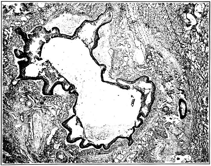
Fig. 1.—Acute bronchitis showing engorgement of blood vessels of mucosa and elevation of epithelium by serum and blood. Autopsy 352.
The blood vessels of the mucosa are engorged. There is sometimes edema or hemorrhage, and in the superficial part of the mucosa polynuclear leucocytes are often fairly abundant. When superficial epithelium has been lost, polynuclears are numerous immediately below the surface of the exposed tissue. Fibrin is often present upon the denuded surface and extends for a short distance into the tissue below. In the deeper part of the mucosa, about the muscularis and especially about and between the acini of the mucous glands, the tissue is infiltrated with lymphoid and plasma cells.
Changes in the mucous glands are invariably present. These changes are distention of ducts and acini with mucous, 147degenerative changes occasionally ending in necrosis of cells, disappearance of acini, dense infiltration of interstitial tissue with lymphoid and plasma cells and finally proliferation of this interstitial tissue. The duct of a mucous gland, dilated and filled with mucus, may be surrounded by lymphoid and plasma cells in great number. Acini, similarly dilated, contain mucus and are composed of cubical cells which have discharged their mucous content. In some instances (e. g., Autopsy 257) the cells of the acini have undergone necrosis; the cytoplasm stains homogeneously and the nuclei have disappeared. Where necrosis has occurred, polynuclear leucocytes may penetrate into the dead cells. In association with degenerative changes in the acini there is abundant infiltration of the interstitial tissue within and about the glands with lymphoid and plasma cells. When the acini have disappeared there is proliferation of fibroblasts and new formation of fibrous tissue, and mucous glands are found in which a few atrophied acini are separated by newly formed fibrous tissue.
With the bronchitis of influenza the small bronchi (with no cartilage or mucous glands) show every stage of transition from early acute inflammation characterized by accumulation of polynuclear leucocytes within the lumen, engorgement of blood vessels, and infiltration of the wall with polynuclear leucocytes, through various stages of destructive changes to complete disappearance of the bronchial wall and formation of an abscess cavity at the site of the bronchus. In the early stages of acute bronchitis, hemorrhage is frequently associated with the lesion. Blood may be abundant within the lumen of the bronchus, and in the mucosa red blood corpuscles often infiltrate the tissue around greatly distended blood vessels, or accumulating below the epithelium, separate it from its basement membrane. Hemorrhage is not limited to the wall of the bronchus, but frequently occurs into the alveoli in a zone encircling the bronchus.
148With acute bronchitis there may be desquamation of epithelial cells with partial or complete loss of epithelial lining. In the smallest bronchi the single layer of columnar cells may be separated in places from the underlying tissue, so that intact rows of cells are found within the lumen. In somewhat larger bronchi, lined by epithelium in multiple layers, superficial columnar ciliated cells may be lost. In some instances superficial epithelial cells appear to have lost their cohesion and are separated by narrow spaces; in these instances, polynuclear leucocytes are often numerous between epithelial cells. Epithelium is occasionally separated from its basement membrane by small accumulations of serum or blood. Occasionally necrosis of epithelial cells with disappearance of nuclei is seen and is doubtless caused by the action of bacteria; the affected cells may be raised from the underlying tissue by accumulated serum (Autopsy 253). The changes which have been described bring about partial or complete loss of the ciliated lining of the bronchial tube.
The severity of changes in the bronchial wall is in direct relation to the extent of destruction of the lining epithelium: when the epithelium remains intact polynuclear leucocytes may be found in considerable number immediately below it, but as the lesion progresses, cells in great part mononuclear, namely, lymphoid and plasma cells, accumulate in large number throughout the wall of the bronchus. There is often abundant cellular infiltration within and about the bundles of the muscular coat. The changes assume the character of chronic inflammation.
When the lining epithelium of the bronchus is lost, fibrin tends to accumulate over the surface of the defect, to which it is firmly attached. It remains separated by a conspicuous space from adjacent intact epithelium over which it may project. This superficial network of fibrin merges with a similar network, extending to a variable depth within the tissue. What may well be described as coagulative 149necrosis has often occurred, and structures, such as white fibrous bundles or wall of blood vessels, are marked out by hyaline material which merges with fibrin. When the walls of the blood vessels which are invariably engorged are involved, the lumen is plugged by a fibrinous thrombus.
Little patches of fibrin adherent to the inner surface of the bronchus may occur in spots where epithelium has been lost; with uniform loss of epithelium the entire circumference may be lined with fibrin forming a circular zone occasionally quite uniform in thickness.
Accumulations of polynuclear leucocytes doubtless bring about conditions which cause solution of fibrin or prevent its formation (when disintegration of leucocytes sets free leucoprotease in abundance). The activity of the infecting microorganisms, usually hemolytic streptococci or staphylococci, may cause complete necrosis of a part or all of the bronchial wall. The cavity which is formed may penetrate into lung tissue that has previously undergone pneumonic consolidation.
Further changes caused by the bronchitis of influenza will be considered under peribronchial hemorrhage and edema, peribronchial pneumonia and bronchiogenic abscess. Purulent bronchitis is almost invariably associated with dilatation of the bronchi, the affected bronchi being distended with pus. With increasing dilatation bronchiectasis becomes evident upon gross examination of the tissue, and is much more advanced in the small bronchi than in the larger cartilaginous passages. This subject will be further considered under bronchiectasis.
In association with the acute bronchitis of influenza the epithelium of bronchi not infrequently looses its superficial columnar ciliated cells and assumes some of the characters of a squamous epithelium being covered by polygonal or flat cells (Figs. 17 and 18). The condition is often described a “squamous metaplasia,” although it doubtless represents a stage of regeneration following injury rather 150than a true metaplasia. The basal cells of the epithelium have a cubical or columnar form; above them the cells become polygonal and as the surface is approached, cells are flat and even scale-like. The nuclei of these superficial cells are often lost. There is no close resemblance to the squamous epithelium of the skin, for intercellular bridges are not seen.
This change may occur within six days after onset of influenza, though in most instances the duration of illness has been two weeks or more. It may affect either large or small bronchi, but it is more frequently found in the latter. Whenever ciliated columnar cells are lost, superficial cells tend to become flat. Epithelium on one side of a bronchus may have a squamous character, whereas that elsewhere is columnar and ciliated. The flat epithelium may undergo thickening so that it is 0.1 mm. or more in thickness. It is noteworthy that regenerating epithelium growing over a denuded surface has the squamous character which has been described (Plate XIV, Fig. 22).
Bacteriology of the Bronchitis of Influenza.—With the pneumonia of influenza, bronchitis is invariably present. Cultures have been made from the right or left main bronchus or from the very small bronchi which contained purulent exudate. A routine method of making the culture has been adopted. The right main bronchus, exposed by drawing the right lung out of the chest and toward the midline, was widely seared with a hot knife; the bronchus was partially cut across through the seared surface with a heated knife and a platinum needle inserted into the lumen. The bacteria obtained named in the approximate order of their relative frequency have been: B. influenzæ, pneumococci, hemolytic streptococci, staphylococci (aureus and albus), B. coli, S. viridans, M. catarrhalis, and diphthoid bacilli which have not been identified. Mixed infections occurred in most instances. The following list arranged by grouping 151bacteria in the order cited above, shows how varied have been the combinations which occur:
B. influenzæ 3
Pneumococci 5
S. hemolyticus 3
Staphylococci 3
B. coli 3
S. viridans 1
B. influenzæ, pneumococci 17
B. influenzæ, S. hemolyticus 18
B. influenzæ, staphylococci 4
Pneumococci, S. hemolyticus 1
Pneumococci, staphylococci 3
S. hemolyticus, staphylococci 4
S. hemolyticus, B. coli 2
Staphylococci, S. viridans 1
B. influenzæ, pneumococci, S. hemolyticus 6
B. influenzæ, pneumococci, staphylococci 15
B. influenzæ, pneumococci, S. viridans 2
B. influenzæ, S. hemolyticus, staphylococci 16
B. influenzæ, S. hemolyticus, M. catarrhalis 1
B. influenzæ, staphylococci, S. viridans 1
Pneumococci, S. hemolyticus, staphylococci 3
Staphylococci, B. coli, S. viridans 1
B. influenzæ, pneumococci, S. hemolyticus, staphylococci 7
B. influenzæ, pneumococci, staphylococci, M. catarrhalis 1
B. influenzæ, S. hemolyticus, staphylococci, B. coli 1
B. influenzæ, S. hemolyticus, staphylococci, S. viridans 1
B. influenzæ, S. hemolyticus, staphylococci, M. catarrhalis 1
B. influenzæ, staphylococci, S. viridans, M. catarrhalis 1
B. influenzæ has been present in the bronchi in 79.3 per cent of instances of pneumonia referable to influenza. Combinations which have been found most frequently are B. influenzæ and pneumococci (17 instances), B. influenzæ and hemolytic streptococci (18 instances), or the same combinations with staphylococci, namely, B. influenzæ, pneumococci and staphylococci (15 instances), and B. influenzæ, hemolytic streptococci and staphylococci (16 instances). There is little doubt that B. influenzæ was not identified in some instances in which it was present; when other microorganisms are very numerous its inconspicuous colonies may be overgrown even though the presence of pneumococci, streptococci 152or staphylococci tends to increase the size of its colonies. Moreover, it is not improbable that the microorganism may disappear from the bronchi. Comparison with observations made upon influenza suggests that multiple methods of examination might have demonstrated a much higher incidence of B. influenzæ. Throat cultures alone made during life demonstrated the presence of B. influenzæ in only 65.7 per cent of patients with acute influenza, whereas when cultures were made from the nose, throat and sputum, and a mouse was inoculated with sputum from each patient, B. influenzæ was found in every instance. After the acute stage of the disease had passed, the number of microorganisms diminished, and in many instances B. influenzæ disappeared from the upper air passages. In some of our autopsies B. influenzæ doubtless present during life has similarly disappeared before death due to pneumonia caused by pneumococci or streptococci. In view of these considerations it is not improbable that B. influenzæ demonstrated by a single culture in 80 per cent of instances has been constantly present.
Table XXVIII represents the incidence of pneumococci, hemolytic streptococci, staphylococci, and B. influenzæ in the bronchi, lungs and blood of those individuals with pneumonia in whom bacteriologic examination has been made at autopsy. The number of cultures made from the bronchi, lungs or blood of the heart is given in the second column of the table and in other columns are given the incidence in number and percentage of the microorganisms which have been mentioned.
Table XXVIII
NO. OF CULTURES PNEUMOCOCCI HEMOLYTIC STREPTOCOCCI STAPHYLOCOCCI B. INFLUENZÆ
NO. POSITIVE PER CENT POSITIVE NO. POSITIVE PER CENT POSITIVE NO. POSITIVE PER CENT POSITIVE NO. POSITIVE PER CENT POSITIVE
Bronchus 121 56 46.3 58 47.9 61 50.4 96 79.3
Lung 153 68 44.4 77 50.3 37 24.2 70 45.7
Blood 218 87 39.9 85 39.0 1 0.5 1 0.5
153Cultures from the bronchus represent the bacteriology of the bronchitis of influenza. Infection of the lung following influenza doubtless occurs by way of the bronchi, so that the bacteria which cause pneumonia are present in the bronchi before they enter the lung tissue. The figures in Table XXVIII, similar to those previously cited, show the high incidence of B. influenzæ, and the occurrence of pneumococci, hemolytic streptococci and staphylococci each present in approximately half of all autopsies.
The figures in Table XXVIII are an index of the capacity of the microorganisms which enter the bronchi to invade the lungs and finally the blood. Pneumococci were present in the bronchi in 46.3 per cent of instances, in the lungs in only slightly less, and in approximately 40 per cent of autopsies they had penetrated into the blood. Hemolytic streptococci enter the bronchi with the same frequency and exhibit an equal ability to penetrate into the lungs and blood. Staphylococci enter the bronchi in half of these individuals, but penetrate into the lungs in only a fourth of the instances. They have entered the blood only once (Autopsy 263) in this instance in association with hemolytic streptococci. B. influenzæ has been present in the bronchi in approximately 80 per cent of autopsies. It is noteworthy that it has been found in the lung in little more than half this percentage of instances and has entered the blood only once (Autopsy 474), in this instance in association with hemolytic streptococci.
In a limited number of autopsies there was purulent bronchitis recognized by the presence of mucopurulent exudate in small bronchi. It has been stated that this group of cases is not sharply separable from other instances of bronchitis, because in some cases death has occurred before a purulent exudate has accumulated or in other instances a purulent exudate has been displaced by edema. Table XXIX shows the bacteriology of instances of purulent bronchitis:
154
Table XXIX
NO. OF CULTURES PNEUMOCOCCI HEMOLYTIC STREPTOCOCCI STAPHYLOCOCCI B. INFLUENZÆ
NO. POSITIVE PER CENT POSITIVE NO. POSITIVE PER CENT POSITIVE NO. POSITIVE PER CENT POSITIVE NO. POSITIVE PER CENT POSITIVE
Bronchus 66 33 50.0 32 48.5 36 54.5 53 80.3
The percentages of various bacteria with purulent bronchitis do not differ essentially from those obtained from all autopsies with pneumonia. B. influenzæ is found in approximately 80 per cent of autopsies. In 16 instances cultures were made from the purulent fluid contained in a small bronchus and the incidence of B. influenzæ (namely, 81.4 per cent) has not differed from that in the main bronchus. In 7 of 8 instances in which cultures were made, both from the right main bronchus and from the purulent fluid in a small bronchus, B. influenzæ was found in one or other in all but one autopsy (87.5 per cent); in this instance (Autopsy 472) respiratory disease began thirty-seven days before death and cultures from large and small bronchi at autopsy were overgrown by B. coli. Since observations upon influenza made during life have shown that B. influenzæ is constantly demonstrable when multiple methods are employed for its detection, the figures just cited give support to the suggestion that B. influenzæ is constantly present in the bronchi with the bronchitis of influenza.
Lobar Pneumonia
The frequency with which the confluent lobular consolidation of bronchopneumonia involving whole lobes or parts of lobes follows influenza has emphasized the desirability of distinguishing carefully between lobar and confluent lobular pneumonia. The pulmonary lesion has been designated lobar pneumonia when it exhibited the well-known characters of this lesion, namely, firm consolidation of large parts of lobes, coarse granulation of the cut surface, 155fibrinous plugs in the bronchi and, on microscopic examination, homogeneous consolidation and fibrinous plugs within the alveoli. With confluent lobular consolidation of bronchopneumonia the consolidated area is in most cases obviously limited by lobule boundaries, and well-defined lobules of consolidation occur elsewhere in the lungs.
Lobar pneumonia occurred in 98 among 241 instances of pneumonia following influenza, namely, in 40.7 per cent of autopsies.
The difficulty of separating lobar and bronchopneumonia following influenza has been increased by the frequent combination of the two lesions in the same individual. There were 34 instances in which lobar and bronchopneumonia occurred together. The anatomic diagnosis of lobar pneumonia was made only when lobes or parts of lobes were firmly consolidated and exhibited the characters of the lesion enumerated above; in several instances, in which there was some doubt concerning the nature of the lesion, microscopic examination was decisive. The associated bronchopneumonic lesions represented all the types which have been associated with influenza. In the group of 34 cases of coexisting lobar and bronchopneumonia, lobular consolidation occurred 10 times, peribronchiolar consolidation 14 times (recognized in all but 4 instances by microscopic examination), hemorrhagic peribronchiolar consolidation 9 times, peribronchial pneumonia 4 times. The intimate relation of these lesions to changes in the bronchi is well shown by the frequent presence of purulent bronchitis. The associated lesions of the bronchi in these cases were as follows: purulent bronchitis in 23 instances; peribronchial hemorrhage in 6; bronchiectasis in 11. The frequency of purulent bronchitis and other bronchial lesions in association with coexisting lobar and bronchopneumonia is in sharp contrast with the occurrence of these lesions in association with lobar pneumonia alone; with 69 instances of 156lobar pneumonia alone purulent bronchitis occurred 17 times and bronchiectasis once.
Lobar pneumonia following influenza passes through the usual stages of red and gray hepatization. Red hepatization was found 16 times, combined red and gray hepatization 28 times, and gray hepatization 20 times. The average duration of pneumonia with red hepatization was 3.7 days, with combined red and gray hepatization 5.1 days and with gray hepatization 7.5 days. These figures, it will be shown later, have some importance in relation to the stage at which hemolytic streptococcus infects lungs the site of lobar pneumonia.
Bacteriology of Lobar Pneumonia.—Table XXX is compiled with the purpose of determining the bacteriology of the bronchi, lungs and heart’s blood in autopsies performed on individuals with lobar pneumonia. In some instances bacteriologic examination of one or other of these organs was omitted; the percentage incidence is an index of the presence of pneumococci, hemolytic streptococci, staphylococci or B. influenzæ in the bronchi, lungs or heart’s blood and measures the invasive power of these microorganisms during the course of lobar pneumonia following influenza.
Table XXX
NO. OF CULTURES PNEUMOCOCCI HEMOLYTIC STREPTOCOCCI STAPHYLOCOCCI B. INFLUENZÆ
NO. POSITIVE PER CENT POSITIVE NO. POSITIVE PER CENT POSITIVE NO. POSITIVE PER CENT POSITIVE NO. POSITIVE PER CENT POSITIVE
Bronchus 44 56.9 14 31.8 22 37 84.1 96 79.3
Lung 53 77.3 13 24.5 8 26 49.1 70 45.7
Blood 87 65.5 11 12.6 1 0.5
Pneumococci, the recognized cause of lobar pneumonia, were found in the lungs in 73.3 per cent of autopsies; failure to find the microorganism in all instances is doubtless the result of its disappearance from the lung, which, it is well known, occurs not infrequently particularly during 157the later stages of the disease. In 65.5 per cent of instances of fatal lobar pneumonia pneumococci have entered the heart’s blood.
Hemolytic streptococci unlike pneumococci were found more frequently in the bronchi than in the lungs; this microorganism which exhibits little tendency to disappear, once it has established itself within the body, found entrance into the bronchi in 31.8 per cent of instances of lobar pneumonia and in 24.5 per cent entered the lungs. Its invasive power is further illustrated by its penetration into the heart’s blood approximately in half this proportion of autopsies.
Staphylococci enter the bronchi in many instances (50 per cent), but relatively seldom (15.1 per cent) invade the lung and rarely if ever penetrate into the blood.
The high incidence, namely, 84.1 per cent, of B. influenzæ in the bronchi is particularly noteworthy; it exceeds that of pneumococci, the well-recognized cause of lobar pneumonia, within the lung. It is found much less frequently within consolidated lung tissue and shows no tendency to invade the heart’s blood. B. influenzæ finds the most favorable conditions for its multiplication within the bronchi.
In view of the frequent occurrence of coexisting lobar and bronchopneumonia it has appeared desirable to determine how far the existence of obvious bronchopneumonia modifies the bacteriology of lobar pneumonia. In Table XXXI the incidence of pneumococci, hemolytic streptococci, staphylococci and B. influenzæ after death with lobar pneumonia on the one hand is compared with their incidence after combined lobar and bronchopneumonia on the other.
Pneumococci are found in the lung more frequently with lobar than with combined lobar and bronchopneumonia. The incidence of hemolytic streptococci and of staphylococci in the lung is on the contrary higher when bronchopneumonia is associated with lobar pneumonia. It is not improbable that these microorganisms have a part in the production of associated bronchopneumonia. The frequency with which microorganisms invade the blood is almost identical in the two groups.
158
Table XXXI
With Lobar Pneumonia Alone
NO. OF CULTURES PNEUMOCOCCI HEMOLYTIC STREPTOCOCCI STAPHYLOCOCCI B. INFLUENZÆ
NO. POSITIVE PER CENT POSITIVE NO. POSITIVE PER CENT POSITIVE NO. POSITIVE PER CENT POSITIVE NO. POSITIVE PER CENT POSITIVE
Bronchus 30 20 66.6 9 30 15 50 26 86.7
Lung 34 29 85.2 7 20.6 3 8.8 18 52.9
Blood 54 36 66.7 7 13
With Combined Lobar and Bronchopneumonia
NO. OF CULTURES PNEUMOCOCCI HEMOLYTIC STREPTOCOCCI STAPHYLOCOCCI B. INFLUENZÆ
NO. POSITIVE PER CENT POSITIVE NO. POSITIVE PER CENT POSITIVE NO. POSITIVE PER CENT POSITIVE NO. POSITIVE PER CENT POSITIVE
Bronchus 14 9 64.3 5 34.3 7 50 11 78.6
Lung 19 12 63.2 6 31.6 5 26.3 8 42.1
Blood 33 21 63.1 4 12.1
The relative frequency with which different types of pneumococci produce lobar pneumonia under the conditions existing when Camp Pike was attacked by an epidemic of influenza is indicated by Table XXXII in which instances of lobar pneumonia alone and of combined lobar and bronchopneumonia are listed separately.
Pneumococcus I and II, which are found approximately in two-thirds of instances of lobar pneumonia occurring in cities, have an insignificant part in the production of these lesions. Pneumococcus IV and atypical Pneumococcus II, which are commonly found in the mouth, are the predominant cause of these lesions, and with Pneumococcus III, also an inhabitant of the mouths of normal individuals, have been the cause of two-thirds of all instances of lobar pneumonia observed in this camp.
159
Table XXXII
With Lobar Pneumonia
NO. OF CULTURES PNEUMOCOCCUS I PNEUMOCOCCUS II PNEUMOCOCCUS II (Atyp.) PNEUMOCOCCUS III PNEUMOCOCCUS IV
NO. POSITIVE PER CENT POSITIVE NO. POSITIVE PER CENT POSITIVE NO. POSITIVE PER CENT POSITIVE NO. POSITIVE PER CENT POSITIVE NO. POSITIVE PER CENT POSITIVE
Bronchus 30 1 3.3 1 3.3 4 13.3 4 13.3 10 33.3
Lung 34 1 2.9 2 5.9 9 26.5 6 17.6 11 32.4
Blood 54 2 3.7 2 3.7 12 22.2 3 5.6 17 31.5
With Combined Lobar and Bronchopneumonia
NO. OF CULTURES PNEUMOCOCCUS I PNEUMOCOCCUS II PNEUMOCOCCUS II (Atyp.) PNEUMOCOCCUS III PNEUMOCOCCUS IV
NO. POSITIVE PER CENT POSITIVE NO. POSITIVE PER CENT POSITIVE NO. POSITIVE PER CENT POSITIVE NO. POSITIVE PER CENT POSITIVE NO. POSITIVE PER CENT POSITIVE
Bronchus 14 2 14.3 1 7.1 3 21.4 3 21.4
Lung 19 1 5.3 5 26.3 6 31.6
Blood 33 2 6.1 3 9.1 4 12.1 12 36.4
There is no noteworthy difference in the occurrence of these types of pneumococci among instances of lobar pneumonia, on the one hand, and of combined lobar and bronchopneumonia, on the other. Different types exhibit no noteworthy differences in their ability to penetrate into lungs and blood.
Hemolytic Streptococcus with Lobar Pneumonia.—There can be no doubt that the concurrent infection with microorganisms other than pneumococcus modifies the progress of lobar pneumonia. With lobar pneumonia alone hemolytic streptococci have entered the bronchi in 30 per cent of instances and have penetrated into the lungs in 20.6 per cent; with associated lobar and bronchopneumonia the same microorganism has entered the bronchi in 34.3 per cent of instances and invaded the lung in 31.6 per cent. Hemolytic streptococci are the only microorganisms other than pneumococci which, in association with lobar pneumonia, 160have found their way from the lungs to the blood stream; more than one-third of all instances of lobar pneumonia in which hemolytic streptococci find entrance into the bronchi die with streptococcus septicemia.
Separation of instances of lobar pneumonia into groups on the basis of the occurrence of red or gray hepatization shows that infection with hemolytic streptococcus is more likely to occur during the early stages of the disease. The average duration of lobar pneumonia with red hepatization has been 3.7 days, with red and gray hepatization, 5.1 days, and with gray hepatization, 7.5 days. Infection with hemolytic streptococcus has occurred in association with red or gray hepatization as shown in Table XXXIII.
Table XXXIII
NO. OF AUTOPSIES NO. WITH HEMOLYTIC STREPTOCOCCUS PER CENT WITH HEMOLYTIC STREPTOCOCCUS
Lobar pneumonia with red hepatization 16 6 37.5
Lobar pneumonia with red and gray hepatization 28 6 21.4
Lobar pneumonia with gray hepatization 20 1 5.0
Notwithstanding the longer duration of the disease and consequent prolongation of exposure to infection, lobar pneumonia, which has reached the stage of gray hepatization, has shown the smallest incidence of infection with hemolytic streptococci. In the stage of gray hepatization there is diminished susceptibility to secondary infection with this microorganism.
Characteristic histologic changes have been found in the lungs of those who have died with lobar pneumonia followed by invasion of lungs and blood by hemolytic streptococci (e. g., Autopsies 273, 430), but with no evidence of suppuration found at autopsy. Within the pneumonic lung occur patches of necrosis implicating both exuded cells and alveolar walls; in some places nuclei have disappeared; elsewhere nuclear fragments are abundant. 161In these patches of necrosis Gram-positive streptococci in short chains occur in immense number. In some instances (e. g., Autopsies 273, 346, 479) interlobular septa are very edematous and often contain a network of fibrin; lymphatics are dilated and contain polynuclear leucocytes in abundance. Streptococci are found within these lymphatics. The histologic changes which have been described represent the earliest stages of abscess formation and interstitial suppuration, lesions almost invariably caused by hemolytic streptococci.
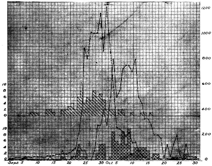
Chart 2.—Showing the relation of (a) date of onset of cases in which autopsy demonstrated lobar pneumonia, indicated by upper continuous line with single hatch, and of (b) date of death of these cases, indicated by lower continuous line with double hatch to (c) the occurrence of influenza, indicated by the broken line, and to (d) the total number of fatal cases of pneumonia, indicated by the broken dotted line. Each case of fatal pneumonia is indicated by one division of the scale as numbered on the left of the chart; cases of influenza are indicated by the numbers on the right of the chart.
Relation of Lobar Pneumonia to Influenza.—Some writers have suggested that lobar pneumonia, heretofore observed during the course of epidemics of influenza, is an 162independent disease with no relation to influenza, both diseases being referable perhaps to similar meteorologic or other conditions. Chart 2, which shows by weeks from September 1 to October 31 the relation of deaths from lobar pneumonia (indicated by double hatch) to deaths from all forms of pneumonia, disproves this suggestion. The two curves follow parallel courses; that representing lobar pneumonia reaches a maximum approximately one week after the outbreak of influenza had reached its height. Lobar pneumonia, like other forms of pneumonia, was secondary to influenza. When a chart is plotted to represent the dates of onset of fatal cases of lobar pneumonia (indicated by single hatch in Chart 2), it becomes evident that the greatest number of these cases of pneumonia had their onset at the beginning of the influenza epidemic, approximately one week before it reached its height. Fatal lobar pneumonia developed less frequently in the latter part of the epidemic; to obtain an explanation of this relation it is necessary to chart separately cases of lobar pneumonia with secondary streptococcus infection, for we have already learned that streptococcus infection was the predominant cause of death in the early period of the influenza epidemic. Exclusion of these instances of secondary streptococcus infection makes no noteworthy change in the character of the chart. Fatal lobar pneumonia, like all forms of fatal pneumonia (p. 140), was more frequent in the first half than in the second half of the epidemic. This difference is referable either to greater virulence of the virus of influenza or to the greater susceptibility of those first selected by the disease or, as more probable, to conditions such as crowding together of patients with influenza, favoring the transmission of microorganisms which cause pneumonia.
Bronchopneumonia
For the purpose of the present study it is convenient to group together instances of bronchopneumonia which have 163been unaccompanied, on the one hand, by lobar pneumonia (p. 155) or, on the other hand, by suppuration, which with few exceptions is caused by hemolytic streptococci or by staphylococci. A group of cases in which lobar and bronchopneumonia have occurred in the same individual have already been considered. In many instances, bronchopneumonia is accompanied by abscess formation or by some other form of suppuration; these lesions will be discussed elsewhere.
Bronchopneumonia unaccompanied by lobar pneumonia or by suppuration occurred in 80 autopsies.
Pneumonic consolidation distributed with relation to the bronchi exhibits considerable variety, and an attempt to define a type of bronchopneumonia characteristic of influenza would be futile. Nevertheless, the bronchopneumonia of influenza has in many instances distinctive characters.
Lesions of bronchopneumonia which are frequently found in the autopsies under consideration may be conveniently designated by descriptive terms, indicative of their location in the lung tissue. These lesions, of which two or more often occur in the same lung, are:
1. Peribronchiolar consolidation with which the inflammatory exudate is limited to the alveoli in the immediate neighborhood of the bronchioles.
2. Hemorrhagic peribronchiolar consolidation in which gray patches of peribronchiolar pneumonia occur upon a deep red background produced by hemorrhage into alveoli. Pfeiffer believed that this lesion was characteristic of influenza.
3. Lobular consolidation with which consolidation is limited to lobules or groups of lobules.
4. Peribronchial pneumonia with which small bronchi are encircled by pneumonic consolidation.
Each one of these lesions will be discussed separately.
Following is a list of the bacteria which have been isolated from the consolidated lung of individuals with bronchopneumonia 164unaccompanied by lobar pneumonia or by suppuration:
B. influenzæ 1
Pneumococci 5
S. hemolyticus 5
S. viridans 1
B. influenzæ, pneumococci 9
B. influenzæ, S. hemolyticus 4
B. influenzæ, staphylococci 4
Pneumococci, S. hemolyticus 1
Pneumococci, staphylococci 2
S. hemolyticus, staphylococci 1
S. hemolyticus, B. coli 1
Staphylococci, S. viridans 1
Staphylococci, B. coli 1
B. influenzæ, pneumococci, staphylococci 1
B. influenzæ, pneumococci, S. viridans 1
B. influenzæ, S. hemolyticus, staphylococci 2
B. influenzæ, pneumococci, staphylococci, S. viridans 1
No microorganisms found 6
47
The similarity of this list to that representing the bacteriology of bronchitis is evident; there is the same multiplicity of microorganisms and the frequent occurrence of mixed infections. B. influenzæ is much less frequently found in the lung. The relative pathogenicity of the large group of microorganisms enumerated above is better indicated by the following list which shows what microorganisms have penetrated into the blood in autopsies performed on individuals with bronchopneumonia:
Pneumococci 20
S. hemolyticus 23
S. viridans 1
Pneumococci, S. hemolyticus 2
No bacteria found 25
Total 71
Table XXXIV shows the percentage incidence of pneumococcus, hemolytic streptococcus, staphylococcus and 165B. influenzæ in the bronchi, lungs and blood and is inserted for comparison with the similar table (Table XXX) showing the incidence of these bacteria in lobar pneumonia.
Table XXXIV
NO. OF CULTURES PNEUMOCOCCI HEMOLYTIC STREPTOCOCCI STAPHYLOCOCCI B. INFLUENZÆ
NO. POSITIVE PER CENT POSITIVE NO. POSITIVE PER CENT POSITIVE NO. POSITIVE PER CENT POSITIVE NO. POSITIVE PER CENT POSITIVE
Bronchus 37 19 48.6 13 35.1 22 59.5 28 75.7
Lung 47 20 42.6 14 29.8 13 27.7 23 48.9
Blood 70 22 31.4 24 34.3
Table XXXIV shows that pneumococci have a less important part in the production of broncho than of lobar pneumonia; with lobar pneumonia this microorganism was found in the lungs in 77.3 per cent of instances and in the blood, in 65.5 per cent, whereas with bronchopneumonia it was found in the lungs in 42.6 per cent and in the blood in 31.4 per cent. Hemolytic streptococci (in lungs and blood) and staphylococci (in lungs), on the contrary, were more common with bronchopneumonia, and doubtless have a part in the production of the lesion. Streptococcus viridans, B. coli and M. catarrhalis, which are not infrequently found in the bronchi (p. 151), occasionally enter the lungs with bronchopneumonia but are rarely found with lobar pneumonia. B. influenzæ has been found in less than 80 per cent of instances in the bronchi and in about half of the lungs, maintaining an incidence approximately the same as that with lobar pneumonia.
Table XXXV shows the types of pneumococci found in association with bronchopneumonia and is inserted for comparison with the similar table (Table XXXII) showing types of pneumococci with lobar pneumonia.
With broncho as with lobar pneumonia pneumococci commonly found in the mouth, namely, atypical II, and Types III and IV, have a more important part in production of the lesion than the so-called fixed types, I and II. Atypical Pneumococcus II has been less frequently encountered with broncho than with lobar pneumonia.
166
Table XXXV
NO. OF CULTURES PNEUMOCOCCUS I PNEUMOCOCCUS II PNEUMOCOCCUS II (Atyp.) PNEUMOCOCCUS III PNEUMOCOCCUS IV
NO. POSITIVE PER CENT POSITIVE NO. POSITIVE PER CENT POSITIVE NO. POSITIVE PER CENT POSITIVE NO. POSITIVE PER CENT POSITIVE NO. POSITIVE PER CENT POSITIVE
Bronchus 37 1 2.7 3 8.1 14 37.8
Lung 47 2 4.3 2 4.3 2 4.3 2 4.3 12 25.2
Blood 70 1 1.4 1 1.4 5 7.1 4 5.7 11 15.9
Peribronchiolar Consolidation.—In many instances of bronchopneumonia, usually in association with lobular or confluent consolidation, small firm nodules of consolidation are clustered about the bronchioles (Fig. 2). These nodular foci of consolidation are usually 1.5 to 2 mm. in diameter, being sometimes slightly smaller or slightly larger. They are usually gray and occasionally surrounded by a red halo; sometimes they are yellowish gray. They are clustered about the smallest bronchial tubes to form groups which are from 0.5 to 1 cm. across. A group of nodular foci of consolidation occupies the central part of a lobule of lung tissue. When pneumonia has been of short duration these foci are fairly soft and not sharply defined, and in many instances this form of bronchopneumonia is first recognized by microscopic examination. When the disease has lasted from ten days to two weeks, the consolidated nodules are very firm and sharply circumscribed, closely resembling tubercles. When they have assumed this character, microscopic examination shows that chronic changes indicated by new formation of interstitial tissue have occurred.