Chapter IV · THE PATHOLOGY AND BACTERIOLOGY OF PNEUMONIA FOLLOWING INFLUENZA (continued 2)
Reading edition
The lesion may be designated peribronchiolar consolidation. It has occurred usually in association with other types of pneumonic lesion in 61 instances, being recognized at autopsy in 18 and by microscopic examination in 43.
167
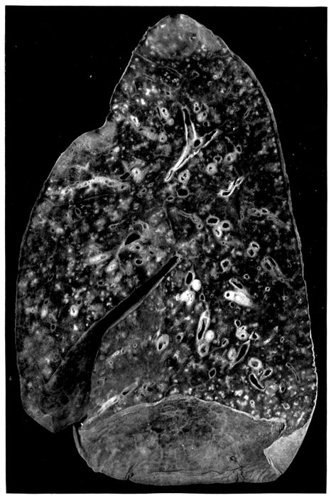
Fig. 2.—Acute bronchopneumonia with nodules of peribronchiolar consolidation and purulent bronchitis. Autopsy 429.
168In association with this lesion there are almost invariably severe lesions of the bronchi. Purulent bronchitis was noted in 47 of the 61 instances, in which this nodular bronchopneumonia was found at autopsy. An index of the severity of the bronchial injury is the frequency with which bronchiectasis has occurred; dilatation of small bronchi was observed in 24 instances. In 10 instances the bronchi were encircled by conspicuous zones of hemorrhage.
In association with this peribronchiolar lesion the lung is often voluminous and fails to collapse on removal from the chest. Pressure upon the lung squeezes from the smallest bronchi, both in the neighborhood of the nodular consolidation and elsewhere, a droplet of viscid, semifluid mucopurulent material. The presence of this tenacious material throughout the small bronchi doubtless explains the failure of the lung tissue to collapse. Interstitial emphysema has been present in some of these lungs.
A red zone of hemorrhage has occasionally been observed about the foci of peribronchiolar pneumonia. A further stage in the same process is represented by hemorrhage into all of the alveoli separating these patches of consolidation. This hemorrhagic lesion, which will be described in more detail later, has been found repeatedly in the same lung with peribronchiolar pneumonia, being present in 8 among the 61 autopsies cited. Lobular bronchopneumonia accompanied the peribronchiolar lesion 27 times and lobar pneumonia accompanied it 20 times.
When an abscess caused by hemolytic streptococcus is associated with peribronchiolar pneumonia, empyema is present, but otherwise pleurisy is absent or limited to a scant fibrinous exudate.
169
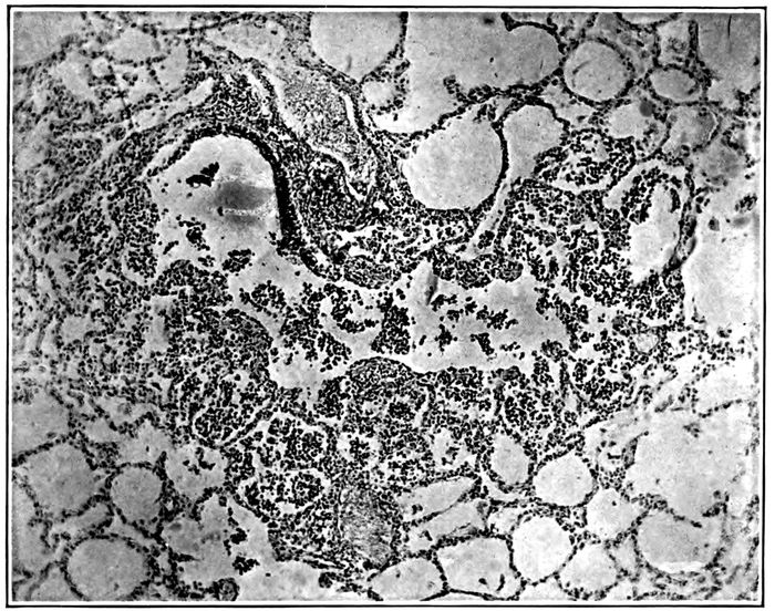
Fig. 3.—Acute bronchopneumonia with peribronchiolar consolidation; a respiratory bronchiole partially lined by columnar epithelium passes into alveolar duct and the adjacent alveoli are filled by polynuclear leucocytes. Autopsy 333.
Histologic examination demonstrates very clearly the relation of this lesion to the bronchioles (Fig. 3). These passages are filled and distended with an inflammatory exudate consisting almost entirely of polynuclear leucocytes. The respiratory bronchioles are beset with alveoli often limited to one side of the tubule and these alveoli are filled with leucocytes. The alveolar ducts, distinguishable from the bronchioles by the absence of columnar or cubical epithelium and by possession of smooth muscle, are similarly filled with leucocytes; the numerous alveoli which form the walls of the alveolar ducts are distended by an inflammatory exudate. In sections which pass through an alveolar duct and one or more of its infundibula, the further extension of the lesion may be determined (Fig. 4). The infundibulum in proximity with the alveolar duct contains polynuclear leucocytes and the same cells are seen in the alveoli which here form its wall, but the intensity of the inflammatory reaction diminishes toward the periphery, so that the distal part of the infundibulum, which is much distended 170and in consequence more readily definable than usual, is free from inflammatory exudate.
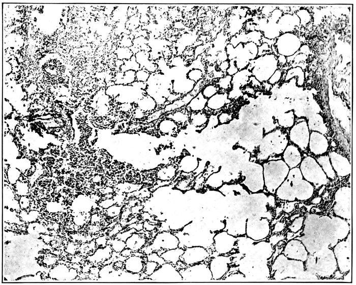
Fig. 4.—Acute bronchopneumonia with peribronchiolar consolidation; a respiratory bronchiole is in continuity with an alveolar duct and two distended infundibula; alveoli about bronchiole, alveolar duct and proximal part of infundibula contain polynuclear leucocytes, the distal part of the infundibula showing no evidence of inflammation. Autopsy 333.
Occasionally there is irregularly distributed hemorrhage and perhaps some edema in the alveoli immediately adjacent to those which form the peribronchiolar focus of inflammation. In such instances small bronchi, that is, air passages, lined by columnar epithelium and devoid of tributary alveoli, may be surrounded by a zone of hemorrhage; immediately surrounding the bronchus, the wall of which shows intense inflammation, alveoli, in a zone of which the radius represents several alveoli, are filled with blood. This hemorrhagic zone is continued from the bronchus over the focus of inflammation which surrounds the bronchiole.
171Another variation in the character of the lesion is doubtless referable to variation in the severity of primary bronchial injury. Alveoli immediately surrounding small bronchi are filled with dense plugs of fibrin. The alveoli which besot the walls of the bronchioles contain fibrin, but the alveolar duct and its tributary alveoli are filled with polynuclear leucocytes.
The bacteria which have been cultivated from the lung in autopsies with peribronchiolar pneumonia are as follows:
Pneumococcus 5
S. hemolyticus 8
B. influenzæ, pneumococcus 5
B influenzæ, S. hemolyticus 7
B. influenzæ, staphylococcus 1
Pneumococcus, staphylococcus 2
S. hemolyticus, staphylococcus 2
B. influenzæ, pneumococcus, S. hemolyticus 2
B. influenzæ, pneumococcus, staphylococcus 1
B. influenzæ, S. hemolyticus, staphylococcus 2
Pneumococcus, S. hemolyticus, staphylococcus 3
No organism 3
Total 41
The following list which shows the bacteria found in the blood is an index to the pathogenicity of pneumococci and hemolytic streptococci:
Pneumococcus 22
S. hemolyticus 20
Pneumococcus, S. hemolyticus 1
No organism 14
Total 57
The percentage incidence of pneumococcus, hemolytic streptococcus, staphylococcus and B. influenzæ in bronchus, lung and blood, given in Table XXXVI, is inserted to indicate with what readiness each one of these microorganisms passes from the bronchus through the lung into the circulating blood.
172
Table XXXVI
PNEUMOCOCCUS HEMOLYTIC STREPTOCOCCUS STAPHYLOCOCCUS B. INFLUENZA
Bronchus 39.4% 57.7% 60.6% 84.8%
Lung 43.9% 61.0% 21.9% 43.9%
Blood 40.3% 36.8% 0. % 0. %
B. influenzæ is present in the bronchi in a very large proportion (84.8 per cent) of those in whom this type of bronchopneumonia has been found at autopsy; it is much less frequently recovered from the lungs. Staphylococci, in part S. albus and in part S. aureus, are less frequently found in the bronchi and are recovered from the lungs in a relatively small proportion of autopsies. The percentage incidence of pneumococci and streptococci in lungs and blood demonstrates the pathogenicity of these microorganisms, for whereas pneumococci and hemolytic streptococci are found in the consolidated lungs in 43.9 and 61.0 per cent of instances of the lesion respectively, they make their way into the blood in 40.3 and 36.8 per cent of instances.
Coexisting infection with pneumococci and hemolytic streptococci has been not uncommon e. g., Autopsy 275 in which both were in the blood; in 2 instances (Autopsies 333 and 378) in which pneumococci were obtained from the blood, hemolytic streptococci were found in the lungs and bronchi; in 3 instances (Autopsies 258, 273 and 445) in which hemolytic streptococci were present in the blood, pneumococci were obtained from the lungs.
In the group of autopsies under consideration, examination of the sputum was made during life and after onset of pneumonia in 11 instances. The microorganisms found in the sputum and at autopsy were as follows:
SPUTUM IN BLOOD, LUNGS OR BRONCHUS AT AUTOPSY
Autopsy 240 Pneum. IV Pneum. IV
246 Pneum. atyp. II, B. inf.
247 Pneum. IV, B. inf. Pneum. IV
250 Pneum. atyp. II, B. inf. Pneum. atyp. II
253 Pneum. atyp. II Pneum. II
285 Pneum. atyp. II, B. Inf. S. hem., B. inf.
288 S. hem., B. inf. S. hem., B. inf.
173 291 Pneum. IV, B. inf. Staph., B. inf.
300 Pneum. atyp. II, B. inf. Pneum. atyp. II, B. inf.
312 Pneum. IV, S. hem., B. inf. S. hem., B. inf.
346 Pneum. IV, B. inf. S. hem., B. inf.
In 2 instances (Autopsies 285 and 346) among this small group of cases, pneumococci but no hemolytic streptococci were found in the sputum several days before death, whereas death occurred as the result of secondary invasion with hemolytic streptococci and no pneumococci were found at autopsy. It is probable that this sequence of events is not uncommon. B. influenzæ finds its way into the bronchi and pneumococci follow it; pneumonia limited to peribronchiolar alveoli may occur in consequence of this invasion. Later hemolytic streptococci may follow the same path and cause death with bacteremia.
Hemorrhagic Peribronchiolar Consolidation.—Peribronchiolar pneumonia accompanied by diffuse accumulation of blood within the alveoli is one of the most frequent complications of influenza. The lung tissue is laxly consolidated, and on section there is a homogeneous dull deep red background upon which are seen small gray spots (1.5 to 2 mm. in diameter) grouped in clusters about the smallest bronchi (Fig. 5). Wide areas of lung tissue are implicated and the lesion is more common in the dependent parts of the lung than elsewhere. In common with other forms of bronchopneumonia the lesion is in most instances associated with changes in the bronchi; in 55 instances of hemorrhagic bronchiolar pneumonia purulent bronchitis was found in 43 instances; it is noteworthy that purulent bronchitis often is not evident in the presence of pulmonary edema and edema is not infrequent with this pneumonic lesion.
Microscopic examination demonstrates the presence of acute bronchitis; the lumina of the small bronchi contain polynuclear leucocytes and red blood corpuscles. Accumulation of blood may separate the epithelium from the basement 174membrane. The mucosa immediately below the epithelium contains polynuclear leucocytes in fair abundance and the blood vessels of the bronchial wall are much engorged. Respiratory bronchioles are distended with polynuclear leucocytes and red blood corpuscles. In a zone about each bronchiole, in areas corresponding to the small gray spots seen upon the cut surface of the lung, the alveoli are filled with polynuclear leucocytes. In the lung tissue intervening between these spots of leucocytic pneumonia the alveoli are distended with red blood corpuscles.
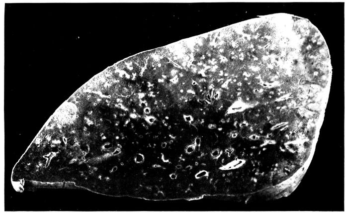
Fig. 5.—Bronchopneumonia with hemorrhagic peribronchiolar consolidation.
In favorable sections it is occasionally possible to follow the bronchiole and alveolar duct, both filled with leucocytes, into an infundibulum. The proximal part of the infundibulum contains polynuclear leucocytes, whereas the distal part and its tributary alveoli are filled with serum and red blood corpuscles.
When the lesion has persisted for a short time there is evidence of beginning migration of polynuclear leucocytes from the blood vessels into the alveoli which are filled with 175blood. The alveolar walls contain numerous polynuclear leucocytes and leucocytes which have entered the intraalveolar blood are numerous in contact with the wall but occur in scant number in the center of the alveolar lumen.
Alveolar epithelium in contact with the blood in the lumen is usually swollen and often uniformly nucleated.
The inflammatory process is evidently transmitted from the bronchioles and to a less degree from the small bronchi to the adjacent alveoli. Polynuclear leucocytes fill the lumen of the bronchiole and the alveoli immediately adjacent; at the periphery of the focus of pneumonia, the alveoli may contain fibrin. In such instances small bronchi (lined by a continuous layer of columnar epithelial cells) may be surrounded by alveoli containing fibrin.
In sections from one part of the lung, the alveoli between the peribronchiolar foci of pneumonia may be uniformly filled with red blood corpuscles, whereas in sections from another part pneumonic foci may be surrounded by a zone of intraalveolar hemorrhage or of hemorrhage and edema outside of which some air-containing tissue occurs. There are transitions between this halo of intraalveolar hemorrhage and edema surrounding each bronchiolar focus and complete hemorrhagic infiltration of all intervening alveoli.
Large mononuclear cells are occasionally fairly numerous within the alveoli containing blood. These cells act as phagocytes ingesting red corpuscles, so that at times they are filled with corpuscles. Disintegration of red corpuscles occurs and brown pigment remains within the cell. It is not uncommon to find numerous mononuclear pigment containing cells which resemble those found with chronic passive congestion of the lungs.
Lungs, the site of hemorrhagic peribronchiolar pneumonia, may undergo chronic changes which will be described elsewhere.
The lesion which has been designated hemorrhagic peribronchiolar pneumonia is that which Pfeiffer regarded as 176the characteristic type of influenzal pneumonia. In the small bronchi containing pus and in lung tissue, Pfeiffer states, influenza bacilli are predominant and present in astonishing number in smear preparations. The demonstration of B. influenzæ by cultures from pneumonic lung is mentioned by him but its association with other microorganisms in such cultures is not discussed.
Microorganisms which we have isolated from the lungs of individuals with hemorrhagic peribronchiolar pneumonia are as follows:
B. influenzæ 1
Pneumococcus 2
S. hemolyticus 10
B. influenzæ, pneumococcus 7
B. influenzæ, S. hemolyticus 3
B. influenzæ, staphylococcus 2
S. hemolyticus, B. coli 3
B. influenzæ, pneumococcus, staphylococcus 2
B. influenzæ, S. hemolyticus, staphylococcus 5
Pneumococcus, S. hemolyticus, staphylococcus 1
No organisms 2
Total 38
With this type of pneumonia B. influenzæ has not been isolated in pure culture; B. influenzæ alone is recorded only once (Autopsy 435), but in this instance the culture has been so obscured by contamination that the occurrence of pneumococci or streptococci cannot be excluded; S. hemolyticus has doubtless been present in this lung, for it has been found in the heart’s blood, in the bronchus, and in the peritoneal exudate of the same individual.
The incidence of pneumococci and hemolytic streptococci in this list does not differ materially from that with peribronchiolar pneumonia unaccompanied by extensive intraalveolar hemorrhage, though hemolytic streptococci are somewhat more frequent with the hemorrhagic lesion. The following table shows the frequency with which pneumococci and hemolytic streptococci have penetrated into the blood:
177
Pneumococcus 11
S. hemolyticus 24
Pneumococcus, S. hemolyticus 1
No organism 12
Total 48
Table XXXVII showing the percentage incidence of pneumococci, hemolytic streptococci, staphylococci and B. influenzæ further emphasizes the similarity between the bacteriology of peribronchiolar pneumonia (Table XXXVI) and the closely related hemorrhagic lesion:
Table XXXVII
PNEUMOCOCCUS HEMOLYTIC STREPTOCOCCUS STAPHYLOCOCCUS B. INFLUENZÆ
Bronchus 44.0% 64.0% 44.0% 72.0%
Lung 31.6% 57.9% 26.8% 52.6%
Blood of heart 25.0% 52.1% 0% 0%
Pneumococci have been found in the lungs (31.6 per cent) and blood (25 per cent), somewhat less frequently than with peribronchiolar pneumonia (43.9 and 40.3 per cent respectively), and hemolytic streptococci have been found in the blood more frequently (52.1 per cent) than with the latter (36.8 per cent) but otherwise the bacteriology of the two lesions corresponds closely. The low incidence of B. influenzæ in the bronchi (72 per cent) with hemorrhagic peribronchiolar pneumonia is perhaps incorrect as the result of the relatively small number of bacteriologic examinations (namely, 25), but the incidence of the same microorganism in the lung has been higher (52.6 per cent) than with nonhemorrhagic peribronchiolar lesion (43.9 per cent).
In some instances infection with hemolytic streptococci has occurred after the onset of pneumonia. The following list compares the results of bacteriologic examination of the sputum made after the onset of pneumonia with that of blood, lungs or bronchus after death:
178
SPUTUM IN BLOOD, LUNGS OR BRONCHUS AT AUTOPSY
Autopsy 237 S. hem. S. hem.
242 Pneum. atyp. II, B. inf. Pneum. atyp. II
247 Pneum. IV, B. inf. Pneum. IV
266 S. hem. S. hem., B. inf.
346 Pneum. IV, B. inf. S. hem., B. inf.
376 (No. S. hem.) S. hem., staph., B. inf.
Instances of secondary infection with hemolytic streptococcus occur in the list, namely, Autopsies 346 and 376.
From the foregoing studies of the bacteriology of peribronchiolar and hemorrhagic peribronchiolar pneumonia the following conclusions may be drawn: (a) B. influenzæ is found in most instances of these lesions in the bronchi and in about half of all instances in the lungs, but does not occur unaccompanied by other microorganisms. (b) In a considerable number of autopsies pneumococcus is the only microorganism that accompanies B. influenzæ; from the lungs it penetrates into the blood from which it is obtained in pure culture. (c) In a considerable number of instances S. hemolyticus accompanies B. influenzæ, and in some of these instances (representing a large proportion of the relatively small number of cases examined during life), examination of the sputum has demonstrated that infection has been secondary to a pneumonia with which no hemolytic streptococci have been found in the sputum.
Lobular Consolidation.—Consolidation of scattered lobules or groups of lobules has occurred in nearly all instances, namely, 71 of 80 autopsies with bronchopneumonia unaccompanied by lobar pneumonia or by suppuration. When death follows shortly after the onset of pneumonia, patches of consolidation have a dull deep red color; blood-tinged fluid escapes from the cut surface which is almost homogeneous or finely granular. The consolidated tissue seen through the pleura, which is raised above the general level, has a bluish red color. Isolated lobules or groups of lobules which have undergone consolidation may be scattered throughout the lungs, but not infrequently there is confluent consolidation of the greater part of lobes, of 179whole lobes or of almost an entire lung. Such lungs are very heavy and may weigh 1,400 or 1,500 grams; bloody serous fluid exudes from the cut surface. The lesion resembles the red hepatization of lobar pneumonia, but confluent patches of pneumonia are usually well defined by lobule boundaries. The tissue is soft and the granulation of lobar pneumonia is absent. In many instances the lobular or confluent areas of consolidation are reddish gray; in some instances consolidated tissue is in places red and elsewhere gray, and in a smaller group of autopsies there is gray consolidation only (Fig. 6). Red lobular consolidation is often seen in those who have died within the first four days following the onset of pneumonia, but is almost equally frequent after from five to ten days; the average duration of pneumonia in these cases was 5.5 days. Combined red and gray consolidation was more frequently found when pneumonia had lasted more than five days, the average duration of pneumonia being 7.3 days. The greater number of instances of gray consolidation were found after seven days of pneumonia, the average duration of the disease being 10.0 days. These figures are cited to show that lobular, like lobar, consolidation passes gradually from a stage of red to gray hepatization, but the change occurs more slowly and is often long delayed.
Lobular pneumonia, which occurred 71 times among 80 cases classified as bronchopneumonia, may be regarded as an almost constant lesion of the disease. It is found not only in association with other lesions of bronchopneumonia, but with lobar pneumonia of influenza as well.
The bacteriology of this lesion shows no deviation from that of the slightly larger group of bronchopneumonia (p. 163). All types of pneumococcus have been found in association with the lesion, Pneumococcus I in 2 instances, Pneumococcus II in 1 instance; atypical Pneumococcus II and Pneumococcus IV have been found much more frequently. Pneumococci have been found in more than a third of these autopsies (42.9 per cent in the lungs, 33.3 per cent in the blood); hemolytic streptococci in less than one-third (28.5 per cent in the lungs, 30.2 per cent in the blood).
180
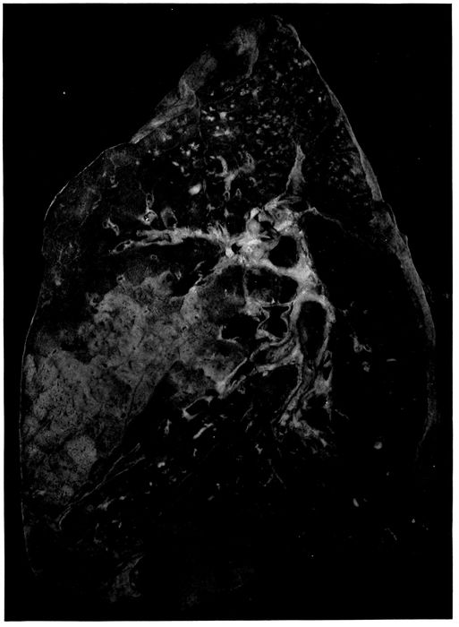
Fig. 6.—Acute bronchopneumonia with confluent gray lobular consolidation in lower part of upper lobe and hemorrhagic peribronchiolar pneumonia in lower lobe; purulent bronchitis.
181The following list shows the bacteriology of a small group of autopsies in which the sputum was examined after onset of pneumonia:
SPUTUM BLOOD, LUNGS OR BRONCHUS AT AUTOPSY
Autopsy 233 Pneum. atyp. II Pneum.
237 S. hem. S. hem.
242 Pneum. atyp. II, B. inf. Pneum. atyp. II
250 Pneum. atyp. II, B. inf. Pneum. atyp. II
253 Pneum. atyp. II Pneum. atyp. II, staph., B. inf.
266 S. hem. S. hem., B. inf.
274 Pneum. IV S. hem.
291 Pneum. IV, B. inf. Staph., B. inf.
312 Pneum. IV, S. hem., B. inf. S. hem., staph., B. inf.
In one instance of streptococcus pneumonia (Autopsy 274) infection with streptococci occurred subsequent to the examination of the sputum made five days before death; pneumococcus was found in the washed sputum.
With lobar pneumonia there was evidence that superimposed infection occurred more frequently during the stage of red than of gray hepatization. With the lobular consolidation of bronchopneumonia this relation has not been found. Among 27 instances of red lobular consolidation, hemolytic streptococcus has occurred 6 times, namely in 22.2 per cent; among 26 instances of red and gray consolidation, 8 times, namely, in 30.7 per cent; among 13 instances of gray consolidation, 5 times, namely, in 38.5 per cent. Infection with hemolytic streptococci is more frequent when the lesion has persisted to the stage of gray hepatization. This difference between lobar and bronchopneumonia is probably dependent in part at least upon the more severe and persistent lesions of the bronchi with bronchopneumonia.
The histology of consolidation which is definitely limited to secondary lobules or groups of lobules varies considerably. When death occurs in the early stage of the lesion, 182consolidated patches are deep red and somewhat edematous, so that bloody serous fluid escapes from the cut surface of the lung and red blood corpuscles are present in the alveoli in great abundance together with polynuclear leucocytes, fibrin and serum in varying quantity. It is not uncommon to find evidence that the lesion has had its origin in the bronchioles and extended from them to other parts of the lobule. Polynuclear leucocytes may be relatively abundant within and immediately about the bronchioles and alveolar ducts, whereas the intervening alveoli and infundibula are filled with red blood corpuscles among which are polynuclear leucocytes and perhaps some fibrin. It may be evident that bronchiolar pneumonia with hemorrhage into intervening alveoli is in process of transformation into a more diffuse leucocytic pneumonia, for polynuclear leucocytes are making their way from the alveolar wall into the blood-filled lumen and, as the result of the presence of blood, remain for a time close to the lining of the alveolus.
When the consolidated lobules have assumed a gray or reddish gray color, polynuclear leucocytes are more abundant and often almost homogeneously pack every alveolus within the boundaries of the lobule. In some instances there is fibrin partially obscured by the presence of leucocytes in great number.
Although fibrin is less abundant with bronchopneumonia than with lobar pneumonia, nevertheless in a considerable proportion of instances it is a very conspicuous element of the inflammatory exudate within the bronchioles, alveolar ducts and alveoli. It is unusual to find the alveolar ducts and alveoli uniformly plugged with fibrin containing leucocytes; there is a variegated distribution of exudate which has little resemblance to that of lobar pneumonia. Occasionally (Autopsies 242 and 247) polynuclear leucocytes fill the bronchioles, alveolar ducts and infundibula, whereas the surrounding tributary alveoli contain fibrin 183and polynuclear leucocytes in moderate number; red blood corpuscles may be present in sufficient number to give a homogeneously red color to the lobular consolidation.
In association with lobular pneumonia, fibrin within the lung tissue undergoes certain changes which outline very sharply the alveolar ducts and the other structures usually ill defined in preparations of the lung. A remarkable appearance is produced by the deposit of hyalin fibrin upon the surface of the alveolar ducts and infundibula. This lesion has been described by LeCount.
Within the alveolar tissue of the lung, spaces are seen lined by a layer of fibrin which stains homogeneously and very brightly with eosin. They are recognized as alveolar ducts by the presence of scattered bundles of smooth muscle in their wall. The layer of hyaline fibrin overlying the surface of the alveolar duct usually forms a continuous lining and covers over the orifices of the alveoli which surround the alveolar duct. These ducts are rendered still more conspicuous by the character of their contents which exhibits a sharp contrast with that of the surrounding alveoli. The alveoli duct occasionally contains a bubble of air, but more frequently it is filled with serum in which red blood corpuscles are sometimes numerous. There is within the lumen scant fibrin and very few cells, among which polynuclear leucocytes are predominant. In the surrounding alveoli on the contrary leucocytes and fibrin are abundant. A similar change is found in the infundibula very clearly defined by their conical form, which is especially well outlined below the pleura or in contact with interlobular septa. The infundibulum is outlined by hyaline fibrin which passes over the orifices of the tributary alveoli and separates the serous contents of the infundibulum from the cellular fibrinous contents of the alveoli about.
The lesion which has been described is often associated with acute bronchitis and bronchiolitis, and the alveoli immediately about the respiratory bronchioles may be filled 184with polynuclear leucocytes. It is very common to find large bubbles of air sharply defined within the purulent contents of the bronchiole. In some lobules the alveolar ducts, infundibula and alveoli intervening between these foci of leucocytic pneumonia are almost uniformly filled with fibrin and polynuclear leucocytes, but in other places the formation of complete layers of hyaline fibrin is in process. Bubbles of air are often seen within the alveolar ducts, and about them is an irregular layer of fibrin formed by the penetration of air into a channel previously filled with a loose network of fibrin containing serum in its meshes. The fibrin compressed against the walls of alveolar duct and infundibulum remains as a compact layer separating these structures from the alveoli which project from their walls. The bubble of air is doubtless later absorbed and replaced by serum, so that many alveolar ducts are filled with serum almost wholly free from cells, whereas alveoli outside the fibrinous membrane contain a network of fibrin with leucocytes in greater or less abundance.
In association with this fibrinous pneumonia, which has been described, hyaline thrombosis of the capillaries is not uncommon. This hyalin material within the capillaries gives reactions of fibrin, and in sections stained by the Gram-Weigert method for demonstration of fibrin, these thrombosed vessels have the appearance of capillaries irregularly injected with a blue material.
The interstitial tissue surrounding consolidated lobules is often edematous; the lymphatics are distended with serum and contain a moderate number of lymphocytes and polynuclear leucocytes.
Among the lungs which have been studied histologically, pneumococcus has been almost invariably associated with the lobular lesions which have just been described, whether hemorrhagic, leucocytic or fibrinous; the histologic changes accompanying infection of the lung with streptococcus will be described later. Pneumococcus has been cultivated from the consolidated lung and is found in section of the lung. B. influenzæ is found in cultures made from the bronchi. Table XXXVIII includes those instances in which the histology of the consolidated lung accords with the description given above.
185
Table XXXVIII
NO. OF AUTOPSY CHARACTER OF LOBULAR CONSOLIDATION PREDOMINANT TYPE OF INFLAMMATORY EXUDATE CULTURE FROM HEART’S BLOOD CULTURE FROM LUNG CULTURE FROM BRONCHUS
242 Red Fibrinous Pneum. atyp. II
244 Red Leucocytic and hemorrhagic Pneum. IV B. inf. Pneum. IV, B. inf.
247 Red and gray Fibrinous Pneum. IV
249 Red and gray Fibrinous Pneum. III
252 Red and gray Fibrinous Pneum. II B. inf. Pneum. II, B. inf., S. vir.
257 Red and gray Leucocytic Pneum. I B. inf., staph.
303 Red Fibrinous Pneum. IV B. inf. Pneum. IV, B. inf., staph.
314 ? Fibrinous Pneum. IV Pneum. IV Pneum. IV, B. inf., staph.
336 Red Fibrinous
395 Red and gray Leucocytic Pneum. atyp. II Pneum. atyp. II
464 Red Leucocytic and hemorrhagic Pneum. I B. inf. Pneum. I, B. inf., staph.
476 Red Leucocytic and hemorrhagic
498 Red and gray Fibrinous S. aur.
506 Red Fibrinous Pneum. IV Pneum. IV S. aur. Pneum. IV, B. inf., S. aur., M. catarrh
186Pneumococcus was found in all but 2 instances, and in one of these (Autopsy 336) the only culture was from the heart’s blood and in the other (Autopsy 498) cultures were unsatisfactory because proper media were not obtainable. Pneumococci of Types I, II, II atypical, III and IV are represented in the list. B. influenzæ has been found in a considerable number of instances in which cultures have been made from the lung and in every instance in which cultures have been made from the bronchi. Staphylococci are often found in the bronchi, but in most instances they do not penetrate into the lung.
Another group of cases of lobular pneumonia are important because in association with necrosis of lung tissue recognized by the microscope hemolytic streptococci have been found in the lungs. In such instances serum is abundant and polynuclear leucocytes are relatively scant though their distribution varies considerably; in some places leucocytes are fairly abundant though elsewhere almost absent, but this distribution bears no obvious relation to the bronchioles. In some instances (Autopsies 274 and 487) red blood corpuscles are numerous but in others (Autopsies 275 and 312) they are inconspicuous. The characteristic feature of the lesion is the occurrence of patches of necrosis within which the nuclei both of exudate and of alveolar walls have partially or completely disappeared. In these areas of necrosis short chains of streptococci are found in immense number whereas in living tissue they are present in moderate number. There has been a relatively inactive inflammatory reaction, great proliferation of streptococci and necrosis of invaded tissue. The bacteriology of instances of lobular pneumonia with necrosis is shown in Table XXXIX.
187
Table XXXIX
NO. OF AUTOPSY CHARACTER OF LOBULAR CONSOLIDATION PREDOMINANT TYPE OF INFLAMMATORY EXUDATE CULTURE FROM HEART’S BLOOD CULTURE FROM LUNG CULTURE FROM BRONCHUS
274 Red Leucocytic and hemorrhagic S. hem. S. hem. S. hem., staph.
275 Red and gray Leucocytic Pneum. IV S. hem. S. hem., B. inf., staph. S. hem., B. inf., staph.
312 Red and gray Leucocytic S. hem. S. hem., B. inf. S. hem., B. inf., staph.
478 Red Leucocytic and hemorrhagic S. hem. S. hem.
188Lobular pneumonia, in some of these instances at least, has been caused primarily by pneumococci; necrosis has been the result of secondary invasion by streptococci. In Autopsy 275 Pneumococcus IV has been obtained from the blood, but in the presence of streptococci has presumably disappeared from the lung and bronchus. In the case represented by Autopsy 274, Pneumococcus IV has been found in the sputum five days before death at the onset of pneumonia, but at this time no hemolytic streptococci have been found. In the case represented by Autopsy 312, Pneumococcus IV, B. influenzæ and a few colonies of hemolytic streptococci have been obtained from the sputum two days after recognition of pneumonia and five days before death.
The hemorrhagic and edematous consolidation of the early pulmonary lesions of influenzal pneumonia is their most distinctive feature. Red confluent lobular pneumonia is frequently found in those who have died within the first week following the onset of influenza. The lungs are voluminous and heavy and may weigh as much as 1,500 grams; the pleura which overlies the consolidated area is blue or plum colored and usually shows scant if any evidence of pleurisy. Scattered patches of consolidation are accurately limited to lobules, but in addition there are large areas often involving the greater part of the lobes and not infrequently situated in the lowermost part of the lower lobes. This confluent consolidation may be obviously limited by lobule boundaries. The consolidated tissue is deep red and laxly consolidated; red serous fluid escapes from the cut surface. The lesion not infrequently occurs in association with hemorrhagic peribronchiolar pneumonia.
The histology of this confluent lesion has been studied in Autopsies 242, 244, 303, 336, 464, 474 and 506. The histology varies, because, in some instances, leucocytes, in other instances, fibrin, is abundant, but the presence of red blood corpuscles in large number within the alveoli gives a red color to the consolidated tissue. In these cases pneumococci, 189associated in the lungs or in the bronchi with B. influenzæ, have been the cause of pneumonia. In two autopsies studied histologically (Autopsies 274 and 478) there was red lobular and confluent pneumonia and the blood and lungs contain hemolytic streptococci demonstrated by cultures; microscopic examination showed the presence of a widespread necrosis of the lung tissue.
In the group of autopsies in Table XL there was red confluent lobular pneumonia. These autopsies are separated from those just cited because there was no histologic examination of the tissue.
Table XL
NO. OF AUTOPSY BACTERIOLOGY OF HEART’S BLOOD BACTERIOLOGY OF LUNGS BACTERIOLOGY OF BRONCHUS
289 Pneum. IV Pneum. IV Pneum. IV, B. inf., staph.
297 Pneum. IV, B. inf. Pneum. IV, B. inf., S. hem. (a few)
306
339 Pneum. IV
364 S. hem.
418 Pneum. atyp. II Pneum. atyp. II, B. inf., S. vir.
424 Pneum. IV.
This group of autopsies confirms the view that the red confluent lobular pneumonia is caused by pneumococci in association with B. influenzæ. Hemolytic streptococci may invade secondarily. In Autopsy 297 a few hemolytic streptococci were found in the bronchus but apparently had not entered the lungs. In the absence of histologic examination it is not possible to determine if the invasion of hemolytic streptococcus (in Autopsy 364) has caused necrosis of the pneumonic tissue.
190
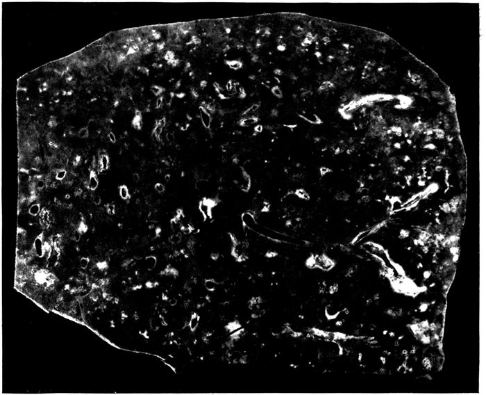
Fig. 7.—Bronchopneumonia with purulent bronchitis and peribronchial hemorrhage.
Peribronchial Hemorrhage and Pneumonia
In a considerable number of instances, namely, in 19 autopsies, hemorrhage about the small bronchi has been recognizable upon gross examination of the lung. A conspicuous zone of hemorrhage 2 or 3 mm. in thickness surrounds small (with no cartilage) often dilated bronchi and on longitudinal section may be tracted for a considerable distance along the bronchus (Fig. 7). In many additional instances peribronchial hemorrhage has been found by microscopic examination. In some instances the peribronchial zone of hemorrhage is firmer than the tissue elsewhere and it is occasionally difficult to determine whether the lesion is hemorrhage or pneumonia. In 7 instances frank red consolidation of peribronchial tissue was recognized at autopsy; this lesion will be considered later under peribronchial pneumonia. Hemorrhage about bronchi, like other evidences of severe injury to bronchi following influenza, is more frequently found in the lowermost parts of the lungs than elsewhere. It is invariably associated with severe bronchitis; the bronchi have contained purulent 191fluid in 15 of 19 instances of peribronchial hemorrhage and in 10 instances the lesion has been associated with dilatation of the bronchi.
Microscopic examination furnishes further evidence of the severity of the bronchial changes which have brought about hemorrhage into the surrounding alveoli. The lumen of the bronchus contains blood and leucocytes; the epithelium is sometimes raised in places from the underlying basement membrane by blood; blood vessels of the bronchial wall are engorged, and there is hemorrhage into the tissue of the bronchus. More frequently the bronchial epithelium is completely lost and the denuded surface is often covered by a layer of fibrin intimately adherent to the inflamed mucosa. Transitions between simple hemorrhage and pneumonia are found, polynuclear leucocytes being mingled with red blood corpuscles. In several instances the alveoli in immediate contact with the bronchial wall have contained fibrin, whereas those in the surrounding zone have contained blood.
Bacteria found in the bronchi in 10 instances of peribronchial hemorrhage have been as follows:
Staphylococci 1
B. influenzæ, pneumococci 1
B. influenzæ, S. hemolyticus 2
B. influenzæ, pneumococci, staphylococci 1
B. influenzæ, S. hemolyticus, staphylococci 4
No organism found 1
The high incidence of B. influenzæ and the frequent association of B. influenzæ and hemolytic streptococci are noteworthy. The instance in which no organisms were found is probably due to a defect in media and should perhaps be excluded from the list.
The percentage incidence of pneumococci, hemolytic streptococci, staphylococci and B. influenzæ in the bronchus, lungs and blood of the heart is an index of the facility with which these microorganisms penetrate internal organs when the bronchi are the site of this hemorrhagic lesion.
192
Table XLI
NO. OF CULTURES PNEUMOCOCCI HEMOLYTIC STREPTOCOCCI STAPHYLOCOCCI B. INFLUENZÆ
NO. POSITIVE PER CENT POSITIVE NO. POSITIVE PER CENT POSITIVE NO. POSITIVE PER CENT POSITIVE NO. POSITIVE PER CENT POSITIVE
Bronchus 10 2 20.0 6 60.0 6 60.0 8 80.0
Lung 13 4 30.8 7 53.8 3 23.1 5 38.5
Blood 17 4 23.5 9 52.9
When these figures are compared with those for all forms of bronchitis no very noteworthy differences are found; the incidence of pneumococci here is less and that of hemolytic streptococci greater. In association with the severe changes present in the bronchi, hemolytic streptococci which enter the lungs almost invariably find their way into the blood.
In 6 instances there has been frank pneumonic consolidation limited to a zone encircling small and medium-sized bronchi which have often been obviously dilated. On cross section these patches of pneumonia are circular, from 1 to 2 cm. in diameter and each contains a bronchus at its center. When the bronchus is cut longitudinally it is evident that pneumonic consolidation forms a cylindrical sheath about the tube. The consolidation varies in color from red to grayish red. In one instance (Autopsy 253) the consolidated tissue has formed a gray zone in contact with the bronchus and is red in a peripheral zone; microscopic examination shows that the alveoli about the bronchus contain fibrin, whereas those at a greater distance contain red blood corpuscles. In this instance, the associated pneumonia in another part of the lung has been somewhat anomalous and has had characters both of lobar and bronchopneumonia, for scattered in the left lung there have been patches of firm consolidation not more than 2 cm. across. The smaller of these patches are deep red, but the larger are coarsely granular and gray in the center. The patchy 193character of the lesion has suggested bronchopneumonia, but the coarse granulation on section and the presence of fibrinous plugs within the small bronchi have presented a close resemblance to lobar pneumonia. This autopsy is one of the few instances in which Pneumococcus II has been found, Pneumococcus II being present in blood and lungs, B. influenzæ, in lungs and bronchi. In 2 additional instances (Autopsies 374 and 392) peribronchial pneumonia, recognizable at autopsy, has been associated with consolidation having the characters of lobar pneumonia. In one instance, Autopsy 374, the right lung has contained two patches of firm, mottled red and pinkish red coarsely granular consolidation each about 6 cm. across, one situated in the upper lobe and the other in the lower lobe. Elsewhere in the lung, in definite relation to dilated bronchi, occur patches of firm, red, coarsely granular consolidation from 1 to 1.5 cm. in diameter when cut transversely. The bronchus in the center has contained purulent fluid. In the opposite lung similar consolidation has been limited to zones about dilated bronchi which contain purulent fluid. Pneumococcus IV has been obtained from the blood of the heart.
The peribronchial pneumonia which has been described occurs in association with evidence of profound injury to the bronchial wall. In 5 of 6 instances purulent bronchitis has been found at autopsy; in half of these instances bronchiectasis has been noted. The epithelium of the bronchus has been found separated from the underlying tissue by serous exudate, blood and leucocytes; epithelial cells undergo necrosis and disappear, the denuded surface being covered by fibrin. Necrosis extends a varying depth into the wall of the bronchus; blood vessels are engorged, and there is in some instances hemorrhage throughout the wall of the bronchus.
The character of the exudate in the alveoli surrounding the bronchus differs considerably in different instances. In some instances (Autopsies 374 and 392) red blood corpuscles 194are predominant in the alveoli in contact with the bronchial wall, whereas in a peripheral zone polynuclear leucocytes are more abundant. In other instances (Autopsies 253 and 402) alveoli next the bronchial wall contain abundant fibrin and these are surrounded by a zone in which the alveoli are filled with blood.
Peribronchial pneumonia is the result of the direct extension of the inflammatory process through the wall of the bronchus; it occurs when the epithelium of the bronchus is destroyed and the underlying tissues are injured, but may be present in a wide encircling zone even when the lesion has not penetrated the bronchial wall. The distribution of the pneumonia demonstrates very clearly that the inflammatory process does not reach the affected peribronchial alveoli by way of the bronchioles tributary to the bronchus.
The bacteriology of these instances of peribronchial pneumonia is noteworthy. (Table XLII.)
Table XLII
AUTOPSY BLOOD LUNG BRONCHUS
253 Pneum. II Pneum. II, B. inf. Staph., B. inf.
374 Pneum. IV
387 Pneum. II, S. hem. Pneum. II, staph., B. inf. Pneum. II, S. hem., staph., B. inf.
392 Pneum. II
402 Pneum. IV, S. hem.
424 ? Pneum. IV
Pneumococcus has been found in every instance either in the lungs or blood. Pneumococcus II, which has been uncommon with the pneumonia following influenza at Camp Pike and has occurred only ten times in more than 200 autopsies, has been present in one-half of these cases. The constant association of the lesion with pneumococcus is particularly significant when a comparison is made between the incidence of pneumococcus with peribronchial hemorrhage, on the one hand, and peribronchial pneumonia on the other; pneumococcus has been present in less than a third 195of the instances of hemorrhage but in all instances of pneumonia.
In addition to the instances in which gross peribronchial consolidation has been noted at autopsy, microscopic examination has demonstrated the presence of fibrinous pneumonia surrounding bronchi in a considerable number of autopsies. In a zone encircling small bronchi (with no cartilage) alveoli are filled by plugs of dense fibrin (Fig. 20) containing in variable number polynuclear leucocytes and mononuclear cells. The width of the zone is often equal or greater than the diameter of the bronchus. Alveoli outside the zone of fibrinous inflammation may contain red blood corpuscles or serum, and desquamated epithelial cells are often abundant.
Of 21 instances of peribronchial fibrinous pneumonia 20 were associated with purulent bronchitis. Further evidence of the relation of the lesion to profound injury to the bronchi is its association with bronchiectasis in 17 instances.
Peribronchial fibrinous pneumonia, like other lesions encircling the small bronchi, bears a direct relation to the severity of microscopic changes in the bronchus. The epithelium of the bronchus is either partially or completely lost. Occasionally epithelium is raised by hemorrhage or leucocytes from the underlying tissue but more frequently it is wholly lost and the surface is covered by a layer of fibrin. In the early stages of the lesion, polynuclear leucocytes may be numerous throughout the bronchial wall, indicating that the inflammatory irritant within the lumen is affecting the entire wall and extending its influence to the surrounding pulmonary tissue. Later lymphoid and plasma cells are more abundant than polynuclear leucocytes. Coagulative necrosis and disintegration of the bronchial wall, proceeding from the inner surface outward, may extend more or less deeply, and fibrinous inflammation of adjacent alveoli is often more extensive about that segment 196of the bronchus which shows the greatest change. In some instances segments of the bronchial wall or even the entire wall has disappeared, so that alveoli containing fibrin form part of the wall of the cavity thus formed. When bronchiectasis has occurred, there are often fissures from the lumen through the entire wall extending into the surrounding lung tissue: here fibrinous pneumonia is particularly conspicuous, occurring in a zone about the edges of the defect. This deposition of fibrin within the alveoli adjacent to the injury doubtless has a part in limiting the distribution of bacterial infection. Nevertheless breaks in the continuity of the bronchial wall are not essential to the production of the lesion and the irritant, which is responsible for the lesion, may penetrate through the bronchial wall to surrounding alveoli and from alveoli to other alveoli immediately adjacent.
With this peribronchial pneumonia the smallest bronchi are distended with pus and their walls are infiltrated with polynuclear leucocytes, lymphoid and plasma cells. In a broad zone encircling the bronchus the alveoli are filled with plugs of fibrin. Bronchioles are similarly distended with polynuclear leucocytes; the alveoli which occur upon the wall of the bronchiole are often limited to one side of the wall and are filled with fibrin. This fibrin occasionally projects into the lumen of the bronchiole and forms a continuous layer in contact with the wall on the same side. The alveolar duct and infundibulum are distended with polynuclear leucocytes. The alveoli upon the wall of the alveolar duct and upon the proximal part of the infundibulum are filled with fibrin. The bronchus, bronchiole, alveolar duct and part of the infundibulum are thus surrounded by a continuous zone of alveoli containing fibrin. The alveoli about the distal part of the infundibulum may be filled with polynuclear leucocytes. Lung tissue between adjacent zones of fibrinous pneumonia may contain serum and desquamated epithelial cells.
197Organization of peribronchial fibrin was found in 10 of the 22 autopsies in which peribronchial fibrinous pneumonia had been found. Fibroblasts have invaded the fibrin and newly formed capillaries have penetrated into it. In some instances the interalveolar septa are thickened and infiltrated with lymphoid and plasma cells, and in 7 instances there was chronic pneumonia with thickening and mononuclear infiltration of the interstitial tissue about the bronchi and blood vessels, and elsewhere. The duration of the fatal illness in 12 instances with no organization was usually from ten days to two weeks, though in 3 instances there was no organization although the respiratory disease had lasted from seventeen to nineteen days (average duration with no organization, 13.5 days). The duration of illness in 10 instances with organization of fibrin was slightly less than three weeks (average 18.9 days). These figures do not accurately represent the duration of pneumonia which usually develops after a period of several days following onset of influenza.
This group of instances of peribronchial fibrinous pneumonia has offered an opportunity to study the bacteriology of pneumonia with organization and to determine if it presents any unusual characters. The bacteriology of autopsies with peribronchial fibrinous pneumonia with no organization is shown in Table XLIII:
Table XLIII
AUTOPSY BLOOD LUNG BRONCHUS
289 Pneum. IV Pneum. IV Pneum. IV, B. inf., staph.
372
376 S. hem. S. hem. S. hem., B. inf., S. aur.
409 0
410 S. hem., B. inf. S. aur.
412 Pneum. II Pneum. II, B. inf.
420 S. hem. S. hem., B. inf. S. aur.
423 S. hem. S. hem., B. inf.
440 0 B. inf., S. aur. B. inf., S. aur.
448 0 0 0
482 0 B. inf., Pneum. IV B. inf., Pneum. IV, S. hem.
489 0 Pneum. IV, B. inf. Pneum. IV, B. inf.
198The bacteriology of instances of peribronchial fibrinous pneumonia with organization of the intraalveolar fibrin is shown in Table XLIV:
Table XLIV
AUTOPSY BLOOD LUNG BRONCHUS
283 Pneum. IV Staph., B. inf. B. inf., Pneum. IV, staph.
291 0 0 B. inf., staph.
398 0
419 0 Pneum. II, B. inf. Pneum. II, B. inf.
421 S. hem. Pneum. IV, S. hem.
422 0 Pneum. II atyp., B. inf.
425 S. hem. S. hem., B. inf., S. alb.
433 0 S. hem., B. inf., S. aur.
460 S. hem. S. hem., B. inf. S. hem., B. inf., staph.
463 0 B. inf., staph. B. inf., staph., Pneum. IV
B. influenzæ has been present in the bronchi in every instance save one in which cultures have been made, and it is probable that in this exceptional instance cultures have remained sterile because the media employed have been defective. The incidence of B. influenzæ in the lung has been unusually high both with and without organization (66.7 per cent with no organization; 77.8 per cent with organization). Streptococci and staphylococci have been found in a considerable proportion of all instances of peribronchial fibrinous pneumonia, but there has been no notable preponderance of these microorganisms when organization has occurred. Organization has been present in instances in which pneumonia is referable to pneumococcus associated with B. influenzæ and unaccompanied by either streptococci or staphylococci (Autopsies 419 and 422). Wadsworth[81] found no organization after inoculation of the lungs of dogs with pneumococcus or with staphylococcus alone, but produced organization when he inoculated animals with both microorganisms.
Injury to bronchi produced in part at least by B. influenzæ exposes the bronchi and lung tissue to repeated infection with a variety of microorganisms; absorption of 199fibrin and regeneration of alveolar epithelium are prevented, resolution fails to occur and organization of fibrin follows.
Suppurative Pneumonia With Necrosis and Abscess Formation
Three varieties of suppurative pneumonia have occurred in association with influenza.
A. Necrosis and suppuration with formation of one or several abscesses usually below the pleura and almost invariably caused by hemolytic streptococci.
B. Interstitial suppurative pneumonia caused by hemolytic streptococcus.
C. Multiple abscesses in clusters caused by staphylococci.
Suppurative pneumonia with necrosis and abscess formation will be discussed in this section. Pulmonary abscesses which occurred in 43 autopsies may be included in this group; in 4 of these autopsies abscess and interstitial suppurative pneumonia occurred in the same individual. These abscesses were much more frequently situated in the lower than in the upper lobes and more often in the right than in the left lung. In most instances there was one or several abscesses situated below the pleura of one lobe; occasionally abscesses occurred in two lobes of the same lung or in both lungs. The distribution was as follows: Abscess in only one lung occurred in right upper lobe in 6 autopsies; middle lobe, 3; lower lobe, 15; left upper lobe, 2; lower lobe, 16. Abscesses occurred in both right and left lower lobes, twice. The usual situation was at the lower and posterior part of the lower lobe at or near the basal edge, less frequently below the posterior border or upon the basal surface of the lobe. These abscesses in almost every instance were found immediately below the pleural surface, so that they appeared upon the pleura as opaque yellow spots usually surrounded by narrow zones of hemorrhage. In one instance (Autopsy 376) the 200abscess cavity was separated from the pleural cavity by remains of the pleura which was as thin as tissue paper and in other instances perforation had occurred (Fig. 9). In Autopsy 480 the abscess cavity which had perforated the pleura was in free communication with a bronchus of medium size.
In most instances of suppurative pneumonia there have been associated lesions of bronchopneumonia which have been peribronchiolar, hemorrhagic or lobular and have exhibited no unusual characters. The abscess or abscesses are situated within an area of pneumonic consolidation which is not limited by lobule boundaries and has not the characters of bronchopneumonic consolidation. In some instances this consolidation is limited to a zone immediately about the abscess, but often it involves the greater part of a lobe. The tissue is laxly consolidated and flabby; on section it has a dull, conspicuously cloudy appearance and is grayish red, pinkish gray or gray; it is homogeneous or very finely granular. Turbid gray fluid, which sometimes resembles thin pus, oozes from the cut surface.
Widespread necrosis of tissue is not infrequently a conspicuous feature of this pyogenic pneumonia (Fig. 8). Upon a cloudy gray background of consolidation are numerous opaque yellowish gray or yellow patches, occasionally 2 or 3 cm. across, giving a mottled character to the cut surface. Upon the pleura these necrotic patches appear as dull opaque yellow spots. They may be surrounded by a zone of hemorrhage. The opaque material is at first firm but may undergo softening, becoming semisolid and finally purulent. Necrotic patches may be scattered throughout a lobe, but fully formed abscesses are with few exceptions immediately below the pleura (Fig. 9).
201
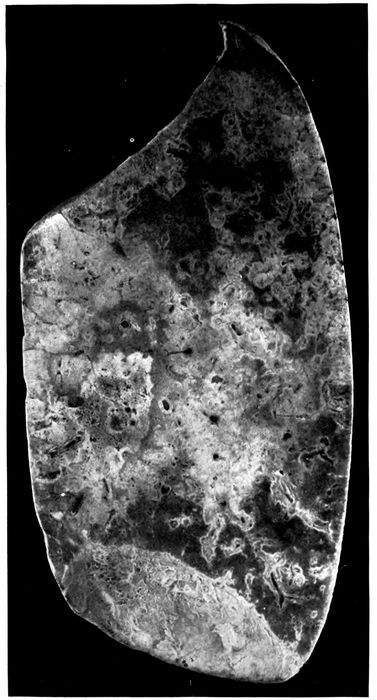
Fig. 8.—Streptococcus pneumonia with massive necrosis. Autopsy 354.
202
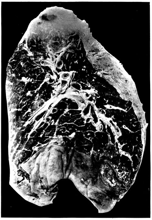
Fig. 9.—Abscess below pleura with perforation caused by hemolytic streptococci. Healing suppurative interstitial pneumonia indicated by yellowish gray lines marking interlobular septa at base of lower lobe. Autopsy 474; right lung. (See left lung, Fig. 10.)
203The duration of illness in cases of pneumonia with abscess varied from a week or less (11 instances) to more than four weeks. The duration of the greater number of cases (17 instances) was between one and two weeks. In one instance onset occurred with symptoms of influenza, pneumonia was recognized two days later, and death occurred only four days after the onset of illness. When the duration of the illness was less than a week the symptoms of onset were in some instances those of pneumonia.
Table XLV shows the incidence of pneumococcus, S. hemolyticus, staphylococcus and B. influenzæ in instances of suppurative pneumonia with abscess formation, 4 instances of abscess with interstitial suppurative pneumonia being excluded:
Table XLV
NO. OF CULTURES PNEUMOCOCCI HEMOLYTIC STREPTOCOCCI STAPHYLOCOCCI B. INFLUENZÆ
NO. POSITIVE PER CENT POSITIVE NO. POSITIVE PER CENT POSITIVE NO. POSITIVE PER CENT POSITIVE NO. POSITIVE PER CENT POSITIVE
Bronchus 24 5 20.8 22 91.6 12 50.0 18 75.0
Lung 36 9 25.0 30 83.3 14 35.6 8 22.2
Blood 37 6 16.2 31 83.8
In over 80 per cent of instances of pulmonary abscess hemolytic streptococcus has been found in blood, lungs and bronchus and, when cultures have been made, in the inflamed pleural cavity as well. Streptococci have been found in immense number in sections from the necrotic lung tissue and the abscesses which have been formed. It is evident that hemolytic streptococci have caused suppurative pneumonia and death, being found in the blood of the heart just as frequently as in the lungs (83 per cent). The relative unimportance of pneumococci is indicated by their low incidence in the blood (16.2 per cent) when compared with that of lobar pneumonia (65.5 per cent) or of bronchopneumonia (31.4 per cent). B. influenzæ has been found in three-fourths of these autopsies in the bronchus, but its incidence in the lungs has been much smaller.
In 3 instances of suppurative pneumonia with abscess formation no hemolytic streptococci were found; they are as follows:
204Autopsy 380.—Bronchopneumonia with gray and red lobular consolidation in right upper and lower lobes; peribronchiolar nodules of consolidation in left lower lobe; abscess, 1.5 cm. across, below the pleura of the posterior border of the left lower lobe near its base; fibrinopurulent pleurisy (300 c.c.) on right side; serous pleurisy (200 c.c.) on left. Pneumococcus III was found in cultures from the blood of the heart from the right lung and with B. influenzæ from the right pleural cavity. No culture was made from the left lung which contained the abscess. In sections of the abscess gram-positive streptococci in chains of 4 to 8 cocci were numerous.
Autopsy 406.—Acute lobar pneumonia with red hepatization of greater part of right lung; patch of consolidation in lower lobe of left lung containing an abscess cavity 2.5 x 1.5 cm.; localized seropurulent pleurisy (375 c.c.) on left side. Pneumococcus IV was obtained from the blood of the heart; a culture from the lung was contaminated. Tissue from the abscess was not saved for histologic examination.
Autopsy 416.—Suppurative pneumonia with necrosis and abscess formation in right lower lobe; fibrinous pleurisy on right side. Pneumococcus IV was obtained from the blood, right lung and right main bronchus. No streptococci were found in sections from the abscess in the right lung.
The foregoing observations demonstrate that suppurative pneumonia with abscess formation following influenza is with few exceptions caused by S. hemolyticus.
The autopsies (Table XLVI) in which pneumococci have been found in association with hemolytic streptococci in the blood or lungs indicate that pneumococci have had a part in the production of fatal pneumonia.
Table XLVI
AUTOPSY CULTURE FROM BLOOD CULTURE FROM LUNGS CULTURE FROM BRONCHUS
258 S. hem. S. hem., Pneum. IV B. inf.
282 S. hem., Pneum. II S. hem., Pneum. II S. hem., B. inf. Pneum. II, staph.
345 S. hem., Pneum. II, staph.
378 Pneum. atyp. II S. hem., Pneum. atyp. II S. hem., B. inf., Pneum. atyp. II
381 S. hem. S. hem., Pneum. II Pneum. IV, staph.
383 Pneum. III S. hem., Pneum. III B. inf.
387 S. hem. Pneum. II, staph., B. inf. S. hem., pneum., staph., B. inf.
These autopsies, notably those in which pneumococci have been found in the blood, suggest that infection with 205pneumococci has preceded suppurative pneumonia caused by hemolytic streptococci. In a small number of instances the sputum was examined in life after onset of pneumonia.
Table XLVII
AUTOPSY SPUTUM CULTURES FROM BLOOD, LUNGS AND BRONCHUS
282 Pneum. IV. B. inf. S. hem., Pneum. II, staph., B. inf.
288 S. hem., B. inf. S. hem., B. inf.
376 (No S. hem., Oct. 8) S. hem., staph., B. inf. (Oct. 11)
In 2 of these 3 cases infection with hemolytic streptococcus occurred subsequent to the onset of pneumonia.
Several observations help to explain the occurrence of abscess in association with the pneumonia of influenza. The fissures which will be described in association with bronchiectasis represent traumatic ruptures of the bronchial wall consequent upon weakening by necrosis and over distention. They expose the injured bronchial wall and the alveolar tissue adjacent to it to infection by the microorganisms contained within the lumen of the inflamed bronchus. Occasionally a favorable microscopic section demonstrates the relation of pulmonary necrosis and consequent suppuration to injuries of the bronchial wall. Peribronchial fibrinous pneumonia occurs about the bronchi of which the epithelial lining has been destroyed, and when a fissure penetrates the bronchial wall fibrinous pneumonia is almost invariably found in a zone about the tear; it doubtless tends to limit the extension of the process. Occasionally, wide areas of necrosis occur within consolidated tissue near the site of the fissure (Autopsy 312 with S. hemolyticus and B. influenzæ, p. 254). Accumulation of polynuclear leucocytes between living and dead tissue may form a line of demarcation (Autopsy 387); finally, fairly large, irregularly formed, abscess cavities are found.
Necrosis and beginning suppuration in contact with the lumen of the bronchus will be described in association with bronchiectasis (Autopsies 312, Fig. 24, and 423, p. 256). In 206the following autopsies upon individuals who have died with pulmonary abscesses, favorable microscopic sections have demonstrated abscess formation in contact with lesions which have penetrated the walls of small bronchi. They help to explain the pathogenesis of abscess in association with influenza.
Autopsy 376.—H. M., white, aged twenty-four, a fireman, resident of Oklahoma, had been in military service one month. Onset of illness occurred October 1, ten days before his death; he was admitted to the base hospital on the fourth day of his illness with the diagnosis of bronchopneumonia.
Anatomic Diagnosis.—Acute bronchopneumonia with patches of lobular and confluent lobular consolidation in both lungs and hemorrhagic peribronchiolar consolidation in right upper lobe; abscess in right upper lobe below pleura; fibrinopurulent pleurisy on right side; purulent bronchitis; bronchiectasis at base of left lobe.
An irregular abscess, 2 x 1 cm., filled with creamy purulent fluid is separated from the interlobular surface of the right upper lobe by a thin membrane representing the pleura. The right pleural cavity contains 200 c.c. of turbid yellow fluid in which is soft fibrin. The bronchi contain purulent fluid in great abundance. The bronchi at the base of the left lower lobe are widely dilated, so that many small bronchi with no cartilage in their wall measure from 3 to 5 mm. in diameter.
Cultures show the presence of hemolytic streptococci in the blood of the heart and in three plates from the lung; B. influenzæ and S. aureus were found in the left bronchus.
The bronchi have wholly or partially lost their epithelium and there is deep erosion of the walls. Cavities containing polynuclear leucocytes occur within the alveolar tissue; in some instances pus containing cavities are surrounded by alveolar tissue, but in other places it is evident, that they have had their origin in bronchi. In a short segment of the circumference the wall of the preexisting bronchus is preserved and consists of squamous epithelium, vascular connective tissue and smooth muscle. The remainder of the bronchus has disappeared and a cavity is produced. The very irregular wall of the cavity is formed by partially destroyed alveoli filled with fibrin and leucocytes.
Autopsy 387.—C. M., white, aged twenty-one, laborer, resident of Mississippi, had been in military service twenty-one days. Illness began on September 22, nineteen days before death, and the patient was admitted to the hospital on the same day with a diagnosis of bronchitis; a diagnosis of bronchopneumonia was made on October 2, nine days before death. The leucocytes on October 3 numbered 8000 (small mononuclear, 36 per cent; large mononuclear, 5 per cent; polynuclear, 59 per cent).
207Anatomic Diagnosis.—Acute bronchopneumonia with consolidation in right upper lobe and hemorrhagic peribronchiolar consolidation in left lower lobe; abscess below pleura in left lower lobe; purulent pleurisy on both sides; edema of mediastinum; purulent bronchitis; bronchiectasis.
There is advanced bronchiectasis, and bronchi with no visible cartilage are dilated to from 4 to 8 mm. in diameter; they contain purulent fluid which wells up from the cut surface. About dilated bronchi there is in places dull red or grayish red consolidation forming an encircling zone. Situated below the pleural surface within an area of consolidation at the posterior border of the left lower lobe there is a spot 3 cm. across where the tissue is yellow and has in places undergone purulent softening. Several smaller abscesses occur nearby.
Cultures from the blood of the heart and from the edematous mediastinum contain hemolytic streptococci. From the abscess are grown S. albus, Pneumococcus II and B. influenzæ. The purulent contents of a small bronchus contains S. hemolyticus, B. influenzæ, S. aureus and a few pneumococci.
Microscopic examination shows that the epithelium of dilated bronchi has disappeared and the denuded surface is covered by fibrin and polynuclear leucocytes; fissures extend from the lumen through the bronchial wall into the surrounding alveolar tissue. A zone of fibrinous pneumonia surrounds these bronchi and fissures in the bronchial wall penetrate into this zone. One dilated bronchus 2.4 mm. in diameter with no cartilage in its wall has vascular connective tissue covered by epithelium on one side, whereas the remainder of the circumference is formed by exposed alveoli filled with fibrin, the bronchial wall having disappeared. A section through a part of the abscess which has been mentioned shows a very irregularly formed cavity approximately 1 x 0.7 cm. Remains of bronchial wall, consisting of very vascular tissue covered by flat epithelium in several layers, indicate the origin of the cavity. Between these remnants of bronchi deep pockets extend into the pulmonary tissue which in the margin of the cavity is the site of fibrinous pneumonia. In one place, in contact with the cavity, a wide area of consolidated tissue has undergone necrosis and both alveolar walls and their contents have lost their nuclei. Leucocytes which are accumulating at the margin of the necrotic patch form a line of demarcation between living and dead tissue.
Abscess may be the result of the profound changes which occur in the bronchi as the result of influenza. Necrosis caused by bacteria within the bronchi weakens and in places destroys the wall. Bacteria penetrate into the surrounding tissue and hemolytic streptococci (or staphylococci) may produce localized abscesses. These abscesses are usually situated near the pleural surface of the lung, because destructive changes causing rupture of the bronchial 208wall occur more frequently in the smaller peripheral bronchi than in the larger bronchi containing cartilage. Abscesses occur more frequently at the bases of the lungs, because the most severe changes in the bronchi occur in the dependent part. (See “Bronchiectasis,” p. 240.)
Healing of Abscess.—The following autopsy is of interest in relation to the treatment of pulmonary abscess and associated empyema.
Autopsy 467.—P. C., white, aged twenty-five, a farmer from Missouri, had been in military service three months. Illness began September 27, thirty days before death, and the patient was admitted the day following onset with headache, backache and cough. Pneumonia with consolidation in the right lower lobe was recognized on the sixth day of illness. On the ninth day 500 c.c. of fluid were withdrawn from the right pleural cavity; there were cyanosis and dyspnea. On the eleventh day 700 c.c. of fluid were withdrawn. On the twelfth day thoracotomy was performed and 100 c.c. of greenish fluid were removed. The patient’s condition improved for a time, but on the twenty-sixth day 1,000 c.c. of straw colored fluid were aspirated from the left pleural cavity and on the twenty-eighth day the same amount of seropurulent fluid was withdrawn.
Anatomic Diagnosis.—Healing abscess of right lower lobe communicating with the pleural cavity; acute purulent pleurisy with closed thoracotomy wound on the right side; purulent pleurisy on the left side; acute bronchopneumonia with lobular consolidation in the left lung; purulent bronchitis; bronchiectasis with formation of spherical bronchiectatic cavities; acute splenic tumor.
At the base of the right chest is a closed thoracotomy wound 2 cm. in length; the right pleural cavity contains 200 c.c. of thick creamy pus and the cavity is lined by a thick tough membrane. The left pleural cavity contains 800 c.c. of white purulent fluid thinner than that on the right side. The right lung is compressed into the posterior and inner part of the chest. The upper lobe is pink and air containing; the posterior and lower part of the lower lobe is red and atelectatic, and fibrous septa are more conspicuous than elsewhere. The pleura of the external surface near the basal edge, in an area 2 cm. across, is depressed and yellowish gray in color. In the center of this area is a small opening communicating with a pocket 0.5 cm. across within the substance of the lung.
In the lower lobe beneath the interlobular surface are two spherical bronchiectatic cavities, each about 1.5 cm. across, with smooth lining in continuity with two branches of the same bronchus of medium size.
Bacteriologic examination showed the presence of S. hemolyticus in the blood of the heart. No growth was obtained from the left lung; the left pleural cavity contained hemolytic streptococci and S. aureus, the latter in 209small number. S. hemolyticus and B. influenzæ were grown from the left main bronchus.
A microscopic section through the abscess and its communication with the pleura shows that its cavity contains polynuclear leucocytes and the wall is formed by granulation tissue covered by fibrin. Some alveoli outside the abscess contain compact balls of fibrin containing a few fibroblasts; this fibrin stains deeply with hematoxylin as if it contained calcium. The surface of the lung is covered by fibrin in process of organization.
In the foregoing instance a pulmonary abscess on the right side has ruptured into the pleura and, completely separated from the adjacent lung by a wall of newly formed tissue, is in process of healing. It shows that these pulmonary abscesses below the pleura may heal provided drainage is established by rupture into the pleural cavity and subsequent evacuation of pleural exudate. It is noteworthy that in this instance empyema extended from the right to the left pleural cavity, both S. hemolyticus and S. aureus were found at autopsy. The thoracotomy wound on the right side was closed at autopsy.
Interstitial Suppurative Pneumonia
A second type of suppurative pneumonia is characterized by acute inflammation of interstitial tissue between the secondary lobules of the lung and by acute lymphangitis; suppuration involves the interstitial septa and the walls of the lymphatics. The lesion is designated by Kaufmann,[82] Beitzke[83] and others acute interstitial pneumonia. Pneumonia dissecans in which solution of interstitial tissue isolates sections of lung tissue is said to be a consequence of the lesion. Many text books of pathology, overlooking the occurrence of this lesion, limit the consideration of interstitial pneumonia to chronic processes in which the interlobular and interalveolar fibrous tissue is increased.
Acute inflammation and edema of the interlobular septa of the lung with no suppuration is often found with both lobar and bronchopneumonia and is occasionally so far advanced 210that it can be recognized on gross examination of the lungs. In a small area interlobular septa are conspicuous as yellowish lines of edematous appearance which may be 1 to 1.5 mm. in thickness and sometimes form a network with rectangular or polygonal meshes. The gelatinous appearance of the edematous fibrous tissue does not suggest suppuration. Microscopic examination shows that the tissue is distended by edema and contains fibrin and polynuclear leucocytes; the lymphatics are distended and contain a network of fibrin within which leucocytes are numerous. Inflammatory edema of the interstitial tissue has been recognized at autopsy four times in association with bronchopneumonia (Autopsy 253 with Pneumococcus II; Autopsy 335, with Pneumococcus IV and S. viridans; Autopsy 477 with S. hemolyticus and Autopsy 498 with S. viridans); twice with lobar pneumonia (Autopsy 343 with Pneumococcus IV and Autopsy 353 with atypical Pneumococcus II); twice with combined lobar and broncopneumonia (Autopsy 273 with S. hemolyticus and Pneumococcus IV and Autopsy 357 with Pneumococcus IV). Edema of interstitial septa was recognized at autopsy in the immediate neighborhood of an abscess three times (Autopsies 277 and 278 with hemolytic streptococci and Autopsy 282 with hemolytic streptococci and Pneumococcus II). In these instances of inflammation and edema the lymphatics are found distended by fibrinous thrombi, and it is probable that occlusion of lymphatics determines the occurrence of inflammatory edema within the surrounding tissue. Inflammation has not proceeded to suppuration.
With interstitial suppurative pneumonia, interlobular connective tissue is marked by conspicuous yellow lines, 1 to 3 or even 5 mm. in thickness, forming a network with polygonal meshes which represent secondary lobules (Figs. 10 and 11). The distended septa not infrequently have bead-like enlargements at intervals and from the cut surface it is often possible to scrape away creamy yellow pus. These lines of suppuration invariably extend up to the pleura and are often broadest immediately below it. Adjacent septa which have not undergone suppuration are much thickened and have the yellowish gray appearance produced by edema.
211
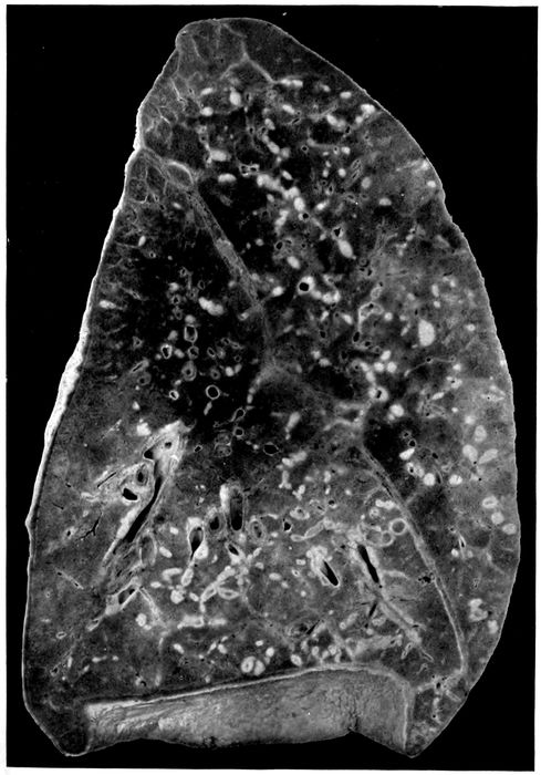
Fig. 10.—Interstitial suppurative pneumonia; interstitial septa are the site of suppuration and lymphatics are distended with purulent fluid; empyema. Autopsy 474, left lung. (See right lung) Fig. 9.
212
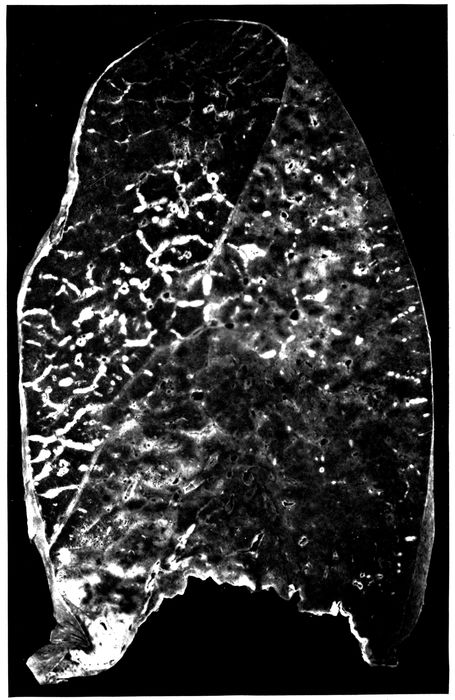
Fig 11.—Suppurative interstitial pneumonia; the left lower lobe is the site of almost uniform consolidation and here interstitial septa and their lymphatics are distended with pus. There is more extensive interstitial suppuration in the upper lobe where consolidation is absent. The cloudy appearance of the consolidated lung is well shown. Autopsy 452.
213Suppurative interstitial pneumonia frequently occurs in association with bronchopneumonic consolidation which may be peribronchiolar, hemorrhagic or lobular, but there is in addition consolidation of the pulmonary tissue between the inflamed septa which may affect part of a lobe, an entire lobe, or parts of several lobes; it does not exhibit the characters of confluent lobular pneumonia.
In approximately half of the cases consolidation, associated with interstitial suppuration, has been lobar in distribution (Fig. 11). The tissue is laxly consolidated, finely granular, and has a cloudy red or gray appearance. The coarsely granular surface of lobar pneumonia is absent. The affected lung may weigh 1,500 or 1,650 grams. Occasionally, interstitial septa of air containing lung tissue is the site of suppurative inflammation or edema. In Autopsy 452 the lower lobe, save a small part at the base, is laxly consolidated; interstitial septa in the consolidated area are yellow, 1.5 to 2 mm. in thickness, beaded and exude purulent fluid on pressure. In the adjacent part of the upper lobe there is a patch of consolidation, and a network of yellow thickened septa extends from it far into the surrounding air containing tissue. The weight of the right lung is 635 grams; of the left, 1,650 grams.
The distribution of interstitial suppuration in 21 instances, including 4 in which the lesion has occurred in the same lungs with abscess formation, has been as follows: right upper lobe, 9 instances; middle lobe, 4; lower lobe, 5; left upper lobe, 7; left lower lobe, 6. In 6 of these autopsies more than one lobe of the same lung has been affected by the lesion; in 2 autopsies parts of both lungs have been affected. Localized abscess of the lung is more common in 214the lower than in the upper lobes, but suppuration of the interstitial tissue is more often found in the upper lobes.
The duration of illness with interstitial suppurative pneumonia has varied from six days to five weeks. In over half of the cases death has occurred during the second week of illness.
The bacteriology of these cases is shown in Table XLVIII.
Table XLVIII
NO. OF CULTURES PNEUMOCOCCI HEMOLYTIC STREPTOCOCCI STAPHYLOCOCCI B. INFLUENZÆ
NO. POSITIVE PER CENT POSITIVE NO. POSITIVE PER CENT POSITIVE NO. POSITIVE PER CENT POSITIVE NO. POSITIVE PER CENT POSITIVE
Bronchus 10 9 90.0 5 50.0 10 100.0
Lung 20 1 5.0 17 85.0 5 25.0 7 35.0
Blood 21 2 9.5 17 81.0
S. hemolyticus has been almost invariably present in lungs, heart’s blood and bronchi. In 16 of 21 autopsies hemolytic streptococci have been obtained from the blood in pure cultures, in one instance associated with pneumococcus. With associated empyema, pericarditis or peritonitis, the same microorganism has been found in the pleural cavities, pericardium or peritoneum. Furthermore, microscopic examination has demonstrated the presence of chains of streptococci in the affected interlobular tissue and in much greater abundance in the distended lymphatics.
Nevertheless in 2 instances no streptococci have been found. These cases are as follows:
Autopsy 330.—Illness began with symptoms of influenza ten days before death; signs of pneumonia were recognized three days before death. There is firm, gray red consolidation of the entire left upper lobe; the interlobular septa are here indicated by yellow lines of obvious suppuration and thick puslike fluid exudes from the cut surface of the consolidated tissue. The upper half of the left lower lobe has undergone gray hepatization, but here there is no distention of the interlobular septa. There is fibrinopurulent pleurisy on the left side with accumulation of 400 c.c. of fluid. Pneumococcus IV is 215obtained from the blood of the heart and from the lung. In the suppurating tissue diplococci which stain by Gram’s method are present in large number; there are a few short chains.
Autopsy 379.—Illness began seven days before death with influenza; signs of pneumonia were first recognized the day before death. The middle lobe of the right lung is firmly consolidated; on section there is mottling of deep red and pinkish red and the cut surface is coarsely granular. The interstitial septa are distended by fluid and are grayish yellow. There is fibrinopurulent pleurisy on the right side with accumulation of 600 c.c. of fluid. Pneumococcus atypical II is obtained from the blood of the heart. A large bacillus unstained by Gram’s method is obtained from the right lung and from the right main bronchus. In the bronchus are a few influenza bacilli. In the suppurating and necrotic tissue of the interstitial septa are found diplococci and chains of 4 to 6 cocci in great number; a few large Gram-negative bacilli are found.
In both these autopsies consolidation had the characters of lobar pneumonia, and pneumococci were obtained from the blood of the heart. It is possible that streptococci failed to grow or while present elsewhere were absent at the spot where cultures were made.
It is noteworthy that B. influenzæ was found in the bronchi in every instance (10) in which cultures were made, but was obtained much less frequently from the lung. In one instance (Autopsy 474) this microorganism was found in the blood in association with hemolytic streptococci. There was suppurative interstitial pneumonia in the left lung and abscess in the right lower lobe with rupture into the cavity and empyema. Hemolytic streptococci and B. influenzæ were found in the bronchus, right pleural cavity and blood of the heart.
In 4 instances (Autopsies 251, 259, 295 and 474) interstitial suppurative pneumonia has been associated with abscess formation. In one instance (Autopsy 251) the right middle lobe has been the site of interstitial suppuration and abscess formation; in another (Autopsy 295) the left lower lobe has been the site of both lesions, but in the other 2 instances suppurative interstitial pneumonia and abscess formation have occurred in opposite lungs. In all 2164 autopsies hemolytic streptococci have been found in the blood of the heart and in lungs or bronchi.
Empyema has been present in all but 3 of 21 instances of interstitial suppurative pneumonia.
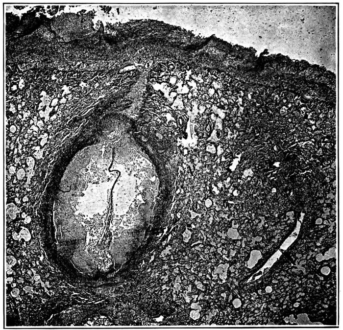
Fig. 12.—Suppurative interstitial pneumonia, showing an immensely dilated lymphatic containing purulent exudate, a short distance below the pleura. Autopsy 474.
Histologic examination of lungs with interstitial suppuration shows that the interlobular septa are distended by serum and contain a conspicuous network of fibrin. Polynuclear leucocytes are present in varying number, and at times densely infiltrate the distended tissue; it is not uncommon to find a zone of densely crowded polynuclear leucocytes along each edge of the septum, whereas the central part contains comparatively few. Occasionally, there is hemorrhage into the distended connective tissue.
217Within the distended septa occur greatly dilated lymphatics filled with polynuclear leucocytes (Figs. 12 and 13). Thrombosis of the distended lymphatics has usually occurred, and a conspicuous network of fibrin in which are polynuclear leucocytes plugs the lumen. Streptococci in chains of variable length are found in the inflamed interstitial tissue, but are present in far greater number within the distended lymphatics.
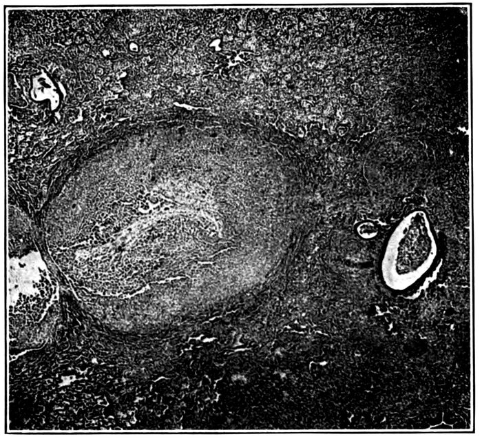
Fig. 13.—Suppurative interstitial pneumonia showing a dilated lymphatic. Autopsy 428.
Necrosis of the cells which fill the lymphatics occurs in spots, usually in the center of the thrombus, and occasionally affects the entire contents of the lymphatic; polynuclear leucocytes have lost their nuclei or in some the nucleus has undergone fragmentation. In these spots the network of fibrin has disappeared. Not infrequently the wall of the lymphatic in a small sector or throughout the circumference 218has undergone necrosis, and spots of necrosis may occur in the interlobular septa distended by inflammatory exudate. Wherever necrosis has occurred, chains of streptococci are present in immense number.
Accumulation of polynuclear leucocytes, necrosis of these cells, solution of fibrin at first in the centers of the lymphatic thrombus and later throughout, occasionally with necrosis of the wall of the vessel, result in the formation of an abscess at the site of the distended lymphatic. These lymphatics, dilated by purulent fluid, may have a diameter from 2 to 3 mm. and may cause considerable compression and collapse of immediately adjacent alveoli. Lymphangitis, distention of lymphatics, thrombosis and finally suppuration may occur in the lymphatic vessels encircling the blood vessels and in those situated in the adventitia of the bronchi of medium size.
The alveoli adjacent to the distended septa are filled by inflammatory products; edema is almost invariably present and the alveoli may contain serum and desquamated epithelial cells; fibrin is often present, but more frequently polynuclear leucocytes are predominant. Not infrequently, abscess formation, recognized microscopically, has occurred in contact with septa most often immediately below the pleura. Polynuclear leucocytes are present in immense number and alveolar septa have disappeared; occasionally, with abscess formation there is more or less widespread necrosis of tissue, cells both of the exudate and of the alveolar walls having lost their nuclei.
Lymphatics in many places are distended and plugged by fibrinous thrombi, whereas elsewhere softening of the thrombus has been brought about by suppuration. Suppuration, both within the lymphatic and in adjacent alveoli, appears to be secondary to lymphatic obstruction. In some instances the lymphatic appears to have undergone distention after the thrombus has formed, for between the 219thrombus and the wall of the lymphatic a channel is occasionally found containing uncoagulated lymph.
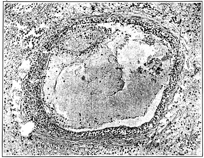
Fig. 14.—Endophlebitis occurring in association with suppurative pneumonia; the intima contains lymphoid cells in great number; at one spot there is a small thrombus adherent to the intima. Autopsy 325.
Acute endophlebitis has been repeatedly observed in association with interstitial suppurative pneumonia (Fig. 14). The lesion usually occurs in veins situated within the septa which are the site of intensely acute inflammation associated with necrosis. The wall of the vein appears to be so injured by the surrounding changes that polynuclear leucocytes and small mononuclear cells accumulate below the endothelium. Throughout the circumference of the veins, often 0.5 to 1 mm. in diameter, the endothelium is separated from the underlying media by polynuclear leucocytes which form a conspicuous zone encircling the lumen. Some cells of lymphoid type are usually present among the polynuclear leucocytes. Polynuclear leucocytes 220are often adherent to the endothelial lining of the vessel and are not infrequently fixed in the process of passing through the endothelium. The lesion may be more severe (Autopsy 325), so that the endothelium has disappeared, and upon the exposed surface fibrin is deposited; within this fibrin polynuclear leucocytes are numerous and nuclear fragmentation has occurred. The middle coat of the vessel usually contains few cells; some polynuclear leucocytes within it may be stretched out as if in process of wandering through the wall.
In other instances the accumulation of cells below the endothelium is almost wholly mononuclear. Cells of the type of lymphocytes occur, but more abundant are slightly larger cells with more abundant cytoplasm. These cells may form a thick zone below the intima throughout the entire circumference of the lesion. It seems probable that these cells, like the polynuclear leucocytes, are derived from circulating blood within the lumen of the vessel, for small cells of the type of lymphocytes are not infrequently found adherent to the lumen and occasionally one is fixed in process of passing through the endothelium.
This endophlebitis appears to be the result of changes outside the vessel; there is usually necrosis of the adjacent tissue and the production of the lesion is favored by lymph stasis; as the result of injury to the vessel wall, polynuclear leucocytes in response to chemotaxis, or with milder irritation, mononuclear cells, wander through the endothelium and accumulate below it perhaps on account of the greater impermeability of the middle coat to the passage of cells.
The lesion described does not occur exclusively with interstitial suppurative pneumonia caused by hemolytic streptococci, but has been found in association with abscess formation (Autopsies 354 and 383) caused by hemolytic streptococci or (Autopsy 322) caused by staphylococci. In 1 instance it has been found with lobar pneumonia (Autopsy 320) caused by atypical Pneumococcus II and in 2 221instances with combined lobar and bronchopneumonia (Autopsy 357 with Pneumococcus IV; Autopsy 392 with Pneumococcus II). In these 3 instances there has been interstitial inflammation, edema and lymphangitis without suppuration.
Interstitial suppurative pneumonia of long standing may occasionally be accompanied by chronic changes which bring about thickening of the interlobular tissue. In the following autopsy acute suppurative inflammation in the left lung has been associated with conspicuous thickening of interlobular septa in the right lung.
Autopsy 474.—I. H., white, aged twenty-one, was a native of Oklahoma and had been in military service one month. His illness began with influenza thirty-six days before death; he was admitted to the base hospital thirty-one days before his death with signs of pneumonic consolidation of the right lower lobe. Evidence of fluid in the right pleural cavity was obtained two weeks before death, and from 100 to 700 c.c. of thick purulent fluid were aspirated on five occasions. Hemolytic streptococci were found in the aspirated fluid.
Anatomic Diagnosis.—Interstitial suppurative pneumonia in left lung; abscess of right lower lobe with rupture into pleural cavity; thickening of interlobular septa of right lower lobe; double purulent pleurisy with thoracotomy on right side; serofibrinous pericarditis.
The right pleural cavity contains 85 c.c. of thick purulent fluid; the right lung (Fig. 9) is collapsed and pushed to the median line, being bound by firm adhesions to the pericardium. Over the external and basal surfaces is a localized cavity walled off by adhesions. An abscess cavity in the lower part of the lower lobe communicates through a perforation in the basal surface of the lung with the pleural cavity and is in free communication with a small bronchus. About the abscess the lung is red and laxly consolidated, but elsewhere air containing; throughout the lower half of the lower lobe, the interlobular septa are marked by conspicuous yellowish gray lines about 1 mm. in thickness. Between these thickened septa the lung tissue contains air. The lung weighs 600 grams. The left lung (Fig. 10) is voluminous and heavy, weighing 1,320 grams. The surface is 222everywhere covered by thickened pleura and fibrin, the pleural cavity containing 150 c.c. of thick purulent fluid. The lung is consolidated varying in color from a fleshy red to yellowish gray. The surface is very conspicuously marked by yellow lines 2 or 3 mm. thick, corresponding to the interlobular septa which have undergone suppuration. The septa have bead-like swellings along their course, and when pus escapes from the cut surface small cavities remain at the site of these swellings.
Bacteriologic examination has shown hemolytic streptococci in the blood, left lung, right and left pleural cavities, and right bronchus. B. influenzæ has been found in the bronchus, in the right pleura and in the heart’s blood. A few colonies of S. aureus have been found on the plate from the right pleural cavity (site of thoracotomy).
Microscopic examination of the right lower lobe shows that the interstitial septa are much thickened by young fibrous tissue infiltrated with lymphoid and a few plasma cells. Large mononuclear cells with granular cytoplasm are very numerous. A lymphatic is much distended and contains a few polynuclear leucocytes and many lymphoid and large mononuclear cells. There is no suppuration. Sections from the right lung show suppurative lymphangitis with suppurative inflammation of interstitial tissue.
The right lung is the site of a healing lesion of the interstitial tissue which has developed simultaneously with acute interstitial suppurative pneumonia in the left lung. Both lesions are doubtless caused by S. hemolyticus. This healing lesion exhibits little similarity to the interstitial bronchopneumonia described by several observers with both measles and influenza.
The following autopsy furnishes further evidence that interstitial suppurative pneumonia exhibits a tendency to heal. Proliferation of endothelial cells lining the inflamed lymphatics gives rise to phagocytic cells which aid in removing the accumulated leucocytes.
223Autopsy 397.—N. P., white, aged twenty-one, farmer, a native of Oklahoma, had been in military service twenty-one days. Illness began twenty-two days before death, the patient being admitted on the day following onset with influenza, pharyngitis and bronchitis. A diagnosis of lobar pneumonia was made fourteen days before death. The left pleural cavity was aspirated twelve days later and 800 c.c. of thick yellow pus were withdrawn. Hemolytic streptococci were found in the sputum five days before death.
Anatomic Diagnosis.—Interstitial suppurative pneumonia in left upper lobe; acute bronchopneumonia with lobular consolidation in right upper lobe; localized purulent pleurisy on left side with compression and atelectasis of left lung; compensatory emphysema of right lung; purulent bronchitis; beginning serofibrinous pericarditis; chronic passive congestion of liver, spleen and kidneys.
The right lung is very voluminous, free from coal pigment and bright pink save over lobular patches of consolidation which have a bluish red color; the bronchi contain mucopurulent material. The anterior surface of the left lung is bound to the chest wall by firm adhesions, but over the external and posterior surfaces of the lung there is a localized cavity containing 1,100 c.c. of turbid fluid. The left lung is collapsed and airless with deep fleshy red color. In the upper lobe there are scattered patches of consolidation 1.5 to 2.5 cm. across where the tissue is grayish red and coarsely granular. In the adjacent tissue interstitial septa are thickened to 1 or 2 mm. and are conspicuous as gray bands. Along their course occur bead-like swellings from which purulent fluid can be scraped. These septa at one point reach the anterior surface of the lung where the pleural cavity is in large part obliterated by adhesions; here there is an encapsulated pocket 4 x 1.5 cm. containing thick creamy pus.
Bacteriologic examination of the blood shows the presence of hemolytic streptococci; cultures from the lungs contain hemolytic streptococci and B. influenzæ.
Microscopic examination shows that interlobular septa are thickened and infiltrated with plasma cells in large number. Leucocytes in the center of much dilated lymphatics have undergone necrosis and have lost their nuclear stain. About the periphery of the lumen and evidently derived from the swollen endothelial cells which surround it, are numerous large mononuclear cells. They act as phagocytes and ingest polynuclear leucocytes. Multinucleated giant cells, derived from these cells, occur. In several places thrombosed lymphatics in process of organization occur; the lumen is filled with compact fibrin which is invaded by fibroblasts and newly formed capillaries.
The process just described is analogous to that which occurs whenever an unopened abscess heals; mononuclear cells accumulate and act as phagocytes ingesting polynuclear leucocytes.
224The following instance of streptococcus empyema is noteworthy because no suppurative pneumonia has been found in association with it. Nevertheless the character of the changes present in the lung indicate that the organ has been the site of an interlobular inflammation which has healed.
Autopsy 499.—J. H. M., white, aged twenty-four, a farmer from Arkansas, had been in military service five months. Onset of illness began two weeks before his admission to the hospital on November 15 with cough, fever, headache and malaise; on admission there was acute bronchitis. Thirteen days after admission the patient developed parotitis (mumps?); five days later and five days before death pleurisy was recognized on the right side and pneumonia was suspected. Death occurred thirty-six days after onset. The temperature on admission was 103.2° F. and remained elevated during one week falling by lysis; from this time until the pleurisy was recognized it was normal and later it remained approximately 103° F.
Anatomic Diagnosis.—Fibrinopurulent pleurisy on right side; fibrinous pleurisy on left side; fibrinopurulent pericarditis; chronic interstitial (interlobular) pneumonia in process of healing; purulent bronchitis; acute splenic tumor; parenchymatous degeneration of kidneys.
The right pleural cavity contains 1,650 c.c. of grayish yellow fluid containing an abundant sediment of softened fibrin. Part of this fluid, more opaque than the remainder is confined in a localized pocket between the inner surface of the lung and the pericardium. The apex and anterior surface of the right upper lobe, over an area about 7 cm. across, is held by fibrinous adhesions to the chest wall; when this adhesion is broken a pocket is exposed 6.5 x 2.5 cm. containing fibrin and fluid. The pericardial cavity is distended by 350 c.c. of turbid yellow seropurulent fluid. The pericardial surfaces are covered by shaggy, tough gray fibrin.
The right lung is collapsed; the lower and posterior part of the upper lobe is deep red and atelectatic. Throughout the upper lobe the interlobular septa are thickened, often 1 mm. across and very conspicuous; in the lower and anterior tip of the lobe is an area where tissue is firm grayish red and heavier than water. The lower and posterior half of the right lower lobe is firm and airless, and the tissue is reddish gray or gray and in places finely granular on section; interlobular septa are conspicuous. Although the lung is cut into thin sections, no abscesses are found. Bronchi throughout the lung contain mucopurulent fluid.
The left lung over its lower half is covered by a thin layer of fibrin. The tissue is crepitant throughout and moderately edematous. Bronchi contain mucopurulent fluid.
Hemolytic streptococci in pure culture are obtained from the blood of the heart, right pleural cavity and pericardium. No growth is obtained on a plate inoculated with material from the right lower lobe. The right bronchus contains hemolytic streptococci and B. influenzæ.
225The pleural surface of the right lung is covered by a thick layer of fibrin which has undergone advanced organization. Fibrous septa within the lung are much thickened by the presence of newly formed fibrous tissue; the interstices of the tissue are distended and contain fibrin into which fibroblasts and new blood vessels have penetrated. Some lymphatics are plugged with fibrin and contain polynuclear leucocytes, lymphoid and large mononuclear cells. In several places organization of these thrombi is beginning. About the blood vessels are thrombosed lymphatics in which polynuclear leucocytes and mononuclear cells are equally abundant. Alveoli immediately adjacent to blood vessels and to fibrous septa often contain fibrin, and alveoli elsewhere contain desquamated cells in abundance.
In association with hemolytic streptococci in the blood, pleura and pericardium, there has been inflammation of the interlobular septa of the lungs with acute lymphangitis; there has been no suppuration and the lesion is in process of healing with new formation of fibrous tissue. It is evident that this lesion, as well as pleurisy with advanced organization, preceded the exacerbation of the patient’s illness which occurred five days before death. The advanced chronic changes found at autopsy indicate that the pulmonary and pleural lesions had their origin during the illness which was present at the time of admission to the hospital. Interstitial pneumonia caused by hemolytic streptococci was of mild character and did not produce suppuration within the lung; nevertheless, hemolytic streptococci which reached the pleura caused empyema.
Suppurative Pneumonia with Multiple Clustered Abscesses Caused by Staphylococci
In the preliminary report of this commission published in The Journal of the American Medical Association, loc. cit., pg. 111, we described suppurative pneumonia with multiple abscesses caused by staphylococci and cited 4 instances of the lesion which followed influenza. Chickering and Park[84] published in a subsequent number of the same journal 226an account of staphylococcus pneumonia, a lesion which has heretofore attracted very little attention.
In a small group of cases abscesses in the lungs have had characters which serve to distinguish them from the abscesses previously described. Small, sharply circumscribed yellow nodules, which in their centers have undergone suppurative softening, form a cluster upon a red, airless background (Figs. 15 and 16). One or more of these groups several centimeters across, occur in the lungs. It is usually evident that the abscesses are clustered about a medium-sized bronchus, but occasionally with increase in the size of the small cavities the lung tissue assumes a honey-combed appearance.
These clustered abscesses occur in association with bronchopneumonia and have been in all instances associated with purulent bronchitis. The mucosa of the small bronchi may be destroyed so that the surface is eroded. These small clustered abscesses are seen as conspicuous yellow spots immediately below the pleura, but there has been no associated empyema. In 2 instances these abscesses were accompanied by fibrinous pleurisy, but in the remaining autopsies the pleura has been normal. The infrequency of empyema is in contrast with its almost invariable presence when a streptococcus abscess is found below the pleura.
Autopsy 280.—Onset of illness with malaise, headache, cough and fever was on September 24, eight days before death. At autopsy there were hemorrhagic peribronchiolar and lobular bronchopneumonia, clustered foci of suppuration in right lung, purulent bronchitis and fibrinous pleurisy. Hemolytic streptococci were obtained from the consolidated lung and from a bronchus. A culture from the right lung was contaminated. In the bronchus were found B. influenzæ and a few staphylococci. Microscopic examination of the abscesses shows that they contain Gram-staining cocci grouped into staphylococcus-like colonies.
Autopsy 286.—Duration of illness, which began September 25 with symptoms of influenza, was nine days. At autopsy there were lobular and confluent patches of bronchopneumonia, clustered abscesses in the right lung below the pleura, purulent bronchitis, and serofibrinous pleurisy localized in the neighborhood of the abscesses. Pneumococcus IV was obtained from the blood of the heart, and Pneumococcus IV, staphylococci and B. influenzæ from the right main bronchus; growth failed to occur on plates from right and left lungs. Microscopic examination shows the presence of clumps of cocci with staphylococcus grouping in the centers of the small abscesses. Section through one abscess shows its continuity with the wall of a bronchus; along one side of the abscess is epithelium composed of flattened epithelial cells in multiple layers continuous with that of the bronchus; the remainder of the abscess wall is formed by disintegrated lung tissue.
227
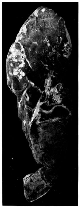
Fig. 15.—Abscesses in two clusters caused by S. aureus in upper part of right upper lobe; confluent lobular consolidation in lower part of lobe. Autopsy 333.
228
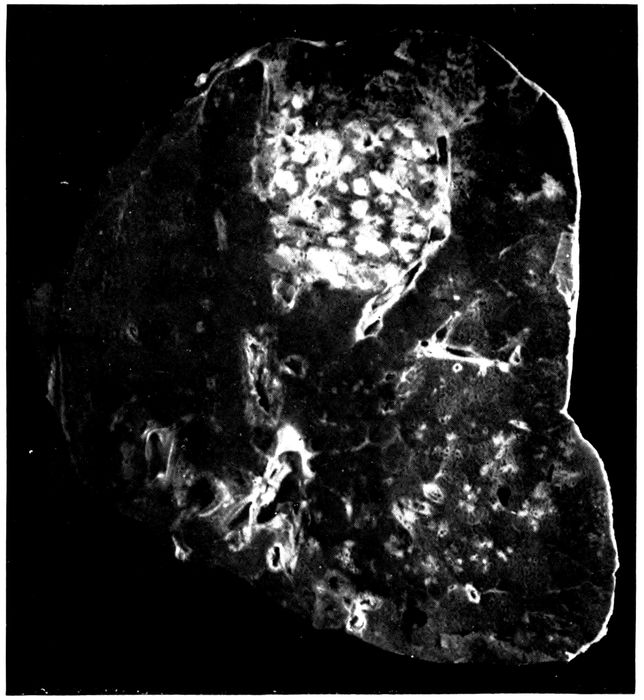
Fig. 16.—Abscesses in cluster caused by S. aureus at apex of right upper lobe. Autopsy 322.
Autopsy 322.—The patient was admitted with influenza eight days before death; signs of pneumonia appeared two days later, and on the following day Pneumococcus IV was obtained from the sputum. At autopsy there were bronchopneumonia with lobar consolidation, abscesses clustered about a bronchus in the right upper lobe and purulent bronchitis. The blood was sterile; S. aureus was obtained from the consolidated part of the left lung; S. aureus and Pneumococcus III from the abscesses of the right lung. Microscopic examination of sections of abscesses showed the presence of Gram-staining cocci in staphylococcus-like colonies, surrounded by necrotic material and polynuclear leucocytes; Gram-negative bacilli resembling B. influenzæ were seen. (See Fig. 16.)