Chapter IV · THE PATHOLOGY AND BACTERIOLOGY OF PNEUMONIA FOLLOWING INFLUENZA (continued 3)
Reading edition
229Autopsy 333.—The onset of influenza was fifteen days before death; a diagnosis of pneumonia was made seven days before death. At autopsy there were confluent bronchopneumonia, clustered abscesses in the right lung and purulent bronchitis (no pleurisy). The blood contained Pneumococcus II atypical. S. aureus and Pneumococcus II atypical were obtained from the abscesses; S. hemolyticus, from the consolidated left lung; S. aureus, B. influenzæ and a few hemolytic streptococci, from the bronchus. (See Fig. 15.)
Autopsy 370.—The patient was admitted seventeen days before death and signs of pneumonia were noted three days after admission. At autopsy there were lobular and confluent bronchopneumonia and small abscesses clustered about bronchi and situated within the gray consolidated lung; purulent bronchitis and patches of atelectasis, with distention of the lungs, so that they failed to collapse on removal. No growth was obtained from the heart’s blood; S. aureus in pure culture was obtained from the abscesses of the right lung; S. aureus, Pneumococcus IV and B. influenzæ were obtained from a small bronchus on the left side.
Autopsy 425.—Illness began with influenza twenty-nine days before death; a diagnosis of pneumonia was made fourteen days before death. At autopsy there were chronic bronchopneumonia with tubercle-like nodules of consolidation with some large patches of consolidation, multiple small abscesses giving a honey-combed appearance to part of the right middle lobe, purulent bronchitis and bronchiectasis. S. hemolyticus was grown from the heart’s blood; S. hemolyticus, B. influenzæ and S. albus from the lung. Sections of an abscess contain clumps of cocci. An abscess cavity has along one side remains of a bronchial wall covered by squamous epithelium; a dilated bronchus, cut longitudinally, terminates in this irregular abscess cavity.
Table XLIX shows the incidence of pneumococci, hemolytic streptococci, staphylococci and B. influenzæ in the foregoing autopsies with abscesses clustered about bronchi:
Table XLIX
NO. OF CULTURES PNEUMOCOCCI HEMOLYTIC STREPTOCOCCI STAPHYLOCOCCI B. INFLUENZÆ
NO. POSITIVE PER CENT POSITIVE NO. POSITIVE PER CENT POSITIVE NO. POSITIVE PER CENT POSITIVE NO. POSITIVE PER CENT POSITIVE
Bronchus 4 2 50.0 2 50.0 4 100.0 4 100.
Lung 6 2 33.3 3 50.0 4 66.7 2 33.3
Blood 6 2 33.3 2 33.3
Staphylococcus shows in the lung the same tendency to produce localized abscesses which it exhibits in other tissues of the body; it invades the lung by way of the bronchi, 230but shows no ability to invade lymphatics, and in the cases we have examined rarely enters the pleura or the blood. In all of these cases B. influenzæ has been found in the bronchi and perhaps precedes the staphylococcus as an invader of the lower respiratory passages. Pneumococci atypical II, Types III and IV have been found in over half of these cases. The significance of this organism is emphasized by the 2 cases in which it has been found in the heart’s blood at autopsy. It appears not improbable that S. aureus has invaded the lung already the site of bronchopneumonia caused by pneumococci.
Notwithstanding the small number of autopsies, the figures in Table XLIX, showing the incidence of pneumococci, streptococci, staphylococci and B. influenzæ, are cited so that they may be compared with the corresponding figures for the usual type of streptococcus abscess (p. 203). The incidence of hemolytic streptococci is relatively low, whereas that of staphylococci approximates 100 per cent. S. aureus was present in great number in the lung of Autopsies 322 and 333 and in pure culture in the abscess of Autopsy 370. Microscopic examination of sections from the abscesses which have been described, demonstrated the presence of Gram-staining cocci in characteristic staphylococcus-like clumps within the exudate of the abscesses; scattered chains of streptococci were not found. In those instances (Autopsies 280 and 286) in which cultures failed to demonstrate staphylococci, microscopic examination demonstrated staphylococcus-like clumps of bacteria within the abscess cavity. Cultures were usually made from the consolidated lung near the abscess where the pleural surface could be seared, rather than from the pus, so that in some instances the microorganism has doubtless escaped detection although present.
In association with the multiple abscesses which have been described, injury to the bronchi and bronchopneumonia have been invariably present. Purulent bronchitis 231has been present in all instances of this lesion; in 2 instances there has been dilatation of the bronchi, and in 1 instance in which the onset of influenza was twenty-nine days before death, there has been advanced bronchiectasis.
Microscopic examination shows that the epithelium of the bronchi is partially or completely destroyed and that destruction of the underlying tissue, with acute suppurative inflammation, penetrates to a greater or less depth into the wall. When the epithelium of the bronchus is wholly destroyed and the lumen is filled and distended with polynuclear leucocytes, a cross section of the tube has the appearance of a small abscess; but more careful examination often shows that the engorged mucosa is still intact. Occasionally, a network of fibrin forms a layer covering the denuded mucosa. Disintegration of the superficial tissue may extend to the muscularis or through it, and may penetrate the wall of the bronchus. The tissue in contact with the exposed surface contains many polynuclear leucocytes and blood vessels plugged with fibrinous thrombi, but deeper in the tissue lymphoid and plasma cells are more numerous. In 2 instances (Autopsies 286 and 425) favorable sections have demonstrated that the wall of an abscess on one side consists of the remains of a bronchus, covered by epithelium composed of squamous cells, Whereas the remainder of the wall, here very irregular, is formed by partially destroyed alveoli plugged with fibrin. The suppurative process has penetrated the wall of the bronchus on one side and extended into the surrounding alveolar tissue. In other instances, abscess cavities occur within the alveolar tissue of the lung and their relationship to bronchi is not evident. In the mass of polynuclear leucocytes which fill the abscess cavity, are clumps of staphylococci in great abundance, usually forming characteristic colonies which are conspicuous with the low power of the microscope.
232
Empyema, Pericarditis and Peritonitis
No sharp line can be drawn between nonpurulent and purulent pleurisy. A diagnosis of empyema has been made when the fluid in the chest has become opaque and fibrin has undergone softening or solution. The lesion has been designated seropurulent when there has been abundant thin, opaque, gray fluid. Pleurisy has been designated fibrinopurulent when the cavity has contained opaque fluid and ragged soft white or yellowish fibrin adherent to the chest wall; this fibrin is evidently in process of disintegration and there may be numerous shreds and flakes of fibrin which subside to the bottom of the fluid. The amount of fluid in the cavity may occasionally exceed 1,700 c.c.; that in both pleural cavities may exceed 2,500 c.c. The lesion has been designated purulent when fibrin has almost wholly disappeared and the cavity contains thick yellowish white fluid. In 4 of 5 instances in which thoracotomy had been performed, empyema has assumed this otherwise uncommon type.
Some inflammation of the pleura is almost constantly found in association with all forms of pneumonia, but in many instances is so slight that it has no noteworthy significance. Table L shows the incidence of various types of pleurisy.
Table L
LOBAR PNEUMONIA BRONCHOPNEUMONIA SUPPURATIVE PNEUMONIA WITH ABSCESS INTERSTITIAL SUPPURATIVE PNEUMONIA
No. % No. % No. % No. %
No pleurisy noted 30 46.9 44 55 1 2.6 1 5.9
Serous pleurisy 5 7.8 9 11.2
Fibrinous pleurisy 10 15.6 5 6.2 1 2.6
Serofibrinous pleurisy 12 18.2 14 17.5 3 7.7
Seropurulent pleurisy 9 23.1 1 5.9
Fibrinopurulent pleurisy 7 10.9 5 6.2 17 43.6 12 70.6
Purulent pleurisy 3 3.7 8 20.5 3 17.6
Total 64 80 39 17
233Empyema has occurred, on the one hand, in 12.4 per cent of instances of lobar pneumonia and in 9.9 per cent of instances of bronchopneumonia alone. It has occurred, on the other hand, in 87.2 per cent of instances of suppurative pneumonia with abscess formation and in 94.1 per cent instances of interstitial suppurative pneumonia. These suppurative lesions are caused by hemolytic streptococci, and when cultures are made from the pleural exudate this microorganism is isolated.
Of 16 instances in which empyema has occurred in association with lobar pneumonia or bronchopneumonia unaccompanied by suppuration in 6 there has been infection with hemolytic streptococci. Empyema has occurred in the absence of hemolytic streptococci only 10 times.
Empyema Caused by Hemolytic Streptococci.—When necrosis preceding abscess formation has occurred in the lung, streptococci are found in immense numbers in the dead tissue. The pleura overlying the abscess undergoes necrosis and occasionally streptococci are particularly numerous upon the pleural surface of the necrotic tissue. In Autopsy 376 a membrane thin as tissue paper, representing the pleura, separated an abscess containing thick pus from the pleural cavity which was the site of empyema. The abscess may rupture into the pleural cavity and at the same time may be in free communication with a bronchus (Autopsy 480). In one (Autopsy 467) instance an abscess which had ruptured into the pleural cavity had completely discharged its contents and was in process of healing, newly formed fibrous tissue being abundant in its wall.
With few exceptions empyema has accompanied subpleural abscess caused by hemolytic streptococci, being found on the side corresponding to the abscess. Among 39 instances of pulmonary abscess, empyema has been limited to the side of the abscess in 23; it has been present on the opposite side as well in 10 instances. In 2 instances there have been abscesses in both lungs; in one (Autopsy 234385 A) there has been double empyema, and in the other (Autopsy 487) empyema only on the left side. In one instance abscess has been recognized by microscopic examination and its location is not recorded. In 5 instances of abscess formation there has been no empyema. In Autopsy 383 there has been no pleurisy noted; in Autopsy 416 there has been fibrinous pleurisy and in Autopsies 277, 290 and 380, serofibrinous pleurisy.
Empyema has been almost invariably found in association with interstitial suppurative pneumonia. This lesion extends by way of the lymphatics up to the pleural surface and is often more conspicuous just below the pleura than elsewhere. Empyema has been absent in only 3 of 21 examples of the lesion and in one of these there has been serous effusion. In 12 instances interstitial suppuration has occurred only on one side and empyema has been limited to this side; in 5 instances with interstitial suppuration on one side there has been empyema on both sides; in 2 instances with interstitial suppuration in both lungs there has been double empyema.
The amount of fluid in the pleural cavity has varied from less than 100 to 1,500 c.c. The fluid has occasionally been seropurulent or yellow, thick and purulent, but in most instances the exudate is best described as fibrinopurulent. There is yellow or yellowish gray purulent fluid containing flakes of soft ragged fibrin.
The foregoing study has shown, on the one hand, that empyema is a frequent complication of streptococcus pneumonia and, on the other hand, that empyema following influenza with relatively few exceptions is caused by hemolytic streptococci. Empyema caused by this microorganism exhibits in some instances characters not seen with other varieties of pleural inflammation. The tissue between sternum and pericardium is often edematous and the adjacent fat has a firm brawny consistence. In some instances the exudate contains blood, and hemolysis has occurred 235so that the fluid has a diffuse red color. The occurrence of multiple pocketed collections of purulent fluid within the pleural cavity is peculiar to streptococcus empyema. These pockets have been found 6 times in association with abscess and 5 times with interstitial suppurative pneumonia. In the presence of an exudate within the pleural cavity, some part of the lung, usually the anterior surface behind the sternum and costal cartilages, is glued by fibrinous adhesions to the parietal pleura. Here occur pockets containing thin purulent fluid and softened fibrin or thicker creamy pus walled off by fibrin about the edges of the pocket. At the site of the lesion the lung, after it is separated from the chest wall, is marked by a shallow depression surrounded by the fibrin which has walled in the pocket. The little cavity thus formed, varying much in size, is usually oval, the long diameter being from 1 to 3 cm. These pleural pockets may occur over the external surface of the lung (Autopsies 452, 455, and 472) or between the internal surface and pericardium (Autopsy 452). Occasionally with partial fibrinous adhesion between the pleural surfaces there are both scattered pockets containing purulent fluid and a larger encapsulated collection of fluid; in Autopsy 455 the pleural surfaces were adherent and there was 100 c.c. of purulent fluid encapsulated in a space over the external surface of the lung, 12 × 8 cm. In Autopsy 452 the lower part of the pleural cavity was encapsulated and contained 650 c.c. of fluid. This tendency of empyema caused by S. hemolyticus to form encapsulated pockets is doubtless of considerable importance in the treatment of the condition.
Stone, Bliss and Phillips[85] have described these encapsulated pockets as “subcostosternal pus pockets” and have maintained that they are formed about the sternal lymphatic nodes. We have found them so widely scattered that this relation seems improbable.
236Pneumococcus Empyema.—Empyema occurred in association with pneumonia referable to pneumococci 10 times, once with Pneumococcus II; 6 times with Pneumococcus atypical II; once with Pneumococcus III and twice with Pneumococcus IV. The lesion was seropurulent once; fibrinopurulent 8 times and purulent once. Fibrin in several instances was somewhat voluminous. In the following instance voluminous masses of fibrin had an important influence upon the attempted treatment.
Autopsy 473.—A. D. P., white, aged twenty-one, a student from Missouri, had been in military service two weeks. He was admitted to the hospital with influenza twenty-eight days before his death, and four days after admission there were signs of pneumonia. Paracentesis was performed on the right side on the eleventh day after admission; 4 c.c. of cloudy fluid which contained Pneumococcus III were obtained at this time and later in the day 800 c.c. were withdrawn. On the thirteenth day attempted withdrawal of fluid from both pleural cavities failed. On the eighteenth day aspiration of the right pleural cavity yielded only 30 c.c. of fluid. On the nineteenth day 400 c.c. of purulent fluid were withdrawn from the right pleural cavity. On the twenty-fifth day there was cyanosis and delirium. Shortly before death aspiration of the right pleural cavity was attempted, but only 4 c.c. of fluid were obtained.
Anatomic Diagnosis.—Chronic bronchopneumonia with lobular and peribronchiolar consolidation in left lung; fibrinopurulent pleurisy on both sides; purulent bronchitis and bronchiectasis.
On removal of the sternum, encysted purulent pleurisy is found between the inner surface of the right lung and the pericardium; there is here 450 c.c. of very thick creamy, greenish yellow pus entirely separated from the remainder of pleural cavity. The external part of the cavity contains 1,450 c.c. of fluid and voluminous masses of firm fibrin which placed in a measuring cylinder occupy 450 c.c. The left pleural cavity contains 400 c.c. of seropurulent fluid in which there is abundant sediment of fibrinous particles.
The right lung is compressed; the bronchi exude purulent fluid. The left lung is voluminous; in the upper and lower lobes there are small yellowish gray nodules of consolidation, grouped in clusters, and gray patches of lobular consolidation occur. Bronchi are dilated and filled with purulent fluid.
Bacteriologic examination shows the presence of Pneumococcus III obtained in pure culture from the blood of the heart and from the right pleural cavity. S. viridans is grown from the left lung; a plate from the right bronchus contained B. influenzæ, S. viridans and a few colonies of staphylococcus and M. catarrhalis.
237The foregoing case is particularly noteworthy because aspiration failed repeatedly to yield more than a few cubic centimeters of fluid, doubtless because the voluminous masses of fibrin present in the cavity prevented escape of fluid. Aspiration was attempted shortly before death, but only 4 c. c. of fluid were obtained; nevertheless, at autopsy the right pleural cavity contained 2,350 c.c. of exudate. Another factor of much importance in relation to treatment is the encapsulation of 450 c.c. of purulent fluid between the inner surface of the right lung and the pericardium. It is possible that free drainage might have emptied the main cavity and perhaps even freed the encapsulated fluid.
Pericarditis.—Among 241 autopsies on individuals with pneumonia following influenza, pericarditis occurred 23 times; these lesions were classified as follows: Serous pericarditis, 1; serofibrinous pericarditis, 9; seropurulent pericarditis, 1; fibrinopurulent pericarditis, 10; purulent pericarditis, 2.
It is noteworthy that in 12 of 23 instances of pericarditis the lesion was associated with S. hemolyticus infection of the lung and whenever in these instances cultures were made (Autopsies 434, 485, 499 and 504) hemolytic streptococci were obtained from the pericardial exudate in pure culture.
The tendency of interstitial suppurative pneumonia to produce pericarditis is especially evident. Among 21 instances of interstitial suppurative pneumonia pericarditis occurred 6 times (28.6 per cent); among 39 instances of suppurative pneumonia with abscess formation, pericarditis occurred twice (5.1 per cent); whereas among all other autopsies, namely, 181, the lesion occurred 15 times (8.3 per cent).
Pericarditis occurred in association with pneumonia referable to Pneumococcus I, once, (Pneumococcus I isolated from the pericardium); to Pneumococcus II, once; to atypical Pneumococcus II, 5 times (twice isolated from the pericardium); 238and to Pneumococcus IV, twice (once isolated from the pericardium).
Peritonitis.—Purulent peritonitis occurred only twice, in both instances in association with pneumonia caused by hemolytic streptococci. Purulent peritonitis was part of a general serositis involving both pleural cavities, pericardium and peritoneum in 2 noteworthy instances:
Autopsy 465.—J. K., white, aged twenty-two, farmer from Oklahoma, had been in military service one month. He was admitted to the hospital with influenza, sore throat and bronchitis twenty-four days before his death. Signs of pneumonia were recognized thirteen days later and at the same time there was otitis media on the right side. Empyema and pericarditis were found three days before death and two days later 1000 c.c. of cloudy fluid were withdrawn from the chest.
Anatomic Diagnosis.—Suppurative pneumonia with consolidation and abscess in right lower lobe below pleura; purulent pleurisy on right, seropurulent pleurisy on left side; beginning serofibrinous pericarditis; fibrinopurulent peritonitis; purulent bronchitis.
The body is emaciated. The right pleural cavity contains 350 c.c. of thick, creamy yellow pus in which are flakes of fibrin; the right lung is collapsed and lies at the back and inner side of the cavity. The left pleural cavity contains 500 c.c. of turbid, yellow, seropurulent fluid in which is soft fibrin. The lower lobe of the right lung is consolidated throughout, flabby, gray red and finely granular on section. Below the pleura of the posterior border is a wedge-shaped cavity with its base 1.5 cm. across, in contact with the pleural surface. About the cavity consolidated tissue has an opaque, yellow color. Bronchi in both lungs contain mucopurulent fluid. The pericardial cavity contains 20 c.c. of turbid fluid; the left auricular appendage is bound by a thin layer of fibrin to the parietal pericardium.
The peritoneal cavity contains 100 c.c. of thick, creamy, yellow, purulent fluid. Between the diaphragm and liver is a layer of fibrin, in places 1.5 cm. in thickness; fibrin is present upon the peritoneum overlying the kidneys and base of mesentery.
Bacteriologic examination shows the presence of hemolytic streptococci, obtained in pure culture from the blood of the heart, right pleural cavity and peritoneum. From the right bronchus are grown S. hemolyticus, B. influenzæ and a few colonies of S. viridans and staphylococcus.
Autopsy 504.—G. R. C., white, aged twenty-eight, farmer from Alabama, had been in military service three months. Onset of illness occurred six days before death, and two days later he entered the hospital with fever (103.4° F.), pains in the abdomen and vomiting. Consolidation at the bases of the lung was recognized on the day following admission and on the day before death 900 c.c. of greenish brown fluid were aspirated from the left pleural cavity.
239Anatomic Diagnosis.—Interstitial suppurative pneumonia with consolidation in left lower lobe; purulent pleurisy on both sides; purulent pericarditis; purulent peritonitis; parenchymatous degeneration of kidneys; acute splenic tumor.
The body is that of a large well-nourished man. The left pleural cavity contains 975 c.c. of creamy, yellow fluid; right pleural cavity contains 425 c.c. of purulent fluid thinner than that on the left side. The left lung is collapsed; the posterior and lower half of the lower lobe is consolidated, flabby, deep red and fleshy in appearance. The interstitial septa are yellow, thickened with bead-like enlargements and contains creamy purulent fluid which flows away and leaves small cavities. This interstitial suppuration is more advanced below the outer surface of the lobe than elsewhere.
The pericardial cavity contains 25 c.c. of creamy, yellow, purulent, fluid; the epicardium is dull, covered in a few places by a small amount of fibrin and below it are ecchymoses.
The peritoneal cavity contains 100 c.c. of thick, yellow pus; the peritoneal surfaces are injected and between the liver and diaphragm is fibrin.
Bacteriologic examination shows the presence of S. hemolyticus in pure culture from the blood of the heart, the lower lobe of the left lung, pericardium and peritoneum. The right main bronchus contains the same microorganism, B. influenzæ and a few staphylococci.
General serositis has been caused by hemolytic streptococci which in one instance have entered the pleura from a subpleural abscess, and in the other from the suppurating interstitial tissue of the lung. In one of these cases the patient entered the hospital with symptoms suggestive of acute peritonitis.
Bronchiectasis
Acute dilatation of the bronchi is a common result of the bronchitis of influenza, and its frequent occurrence is an index of the severity of the changes in the bronchial wall. In some instances the smaller bronchi in well-localized areas are uniformly dilated; in other instances, large cavities, several centimeters in diameter, are formed and all transitions between the two extremes occur.
The occurrence of bronchiectasis following influenza is mentioned by Leichtenstern[86]. He states that evidence of 240bronchiectasis can persist for weeks or months and nevertheless end with complete restitution of the lungs to normal. Lord[87] has described instances of bronchiectasis occurring in association with infection by B. influenzæ and Boggs[88] has recorded similar observations.
We have had abundant opportunity to observe early stages in the production of bronchiectasis and to study the much discussed pathogenesis of the condition.
The following figures show the predilection of bronchiectasis for the left lung and for the lower lobes: Bronchiectasis occurred 30 times in the left lung alone, 9 times in the right lung alone and 13 times in both lungs, the total being 52. Among 30 instances in which the lesion occurred only in the left lung, in 24 it was limited to the lower lobe, and in 15 of these 24 instances to the base of the lower lobe. Among 9 instances in which dilatation of bronchi occurred only in the right lung, it was limited to the lower lobe in 4 instances and to the base of the lower lobe in 2 of these 4 instances.
When the lesion is limited to the base of the lower lobes small bronchi with no recognizable cartilage in their wall are dilated to a diameter of from 3 to 6 cm. and are distended with thick mucopurulent fluid. The tenacious character of the bronchial contents and the action of gravity doubtless have a part in the production of the dilatation. In several instances dilatation of the bronchi was limited to the basal parts of both upper and lower lobes.
When bronchiectasis occurs throughout a whole lung, usually the left, or in both lungs, the lesion is more advanced and conspicuous (Fig. 26). There is diffuse dilatation of small and medium-sized bronchi. Dilated bronchi with deeply injected mucosa and filled with yellow mucopurulent fluid, are seen throughout the sectioned lung. A bronchus cut longitudinally may have a nearly uniform 241diameter of from 5 to 9 mm. for a distance of 5 or 6 cm., maintaining this diameter to within 1 cm. of the pleural surface, where normally only small bronchi occur.
More advanced bronchiectasis is represented by the occurrence of spherical bronchiectatic cavities, having a diameter from 1 to 2.5 cm. In some instances there have been two or three of these cavities but occasionally there may be many. Cylindrical dilatation of the bronchi usually occurs widely distributed in the lungs. In Autopsy 440 a small bronchus, cut longitudinally, was dilated to a diameter of 5 mm. for a distance of 5 cm. and terminated in a spherical cavity 2 cm. in diameter; there was another smaller spherical cavity nearby and dilated bronchi occurred elsewhere. In Autopsy 467, in the upper part of the lower lobe, two spherical cavities 1 and 1.5 cm. in diameter communicated with a bronchus of medium size.
Autopsies with bronchiectasis are listed in the order of the duration of illness to show the parallel increase in the severity of the lesion (Table LI). In 2 instances (Autopsies 244 and 314) bronchiectatic cavities surrounded by firm fibrous tissue have evidently existed before the onset of the fatal illness, which has lasted in one instance approximately four and in the other six days; these autopsies have been omitted from the table.
The table shows that bronchiectasis observed within twelve days after onset of illness with symptoms of influenza is moderately advanced and almost invariably limited to the left lower lobe and usually to the base of the lobe. Advanced dilatation, indicated by the formation of spherical or cylindrical cavities, occurs with increasing frequency as the duration of the respiratory disease increases.
Bronchiectasis has been almost invariably associated with purulent bronchitis. The dilated bronchi contain mucopurulent material and throughout the lungs the same condition is usually widespread. Among 137 instances of purulent bronchitis bronchiectasis consequent upon influenza has been present in 50.
242
Table LI
NO. OF AUTOPSY DURATION OF ILLNESS IN DAYS TYPE OF PNEUMONIA LOCATION OF BRONCHIECTASIS CHARACTER OF BRONCHIECTASIS BACTERIA IN BRONCHUS
394 5 ? Broncho Rt. base Dilatation
359 7 + Lobar and broncho Lt. lower lobe Dilatation
322 8 Abscess (staph.) Lt. base Dilatation
325 8 Interst. suppuration Lt. base Dilatation S. hem., B. inf., staph.
352 8 Lobar and broncho Lt. lower lobe Advanced dilatation
429 8 ? Broncho Rt. base Dilatation
288 10 Abscess Lt. base Dilatation S. hem., B. inf.
374 10 Lobar and broncho Rt. and lt. lungs Advanced dilatation
376 10 Abscess Lt. base Dilatation S. hem.
437 11 Lobar Rt. lower lobe Advanced dilatation
482 11 Broncho Lt. base Dilatation B. inf., Pneum. IV, S. hem.
489 11 Lobar and broncho Lt. lung Dilatation B. inf., Pneum. IV.
287 12 Lobar and broncho Lt. lower lobe Advanced dilatation Pneum. IV., B. inf., staph.
289 12 Broncho Lt. lower lobe Advanced Pneum. IV., B. inf. staph.
295 12 Interst. sup. and abscess Rt. lung Advanced dilatation S. hem., B. inf.
336 12 Broncho Lt. base Dilatation
375 12 Broncho Rt. and lt. bases Dilatation
422 12 ? Lobar and broncho Lt. base Dilatation
381 13 Abscess Lt. base Spherical
391 13 Lobar and broncho Lt. lung Dilatation
401 14 ? Lobar and broncho Rt. and lt. lungs Spherical
402 14 Chronic broncho Rt. lower lobe Dilatation
410 14 ? Abscess Rt. upper lobe Dilatation
333 15 Abscess (staph.) Lt. upper lobe Dilatation S aur., B. inf. S. hem.
389 15 Interst. suppuration Lt. lung Advanced dilation
412 15 Lobar and broncho Lt. lower lobe Cylindrical
398 16 Broncho Rt. and lt. lungs Advanced dilatation
423 16 Broncho Lt. base Dilation
243488 16 Abscess Lt. lower lobe Dilatation S. hem., Pneum. atyp. II.
312 17 Broncho Rt. and lt. lungs Dilatation S. hem., B. inf. staph.
372 17 Broncho Rt. lung Dilatation
385 C 17 Interst. suppuration Lt. base Dilatation
448 17 Broncho Lt. lung Dilatation
460 17 Abscess Lt. lower lobe Spherical S. hem., B. inf., staph.
291 18 Broncho Lt. base Advanced dilatation B. inf., staph.
296 18 Abscess Lt. base Dilatation S. hem., B. inf.,
387 19 Abscess Rt. and lt. lungs Advanced dilatation S. hem., B. inf., S. aur. Pneum. II.
421 19 Chronic broncho Rt. lung Advanced dilatation
440 19 Chronic broncho Rt. and lt. lungs Spherical B. inf., S. aur.
419 20 Broncho Rt. lung Dilatation Pneum. II, B. inf.
463 20 Chronic broncho Rt. and lt. lungs Spherical B. inf., staph., Pneum. IV
431 23 Chronic broncho Lt. base Dilatation
468 23 ? Lobar and broncho Lt. lung Dilatation S. aur., B. inf., S. vir.
465 25 ? Broncho Lt. base Dilatation S. hem., B. inf., staph., S. vir.
445 27 Broncho Lt. lower lobe Spherical S. aur.
449 27 Abscess Rt. and lt. lungs Spherical S. hem., B. coli.
378 28 Abscess Lt. base Cylindrical S. hem., B. inf., Pneum. atyp. II.
473 28 Chronic broncho Lt. lung Advanced dilatation B. inf., S. vir., staph., M. catarr.
425 29 Abscess (staph.) Rt. and lt. lungs Cylindrical
467 30 Abscess Rt. lower lobe Spherical S. hem., B. inf.
472 37 Chronic broncho Rt. and lt. lungs Advanced dilatation B. coli
487 55 Abscess Rt. and lt. lungs Cylindrical B. inf. S. hem.
244The bacteriology of autopsies with bronchiectasis is shown in Table LII.
Table LII
NO. EXAMINED PNEUMOCOCCUS S. HEMOLYTICUS STAPHYLOCOCCUS B. INFLUENZÆ
NO. POSITIVE PER CENT POSITIVE NO. POSITIVE PER CENT POSITIVE NO. POSITIVE PER CENT POSITIVE NO. POSITIVE PER CENT POSITIVE
Bronchus 29 9 31.0 15 51.7 16 55.2 23 79.3
Lung 37 16 43.2 18 48.6 10 27.0 19 51.4
Blood 50 12 24.0 22 44.0
Comparison of the percentage incidence of the organisms which have to be found associated with bronchiectasis and with purulent bronchitis unaccompanied by bronchiectasis shows that there is no noteworthy difference in the occurrence of pneumococci, hemolytic streptococci or B. influenzæ within the bronchi. When allowance is made for the difficulty of demonstrating B. influenzæ in the presence of a large number of other microorganisms, it is not improbable that this organism has been constantly present in the purulent contents of the bronchi with purulent bronchitis, with and without bronchiectasis. Pneumococci, streptococci and staphylococci are each present in the bronchi in about one-half of the instances of bronchiectasis and mixed infections are very common, S. viridans, B. coli and M. catarrhalis being occasionally found in the bronchi. The table shows that pneumococci, streptococci and staphylococci show no greater tendency to enter the lungs and blood when bronchiectasis and purulent bronchitis coexist than with purulent bronchitis alone.
Moderate dilatation of the small bronchi at the base of the left lung was found in several instances eight days after onset of symptoms referable to the respiratory passages. Advanced, diffuse dilatation of the bronchi was seldom seen before the lapse of two weeks, and bronchiectasis with formation of spherical or cylindrical cavities 245was found with few exceptions three weeks after onset of the fatal illness. Long continued, purulent bronchitis does not necessarily produce dilatation of the bronchi. It is noteworthy that the average duration of the fatal illness in 137 instances of pneumonia and purulent bronchitis with no bronchiectasis was 12.5 days, whereas the average duration of 49 instances of pneumonia with purulent bronchitis and bronchiectasis was only 16.5 days.
Bronchiectasis is almost invariably associated with purulent bronchitis in which tenacious mucopurulent fluid accumulates in the bronchi. It begins at the bases of the lower lobes and is usually more advanced here than elsewhere. Mechanical distention of the small bronchi by viscid fluid, expelled with difficulty, brings about their dilatation and gravity appears to have a part in accentuating the process. Histologic examination of the changes accompanying bronchitis show that lesions which penetrate into the muscular layer and presumably weaken the bronchial wall are not uncommon and partial or complete destruction of the wall may result. To what extent infiltration of the muscular wall by polynuclear leucocytes or by lymphoid and plasma cells is accompanied by changes which weaken the wall may be questioned. When the epithelial lining of the bronchus is destroyed coagulative necrosis of the underlying tissue occurs and may extend a variable distance into the bronchial wall, not infrequently penetrating into or entirely through the muscular layer. These changes furnish an explanation of the occurrence of bronchiectasis following influenza.
246
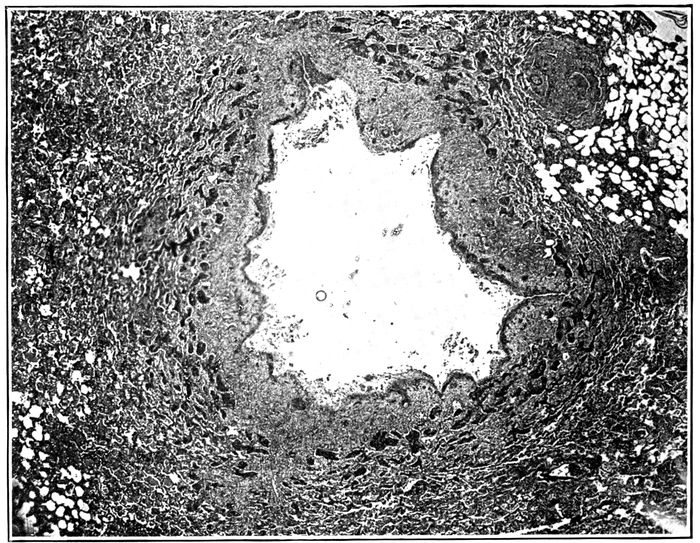
Fig. 17.—Acute bronchiectasis showing fissures penetrating into bronchial wall and at one place entering surrounding alveolar tissue; the surrounding alveoli are filled with fibrin. Autopsy 425.
Acute bronchiectasis may be found following influenza after the illness has lasted eight or ten days. There is no increase of fibrous tissue. Small bronchi with no cartilage, which in normal lungs have a diameter approximating 1 mm., are dilated to 3 mm. or more. The surface epithelium is wholly or partially lost. Necrosis occurs in places and extends deep into the tissue, destroying muscle and often penetrating the entire thickness of the wall which in these small bronchi consists in large part of fibrous tissue containing greatly engorged blood vessels. In this necrotic material nuclei are absent and the tissue containing fibrin stains deeply with eosin. In it occur fissures or tears which extend from the lumen a variable distance, very frequently penetrating the entire thickness of the wall and entering adjacent alveoli (Figs. 17 and 19). Alveoli thus exposed almost invariably contain plugs of dense fibrin. Where these rents have occurred, adjacent edges of the bronchial wall, held together by underlying lung tissue, have separated from one another, so that the circumference of the bronchus has been increased (Fig. 18). These breaks in the continuity of the wall may occur in several places, so that a fourth or a third of the circumference may be formed 247by exposed alveolar tissue which has become the site of fibrinous pneumonia (Fig. 20). During life, though the inflamed bronchus is filled by mucopurulent exudate, distention of loose alveolar tissue, uniting the interrupted bronchial wall, is doubtless greater than it appears in the lung fixed by hardening fluids.
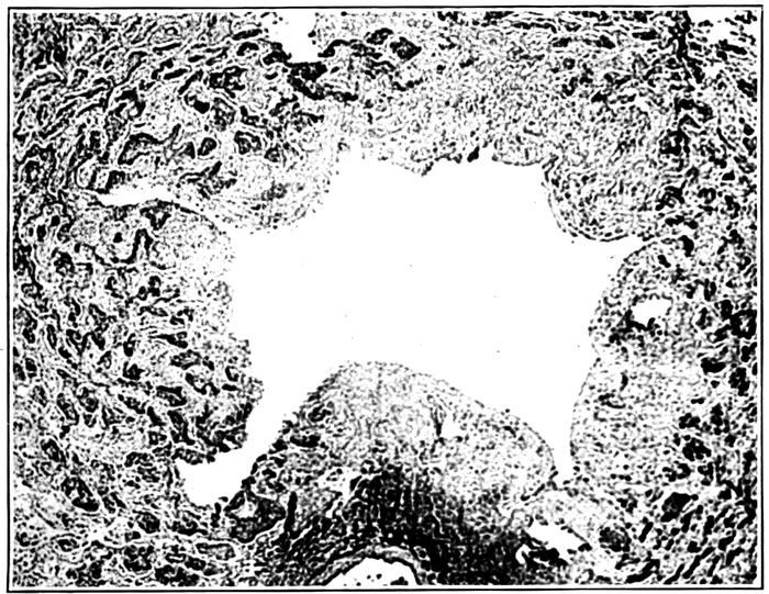
Fig. 18.—Acute bronchiectasis showing fissures in the bronchial wall extending into neighboring alveoli which in zone about are filled with fibrin; one fissure has separated widely; peribronchial fibrinous pneumonia (fibrin is black). Autopsy 425.
Recently dilated bronchi have an irregularly stellate lumen as the result of clefts penetrating at intervals into or through the bronchial wall (Fig. 26). Longitudinal fissures mark the lining of these dilated bronchial tubes.
When the fatal illness has lasted more than two weeks, abundant new formation of fibrous tissue occurs in a zone surrounding the dilated bronchus. Adjacent alveolar walls are thickened by young fibrous tissue. Alveoli, much diminished in size, are filled by hyaline fibrin into which fibroblasts 248and newly formed blood vessels have penetrated. These changes are limited to a wide zone in immediate contact with the dilated bronchus, whereas at a greater distance alveolar walls have undergone no thickening and alveoli contain no fibrin.
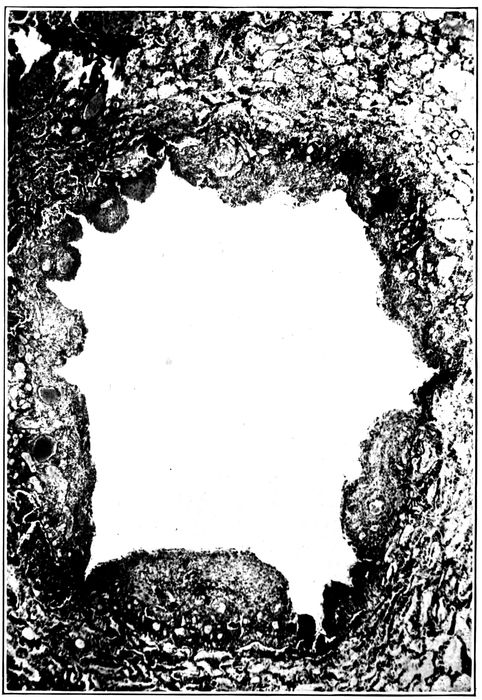
Fig. 19.—Acute bronchiectasis; the bronchial wall indicated by engorged mucosa shows a varying degree of destruction, fissures extending into and through the bronchial wall. Autopsy 352.
249
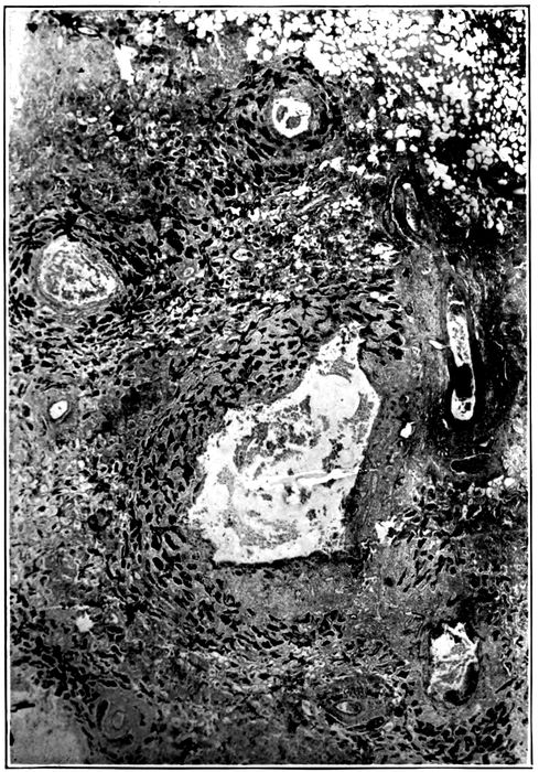
Fig. 20.—Acute bronchiectasis; with destruction of bronchial wall exposing alveoli filled with fibrin; peribronchial fibrinous pneumonia is seen about several bronchi present in the section; Gram Weigert fibrin stain. Autopsy 425.
This stage is well represented by Autopsy 421 after an illness of nineteen days. Bronchiectatic cavities, from 3 to 6 mm. in diameter, are numerous in sections of the lung; their lumina are irregular in outline and often irregularly stellate. Microscopic examination shows the presence of 250clefts which interrupt the bronchial wall at intervals throughout its entire circumference. The original wall is well indicated by the very richly vascularized connective tissue containing scattered muscle bundles and is infiltrated with lymphoid and plasma cells in great number. Where fissures have occurred the adjacent edges of the interrupted wall have separated from one another, leaving a wide interval where underlying alveolar tissue is exposed. Two changes tend eventually to render the fissures inconspicuous, namely, regeneration of epithelium and new formation of fibrous tissue. Exposed alveoli filled with fibrin are in process of organization and epithelium which has assumed a squamous type has grown down over the exposed surfaces of the interrupted bronchial wall. It has begun to cover or in some instances has completely covered the surface of rents entering alveoli plugged with fibrin (Fig. 21). In the periphery of the bronchus alveolar walls are thickened and infiltrated with lymphoid and plasma cells. The same changes affect bronchi containing cartilage which is undergoing atrophy.
The reinforcement of the fissured bronchial wall by new formation of fibrous tissue, by thickening of the interalveolar walls and by organization of fibrin within the alveoli is well shown after four weeks (Autopsy 425; Fig. 28). There are spherical bronchiectatic cavities more than a centimeter in diameter surrounded by a dense fibrous wall in which are atrophied alveoli lined by epithelium of cubical form. Occasionally, the fibrous wall is interrupted and alveoli, plugged with organizing fibrin, are in immediate contact with the lumen. When these plugs of fibrin which are slowly absorbed disappear, evidence of preexisting rents in the bronchial wall are lost, and there are in this lung bronchiectatic cavities of which the wall is a continuous circle of dense fibrous tissue.
251
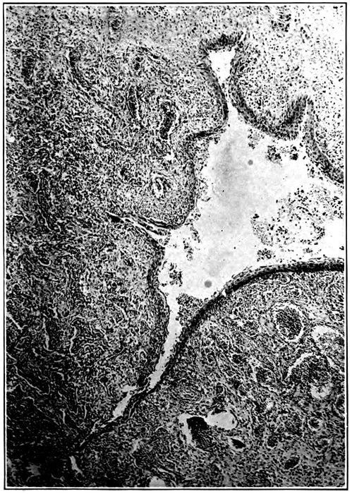
Fig. 21.—Bronchiectasis with fissures extending through the bronchial wall into alveolar tissue which is the site of fibrinous pneumonia; epithelium has grown down into these fissures and has covered the exposed surfaces. Autopsy 463.
252
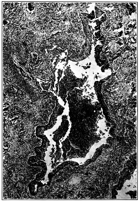
Fig. 22.—Regeneration of epithelium over fissures which have been formed in the wall of a bronchus; the epithelium in the neighborhood of and within the fissure is squamous.
Epithelium lining the dilated bronchi is at times completely destroyed (Fig. 28), but more frequently it persists in part. That which remains has almost constantly the character of squamous epithelium (Figs. 22 and 23). The lowermost cells are cubical; those above them are polygonal, tending to become flatter as the surface is approached; upon the surface are cells often much flattened and occasionally they have lost their nuclei and stain deeply with eosin as the result of superficial necrosis. The change should not be regarded as metaplasia, for the epithelium assumes this squamous type when the superficial columnar cells have been lost. Actual necrosis of superficial ciliated columnar cells is occasionally seen (Autopsy 352); injured cells have separated from one another and desquamated 253into the lumen of the bronchus. The epithelium which remains after the superficial cells are lost consists of cells which become flatter from base to surface, but the intercellular bridges characteristic of the epithelium of the skin are not found. When epithelium is in process of regeneration, a layer gradually diminishing in thickness extends over the denuded surface, the advancing edge being formed by very flat cells in a single layer. The epithelium growing into fissures which have penetrated the bronchial wall may completely cover the exposed alveolar tissue. The newly formed epithelium may follow a fissure into an alveolus which has been opened and come into contact with the fibrin which fills the alveolus.
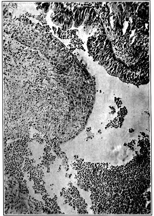
Fig. 23.—Squamous epithelium growing over the defect in the bronchial wall shown in Fig. 22 more highly magnified; squamous epithelium is present above and columnar epithelium below.
254Bronchiectasis usually affects the small bronchi with no cartilage. It is not uncommon to find greatly dilated bronchi with no cartilage in close proximity to cartilage containing bronchi of smaller caliber. In one instance (Autopsy 421) a bronchus of medium size with cartilage measured 3 mm. in diameter, whereas two bronchi with no cartilage were dilated to 4 and 6 mm., respectively. Nevertheless, larger bronchi are occasionally the site of superficial loss of epithelium, necrosis extending into the bronchial wall, formation of fissures and stretching of the wall at the spot which is weakened. In association with these changes atrophy of the cartilage may occur (Autopsies 421, 425, 440, 463). Plates of cartilage in process of atrophy are readily recognized by their irregularly indented outline and often by their small size. The fibrous tissue surrounding the cartilage is the site of chronic inflammation and is densely infiltrated with lymphoid and plasma cells among which polynuclear leucocytes are scant. Nevertheless, polynuclear leucocytes are abundant in immediate contact with the cartilage and appear to have an important part in the solution of its matrix, for about them occur indentations of the edge. Leucocytes penetrate into the cartilage.
The necrosis and tears which occur in the wall of the bronchus are not always limited to the bronchus, but may extend deeply into the surrounding tissue. In Autopsies 312 (Fig. 21) and 423 wide areas of necrosis have penetrated deeply into the tissue about the bronchi.
Autopsy 312.—Illness began with influenza on September 26, seventeen days before death; a diagnosis of lobar pneumonia with consolidation of 255the right lower lobe was made ten days after onset and Pneumococcus IV, B. influenzæ and S. hemolyticus were found in the sputum. At autopsy there was bronchopneumonia with red and gray lobular and confluent lobular patches of consolidation and right and left serofibrinous pleurisy; there was purulent bronchitis; no abscesses were seen. Small bronchi throughout both lungs were dilated and often surrounded by a zone of hemorrhage.
Hemolytic streptococci were found in the heart’s blood, in the pleural exudate, consolidated lung and bronchus; B. influenzæ was found in the lung and in a small bronchus, and staphylococci in the contents of a small bronchus.
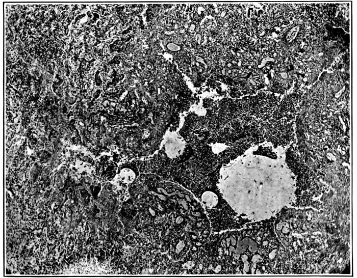
Fig. 24.—Acute bronchiectasis with fissures extending through bronchial wall which is marked by great engorgement of blood vessels; at one point a fissure has penetrated deep into the alveolar tissue and formed a small cavity containing purulent exudate and surrounded by fibrinous pneumonia. Autopsy 312.
Bronchi which are the site of acute inflammation have lost their epithelium wholly or in part, and deep fissures penetrate the entire thickness of the bronchial wall, extending into the surrounding lung tissue which is the site of fibrinous pneumonia. In some instances plugs of fibrin within the alveoli are bisected by these tears. There is some superficial necrosis along the edge of each fissure, in several places extending outward from defects in the walls of small bronchi dilated to approximately 1.5 mm. There are wide patches of necrosis affecting both alveolar walls and contents 256of alveoli and extending 2 mm. into the lung tissue. When a fissure has penetrated from the lumen of the bronchus into necrotic tissue (Fig. 21), polynuclear leucocytes have accumulated within the necrotic tissue, disintegration of tissue occurs, and a small cavity communicating with the bronchus is formed.
Autopsy 423.—C. H., white, aged twenty-five, resident of Oklahoma, had been in military service one month. Death occurred sixteen days after onset of influenza.
Anatomical Diagnosis.—Chronic bronchopneumonia with peribronchiolar consolidation throughout right lung and in left lower lobe; right purulent pleurisy; purulent bronchitis; bronchiectasis at base of left lung.
The right lung weighs 1,260 grams; in the upper lobe are yellowish gray nodules having the appearance of tubercles clustered about small bronchi; in places similar nodules occur upon a background of pinkish gray consolidation occupying the greater part of the lower lobe. Bronchi contain purulent fluid. The left lung weighs 760 grams; it is edematous and small, yellowish gray nodules of consolidation in the lower lobe are clustered about terminal bronchi. Bronchi at the base of the lower lobe are dilated.
Bacteriologic examination shows the presence of hemolytic streptococci in the blood of the heart; hemolytic streptococci and B. influenzæ in the lung.
Microscopic examination shows that the walls of the bronchi are infiltrated with lymphoid and plasma cells; these cells are very numerous in peribronchiolar patches of consolidation. A small bronchus 1 mm. in diameter has squamous epithelium along one side; on the opposite side, the wall is completely absent and there is superficial necrosis of exposed alveoli filled with fibrin. A deep fissure passes from the bronchus into the consolidated tissue; its edges are necrotic and it is filled with polynuclear leucocytes. A small cavity in contact with the bronchus has been formed. In another part of the lung a distended bronchus has lost its epithelium on one side, and here alveoli filled with fibrin form the wall of the bronchus which is filled with leucocytes. Extending outward from the eroded wall is a focus of necrosis where both alveolar walls and contained exudate have lost their nuclei.
The necrosis which has had its origin in the bronchi is soon followed by accumulation of polynuclear leucocytes, softening and disintegration of tissue. Discharge of the disintegrated tissue through the bronchi results in the formation of a small cavity continuous with the bronchus. These changes are well illustrated by the bronchiogenic abscesses which have been described elsewhere (Autopsies 376, p. 206, and 387, p. 206). When disintegrated tissue 257is discharged by way of the bronchi no accumulation of pus occurs, but cavities will be formed, in part by dilation of bronchi, in part by erosion of the adjacent lung tissue. Histologic examination shows that these changes have produced the advanced bronchiectasis found in Autopsy 445 (Fig. 25).
Autopsy 445.—W. F., white, aged twenty-three, from Mississippi, had been in military service one month. His illness began September 22, twenty-seven days before death, with severe coryza, weakness, nausea and vomiting; great pain in bones, cough and sore throat. He was admitted to the base hospital one week later with diagnosis of influenza and bronchitis. On October 3, sixteen days before death, signs of consolidation were found on the left side over the back and a diagnosis of lobar pneumonia was made. On October 18 there was severe headache, pupils were dilated, and there was rigidity of neck; lumbar puncture was made and pneumococci were found in the fluid obtained. Death occurred on the following day.
Anatomic Diagnosis.—Bronchiectasis with unresolved pneumonia limited to the left lower lobe; acute bronchopneumonia with peribronchiolar consolidation in right lung; purulent bronchitis, peribronchial hemorrhage and organizing bronchiolitis in right lung; adherent pleura on left side; purulent meningitis.
The left upper lobe is crepitant throughout. The outer and posterior two-thirds of the left lower lobe is riddled with cavities often rounded and varying in diameter from 0.5 to 3 cm. but not infrequently irregular in shape and in communication with adjacent cavities (Fig. 25). In places cavities pass in a tortuous course from pleura to the midpart of lung. The lining of these cavities is usually smooth, but in places is covered by gray necrotic material. Communication between the cavities and medium-sized bronchi is occasionally found. The lung tissue between the cavities is in part grayish red and consolidated, in part pink and air containing. The right lung is edematous throughout; the bronchi in the lower part of the right lung contain purulent fluid and are in places surrounded by zones of hemorrhage.
The spleen is very large (14 × 11 × 5 cm.) and firm.
The spinal fluid is cloudy and blood vessels over the lumbar enlargement and lower thoracic region are congested; in the upper thoracic region the cord is covered by purulent exudate.
Bacteriologic examination demonstrates the presence of hemolytic streptococci in the blood of the heart; plates from the left lung contain a few colonies of S. aureus and Pneumococcus IV; plates from the right main bronchus contain S. aureus and a large bacillus which does not stain by Gram’s method. Three plates from the spinal meninges contain Pneumococcus IV.
258
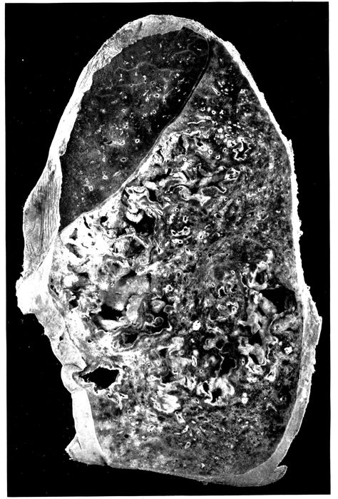
Fig. 25.—Advanced bronchiectasis throughout lower left lobe. Autopsy 445.
259Microscopic examination shows that the cavities which have been described are lined by very vascular connective tissue containing many cells; there is no epithelial lining and the surface is in places covered by fibrin. On the surface polynuclear leucocytes are numerous, but immediately below, large mononuclear cells occur and frequently contain one or several ingested polynuclear leucocytes. None of the structures peculiar to the bronchi can be identified in the wall of these cavities, and in many places it is evident that lung tissue has undergone destruction, for in places the lining of vascular connective tissue is interrupted and an extension of the cavity penetrating into the lung substance is surrounded by alveoli filled with fibrin; in contact with the cavity there is some necrosis.
The cavities communicate with the bronchi and are lined in part by vascular connective tissue which may in part represent preexisting bronchial walls, but no epithelium is present and the relation to the bronchi cannot be established with certainty. These cavities have extended by necrosis which has broken the vascular connective tissue of their wall and penetrated into adjacent lung tissue. Death has been the result of purulent meningitis caused by pneumococcus, and the histologic changes in the walls of the cavities suggest that the activity of the inflammatory reaction here is subsiding, for large mononuclear cells are numerous and are ingesting polynuclear leucocytes. The changes described would, if continued, result in the formation of cavities lined by fibrous tissue and resembling many of those formed as the result of dilatation of the bronchi.
A study of the progress of the changes which result in the formation of bronchiectatic cavities has shown how the inflammatory irritant within the bronchus destroys the epithelium of the bronchus, penetrates into the deeper tissues and produces fissures which extend through the entire thickness of the bronchial wall at one or usually several places. These longitudinal fissures, which at first often give a stellate outline in cross section to the cavity of the affected bronchus, permit the separation of the edges of the fissure, so that an increase in the circumference occurs. The base of the fissure is formed by surrounding alveolar tissue and its edges are the site of necrosis. Tears may extend into the surrounding alveolar tissue, thus permitting further stretching of the bronchial wall. The consequences 260of rupture of the small bronchi into the adjacent alveoli are to some extent overcome by the inflammatory reaction which plugs the adjacent alveoli with fibrin.
Compression of the lungs by forced expiration, even though the glottis were closed as in coughing, would not dilate the bronchi, because pressure outside and within the bronchi would be equally elevated (Thornton and Pratt[89]). The pressure within the bronchi does not differ greatly from atmospheric pressure, whereas the negative pressure within the pleural cavity may vary from approximately 6 mm. of mercury during quiet inspiration to 30 mm. with forced inspiration. Excess of pressure upon the inner surface of the bronchial walls will vary with coughing and other respiratory efforts, between these limits depending upon the readiness with which pressure is equalized within and without the bronchi by penetration of air into the alveoli. The presence of viscid mucopurulent fluid within bronchioles will obstruct these tubules and retard the entrance of air into alveoli.
Weakening of the bronchial wall by the changes which have been described will cause lasting dilatation of the bronchi. Whatever increases pressure within the bronchi will increase the tendency to dilatation; the bronchi being filled with mucopurulent exudate dilatation usually appears first at the bases of the lung, since gravity increases intrabronchial pressure here. New formation of fibrous tissue within the wall of the bronchus, thickening of adjacent alveolar walls, and organization of fibrin reinforce the weakened bronchial wall and limit the dilatation which follows injury to the wall. Regeneration of epithelium covering the dilated tube will further obscure the early changes which have made dilatation possible. The changes which weaken the bronchial wall permit dilatation at a time when there is no new formation of fibrous tissue. When the bronchial lesion has persisted several weeks, 261chronic pneumonia is associated with it. It has been suggested that the contraction of newly formed fibrous tissue within the substance of the lung might cause bronchi to be enlarged by traction upon their walls. Newly formed connective tissue is most abundant in the wall of the bronchiectatic cavity, and here contraction would tend to diminish the size of the cavity.
Unresolved Bronchopneumonia
Chronic bronchopneumonia is characterized by changes similar to those associated with chronic inflammation in other parts of the body, namely, by thickening of the interstitial tissue of the lung, by accumulation of mononuclear cells, by proliferation of fibrous tissue and by organization of exuded fibrin. In a few instances these changes have begun at the end of two weeks after onset of influenza, but they have been little advanced until three weeks has elapsed; advanced chronic inflammation has occurred after from four to eight weeks. Chronic inflammation primarily affects those structures which are most severely injured by the acute lesion and is most conspicuous in immediate proximity to the small bronchi and bronchioles; the perivascular and interlobular connective tissue are secondarily involved. Corresponding to each of the lesions of the alveolar tissue which have been found with bronchopneumonia, namely, peribronchiolar, hemorrhagic peribronchiolar, lobular and peribronchial consolidation, there is a chronic lesion which develops when pneumonia has failed to resolve.
The term interstitial bronchopneumonia has been used by MacCallum to designate a lesion which he has found in association with measles at Fort Sam Houston. This name he states does not describe accurately the early stage of the lesion, for its interstitial character is not evident at first. In his monograph on “Epidemic Pneumonia in the Army Camp,” published in 1919, MacCallum describes and pictures 262instances of the lesion which we have designated interstitial suppurative pneumonia and classifies them as interstitial bronchopneumonia. We have shown that this lesion, which is the result of infection of the lymphatics with S. hemolyticus, bears no necessary relation to the lesion which is characterized in its early stage by peribronchiolar pneumonia and in its later stages by chronic inflammation with mononuclear infiltration and proliferation of the peribronchial, perivascular and interalveolar tissue. At Fort Sam Houston, nearly every patient with measles was infected with hemolytic streptococci; we observed, following influenza, similar prevalence of hemolytic streptococci in certain wards in the base hospital at Camp Pike. Among the cases at Fort Sam Houston there were doubtless instances both of interstitial suppurative pneumonia caused by hemolytic streptococcus and of chronic bronchopneumonia not referable to this microorganism.
Studying pneumonia following influenza at Camp Lee, Va., and later at Camp Dix, N. J., during the fall of 1918, MacCallum reached the conclusion that “interstitial bronchopneumonia” following influenza was caused by B. influenzæ of Pfeiffer. This lesion attributed to B. influenzæ differed from that previously referred to hemolytic streptococcus in the following characters: the lymphatic channels in the bronchial walls and widened interlobular septa are inconspicuous and none are found distended with exudate; there is no intense infection of the pleura, and polynuclear leucocytes are inconspicuous in the alveolar exudate and in the walls of the bronchi. It seems probable these differences are explained by the absence of hemolytic streptococci which tend to invade lymphatics and produce severe inflammatory changes in the pleura.
Chronic Bronchitis.—The earliest changes in the bronchial wall with bronchitis of influenza are hyperemia, leucocytic infiltration and hemorrhage, and they may occur even though the lining epithelium remains intact. Epithelium 263frequently undergoes partial or complete destruction, and with this severe injury the influence of the inflammatory irritant may extend directly through the wall of the bronchus, for in some instances there is hemorrhage into all the alveoli in a zone encircling the bronchus. Since these alveoli have only indirect communication with the affected bronchus through alveolar tissue not involved in the inflammatory process, it is evident that the surrounding hemorrhage is secondary to the lesion of the bronchus. Fibrinous inflammation in other instances, similarly localized in a zone of alveoli encircling a bronchus, is doubtless the result of direct extension of the inflammatory process through the bronchial wall. After the disease has existed during two or three weeks inflammation is still active immediately below the inner surface of the bronchus; here polynuclear leucocytes are numerous whereas in the deeper parts of the mucosa and about the muscularis leucocytes are scant but lymphoid and plasma cells are very numerous. The severity of the inflammatory reaction may be judged by the abundance and extent of this cellular reaction and is in close relation to the intensity of the changes affecting the mucous membrane of the bronchus. Infiltration of the entire bronchial wall with lymphoid and plasma cells is almost invariable when the primary injury to the bronchus has destroyed the epithelial lining, and this infiltration is not limited to the bronchial wall but extends outward into the contiguous alveolar septa which are thickened by it. The sheath of the pulmonary artery which accompanies the bronchus exhibits a similar change, and the alveolar septa, as a fringe about it, are thickened and infiltrated with mononuclear cells. Interlobular septa continuous with the bronchus often show some infiltration.
A later phase in this series of changes is represented by new formation of fibrous tissue. The bronchial walls and interalveolar septa are thickened by proliferating fibrous tissue, young fibroblasts and newly formed collagen fibrils 264being abundant (Fig. 28; also Fig. 30). This increase of fibrous tissue is especially noteworthy immediately surrounding the walls of the small bronchi, which are often considerably dilated, and about the smaller of those bronchi which have cartilage; with thickening of alveolar walls immediately adjacent to the bronchus every stage in the obliteration of the alveoli may be found. Their walls are thickened and their lumina are diminished in size and often flattened in a direction concentric with the bronchus. Such atrophied alveoli lined by cubical epithelial cells occurring within the thickened peribronchial fibrous tissue give evidence that this tissue has replaced alveoli. Alveoli surrounding and within the new fibrous tissue are frequently filled with fibrin, and organization indicated by penetration of fibroblasts and capillaries into the fibrin may be far advanced. There is some increase of perivascular and interlobular tissue. The bronchiectasis which is almost invariably found with unresolved bronchopneumonia has been described. Squamous transformation of epithelium (page 251) is frequently found in association with the chronic bronchitis of unresolved pneumonia.
Organizing Bronchitis and Bronchiolitis.—When the bronchial epithelium is destroyed, fibrin is deposited upon the denuded surface and may partly or completely fill the lumen of the bronchial tube. The plug of fibrin is adherent to the underlying tissue wherever epithelium is lost but is separated from the bronchial wall by a well-defined space where epithelial lining is still intact. Fibroblasts promptly migrate from the wall of the bronchiole into this fibrin, and fibroblasts, fixed during ameboid movement, are irregularly elongated in a direction toward the fibrin.
Organization of fibrin occurs within the smallest bronchi (diameter 0.3 to 0.5 mm.) or within respiratory bronchioles. It has been found in 8 autopsies. In one instance it has been present eleven days after the onset of influenza, but usually it is seen three or four weeks after onset of symptoms 265of respiratory disease. In the early stages of the lesion a plug of fibrin within the lumen of the bronchus or bronchiole is invaded by fibroblasts, plasma cells and newly formed capillaries. These capillaries have their origin in the wall of the tube and enter the fibrin at points where in consequence of loss of epithelium fibrin is continuous with the connective tissue. When the bronchiole is cut longitudinally, partially or completely organized fibrin may be found adherent at several places with intact epithelium, sometimes beautifully ciliated, between the sites of attachment. The fibrin is finally replaced completely and the lumen of the bronchiole contains a mass of organized fibrous tissue in which young fibroblasts and plasma cells are numerous.
The lesion has been associated with chronic bronchopneumonia in 6 of 8 instances. In Autopsy 445, p. 257, organizing bronchitis and bronchiolitis occurred in the right lung unassociated with other chronic lesion, although there was advanced bronchiectasis with fibrous induration in the left lung. In Autopsy 499 (p. 224) organizing bronchiolitis occurred in association with chronic changes which appear to have followed interstitial suppurative pneumonia caused by S. hemolyticus. Other severe lesions of the bronchi have accompanied organizing bronchitis and bronchiolitis. Purulent bronchitis has been present in 7 of 8 instances; bronchiectasis in 5 of 8 instances.
The bacteriology of autopsies with organizing bronchitis and bronchiolitis is shown in Table LIII.
The bacteriology of these cases presents no constant feature. Invasion of the blood by S. hemolyticus has been present in a large proportion of cultures, namely, in 5 of 7 (71.4 per cent). In one of the 2 instances in which hemolytic streptococci have been found, neither in the blood nor lungs, Pneumococcus III has been found in the blood and S. viridans in the lungs and bronchus; in the other, S. aureus has been found in the lung and bronchus. Staphylococci have been found frequently in the bronchi (60 per cent) and in the lungs (50 per cent). B. influenzæ has been present in the bronchi in the usual proportion of instances (80 per cent). The lesion has occurred in the presence of B. influenzæ combined with streptococci or staphylococci.
266
Table LIII
AUTOPSY DURATION OF ILLNESS BLOOD LUNGS BRONCHUS
420 11 days S. hem. S. hem., B. inf., S. aur.
402 14 days Pneum. IV, S.hem.
370 17 days S. aur. S. aur., Pneum. IV, B. inf.
457 17+ days Pneum. IV, B. inf.
421 19 days S. hem. Pneum. IV, S. hem.
445 27 days S. hem. Pneum. IV, S. aur. S. aur.
473 28+ days Pneum. III S. vir. B. inf., S. vir., staph., M. catarr.
499 36 days S. hem. S. hem. B. inf.
Thrombosis of lymphatics in the wall of bronchi adjacent to blood vessels and in interlobular septa occurs, and occasionally organization of the fibrinous plug within the lymphatic is in progress (Autopsies 283, 425 and 463). Fibroblasts and capillaries penetrate from the wall of the lymphatic into a mass of hyaline fibrin which fills the lumen.
Unresolved Bronchopneumonia.—The most common type of pneumonic lesion following influenza is characterized by acute inflammation of the alveoli immediately adjacent to the bronchioles and the lesion is associated in many instances with hemorrhage or edema. If this lesion persists unresolved during several weeks, evidences of chronic inflammation are found. Peribronchial, perivascular and interlobular connective tissue is thickened and richly infiltrated with lymphoid and plasma cells, large mononuclear cells and many young fibroblasts. Interalveolar septa adjacent to the walls of bronchi and between alveoli surrounding inflamed bronchioles are implicated in the process. 267Interstitial changes characterize the lesion only in its late stage. It appears undesirable to give the name “interstitial pneumonia” to the early stage of a lesion which begins and in most instances terminates as an acute relatively superficial inflammation of the bronchi, bronchioles and peribronchiolar alveoli.
Chronic bronchopneumonia is often overlooked at autopsy because newly formed connective tissue is not present in sufficient quantity to attract attention (Fig. 26). When the lesion is advanced conspicuous gray white patches of fibrous tissue may be seen about the bronchi (Autopsy 487; Fig. 27) and interlobular septa may be obviously thickened (Autopsy 472). The most distinctive feature of the lungs is the presence of small, firm, gray or yellowish gray nodules of consolidation which resemble miliary tubercles. They represent the peribronchiolar patches of bronchopneumonia present during the acute stage and have assumed the well-defined outline and firm consistence of tubercles because polynuclear leucocytes and red blood corpuscles have in large part disappeared, interstitial tissue is increased, and exudate is in process of organization. These nodules are grouped in clusters about the small bronchi.
With unresolved bronchopneumonia the lungs are very voluminous and fail to collapse after they are removed from the chest and in some instances even after incision. The air containing tissue is usually dry. In our autopsies the lungs have been pink in color and often free from coal pigment, because those suffering with pneumonia have been in considerable part men from rural districts. Thick mucopurulent material exudes from the small bronchi which have been cut across; purulent bronchitis has been present in 20 of 21 instances of chronic bronchopneumonia. Bronchiectasis has been present in 13 instances; dilatation is often advanced, so that throughout the lungs are found bronchi with no cartilage distended to a diameter of 0.5 cm. In addition to the firm peribronchiolar tubercle-like nodules of consolidation there are scattered patches of gray lobular or confluent lobular consolidation. Yellowish nodules, grouped about bronchi and resembling those found elsewhere in air containing tissue, are occasionally seen scattered upon the cut surface of a patch of gray, confluent lobular consolidation (Autopsies 421, 423, 431).
268
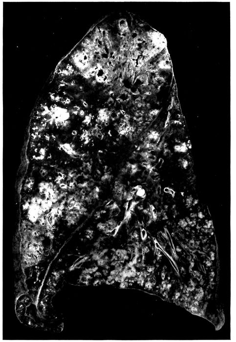
Fig. 26.—Unresolved bronchopneumonia with tubercle-like nodules of peribronchiolar consolidation best seen in lower lobe; bronchiectasis. Autopsy 425.
269Microscopic examination demonstrates the presence of those changes which have been described in association with chronic bronchitis and bronchiectasis. There is abundant new formation of fibrous tissue about the bronchi of small and medium size, thickening of adjacent interalveolar walls and incorporation of alveoli into the thickened bronchial wall (Figs. 27, 28, 30, and 31). In half of the instances of chronic bronchopneumonia there has been peribronchial fibrinous pneumonia, and organization of fibrin within the alveoli is usually well advanced. In one instance (Autopsy 487; Figs. 27 and 28) after an illness of fifty-five days this process has resulted in the formation of conspicuous patches of firm, grayish white fibrous tissue surrounding dilated bronchi. Organization of fibrinous exudate within the lung has not been limited to the alveoli but has occurred in the bronchioles as well. Organizing bronchiolitis has been present in 5 instances (Autopsies 370, 402, 457 and 473).
Increase of fibrous tissue occurs about the blood vessels and in the septa between the lobules, which are infiltrated with mononuclear wandering cells and fibroblasts. Dilatation and thrombosis of the lymphatic vessels have occurred in both situations, and in 3 instances (Autopsies 283, 425 and 463) organization of these fibrinous thrombi has occurred.
270
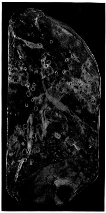
Fig. 27.—Unresolved pneumonia with peribronchial formation of fibrous tissue; bronchiectasis. Autopsy 487.
271
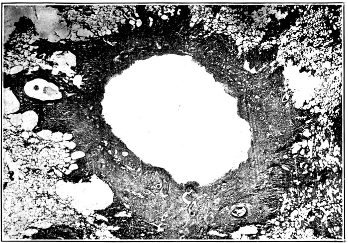
Fig. 28.—Unresolved pneumonia with bronchiectasis showing new formation of fibrous tissue about a greatly dilated bronchus of which the epithelial lining has been lost. Autopsy 487.
Thickening, cellular infiltration and fibrosis of the bronchial walls with interstitial inflammation and fibrosis of immediately adjacent alveolar septa are found about the ramifications of the bronchial tree and may be followed to the smallest bronchi. When the respiratory bronchioles are reached it will be found that the alveoli which stud their walls are implicated in the change. The fibrin which they contain is infiltrated with lymphoid and plasma cells, and with progress of the lesion is invaded by fibroblasts and capillaries. Infiltration and fibroid thickening extends from the bronchiolar wall to the alveolar septa continuous with it (Fig. 31 with measles). Similar changes occur about the alveolar ducts, and about the orifices of the tributary infundibula (Fig. 32), peribronchiolar foci of acute inflammation having assumed the characters of a chronic inflammatory process. Fibrin within the alveoli contains round cells and fibroblasts. With thickening of alveolar walls the alveolar lumina may be much diminished in size and often persist as spaces lined by cubical cells. Polynuclear leucocytes are usually numerous within the alveolar duct and 272in a few alveoli immediately adjacent to it, but elsewhere throughout the focus of inflammation round cells are predominant. The changes which have been described correspond with the transformation of ill-defined, gray or reddish gray spots of consolidation grouped about the terminal bronchi into firm sharply defined grayish white nodules having the consistence and appearance of miliary tubercles.
One of the most constant characters of pneumonia following influenza is its hemorrhagic character. In the earlier stages of pneumonia phagocytosis of red blood corpuscles by large mononuclear cells is frequently seen. In association with the chronic changes which have been described, large mononuclear cells filled with brown pigment, doubtless formed from red corpuscles, are often found within the alveoli. These pigment containing cells are similar to those commonly associated with chronic passive congestion of the lungs.
In one instance (Autopsy 457) hemorrhagic peribronchiolar pneumonia has been found in process of organization. The bronchioles and alveoli adjacent to them contain polynuclear leucocytes, but intervening alveoli almost uniformly contain blood and are the site of new formation of connective tissue. Interalveolar septa are thickened and alveoli which are lined by cubical epithelium are often diminished in size. In many places fibroblasts have penetrated in considerable number into the blood within the alveoli and occasionally newly formed capillaries are found within them.
Lobular patches of pneumonia are often found in process of organization (Autopsies 370, 421, 423, 433, 463, 472 and 473). Microscopic examination shows that whole lobules well defined by thickened septa are the site of chronic interalveolar inflammation and intraalveolar organization of exudate, whereas adjacent lobules are air containing and relatively normal. In the earlier stages of the process fibrin present within the alveoli is invaded by fibroblasts, 273mononuclear wandering cells and blood vessels but in the later stages fibrin has disappeared; the lumina of the alveoli are occupied by cellular fibrous tissue and in places the thickened alveolar walls and intraalveolar fibrous tissue have been fused to form wide patches of new tissue.
With chronic bronchopneumonia confluent lobular consolidation occasionally has a gray ground upon which are scattered small yellow spots clustered about the small bronchi (Autopsies 421, 423 and 431). Microscopic examination has shown that the yellowish spots correspond to dilated bronchioles filled with purulent exudate and surrounded with alveoli containing many polynuclear leucocytes. In the interstitial tissue about the bronchiole and between adjacent alveoli plasma cells are often present in great number. Between these spots of subacute bronchiolar inflammation lung tissue is the site of interalveolar proliferation of fibrous tissue and intraalveolar organization of exudate.
In all instances of chronic bronchopneumonia there has been peribronchial pneumonia in a zone encircling small bronchi with no cartilage and the smallest of the bronchi which have cartilage in their wall; thickening of interalveolar septa, organization of peribronchial fibrinous pneumonia and partial disappearance of alveoli have been described. In the following autopsy peribronchial fibroid pneumonia has been so advanced that conspicuous patches of gray white tissue surrounding bronchi have replaced in some parts of the lung a considerable part of the lung substance.
Autopsy 487.—W. C., white, aged twenty-seven years, a farmer from Mississippi had been in military service twenty-one days. Illness began on September 17, fifty-five days before death, with chill, fever, cough, backache, pain in the chest and coryza. The patient was admitted two weeks after onset with the diagnosis of influenza. Eight days later his sputum was blood tinged and there were signs of bronchopneumonia. One month after admission the patient developed a rash and a diagnosis of scarlet fever was made.
274Anatomic Diagnosis.—Chronic bronchopneumonia with peribronchial fibroid induration; bronchiectasis; purulent bronchitis; abscesses at the bases of both lungs; seropurulent pleurisy on the left side.
The body is much emaciated. The left pleural cavity contains 650 c.c. of opaque, dull yellow, thin, purulent fluid. The surface of the left lung is covered in spots by white partially organized fibrin.
On section of the right lung (Fig. 27) the tissue is found in great part air containing but there are numerous firm, gray patches, irregular in shape and from 1 to 2 cm. across. In these spots the tissue is tough and resembles fibrous tissue; within them are much dilated bronchi. In the central part of the upper lobe is a group of cavities with smooth wall, the largest of these cavities being 12 mm. in diameter; immediately adjacent are dilated bronchi. Between and surrounding these cavities is gray tissue, like that described above. Below the outer surface of the upper lobe is an extensive area 7 cm. from above downward, thickly studded with bronchiectatic cavities, in the walls of which there is tough fibrous tissue. In the middle lobe are several dilated bronchi, the largest of which is 7 mm. in diameter, and elsewhere occur dilated bronchi with thickened walls. At the base of the lung below the pleura are two abscesses, which are yellow in the center and surrounded by hemorrhagic tissue. At the posterior part of the lower lobe there are numerous firm, nodular, yellowish spots grouped in clusters upon a background of red, air containing tissue. The bronchi throughout the lung contain mucopurulent fluid.
In the left lung patches of fibrous tissue are more numerous than on the right side and are irregular in shape, from 1 to 2 cm. across and most abundant in the center of the upper lobe. This fibrous tissue is in great part gray but in places it has a yellowish tinge. The bronchi everywhere are moderately dilated. At the base of the lung below the pleura is an abscess.
The other organs show no noteworthy change.
Bacteriologic Examination.—The fluid in the left pleura and right main bronchus contain S. hemolyticus. B. influenzæ is found in the right lung and right main bronchus.
Microscopic examination shows that the patches of dense fibrous tissue seen at autopsy almost invariably surround dilated bronchi with no cartilage in their walls (Fig. 28) and with a diameter of from 1 to 2 or more millimeters. These bronchi have lost their epithelial lining; they contain polynuclear leucocytes, and their wall in contact with the lumen is infiltrated to a varying distance with the same cells. Their inner surface is very irregular, and superficial necrosis occurs. The limits of the preexisting bronchial wall is no longer recognizable in the dense surrounding fibrous tissue richly infiltrated with lymphoid and plasma cells. In contact with the bronchus, often in a wide zone, all traces of alveoli have been destroyed, but further outward alveoli are represented by spaces lined by cubical epithelium. At the periphery of the zone of fibroid induration alveolar walls are much thickened and richly infiltrated with mononuclear wandering cells; the lumina of the alveoli contain plugs of organized fibrous tissue often covered by flat or cubical epithelium. In the surrounding tissue a few small bronchi are lined by columnar 275epithelium; there is scant new formation of fibrous tissue but the alveolar walls are thickened and infiltrated with cells. Epithelium of the larger bronchi with cartilage in their walls is usually intact and there is about them little peribronchial inflammation.
Advanced induration about the bronchioles represents a late stage of chronic peribronchiolar pneumonia. A bronchiole cut transversely is found in the center of a focus of induration situated within relatively normal air containing lung tissue. Next the bronchiole which in some instances has wholly or partly lost its epithelium there is very cellular fibrous tissue; further from the bronchiole alveoli are much diminished in size, lined by flat or cubical epithelium and separated by thick cellular walls. Plugs of cellular fibrous tissue sometimes fill the alveolar duct. In favorable sections, cut in a plane which shows the alveolar duct opening out into infundibula, it is found that newly formed fibrous tissue surrounds the alveolar duct and extends into the walls of its tributary alveoli; alveoli may be obliterated by this fibrous tissue. Induration of alveolar walls is evident along the proximal part of the infundibula which are readily demonstrable because they are much dilated. (See Fig. 32.) The distal parts of the infundibula are surrounded by alveoli with delicate walls.
One bronchus retains along one side part of its epithelium which has assumed a squamous form. In other places the wall has undergone necrosis which at one spot extends deeply into the surrounding tissue. Necrotic tissue in another part of the circumference is infiltrated with polynuclear leucocytes and separated from the surrounding tissue by a space filled with leucocytes. An abscess communicating with the bronchus is thus formed.
The foregoing instance is an example of the chronic fibroid pneumonias with bronchiectasis which occur as sequelæ of the epidemic of influenza. It is not improbable that a considerable number of those who suffer with chronic bronchitis and bronchiectasis following influenza have less extensive lesions similar to those which have been described.
276Bacteriology of Unresolved Bronchopneumonia.—Bacteria found in the bronchi in 10 instances of chronic bronchopneumonia have been as follows:
Bacteria in Bronchi with Chronic Bronchopneumonia
B. coli 1
B. influenzæ and pneumococcus 1
B. influenzæ and S. hemolyticus 2
B. influenzæ and staphylococcus 1
S. hemolyticus and B. coli 1
B. influenzæ, pneumococcus and staphylococcus 3
B. influenzæ, S. viridans and M. catarrhalis 1
Bacteria found in the lungs in 17 instances of chronic bronchopneumonia were as follows:
Bacteria in Lungs with Chronic Bronchopneumonia
B. influenzæ 1
Staphylococcus 1
S. viridans 1
B. influenzæ and pneumococcus 1
B. influenzæ and S. hemolyticus 3
B. influenzæ and staphylococcus 3
Pneumococcus and S. hemolyticus 1
S. hemolyticus and B. coli 2
B. influenzæ, S. hemolyticus and staphylococcus 3
No organism found 1
A noteworthy feature of these lists is the multiplicity of microorganism found, namely, B. influenzæ, S. hemolyticus, pneumococcus, staphylococcus, S. viridans, B. coli, and M. catarrhalis. More than one microorganism is usually found in both bronchus and lung. In the one instance (Autopsy 472) in which B. coli alone has been found in the bronchus, B. coli and S. hemolyticus have been found in the lung and hemolytic streptococcus in the blood; it is evident that B. coli alone has not been responsible for the lesion. In one instance (Autopsy 487) B. influenzæ alone has been found in the lung but hemolytic streptococci have been found in the bronchus, pleura and blood of heart; with S. aureus alone in the lung (Autopsy 370), S. aureus, Pneumococcus IV and B. influenzæ have been found in the bronchus. With S. viridans alone in the lung (Autopsy 473), 277Pneumococcus III has been found in the pleura and in the blood of the heart and has doubtless had an important part in the production of pneumonia; S. viridans, M. catarrhalis and B. influenzæ have been found in the bronchus in this instance.
No single microorganism is associated with the lesions but combinations of B. influenzæ with hemolytic streptococci or staphylococci are common (over 50 per cent). In Autopsy 422 B. influenzæ and Pneumococcus atypical II have been present in the lungs. Among 10 instances in which cultures have been obtained from the bronchus B. influenzæ is found 8 times, and in the 2 instances in which it has not been identified B. coli has been present. B. influenzæ has seldom been found (Table XXVII) in the presence of B. coli, and it is not improbable that B. coli outgrows and obscures the presence of B. influenzæ.
Table LIV shows the per cent incidence of pneumococci, hemolytic streptococci, staphylococci and B. influenzæ in the bronchus, lung and heart’s blood with chronic bronchopneumonia and serves as an index of the readiness with which each of these microorganisms passes from bronchus to lung and from lung to the blood in this disease.
Table LIV
PNEUMOCOCCUS PER CENT POSITIVE HEMOLYTIC STREPTOCOCCUS PER CENT POSITIVE STAPHYLOCOCCUS PER CENT POSITIVE B. INFLUENZÆ PER CENT POSITIVE
Bronchus 40.0 30.0 50.0 80.0
Lung 12.5 56.2 37.5 68.7
Blood 16.6 55.6 0 0
Comparison of Table LIV with the analogous figures for acute bronchopneumonia shows little noteworthy difference. Pneumococci are less frequently found in the lung (12.5 per cent) and in the blood (16.6 per cent) with chronic bronchopneumonia than with acute bronchopneumonia (lung 43.9 per cent; blood, 40.3 per cent). Hemolytic streptococci and staphylococci are not more frequently 278found with unresolved than with acute bronchopneumonia and failure to resolve cannot be referred to either or to both microorganisms, for bronchopneumonia not infrequently remains unresolved in their absence. B. influenzæ is present in the bronchi in at least 80 per cent of instances and perhaps in all; it is usually combined both in the lungs and in the bronchi with one of the pyogenic cocci.
The severity of the injury to the walls of bronchi resulting in continued infection with a variety of bacteria, appears to be the factor determining failure of resolution and the persistence of bronchopneumonia.
The Relation of Unresolved Bronchopneumonia to Interstitial Suppurative Pneumonia Caused by Hemolytic Streptococci.—Hemolytic streptococci have been present in a considerable proportion of those who have had unresolved bronchopneumonia and its occurrence in the bronchi, lung and blood of the heart indicates that it has had an important part in causing death. Unresolved bronchopneumonia, following measles, designated by MacCallum “interstitial bronchopneumonia” in a series of autopsies at Fort Sam Houston in the spring of 1918, was constantly associated with hemolytic streptococci. Among the lesions described as interstitial bronchopneumonia was at least one which was evidently what we have designated interstitial suppurative pneumonia. Lymphangitis was not infrequently found with “interstitial bronchopneumonia” following measles. At Camp Lee and Camp Dix, following the epidemic of influenza, MacCallum found “interstitial bronchopneumonia” with no hemolytic streptococci and noted that lymphatics in the interstitial septa were inconspicuous and that none was found distended with exudate; empyema was not present.
We have shown that interstitial suppurative pneumonia is an acute lesion caused by hemolytic streptococci. Unresolved bronchopneumonia is accompanied by chronic pneumonia and has no necessary relation to this microorganism.
279In a foregoing section we have described instances of interstitial suppurative pneumonia unaccompanied by chronic changes, and in the present section we have described instances of unresolved bronchopneumonia with no infection by hemolytic streptococci. We have pointed out that the incidence of streptococcus infection with unresolved bronchopneumonia does not materially differ from that with acute bronchopneumonia even though the greater duration of the disease gives more opportunity for infection. In some of the autopsies made by MacCallum at Fort Sam Houston, lesions of streptococcus infection doubtless coexisted with unresolved bronchopneumonia.
In the 3 autopsies described below, interstitial suppurative pneumonia with empyema caused by hemolytic streptococcus occurs in association with unresolved bronchopneumonia.
Autopsy 420.—J. E. S., white, aged thirty-two years, born in England and resident of Los Angeles, Cal., had been in military service one month. Onset of illness began on October 3, eleven days before his death. He was admitted to the hospital on the following day with the diagnosis of influenza and acute bronchitis. Pneumonia believed to be lobar was recognized eight days after admission.
Anatomic Diagnosis.—Unresolved bronchopneumonia with hemorrhagic peribronchiolar consolidation in right lung; interstitial suppurative pneumonia with consolidation in left upper lobe; fibrinopurulent pleurisy; purulent bronchitis.
The left pleural cavity contains 200 c.c. of turbid yellow fluid in which are flakes of fibrin. In the inner and upper part of the left upper lobe there is an area of consolidation where the tissue has a cloudy, pinkish gray color and is finely granular on section. Here the interstitial septa are distended by edema, so that they are in places 0.5 c.c. across; in some spots they have a bright yellow color. In the posterior parts of the middle and lower lobes there is flabby consolidation where the tissue has a cloudy, red color with scattered ill-defined yellow spots.
Bacteriologic examination shows the presence of hemolytic streptococci in the blood of the heart; hemolytic streptococci with B. influenzæ and S. aureus in the left lung and S. hemolyticus with S. aureus in the right lung.
Microscopic examination shows that bronchi, bronchioles, alveolar ducts and the greater part of the infundibula are filled with polynuclear leucocytes, whereas the alveoli surrounding these structures contain fibrin. The walls of the small bronchi are thickened and contain mononuclear cells; the adjacent 280alveolar walls are similarly infiltrated and thickened and the fibrin within them is undergoing organization, being invaded by plasma cells, fibroblasts and newly formed blood vessels. In some sections interstitial septa are distended by edema and contain fibrin in abundance; in places the tissue contains polynuclear leucocytes closely packed together. There are lymphatics greatly distended by polynuclear leucocytes with some fibrin, lymphocytes and red blood corpuscles.
Autopsy 428.—D. B., white, aged twenty-five, a farmer from Oklahoma, had been in military service three weeks. Onset of illness was on September 21, twenty-five days before death, with fever, cough and mucopurulent expectoration. The patient was admitted with the diagnosis of acute bilateral bronchitis. Four days later bronchopneumonia was recognized, and subsequently there was otitis media and empyema; 600 c.c. of thin, purulent fluid were aspirated from the right chest three days before death.
Anatomic Diagnosis.—Unresolved bronchopneumonia; suppuration of interstitial tissue of upper right and lower left lobes; purulent bronchitis; fibrinopurulent pleurisy; thoracotomy wound at the base of the right chest; collapse of both lungs; serofibrinous pericarditis.
The left pleural cavity contains 550 c.c. of turbid seropurulent fluid in which are numerous flakes of soft fibrin. The right pleural cavity contains 150 c.c. of similar fluid. The mediastinum is edematous. The pericardial cavity contains 50 c.c. of yellow fluid.
The right lung is moderately collapsed. In the upper and lower lobes are small patches of red, lobular consolidation. The upper third of the upper lobe is laxly consolidated and near its inner surface the interstitial septa are thickened to from 1 to 1.5 mm. in width, and at intervals occur bead-like swellings from which creamy purulent fluid exudes upon the cut surface. In the left lung small patches of gray consolidation occur throughout the lower lobe and here the interstitial septa are thickened, beaded and contain purulent fluid.
Bacteriologic examination shows that the blood contains S. hemolyticus; from the right lung and from the right main bronchus hemolytic streptococci and B. influenzæ are grown.
Microscopic examination shows that the epithelium of the bronchi has undergone hypertrophy; the wall is infiltrated with lymphoid and plasma cells and thickened by new formation of fibrous tissue; there is similar thickening of adjacent alveolar septa and alveoli, often lined by cubical cells, are diminished in size. Connective tissue about the blood vessels and the interstitial septa are thickened and infiltrated with mononuclear cells. In parts of the lung the interstitial septa are edematous and contain polynuclear leucocytes, in some places in great number. Lymphatics are greatly dilated and filled with polynuclear leucocytes which in the center of some lymphatics have undergone necrosis. In one place a small abscess is in contact with a distended lymphatic. Lymphatics contain Gram-staining cocci in pairs and short chains, present in immense number where necrosis has occurred.
Autopsy 433.—B. J., white, aged twenty-seven, from Arkansas, has been in military service one month. Onset of illness was on September 28, nineteen 281days before death, with cough and expectoration. Pneumonic consolidation was recognized two days later and 20 c.c. of cloudy fluid were aspirated from the left chest on the same day. Hemolytic streptococci were found in a culture from the throat nine days before death.
Anatomic Diagnosis.—Unresolved bronchopneumonia with peribronchiolar and confluent lobular consolidation; interstitial suppuration of the right lower lobe; purulent bronchitis; fibrinopurulent pleurisy.
The right pleural cavity contains 700 c.c. of yellowish gray purulent fluid containing flakes of fibrin. The left pleural cavity contains seropurulent fluid localized over the external part of the lung.
The right lung is voluminous and free from consolidation save at the lower and posterior part of the lower lobe where the tissue is deep red and studded with firmer spots of yellow color clustered about the bronchi. In places the interstitial septa are thickened and yellow. Surrounding some of the bronchi near the apex of the left lung are red patches of consolidation.
Culture from heart’s blood remained sterile. S. hemolyticus was grown from right pleural cavity, and S. hemolyticus and B. influenzæ were grown from the right lung. Culture from the left lung contained S. aureus and contaminating microorganisms.
Microscopic examination shows the presence of peribronchiolar patches of pneumonia in which there are few polynuclear leucocytes and many lymphoid and plasma cells; the alveolar walls are thickened and infiltrated with mononuclear cells. In some sections the tissue is wholly consolidated and the site of advanced organizing pneumonia. Interlobular septa and connective tissue about blood vessels are thickened and cellular. Small bronchi have lost their epithelial lining, their walls are thickened and there is peribronchial organizing pneumonia. In some sections the lymphatics are immensely dilated and distended with polynuclear leucocytes. There is necrosis of the walls of the lymphatics and of the polynuclear leucocytes within the lumen.
In the discussion of acute bronchopneumonia it has been shown that S. hemolyticus is not infrequently a secondary invader of a pneumonic lesion perhaps caused by pneumococci. With progress of the disease hemolytic streptococci persist. In the autopsies with unresolved pneumonia just described, hemolytic streptococci have found their way into the lymphatics and produced suppurative lymphangitis with inflammation of the interstitial septa of the lung.
282