Chapter III · THE EXTERNAL AND ANATOMICAL STRUCTURE OF ANTHROPOID APES, COMPARED WITH THE HUMAN STRUCTURE.
Reading edition
In order to complete as far as possible the description which we propose to give of the general natural history of these remarkable animals, it is necessary to examine their anatomical structure. Yet it is not so much our aim to give a detailed and exhaustive description of their anatomy, as to glance rapidly at those peculiarities of their inner structure which catch the eye. It seems to me expedient in this case to follow the method of systematic and descriptive anatomy, and to take the several natural organs in succession. This method, which has long prevailed for studying the structure of the human body, should also be our guide in our researches in comparative anatomy. Our readers need scarcely be told that the anatomy of anthropoids is only a small branch of the comparative anatomy of vertebrate animals in general.
I begin by considering the bony structure of anthropoids, and, in particular, of the gorilla. And it will be well to note the important differences between the structure of the skull of a young and aged male, and of a young and aged female gorilla.
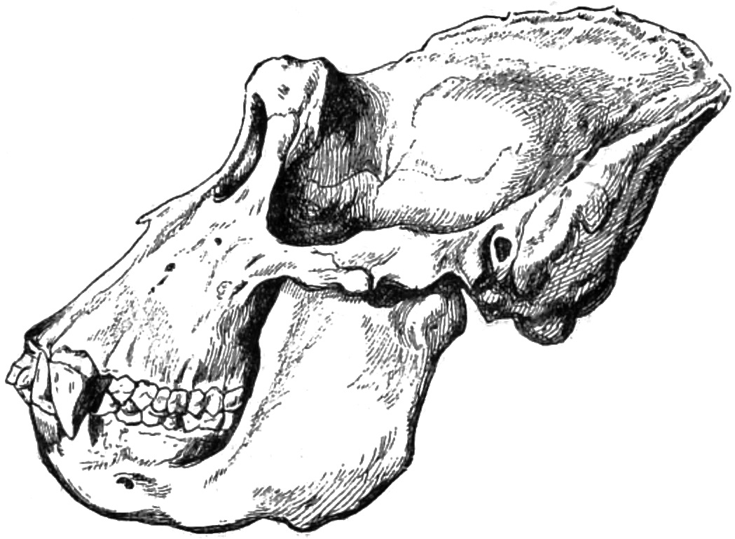
Fig. 15.—Skull of an aged male gorilla in profile.
The skull of the aged male animal is large and heavy. Its average weight is one and a quarter kilogrammes. The longitudinal diameter, from the alveolar point of the upper jaw to the occipital point, may be as much as 294 mm. The overhanging orbits are high in front, and flattened off behind, and their upper edges unite to form a ridge in the middle of the face. To these the back parts of the orbits are attached, in shape like a truncated cone, round and prominent in front, and narrowing into bony capsules in the direction of the brain-pan. They open directly in front, and the aperture is generally in the form of a regular square. The edges are seldom so blunted off as to present a figure somewhat approaching to a circle (comp. Figs. 15, 16). The frontal bone, which in the young of both sexes is high, broad, and arched, becomes depressed in the centre in the aged male. The temporal ridges, thickened to a hem, pass over this to the coronal crest.
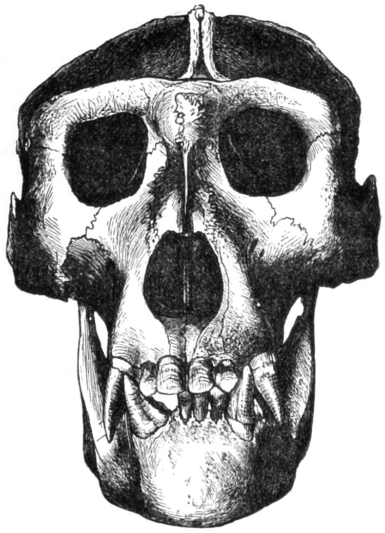
Fig. 16.—Front view of the skull of an aged male gorilla.
This crest is highly characteristic. It begins in the region of the frontal bone, and, rising abruptly, unites itself with the transverse occipital crest. It is of varying height,12 but is rarely altogether absent in an adult male animal. On the top of this coronal crest we may see the two well-developed bony ridges which almost touch each other, and which indicate the upper limits of the temporal muscles on either side. In young animals these ridges tend downwards over the sides of the head, below the vertex of the skull. Their position and direction vary with the growth of the skull, and correspond with that of the coronal crest. The transverse occipital crest is of considerable height in the case of aged and vigorous animals, and is frequently somewhat concave in front, and convex at the back. The fore surface of this crest is formed of the two parietal bones, the hinder surface of the squamose portion of the occipital bone. The lambdoidal suture is on the top of this occipital crest, and in this case, as in that of other mammals, including man, it unites the parietal bones with those of the occiput. The point of union between the coronal and occipital crests divides the latter into two symmetrical lateral halves, curving outwards and downwards. The high, wide squamose portion of the occipital bone is somewhat flattened behind, or more rarely arched, while it is abrupt at its base and in some degree in front. Six curved lines, three on either side, opposite each other, sometimes mark the limits of the attachments of the cervical muscles on the head. The mastoid process of the temporal bone is present, but Brühl could find no trace of a styloid process on the skulls of gorillas and chimpanzees.
The squamous portion of the temporal bone is often connected with the frontal bone by the process termed Virchow’s frontal process of the temporal bone. The nasal bones are high, very narrow in their upper part, and widening below. When they are united in the centre of the nasal bridge, a sloping, keel-shaped projection may often be observed. The inferior turbinated bones of the nasal cavity are remarkable for their size. In the skulls of young animals the inter-maxillary bones, which are in all anthropoids early united with those of the same region, stand up high and peaked between the nasal bones and those of the upper jaw.
The crowns or prominent external surfaces of the enormous canine teeth project in the centre of the face on either side like pillars, just below the nostrils, and extend above and below the row of teeth in the two upper jaws (see Fig. 16). In this way the crowns of the canine teeth form a retreating triangular space, of which the base-line of the equilateral triangle corresponds with the row of teeth. The chin part of the lower jaw, in a front view, also takes the form of an equilateral triangle. In the latter case the base-line is covered by that section of the row of teeth containing the incisor teeth. The sides of the triangle are covered by the converging canine teeth (see again Fig. 17). The incisor teeth, enclosed between the latter, in that part of the lower jaw already described, are retreating. The rami of the lower jaw are high and very wide. The angle of the lower jaw is obtuse (Fig. 15). The front or coronoid process and the back or condyloid process of the ramus of this bone are separated from each other by a deep, hollow cleft. The condyloid process projects abruptly above, but is less marked behind.
When we consider the internal form of the skull of an aged male gorilla, the first thing that strikes us is the marked development of the frontal sinuses, and especially their width in the region of the nasal portion of the frontal bone. We next observe the wings of the sphenoid bone, and that these large concave apophyses are provided with spaces only slightly separated from each other. These sinuses are not only plainly connected with each other, but with the sphenoidal sinuses. There is a broad sinus in the malar bone, provided with vestibules, and this has a deep communication with the maxillary sinus, or antrum of Highmore, embedded in the body of the upper maxillary bone. There are, finally, sinuses at the point of junction between the coronal and occipital crests.
The maxillary region of the cranium of the young male gorilla is already somewhat prognathous, and the keel-shaped elevation of the bridge of the nose is also very apparent, but the development of these parts is not nearly so advanced as in the aged male. The whole contour of the cranium is oval, and without the high crests so characteristic of the aged male animal. It is well known that the Swedish anatomist and anthropologist Anders Retzius has classified the skulls of different races of men as long-headed (dolichocephali) and short-headed (brachycephali). In the former class, the length is considerably greater than the height; while in the latter, the difference is either slight or non-existent. The skulls of the dolichocephali are long and oval; those of the brachycephali are short, round, or square. In addition to this division, which is of great value in the rapid and superficial, yet sound classification of racial skulls, Retzius has constituted another. He has characterized skulls of which the profile is straight, or nearly straight, as orthognathous (rechtzähnige); and those of which the maxillary region is very prominent, as prognathous (schiefzähnige). These orthognathous and prognathous skulls may be either dolichocephalic or brachycephalic.13
In applying this classification by Retzius to anthropoids, the gorillas and chimpanzees have been characterized as dolichocephalic and prognathous, the orang-utans and the gibbons as brachycephalic and prognathous. Several scientific men have sought to establish the noteworthy distinction that dolichocephalic anthropoids are found in Africa, and brachycephalic anthropoids in Asia. This distinctive characteristic is held to agree with the geographical and ethnological conditions of the continents in question.14 Virchow remarks in a later work that the skull of a gorilla becomes longer with every year of life, but that this is not so much due to the cranium as such, as to its bony outworks, such as the strongly developed supra-orbital arches, the enlargement of the frontal sinuses, etc. Measurements rather tend to show that the young gorilla is brachycephalic, but that this characteristic diminishes with increasing age, at any rate, if the external excrescences are taken into account. But it is quite otherwise when the furthest point of measurement is taken from the frontal arch, not from the nasal prominence. In such a case the increase of the brachycephalic condition is established.15
In the skulls of such young males as those here mentioned, the temporal ridges, which in aged animals are in close proximity in the region of the developed bony crests, have already in some cases begun to approach each other, but they are still far apart. In young specimens we can distinguish, on each side of the parietal bones, two temporal ridges, opposite each other, and taking a nearly parallel course. The upper ridge, which loses itself on the external surface of the mastoid process, which is already developed, corresponds to the junction of the fascia of the cranial muscles (Galea aponeurotica musculi epicranii) with the fascia enclosing the large temporal muscles. The lower ridge, which is gradually merged in the upper edge of the zygomatic process of the temporal bone, forms the demarcation of the fleshy origin of the temporal muscle. This corresponds to the spot at which the two layers of the temporal fascia unite. In a very young male these temporal ridges can be only faintly traced; they become more strongly marked as his growth advances, and as they approximate more closely to each other on the vertex of the cranium. I have examined a skull of which the sutures were still open, and could already trace the development of the coronal crest in two divisions, separated from each other by a longitudinal furrow. The upper edges of these divisions corresponded to the two temporal ridges, which were in close proximity to each other. If the animal had not died at this stage of its development, it is probable that, with advancing growth, the two divisions of the crest would have been welded into one structure. Such a condition only characterizes a transitory stage of development, repeated in each individual.
In the centre of the vertex of the cranium, where the longitudinal crest of which we have so often spoken is subsequently developed, we may often observe on the sagittal suture of the cranium of a young male a longitudinal swelling, which increases very gradually. In the region of the two upper semicircular curved lines (lineæ semicirculares s. nuchæ supremæ), on the squamous occipital portion, or between these and the two central cervical lines, a transverse swelling is early developed; this swelling sometimes extends to the lambdoidal suture, or, at any rate, to its neighbourhood. This bony excrescence, of which the anatomical term is Torus occipitalis transversus, corresponds to the first layer of the transverse occipital crest so characteristic of the old male gorilla (see Fig. 15).
In several skulls of young gorillas, in the region of the coronal suture, a small, insulated, intermediate bone may be observed (Virchow’s os epiptericum) between the squamous portion of the temporal bone and the greater wing of the sphenoid, with which it is sometimes completely welded. In this case there is, above the os epiptericum, a direct connection between the temporal and frontal bones by means of the frontal process (Virchow’s processus frontalis squamæ temporalis), which is not rare in anthropoids.16 This process often owes its origin to the os epiptericum, which is in its early stages attached to the temporal bone. I shall have to refer again to this frontal process.
The orbits are more rounded in young than in aged skulls; in the latter they are always angular, although the angles, especially the upper and external angles, may be more or less blunted. Virchow remarks that in the skull of a very young gorilla the height of the orbit exceeds its width, and that at that age the skull is therefore high. In the aged male gorilla the height of the orbit, according to the several measurements I have taken, varies between 39 to 52 mm., and the width between 37 to 45 mm.
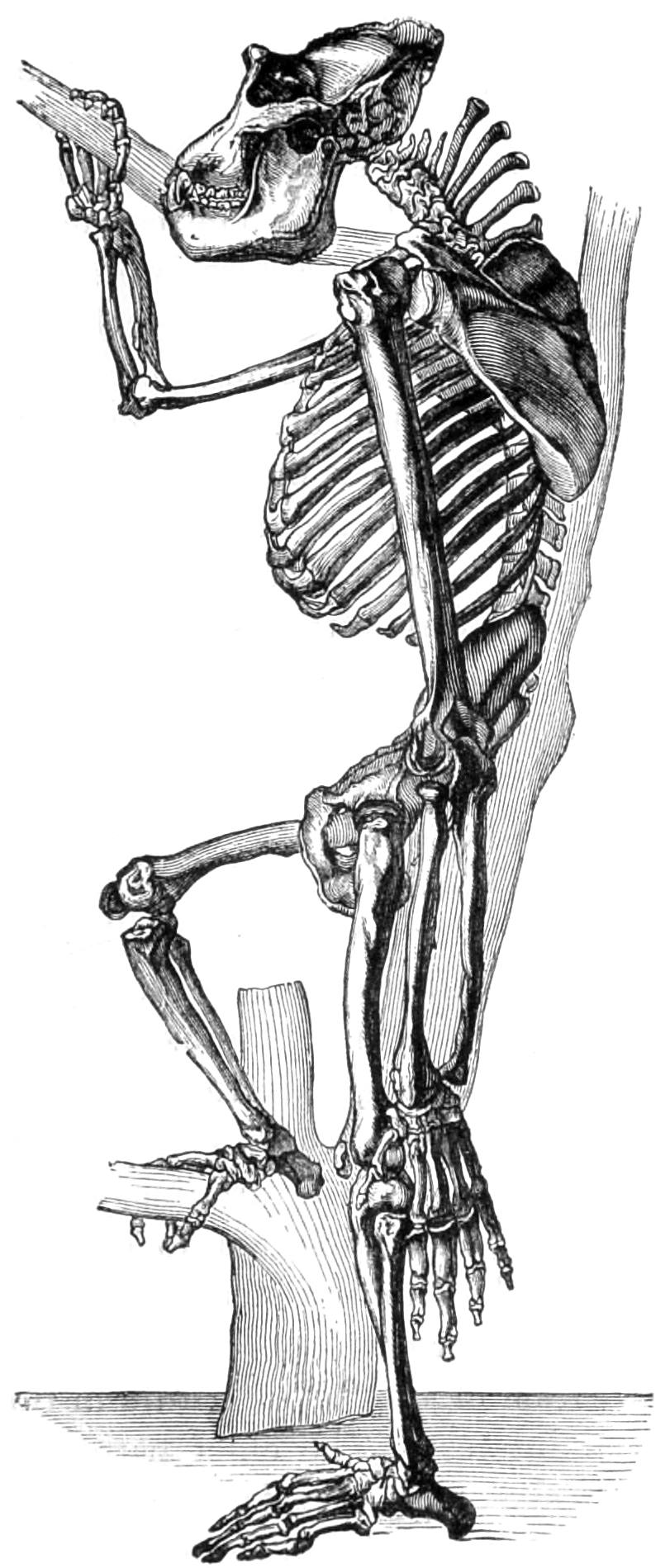
Fig. 17.—Skeleton of an aged male gorilla.17
The rest of the skeleton of the aged male gorilla corresponds in its powerful and massive form with the general structure of the body, which is remarkable for its height and strength (see Fig. 16). In the skeleton of the trunk there are seven cervical, thirteen dorsal, and four lumbar vertebræ, thirteen ribs, and, even in aged animals, a sternum composed of several pieces of bone. The cervical vertebræ display long spinous processes, which are most strongly developed between the fourth and seventh vertebræ. The extremities of this colossal structure, combined with the elevation of the occipital region, present a convex outline when seen from behind. This structure provides the point of insertion and support for the powerful cushion of cervical muscles. The dorsal vertebræ, which increase in height, width, and depth as they stand lower on the column, taper, and are keel-shaped at their junction with the cervical vertebræ. The central parts of the widely arched ribs, which are thirteen or sometimes fourteen in number, are very thick and powerful in the aged male. Only seven pairs of ribs are attached by the costal cartilages to the sternum, and two other costal cartilages are in proximity with them. The other cartilages are only rudimentary, and the terminations in the muscular system of the belly are free. There are, indeed, variations from the type here established, and from ten to eleven ribs are sometimes attached to the sternum by thread-like strips of ligament or cartilage.
The formation of the pelvic girdle in this animal is of special interest. The chief parts of this portion of the skeleton—that is, the hip, pelvic, or innominate bones—are high, tapering in their lower part, and broad and flat above, where they terminate in the crest of the ilium, which describes a quarter of a circle. There is, for the most part, only one small superior iliac spine, and the ischii are somewhat turned outwards, and furnished with broad, rounded tuberosities, and for the most part with only a single large sacro-sciatic notch. The horizontal rami of the pubes are narrow, while the descending rami are wide. The os sacrum is narrow, and shaped like a protracted cone, turning abruptly outwards, and resembling the basal joint of a true tail. The coccyx appears to be the rudiment of a genuine tail.
The bones of the shoulder-girdle present interesting peculiarities. The clavicles are long and slender, with a leaf-shaped, flattened end articulating with the scapula, and a thickened end articulating with the sternum. The scapula is a very large triangular bone, resembling the human scapula in its general form, and the supra- and infra-spinous fossæ are not strongly marked. The long and powerful humerus has its head inclined at an angle of sixty degrees towards the axis of the shoulder. Frequently, but not invariably, the lower, flattened extremity of the humerus is pierced on one or both sides above its rounded eminence, and this is termed by Darwin the intercondyloid foramen.
The radius has a powerful head, and a shaft considerably curved outwards, while it is, on the other hand, curved backwards and inwards at the elbow. The bones of the carpus, metacarpus, and phalanges are remarkably long, broad, and deep. The development of the femur corresponds to that of the whole skeleton. Its middle piece or shaft is curved in front and flattened behind. The shaft of the tibia is generally rounded off, but is sometimes rather laterally compressed.
The os calcis of the foot is slender, curved outwards in the centre and inwards behind the astragalus. The head, with its cuneiform extremity, is of a transverse oval shape, turned inwards. The scaphoid bone, which is generally in connection with this projection, takes the same direction towards the inner side of the foot. This peculiar contortion causes the tarsus of the gorilla to appear almost as if it had been subjected to a deviation or fracture of its longitudinal axis.
In young and adult males, as well as in young females, the structure of the bones is generally less massive than in aged males. In the female skeleton the strongly developed depressions and ridges, especially in the bones of the extremities, are absent. The head of the ulna is, for example, less deeply set in the case of a female, and its projections are smaller than in the male animal. In the female, also, the head of the radius is smaller, and the triangular shape of its shaft is less strongly marked. The pelvic bones of a female gorilla are wider, flatter, and less concave on their very projecting inner surface. They diverge more widely from each other, and this is also the case with the tuberosities of the ischium. The pubic arch is less depressed than in the male gorilla. Although the spinous processes of the vertebræ attain to some length and thickness, their development in the female is not so great as it is in the male sex.
The bony structure of the chimpanzee offers many points of resemblance to that of the gorilla, while it differs in certain particulars from the structure of other anthropoids. And first, the size of the skeleton is smaller than that of the gorilla, which is in agreement with the smaller relative size of the body of the chimpanzee.
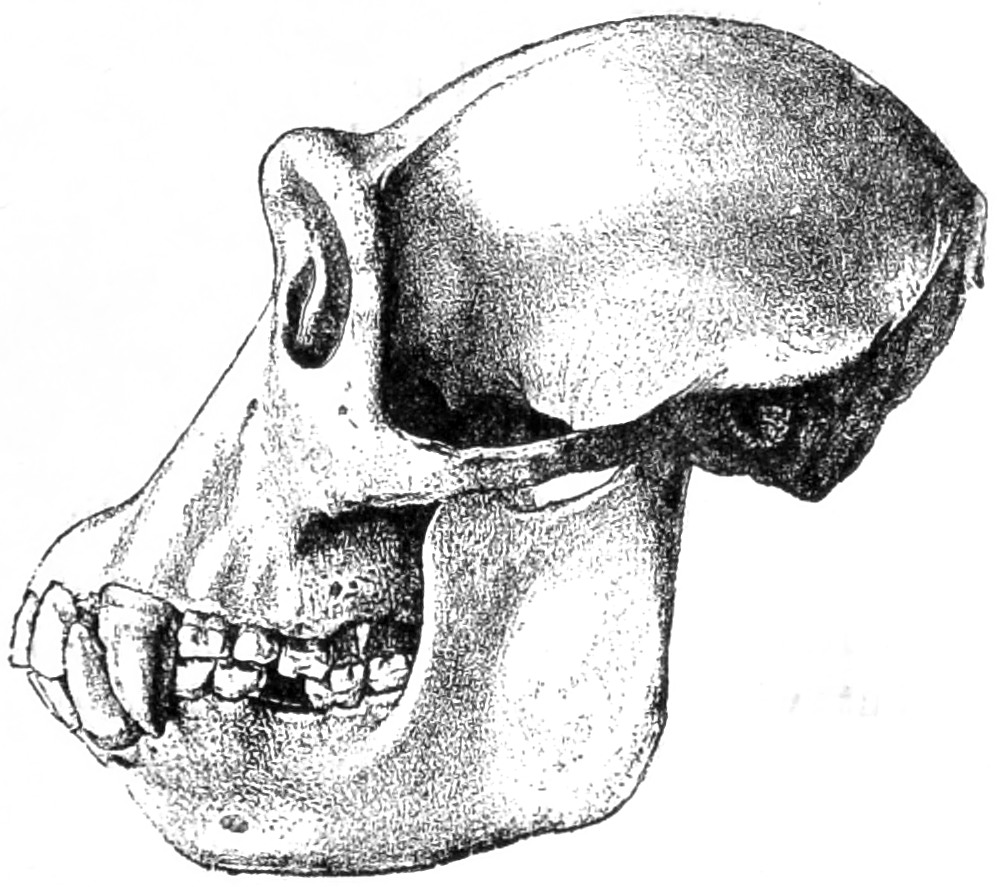
Fig. 18.—Skull of an aged male chimpanzee.
We must begin with a general view of the skull of the chimpanzee. In both sexes the frontal regions are smaller, while the coronal region is more rounded than in the gorilla. The high bony crests and prominent supra-orbital arches are wanting in the chimpanzee; the peculiar character of the bony ridges, projecting like tubes from the other parts of the skull, is less marked, and they belong more directly to the frontal region (see Fig. 18). The bony bridge of the nose is more concave in the chimpanzee; the jaw-bones are smaller and less compressed in the centre than they are in the gorilla.
When we undertake to describe the skull of the chimpanzee in detail, it becomes necessary to consider separately the skulls of aged and young males, and of aged and young females; for in this case also the distinctions of sex and age are very evident. On the skull of an aged male chimpanzee the temporal ridges are not much developed on the coronal arch. They meet on this arch from 60 to 90 mm. behind the orbits, and form only a small coronal crest. The transverse occipital crest is somewhat developed, and at its point of union with the coronal crest the temporal ridges divide to form its upper edges. This is the case not only with the Rio Quillu skull, from which Fig. 18 is taken, but with that of the so-called troglodyte Tschègo given by Duvernoy.18 In some other specimens belonging to aged male animals the presence of a coronal crest cannot, however, be detected. In these the temporal ridges are very small, and more or less distant from each other. While the transverse occipital crest maintains an almost uniform height on the gorilla skull, like a detached ridge, it is only slightly elevated behind in those chimpanzee skulls in which the crest is partially developed. In the gorilla male this ridge divides the squamous occipital portion, which is sometimes bevelled, sometimes slightly convex; in the male chimpanzee this part is more decidedly arched, and takes the form of a half-oval. The mastoid processes are also present in the chimpanzee. The external occipital crest and the curved lines are generally apparent. The styloid processes are more plainly traced than in the gorilla. In the latter, as well as in the chimpanzee, there is a blunt, tubular process of the temporal bone, opposite to another bony process, issuing from the occipital bone. This has been observed by Virchow, and is termed by him the carotid process (Processus caroticus).
The orbits of the chimpanzee are generally more rounded, with a distinctly circular rim, while the nasal bones are as long and narrow as in the gorilla. The region of the jaws is very prognathous; the external nasal openings are rounder and smaller than in the gorilla. The crowns of the canine teeth project in the same pillar-shaped form (Fig. 18). The triangular space enclosed by these and by the row of teeth in the upper jaw is often very wide and projecting, even more so than in the gorilla. But whereas in the latter the canine teeth are shaped almost like a three-sided pyramid, in the chimpanzee they are more rounded and conical. In the general structure of the teeth of both species there are certain differences of which we shall speak presently.
The brain-pan of a young male chimpanzee is still more arched than it is in aged animals. The temporal ridges are still far apart. The transverse occipital crest displays near the mastoid process well-defined wing-shaped indentations. In the skulls of very young males the transverse occipital swelling of which we have spoken (Torus occipitalis transversus) is already developed. The orbits are distinctly detached from the skull; the bridge of the nose is depressed; the crowns of the canine teeth are, in conformity with the still slight development of the teeth themselves, less marked, and the triangular space enclosed by the teeth is less convex than in older animals.
The skull of the adult chimpanzee is, in its coronal and occipital parts, more uniformly arched, narrower, and more elongated than in aged males. The transverse occipital ridge usually develops itself in the region of the upper curved lines, or in the bony parts enclosed between these and the central lines. The nasal and upper maxillary region is depressed. That section of the upper jaw which contains the incisor and canine teeth is small. In the skulls of all chimpanzees, of whatever sex or age, the body of the lower jaw is comparatively small, with two low but wide rami, of which the coronoid and condyloid processes are divided from each other by a comparatively wide cleft. The rami of the chimpanzee’s lower jaw are still more abruptly retreating than is usually the case in the gorilla.
The skull of a very young female gorilla is shaped almost like a half-sphere. The orbits are scarcely detached from the forehead; the want of elevation of the orbital arch, and the slighter prognathism of the jaw, is marked by the deep depression between it and the nose and forehead (Fig. 20).
The cancellous texture of the bones of the chimpanzee’s skull admits of a whole system of cavities communicating with each other, which are of the nature of the so-called sinuses present in the frontal, sphenoid, ethmoid, and maxillary bones of the human skull. In the chimpanzee, however, the sinuses are more extensive than in man, or even than in the gorilla. The large cavities of the forehead communicate with those of the nose and jaws. The sphenoidal sinuses and ethmoidal cells are large and deep. The greater wings of the sphenoid bone and its pterygoid processes are provided with considerable cavities. The mastoid cells of the temporal bones are in connection with the cells of the greater wings and pterygoid processes of the sphenoid bone, and also extend through the squamous portions and zygomatic processes of the temporal bones, losing themselves in their upper part in the smaller cells of cancellous bone which are found between the outer and inner walls of the skull. These are of more uniform shape and size.
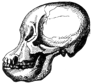
Fig. 19.—Skull of a very young female chimpanzee.
The skeleton of the chimpanzee, in accordance with the smaller size of the species, is relatively of a slenderer build than that of the gorilla. The spinous processes of the seven cervical vertebræ are more slightly developed, and have undivided extremities. The transverse processes of the fifth and sixth cervical vertebræ are almost of the same shape as cervical ribs. There are thirteen dorsal vertebræ, somewhat laterally compressed: this compression is greater than in man and in the gorilla. The four lumbar vertebræ of the chimpanzee are furnished with long, thin, riblike transverse processes. The so-called mammillary processes of the final vertebra are strongly developed in the male. The intervertebral foramina are small, as they are also in the gorilla and orang-utan. The thirteen ribs of the chimpanzee remind us of the human structure. The collar-bone is slightly curved, as in the gorilla. There is a marked difference between the sexes in the structure of the scapula which is broad and three-sided in the male, small and leaf-shaped in the female.
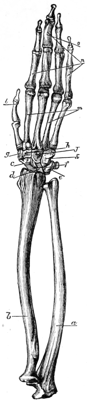
Fig. 20.—Skeleton of the forearm and hand of the Central African bam-chimpanzee. a, Ulna. b, Radius. c, Scaphoid bone. d, Semi-lunar bone. e, Cuneiform bone. f, Pisiform bone. g, Trapezium. h, Os magnum. j, Trapezoid. k, Unciform bone. l, Phalanges of thumb. m, Metacarpal bones. n, Phalanges.
The humeri have slender shafts, with well-developed condyles and ridges. The bones of the forearm are much curved, so that the interval between them is, as in the gorilla, somewhat wide. From the wrist to the final phalanges the hand is more slender than in the gorilla.
The pelvis in this species of ape has high, narrow ilia, spreading in their upper parts, and projecting forwards, so as to form the cavity of the abdomen, and, especially in the male sex, the anterior spines of the ilium are more strongly developed than in the gorilla and orang-utan. The ischiatic tuberosities are of a spreading form, and diverge considerably from each other. The pubic arch is deeply hollowed, but the point of juncture is elevated. As in the gorilla, the os sacrum resembles the basis of a tail, but it is less developed and less conical in form.
In the chimpanzee, as well as in other anthropoids, the coccyx gives altogether the impression of a laterally compressed and rudimentary tail. This is especially the case in young animals, in which the coccyx always appears to be very narrow and prolonged. In older animals this part gradually widens, yet without losing its resemblance to a rudimentary tail.
The head of the femur resembles a section of a sphere, of which the upper part is sometimes wanting. Its shaft, which is curved in front, is much slenderer in the female than the male. The patella is oval. In the tibia the narrow shaft is laterally compressed, and bent inwards. The bones on the inner side of the foot take a backward direction, while those on the outer side, attached to the fibula, turn outwards.
In the ankle-joint the head of the astragalus is much arched, and turned inwards. The scaphoid bone is thick and deeply hollowed. The metatarsal bones and phalanges have a considerable upward convexity (Fig. 21).
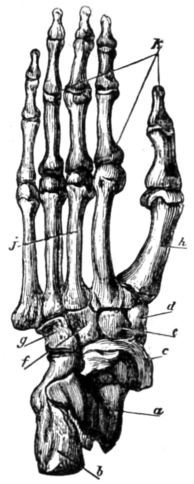
Fig. 21.—Skeleton of foot of the Central African bam-chimpanzee. a, Astragalus. b, Os calcis. c, Scaphoid bone. d, e, f, Cuneiform bones. g, Cuboid bone. h, First metatarsal bone. j, Second to fifth metatarsal bones. k, Phalanges.
The skeleton of the orang has also its special characteristics. We have already remarked, in describing the external form of the heads of these animals, that the skull is high and projecting, and retreating in its hinder part. In the old male orang this part of the bony structure is of smaller size than in the old male gorilla. The arch of the cranium is shorter and rounder than in that animal and in the chimpanzee. The central longitudinal crest of the vertex is present, but in accordance with the more spherical shape of the coronal part of the cranium, this crest is more arched above than in the gorilla, in which it slopes gently upward to the transverse occipital crest, which rises high and peaked from the back of the head. This latter crest is indeed developed in the orang, but it is not so high, and is more retreating. In consequence of this formation, the upper posterior part of the gorilla-skull appears in profile to be much more abrupt and peaked than that of the orang. In the latter, also, the orbital arches are not so high and abrupt, and not so much detached from the rest of the skull. In the orang the squamous occipital portion declines abruptly in front and below, yet it is generally more arched than in the gorilla. The orbits of the orang, which are sometimes rounded, sometimes more square, are divided from each other by a narrow partition. The space between them and the anterior nares is not so great as in the gorilla. While in the last-named animal the space between the root of the nose and the teeth of the upper jaw-bone is convex, in the chimpanzee it is generally vertical, and in the orang it is depressed (Fig. 22). The maxillary parts, furnished with strong canine teeth, are very prognathous, yet hardly to the same extent as in the chimpanzee. The body of the lower jaw is high, and its rami are high and wide. The bony crests of which we have spoken are absent in the female. The coronal part and the squamous occipital parts are arched; the upper jaw is smaller, and the lower jaw is also less massive, than in the male animal. In very young animals the predominance of the strongly arched cranium over the countenance is apparent, and the increase of size in the latter occurs gradually (Fig. 23).
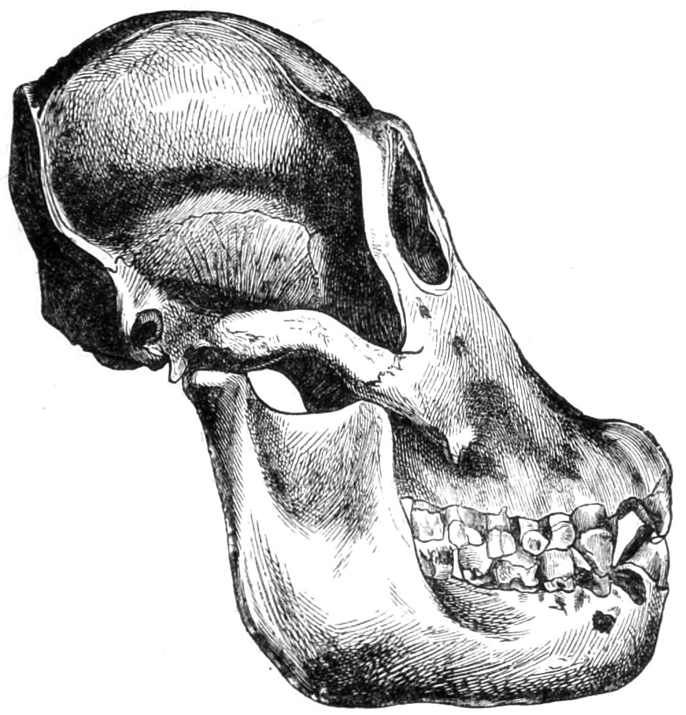
Fig. 22.—Skull of middle-aged female orang.
The anterior nares are narrow at the top, and wide at their base. They are more decidedly pear-shaped (Apertura pyriformis) than those of the gorilla and chimpanzee. In the latter animals these apertures are generally wider and more uniformly rounded. Bischoff justly observes that the bony part below the orbits, which in the gorilla is wide above, tapering away in the lower part of the face, is narrower and more vertical in the orang. The nasal bones of the orang are high and of moderate width. Brühl mentions the styloid process of the orang’s skull, which is, however, somewhat abortive when we compare it with that of the human skull. It has its origin in a tolerably deep groove. On the other hand, Brühl, as we have already observed, can find no trace of the styloid process in the skulls of the gorilla and the chimpanzee!19
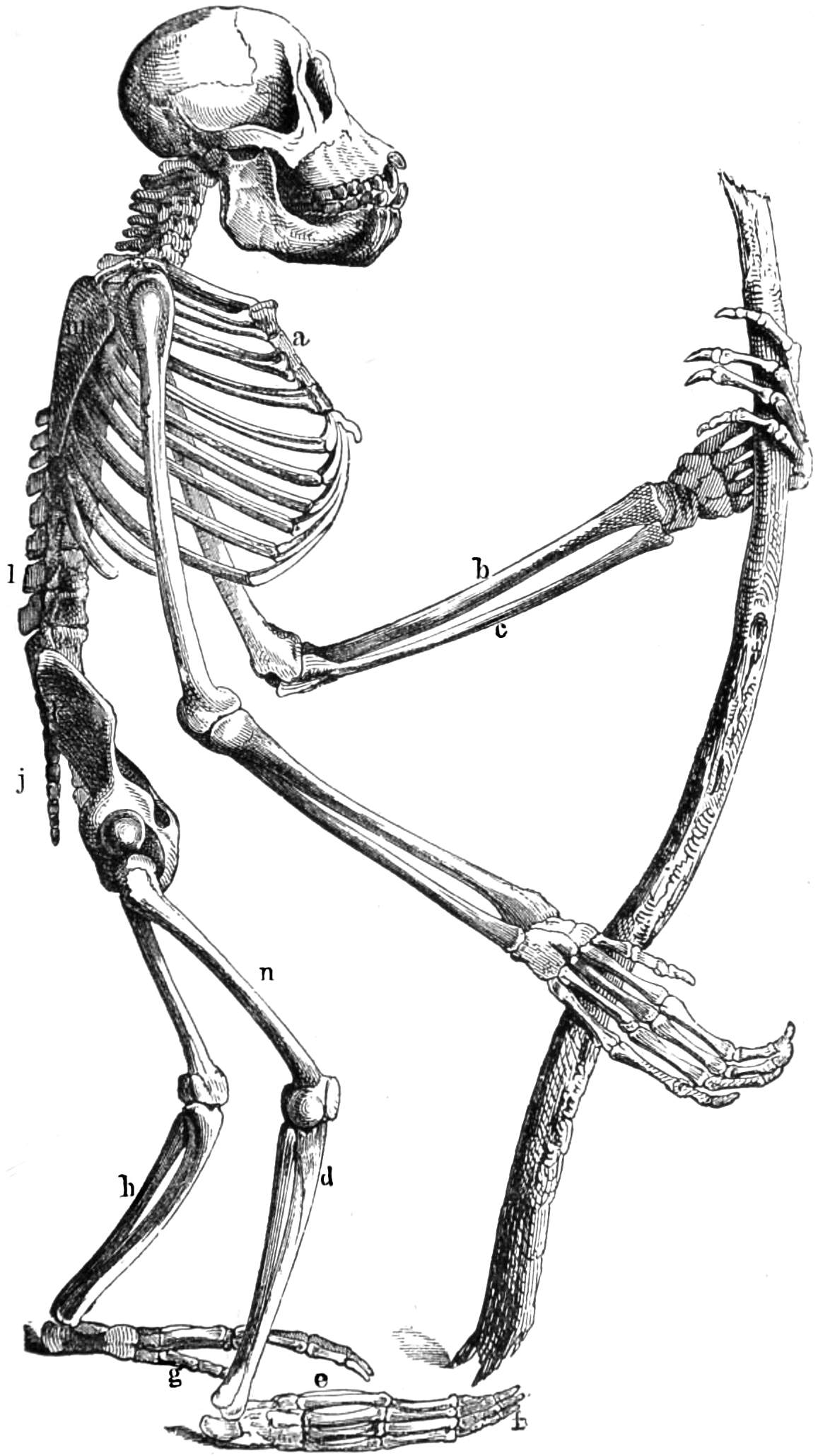
Fig. 23.—Skeleton of young orang-utan. a, Sternum. b, Radius. c, Ulna. d, Tibia. e, Metacarpus. f, Phalanges. g, Great toe. h, Fibula. j, Hip-bones. k, Coccyx. l, Vertebral column. m, Scapula. n, Femur.
There are many large-celled bony cavities in the orang’s skull. These may be observed in the greater wings and pterygoid processes of the sphenoid bone, in the mastoid and squamous parts of the temporal bones, in the lachrymal bones, in the body, and in the condyles of the occipital bone, and in the zygomatic arch. The larger fore-cells on the squamous part of the temporal bones are connected by a wide aperture with the sinuses of the greater wings and pterygoid processes of the sphenoid bone. A sinus which may be observed on the greater wing generally communicates by a large round hole with the temporal cells. There is generally, but not always, a communication between the sinuses of the greater wing and pterygoid process and the nasal cavity. These cavities sometimes communicate with each other through a wide aperture at the base of the nose. The squamous part of the temporal bones has a cellular sinus, which communicates with the cells of the mastoid process, in its lower part with the tympanum, and in its fore-part with the ossicles of the lower wall of the tympanum. The maxillary sinuses are in connection with the cells of the lachrymal bone. There is nothing in the orang’s skull corresponding to the Vidian canal of the sphenoid bone, but it may be traced in the gorilla and the chimpanzee.
The vertebral column of the orang has not the same colossal spinous processes which distinguish that of the gorilla. It differs also in many other, though less striking, particulars both from the gorilla and the chimpanzee. In the orang there are generally twelve dorsal vertebræ, tapering in their lower parts; while their long, thick, transverse processes, which are full of knots, take an upward direction. The upper articular processes of the four lumbar vertebræ present short and rather insignificant mammillary processes. The sternum of the young orang is generally formed of one large upper bone, with six smaller bones below. In older animals the body of the sternum appears to consist of a tier of three bones connected together. The ribs resemble those of the human skeleton, the clavicle is long and straight, and the scapula also resembles that of a man in form. The flat pelvic bones of the orang also turn outwards; the ischiatic bones are short, with spatula-shaped tuberosities; the pubic arch is high, and the obturator foramen is narrow and oval. The sacrum and coccyx do not resemble a rudimentary tail so much as in the case of the anthropoids we have already described. We are reminded of the human structure in the humerus, of which the shaft is much curved behind, and on its outer side. The ulna is very slender, and provided with a protracted, jagged styloid process. The neck of the radius is tapering, while its shaft is arched like that of the ulna, and the anterior border and oblique line are sharp. The wrist, metacarpus, and fingers are long and narrow.
The femur of the orang is remarkable for its large head, shaped like a section of a sphere, and its slender shaft. The latter is less bent than in the gorilla. The patella, which, in my opinion, should be classed among the so-called sesamoid bones, is in this case of an irregular form. The shank and foot-bones are remarkably slender. The scaphoid is tapering; the head of the astragalus does not turn inward so much as in the gorilla. The hinder surfaces of the metatarsal bones and of the phalanges turn decidedly outwards.
We have now to consider the bony structure of gibbons, in which there are many specific variations which our space will not allow us to consider in detail, but a slight sketch of their organic system must be given. The brain-pan of this animal’s skull is of an oval shape, without the crests so characteristic of other anthropoids, and even in the aged males of this species their development is so slight as to be scarcely perceptible. The occipital bone of male animals is, indeed, generally rounded, and the whole occipital portion is somewhat compressed in a downward direction, while the coronal region is at the same time flattened. The cranium gradually widens behind, so that, when seen from above, its form is somewhat pear-shaped. In aged males the orbits project from the low, retreating frontal bone, and are surrounded by a bony, circular rim.
The face is not very prognathous, and the short wide nasal-bones form a wide, depressed partition between the orbits. The edges of the jaw-bones describe a parabolic curve and are considerably elongated. The palate is consequently long and narrow. The rami of the lower jaw are wide and low, and their coronoid processes are only slightly developed. In aged males the teeth, and especially the canine teeth, are long and projecting; yet, comparatively speaking, they never attain to the great development of those of other anthropoids.
The number of vertebræ seems to be subject to considerable variation even in the same species, and various estimates are given by different naturalists. Müller, for example, has said that in several species (Hylobates syndactylus, H. leuciscus, H. variagatus, and H. concolor) there are thirteen dorsal, five lumbar, six sacral, and four coccygeal vertebræ. Cuvier counted in the siamang, thirteen dorsal, five lumbar, four sacral, and three coccygeal vertebræ. In Hylobates agilis I counted thirteen dorsal, six lumbar, five sacral, and four coccygeal vertebræ. Hylobates syndactylus has long coccygeal bones, and an elongated os sacrum, which gives the impression of serving for the application of a short tail, or, indeed, of being in itself a rudimentary tail. In other respects the cervical, dorsal, and lumbar vertebræ differ little in structure from those of man.
The ribs on the sternum, which widens abruptly outwards, are strongly arched. The lowest of these project, owing to the width of the shaft. In the sternum there is a want of proportion between the smallness of its body and the size and width of its extremity. The ensiform appendix of this bone is long and wide, and spatula-shaped at its lower extremity. In the shoulder-girdle the clavicles are very slender, and much arched. The scapulæ, on the other hand, are high and narrow, spatula-shaped, and provided with a steeply projecting acromion process, a strongly developed coracoid process, and deep glenoid cavities. The upper limbs are, in conformity with the general structure of these apes, very slender; the shafts of the bones of the upper and forearm are elongated, with small extremities. The condyles are small, especially those of the elbow. The bones of the wrist, the metacarpus, and the fingers are also long and slender.
In the pelvis we note that the ilia are narrow below, and expand in the form of a spatula above, and that their position is almost vertical. Their inner surfaces are only slightly concave, and are directed somewhat forwards. The ischiatic bones are low, with wide, flattened, rugged tuberosities, and rounded foramina obturatoria. The ischiatic rami project forwards in an almost horizontal direction. There are large prominences on the pubic arch of the siamang.
The leg-bones are much shorter than those of the arm. The heads of the femurs stand out plainly from their short necks and large trochanters, as segments of perfect spheres. In this case, as in that of other anthropoids, the third trochanter (trochanteres tertii), often so apparent in the human femur, is barely indicated. The shank-bones are arched. The tibia is often laterally compressed, so that its transverse section forms a scalene triangle. The malleoli are compressed from before backwards. The elongated heel-bones appear to be laterally compressed. The canal between the astragalus and the os calcis (Sinus tarsi) is very wide. The metatarsal bones and phalanges have large bases, long slender shafts, and heads projecting on the under side. Even the final phalanges are long and slender.
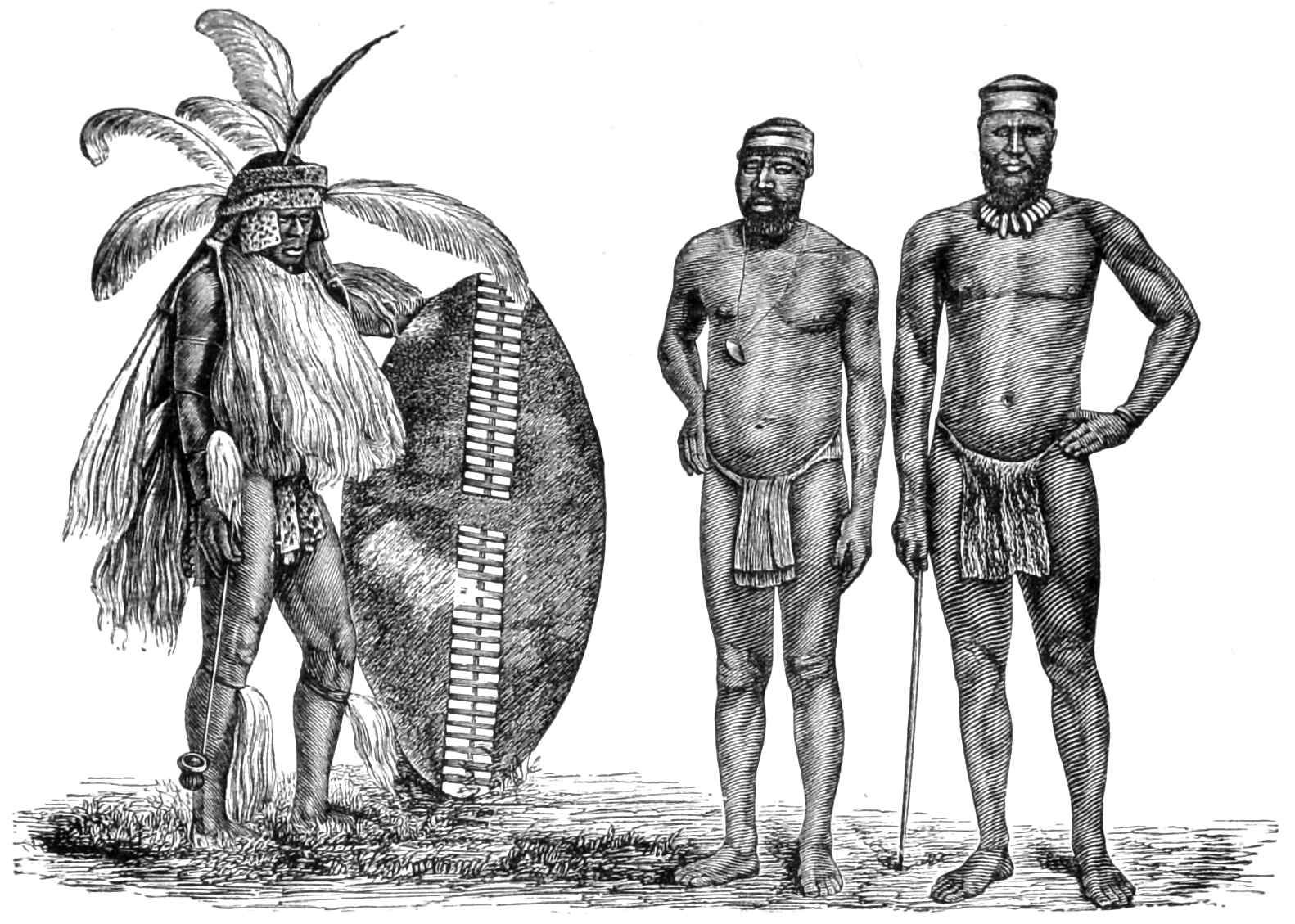
Fig. 24.—The Zulu king, Ketchwayo, in fighting array, with two of his men.
We shall now find it profitable to compare the external characters of anthropoids with those of man. We are sometimes disposed to see the true likenesses of anthropoid apes in dark-skinned, naked savages. These savages are often insufficiently fed, the skin is wrinkled, the face, even at an early age, is deeply furrowed, and their general appearance is neglected. The dark silhouette of such people stands out so distinctly against a clear background, their habit of life is so rude, their attitudes impress us so disagreeably, that we are involuntarily led to make such a comparison. This tendency unfortunately gives a wide field for exaggeration among dilettanti naturalists, and such as are zealous to establish a preconceived theory. A conscientious inquirer must, however, be cautious, and avoid too great generalization in such comparisons. For instance, much has been said of the pithecoid structure of all African negroes, yet this only applies to some peculiarly hideous races, in a state of physical degradation. There are many negro tribes in different parts of Africa which are remarkable for their well-formed bodies, and for a not ignoble bearing. The warlike demeanour of the natives of Ashanti, Dahomey, and Ibos is well known. Although the Hausanese are flat-nosed and thick-lipped, yet when armed and dressed in uniform, as we see in the photographs of Captain Glover’s force, their military bearing is very apparent. The tribes of Schilluk, Nuehr, Bari, Niam-Niam, and A-Bantu present examples of distinguished warriors, however rude and savage. Dabulamanzi, commander of the Zulus at the butcheries of Isandlhwana and Ulundi, and his chiefs, give me, in a photograph in my possession, the impression of gallant warriors, however uncivilized. In all these cases it is difficult to establish the resemblance to anthropoid apes (see also Fig. 24).
The Papuans, especially on the Australian continent, are generally classed with the African negroes in such comparisons. We admit that a horde of Australian blacks, degraded by hunger and fatigue, emaciated and dirty, may, as they roam through the shadeless woods, the steppes and thick scrub of their native country, present a strange and brute-like appearance. And if the foreign intruder takes a coarse pleasure in giving drink to these savages, their immodest gestures may afford a revolting impression of their bestial nature. Yet the habits even of these dark-skinned savages are altogether different under more favourable conditions. Although of small stature, they are not badly proportioned, and their manners and bearing are capable of improvement, so that they can act as native police, messengers, etc. This was the case also with the natives of Queensland, Australia, whom I saw in the Zoological Gardens, Berlin, throwing the boomerang. Even in these tamed savages, however, we must note the projecting orbits, the deep depression between the forehead and nose, and the flatness of the latter organ. There are aged, wrinkled bushmen, negroes, Papuans, Malays, Japanese, and Mongols of inland Asia whose countenances are altogether pithecoid. And such a cast of face may even be found in Europe.
Some years ago, Mr. Bond, a land-surveyor in British India, asserted that he had found the missing link between man and apes in the mountainous district of the Western Ghauts. And indeed, the race he describes seems to have a strong resemblance to apes. “The forehead is low and retreating. The lower part of the face projects like the muzzle of an ape; the legs are short and bent outwards. The trunk and arms are comparatively long. The hands and fingers are contracted so that the latter cannot be freely extended; a thick skin covers the hollow of the hand and the fingers, especially their tips; the nails are small and imperfect; the feet are broad, and covered both on their backs and soles with a thick skin. This tribe seems to worship nature. They have no fixed dwellings; they live chiefly on roots and honey, and exchange the latter, together with wax and other productions of their forests, for tobacco, clothes, and rice.”20
Nothing more, so far as I am aware, has been published concerning this race. The description given above leaves much to be desired. The assertion respecting the contracted fingers is obscure, and such a condition is directly opposed to any resemblance with the flexible hand of apes.
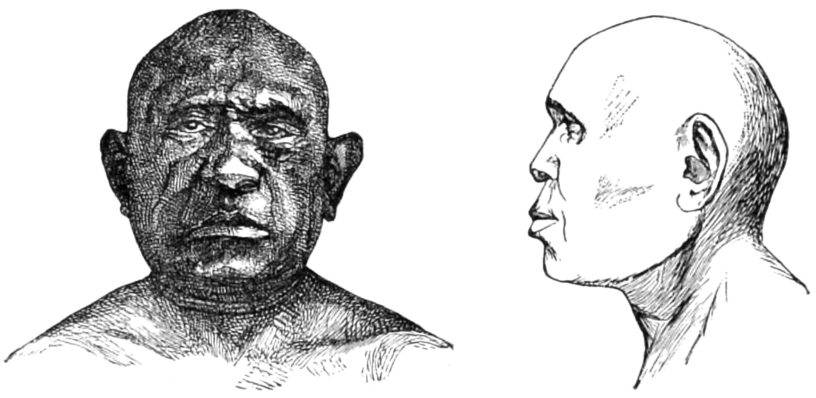
Fig. 25. Aidanill, hairless Australian.
Fig. 26. The same in profile.
Let us turn from a tribe of which the existence is still dubious, to consider the portraits we subjoin of a man and woman, aborigines of Queensland, in a district watered by the Ballone. These are Aidanill, the brother, and Dewan, the sister, members of a hairless family. The indefatigable Miklucho-Maclay went to Gulnarber, 140 miles from Tulba, in order to examine them, and took the photographs from which our illustrations are taken.21
A likeness to the chimpanzee, when deprived of its hair, may be traced in the keel or roof-shaped form of the skull; in the prominence of the supra-orbital arches; in the deep depression between the forehead and nose, of which only the centre of the bridge has a slight vertical elevation; in the broad, flattened nostrils, bounded by deep furrows; in the wide, fleshy mouth, and the large, laterally projecting ears. Gratiolet and Alix give such a head in their treatise on Troglodytes Aubryi (Figs. 25, 26, 27). When we add to this the dark brown skin, the deeply furrowed countenance, and the dark brown eyes, as they are described by Miklucho-Maclay, the external resemblance between many of the Australian aborigines and apes becomes more marked.
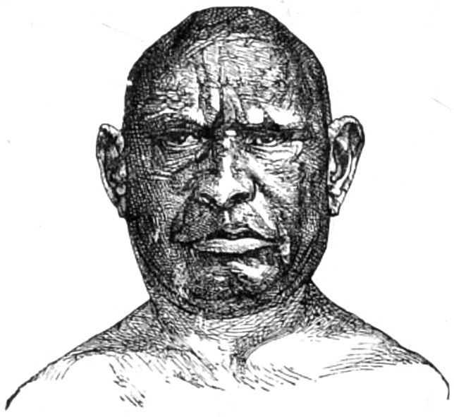
Fig. 27.—Dewan, Aidanill’s sister.
Projecting ears are common among men of different races, and I have observed them in Europeans who are otherwise well formed. Even in this latter case the effect is ape-like. Much has been said of the resemblance which may often be observed between the human ear and that of apes. It is admitted that hardly any part of the organism varies so much in its characteristics as the external ear. This is the case with anthropoids, and almost more frequently with men. Individuals of all nations are found with defective development of this or that characteristic helix, angle tragus, notch concha, and fossa, with lobules imperfectly formed or altogether absent. I have frequently observed such misshapen ears, which vary from the perfect type, and bear a certain resemblance to the ear of apes, among the hard-featured peasantry of Germany, Switzerland, France, Italy, and Poland, who cannot be said to count beauty as part of their inheritance. In Africa I found this defective formation more common among the Maltese, Greeks, and Turks who were living in the country, than among the fellaheen, Berbers, and negroes. The latter have been unjustly charged with the possession of “hideous ape-like ears,” whereas, among the African races, these organs are, in the majority of cases, of a pleasing form. With respect to the Australian blacks, and to the Malay, Mongolian, and Indian races, I cannot rely on my personal observation. According to my very limited experience, there is much individual variation among these races, and ears of the hideous, ape-like formation might be sought for with success. The specific resemblance to apes can, indeed, only be ascertained by one who is accurately acquainted with the organism of these animals. These and similar ideas are often expressed by the unlearned, who do not really understand the characteristics in question.
Darwin speaks of the anthropoid form of the ear in the chimpanzee and orang.22 “The ears of the chimpanzee and orang are curiously like those of man, and I am assured by the keepers in the Zoological Gardens that these animals never move or erect them, so that they are in an equally rudimentary condition, as far as that function is concerned, as man. Why these animals, as well as the progenitors of man, should have lost the power of erecting their ears, we cannot say. It may be, though I am not quite satisfied with this view, that owing to their arboreal habits and great strength they were but little exposed to danger, and so during a lengthened period moved their ears but little, and thus gradually lost the power of moving them. This would be a parallel case with that of those large and heavy birds, which from inhabiting oceanic islands have not been exposed to the attacks of beasts of prey, and have consequently lost the power of using their wings for flight.
“The celebrated sculptor, Mr. Woolner, informs me of one little peculiarity in the external ear which he has often observed both in men and women, and of which he perceived the full signification. His attention was first called to the subject whilst at work on his figure of Puck, to which he had given pointed ears. He was thus led to examine the ears of various monkeys, and subsequently more carefully those of man. The peculiarity consists in a little blunt point, projecting from the inwardly folded margin, or helix. These points not only project inwards, but often a little outwards, so that they are visible when the head is viewed from directly in front or behind. They are variable in size and somewhat in position, standing either a little higher or lower; and they sometimes occur on one ear and not on the other. Now the meaning of these projections is not, I think, doubtful; but it may be thought that they offer too trifling a character to be worth notice. This thought, however, is as false as it is natural. Every character, however slight, must be the result of some definite cause; and if it occurs in many individuals deserves consideration. The helix obviously consists of the extreme margin of the ear folded inwards; and this folding appears to be in some manner connected with the whole external ear being permanently pressed backwards. In many monkeys, which do not stand high in the order, as baboons and some species of macacus, the upper portion of the ear is slightly pointed, and the margin is not at all folded inwards; but if the margin were to be thus folded, a slight point would necessarily project inwards and probably a little outwards. This could actually be observed in a specimen of the Ateles beelzebuth in the Zoological Gardens; and we may safely conclude that it is a similar structure—a vestige of formerly pointed ears—which occasionally reappears in man.”
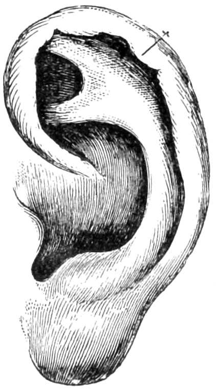
Fig. 28.—Human ear.
I subjoin an illustration of the human ear, in which the pointed tip mentioned by Darwin may be easily discovered. This point may also be perceived in the ears of anthropoids, and especially in those of the orang-utan. Meyer has attempted to show that this Darwinian pointed tip is only due to the abortive development of part of the helix, and in this case we should not regard the occurrence as an ape-like pointing of the helix, but rather as its partial interruption owing to the pathological condition of that organ.23 In a later edition of his work, Darwin admits, in reply to Meyer, that this explanation may apply to many cases in which there are several very small points, or when the whole of the helix is sinuate. In one case, photographed by Darwin, the prominence was so large that, if we were to assume with Meyer that the ear would have been normal if the cartilage had been uniformly developed along the whole extent of the helix, the latter must have occupied a third part of the ear. Two cases were mentioned to Darwin in which the upper edge of the ear had no inner fold, and was so pointed that it was very like that of an ordinary mammal. The ear of the fœtus of an orang given in Darwin’s illustration appears to be pointed, although in the adult animal that organ is very like the human ear. The Darwinian tip may also be seen in the fœtus of an orang described and illustrated by Salvatore Trinchese in the Annali del Museo civico di Storia Naturale di Genova (1870). The tip of the helix is pointed in very young individuals of the gibbon species, especially in Hylobates Lar. Among the lower apes the pointed ear is very common (see Fig. 29).
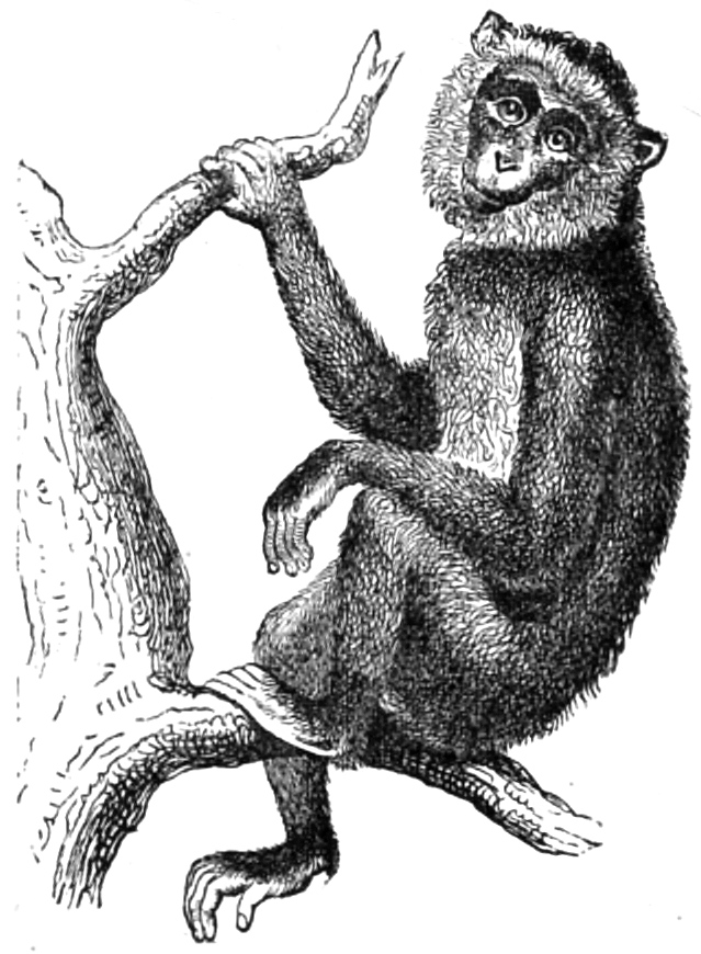
Fig. 29.—Magot (
Innuus ecaudatus
).
The eyelids of anthropoids greatly resemble those of man in their structure. In adult gorillas and chimpanzees there is always a semilunar fold (plica semilunaris) corresponding to the membrana nictitans, or third eyelid of birds. In man there exists, instead of this, only a rudimentary apparatus, the caruncula lachrymalis. In some individuals it attains to a considerable size, as I have observed in the fellaheen, Berbers, Shillook, and other tribes. On the other hand, the conversion of the caruncula into a true, although only rudimentary, plica semilunaris has not been observed by me in the human eye. Miklucho-Maclay describes the caruncula in Melanesians (the Papuans of New Guinea), in the Orang-Sakay (of the Malay peninsula), and in the Mikronesians (of the island of Japan and of the Palau archipelago), as two or three times as wide as that of the average European.24
The eye of the young male gorilla which was kept alive in the Berlin Aquarium from 1876–77 was carefully examined by me in June, 1877. I found that the sclerotic membrane of the eyeball was whitish, surrounded by a dark brown ring. A second darker ring, sharply defined, surrounded the cornea. The iris was of a yellowish brown. The sclerotic membrane, however, gradually deepens in colour so as to give the effect of a uniform dark brown. The iris retains a light brown colour for a longer period, but it darkens with age. In an aged animal there is no brightness in the eye, except from reflected light. In the chimpanzee the iris is light brown, verging on yellow; and this is also the case in the orang.
The expressionless, indifferent look of anthropoids has often been observed, and undoubtedly chimpanzees and orangs generally gaze placidly before them. I have, however, observed an animated expression in the eyes of the former species, and W. L. Martin has also observed a flash and brightening of their eyes. I shall never forget the expression of malicious anger in the eyes of the female animal Mafuca, at Dresden, as soon as she was teased. The expression of the eyes of the gorilla in the Berlin Aquarium also changed frequently, especially when he was about to perform some mischievous trick, or when he was provoked to anger. The expression of this animal was very human, but necessarily it could only recall the darkly coloured eyes of negroes and other black races. In 1876 there were two very young orangs in the Berlin Aquarium, one hairy and the other hairless. These animals clung together in a close embrace. If they were separated, their eyes became bright and restless, and they again sought to embrace each other while uttering plaintive cries. On tickling one of the animals under the chin, it made a most absurd grimace, and its eyes brightened, as Martin has observed in similar cases. The eyes of the gibbons which I have observed had a thoroughly mild and placid expression, rarely animated by any fire.
The instance we have mentioned of hairless Australians is the more remarkable since these aborigines are for the most part distinguished for their luxuriant growth of hair. The Australian blacks and the Ainos of Yedo are, as a rule, perhaps the most hairy races in the world. It is known, however, that in all countries and climates exceptional cases are found of individuals whose bodies are wholly or partially covered with hair, and these conditions sometimes affect whole families. Interesting historical and morphological researches respecting these hairy men have recently been made by von Siebold, Ecker, Virchow, Bartels, and Ornstein. In many of these cases we are presented with decidedly brute-like phenomena. The Mexican woman Julia Pastrana displays the strongest resemblance to apes. Other hairy men remind us at the first glance of some of the canine species. In all races the women are less hairy than the men. Darwin states that in the females of some species of apes the under side of the body is less hairy than in the males, and this is also the case with anthropoids, especially with the chimpanzee.
The beard is, as we know, common to man and apes. Among apes it is more strongly developed in the male than in the female, and this is also the case in the human species. Darwin points out that the growth of the beard both of men and apes occurs at the period of their sexual maturity, and also that there is a remarkable parallel between men and apes in its colour. For when the human beard varies in colour from the hair of the head, which is frequently the case, it is, without exception, of a lighter, and generally of a reddish hue. Darwin observed this in England, and Hooker found no exception to the rule in Russia. J. Scott carefully observed the numerous races which are to be found in Calcutta, as in other parts of India, namely, the two Sikh races, the Bhoteas, Hindus, Burmese, and Chinese. Although most of these races have very little hair on the face, Scott found that in all cases without exception, in which there was any difference in colour between the hair of the head and the beard, the latter was of a lighter shade. In apes the colour of the beard often differs widely from that of the hair of the head, and in such cases it is always of a lighter shade, often white, sometimes yellow or reddish.
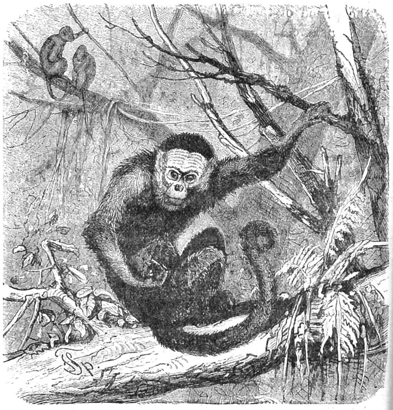
Fig. 30.—Capucin ape (
Cebus capucinus
).
“It is well known,” says Darwin, “that the hair on our arms tends to converge from above and below to a point at the elbow. This curious arrangement, so unlike that in most of the lower mammals, is common to the gorilla, chimpanzee, orang, some species of Hylobates, and even to some few American monkeys. But in Hylobates agilis the hair on the forearm is directed downwards or towards the wrist in the ordinary manner; and in Hylobates lar it is nearly erect, with only a very slight forward inclination; so that in this latter species it is in a transitional state. It can hardly be doubted that with most mammals the thickness of the hair and its direction on the back is adapted to throw off the rain; even the transverse hairs on the forelegs of a dog may serve for this end when he is coiled up asleep. Mr. Wallace remarks that the convergence of the hair towards the elbow on the arms of the orang (whose habits he has so carefully studied) serves to throw off the rain, when, as is the custom of this animal, the arms are bent, with the hands clasped round a branch or over its own head. We should, however, bear in mind that the attitude of an animal may perhaps be in part determined by the direction of the hair; and not the direction of the hair by the attitude. If the above explanation is correct in the case of the orang, the hair on our forearms offers a curious record of our former state; for no one supposes that it is now of any use in throwing off the rain, nor in our present erect condition is it properly directed for this purpose.”25
Darwin also remarks that it is erroneous to deny that apes have eyebrows. In fact, long bristly eyebrows are present in all anthropoids—not growing thickly together like those of men, but scattered among the shorter and thicker growth of hair which clothes the parts above the orbits; nor do they maintain any definite direction. In the white-handed gibbon, these eyebrows are remarkable for their length and stiffness. A growth of hair corresponding to eyebrows may, indeed, be observed above the upper eyelids of all mammals, including seals and pachydermata. On the upper lip of gorillas, chimpanzees, and orangs we may also observe a number of somewhat longer, stiff, and bristly hairs which stand apart from the otherwise short hairs on the lips, and give the impression of a cat’s “whiskers.” In Hylobates albimanus I observed that these vibrissæ attain to a considerable length (Fig. 10).
The external form of the trunk of anthropoids, taken as a whole, does not greatly differ from that of man. We have not, indeed, the well-formed human torso, with its graceful lines; and the formation of the posteriors, together with a want of expansion about the hips, displeases us in its departure from the human type (see Figs. 1 and 6). We shall not be disposed to compare the torso of the Apollo Belvedere, or of the Olympian Hermes with that of a gorilla or chimpanzee. Yet the torso of a powerful male gorilla, from which the hair has been removed, may be favourably compared with that of one of the large-bellied, lean-armed weaklings who are everywhere to be found as living caricatures of the human species.
The neck of anthropoids is generally short and thick. In the gorilla that part of the body has a great backward convexity, owing, as we have said, to the great development of the spinous processes of the cervical vertebræ, and of the muscles attached to them. A short, thick throat, and considerable development of the neck, a bull-neck, as it is called, is also not unfrequent in man. This peculiarity is sometimes supposed to be one of the national characteristics of the African blacks. Burmeister says that “the negro’s thick neck is the more striking, since it is generally allied with a short throat. In measuring negroes from the crown of the head to the shoulder I found the interval to be from nine and a quarter to nine and three-quarter inches. In Europeans of normal height, this interval is seldom less than ten inches, and it is more commonly eleven inches in women, and twelve in men. The shortness of the neck, as well as the relatively small size of the brain-pan, and the large size of the face may the more readily be taken as an approximation to the simian type, since all apes are short-necked, and the relative distance of these animals is somewhat further from the negro than that of the negro from the European. This shortness of the neck in the negro explains his greater carrying power, and his preference for carrying burdens on his head, which is much more fatiguing to the European on account of his longer and weaker neck.”26
Burmeister’s assumption on this subject is, however, much too general. It does not apply to many of the negro races—at any rate, not to those of the Upper Nile valley. A long, thin neck is the characteristic of the Funje, Shillooks, Denkas, Baris, and other large tribes of those regions. Among these people the interval between the top of the head and the shoulder is from ten to eleven, and even from eleven to twelve inches (240 to 260 mm., and 260 to 286 mm.). Burmeister has been thinking exclusively of the Brazilian blacks. Yet I am unable to trace the typical short neck, either in the well-known portraits of slaves by Maurice Rugendas,27 or in the collection of photographs of Brazilian negroes which is in my possession. This characteristic is also absent, even in many portraits of West African and Mozambique blacks, tribes from which the slave population of Brazil has been chiefly drawn. Many Mongolians, Malays, Papuans, and Polynesians have short, thick necks, but this characteristic is more rare among the American aborigines and among Europeans. If we are to recognize an approximation to the simian type in this formation, it is one common to several nations, and it is not confined only, nor even chiefly, to the negro races.
The remarkable elongation of the upper limbs of anthropoid apes cannot be compared with the length of the corresponding limbs in men. For although among negroes and the members of other primitive peoples we may occasionally observe unusually long arms, yet these are individual peculiarities which are also found among Europeans, and cannot be counted among racial characteristics.
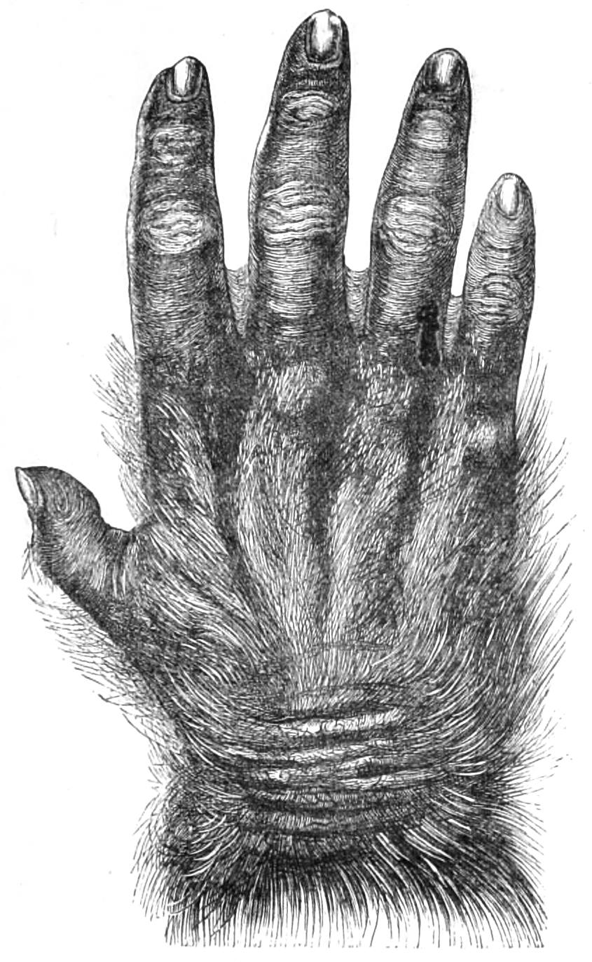
Fig. 31.—Hand of a very aged male gorilla.
The hand of the orang and the gibbon is too long and narrow to be directly compared with the human hand. The chimpanzee and the gorilla, especially the latter, have hands more like those of man. In the case of an adult male gorilla the first glance at this member reminds us of the knotty fist of a black dock labourer or lighterman, like those who, at Rio de Janeiro, Bahia, or La Guayra, lift the heavy bags of coffee and place them on their heads or on their herculean shoulders. Much has been said of the enlargement of the connective skin between the bases of the fingers of a negro hand, and of the pointed extremities of the fingers. Van der Hoeven, in his well-known treatise, De Natuurlijke Geschiedenis van den Negerstam, has described and drawn the hand of an Ashanti boy, formed in this manner. Hence there is a disposition to recognize in this peculiarity an important characteristic of the negro race. As in the hand of the gorilla, the connective web between the bases of the fingers is also extensive, and the ungual phalanges taper at their extremities, there is also an inclination to ascribe an expressly anthropoid character to the negro hand. Yet this structure of the fingers is by no means universal among the negroes. An enlargement of the connective web is not indeed uncommon, but its extent varies considerably. Nor is it wanting in the fingers of other races. An attentive observer will be able to trace it in the labouring population of country districts in Europe. I have myself frequently observed this characteristic in Canton Wallis, and in the Lombard and Genoese provinces, through which I travelled on foot in 1869 and 1871, when I devoted special attention to this point. In Fig. 32 I give a negro hand of a type which seems to be common among the blacks in the inland districts of North-eastern Africa. It can hardly be denied that the form of this hand, which is certainly not flattered, possesses the characteristics of a thoroughly human organization.
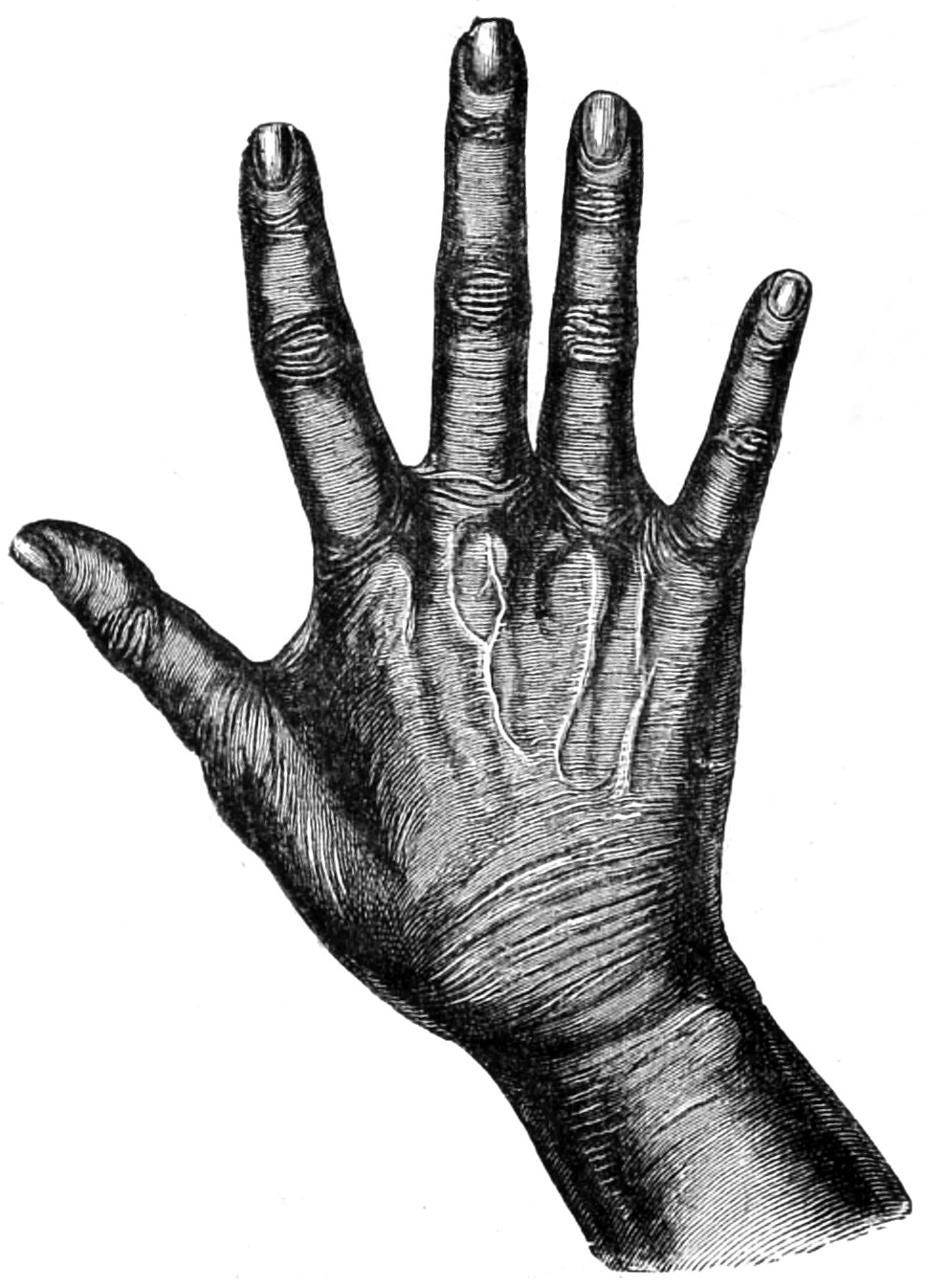
Fig. 32.—Hand of a Hammegh from Roseres, on the Blue Nile.
With respect to other primitive peoples besides negroes, we have not at present sufficient information, and we ought therefore to beware of premature generalization. The thin shanks, with imperfectly developed calves, found among many primitive races, and especially among the African and Australian blacks, are often and not unjustly adduced as an instance of their ape-like formation. In fact, the general uncomeliness of these parts in the races in question is one of their significant characteristics.
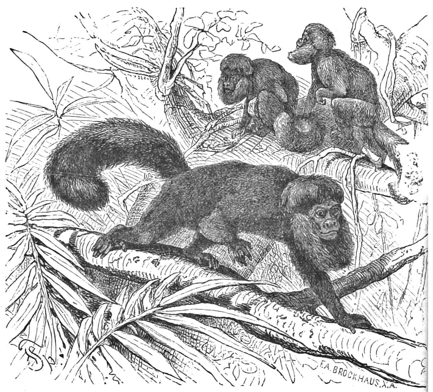
Fig. 33.—Satan’s ape (Pithecia Satanas). Shows the formation and mode of using the feet in apes of the New World.
The anthropoid foot resembles in structure those of other apes, including those of the New World, and as a rule it differs from the human foot in the flexibility of the great toes. It has, however, been justly observed that many individuals of different races have been able to use the great toe almost as if it were a thumb. Such persons may be found everywhere. Men who have been born without arms, or who have been deprived of them during life, have been able to use their feet like hands, as some compensation for this privation. The most surprising instance of our time has occurred in the violinist without arms, whose performances are heard in various continental capitals. Another, mentioned by Bär, was able to write with his feet. But even people who have the full use of their upper limbs can often grasp with the great toe as if it were a thumb, so as to pick up small objects from the ground, or draw them towards them. Constant practice in such feats produces a certain dexterity. Negroes, Malays, Polynesians, and Indians make use of their outstretched great toes in climbing with as skilful a gripe as our schoolboys and sailors are also able to do in gymnastics, or in climbing up the masts. Among such people the distinction between the foot of man and apes is less marked, since, even when at rest, the great toe is apt to be somewhat detached from the others. This may be seen in A. Buchta’s excellent photographs of individuals of the Central African tribe, the Makraka. Haeckel justly observes that there is no marked physiological distinction between the hand and foot which can be established on a scientific basis. In order to make such a distinction it is necessary to consider their morphological characteristics.28
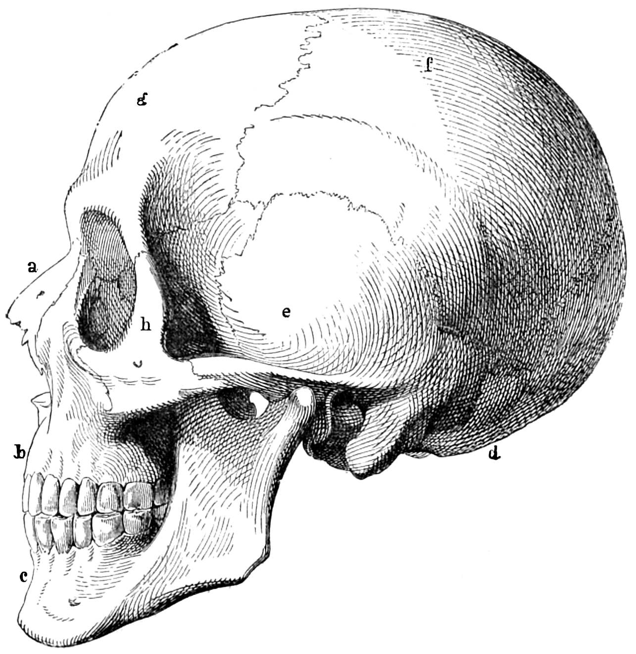
Fig. 34.—Human skull. a, Nasal bone. b, Upper jaw. c, Lower jaw. d, Occipital bone. e, Temporal bone. f, Parietal bone. g, Frontal bone. h, Malar bone.
Structure of the skeleton.—In comparing the skulls of anthropoids with those of men, we should, in the case of the gorilla, chimpanzee, and orang-utan, content ourselves with young specimens rather than with the skulls of adults. In aged apes of these species, the colossal development of the bony crests of the skull, as well as that of the jaws, the prominence of the orbital rim, and the flattening of the occipital bone, present distinctions of such a searching character that we are greatly hindered in the pursuit of the comparative method. But during the process of development the anthropoid skeleton admits of a direct comparison with that of man. In a young animal the rounded skull suggests a parallel between it and the human head. It must be admitted that we find, especially in primitive peoples, many human skulls which in their whole plastic form differ little from the skulls of young gorillas, chimpanzees, and orangs. Even in the way the occipital bone is rounded off, young anthropoids and men are often found in a similar stage of development. The squamous occipital portion in a young negro, Papuan and Malay, is indeed often flatter and more bevelled than it is in a young gorilla or chimpanzee.
We must not, however, assume that the two individuals brought into comparison are of precisely the same age, since such a point cannot easily be ascertained, even when subjects for examination are afforded by one of our larger museums. Savages are seldom able to give their precise age, and the attempt to do so often relies on insufficient data. The direct examination of the skull will afford some information on this point; but the conditions of growth in anthropoids are not so well known as to admit of an accurate estimate. We have to rely on the state of the teeth, on the stage at which the development of the bony crests has arrived, etc., in order to form an approximate estimate of the age of the skull.
On the squamous occipital portion the arrangement of the curved lines which are the boundaries to the attachments of the cervical muscles, is common to men, to anthropoids, and to other apes. Only indications of these lines are to be found in the lower order of mammals. In the human skull there is sometimes a formation belonging to the squamous occipital portion which has a distinctly pithecoid or ape-like character. This is the occipital swelling we have already described (Torus occipitalis transversus), which may be either enclosed by the two upper curved lines, or lie between these and the central curved lines, or may be altogether in the region of the latter. This swelling extends in a gradual manner above and below its bony support. Its edge may be more or less sharp, more or less like a crest in its development, wider or narrower, with or without a central eminence, but its appearance is always striking. In young male and female gorillas, orangs, and chimpanzees this formation represents the completely formed transverse occipital crests, which are found for the most part in aged male animals of these species. These swellings may also be observed on the skulls of adult men of all times and all nations. They are by no means rare in the skulls which are in ordinary use at the Berlin School of Anatomy, and they are remarkably common in many groups of skulls. They are frequent among the skulls, for the most part without their lower jaws, which the late Dr. Sachs disinterred in a Mohammedan burial-ground of the thirteenth century, near Cairo. These are the remains of Mohammedans of different ranks, but, for the most part, of the peasantry or fellaheen. Ecker was able to trace the sagittal crest in the skulls of Australian males, while it is absent in the females. Similar indications of the bony crest have been observed by me in the roof-shaped or scaphocephalic skulls of many negroes, but in these cases I am not aware whether there is a corresponding distinction of sex. It can hardly be denied that this bony prominence is a human characteristic.
Broca has given the term pterion to the H-shaped connection formed by the sutures between the parietal bone, the greater wing of the sphenoid bone, the squamous portion of the temporal bone, and the frontal bone. One of the most common disturbances in the symmetry of the connecting suture, as we have already briefly mentioned, arises from the insertion of a frontal process of the squamous portion of the temporal bone between the lower angle of the parietal bone, the fore-part of the frontal bone, and the greater wing of the sphenoid bone. This process of the temporal bone varies in size, and may occur on one or both sides. A similar formation is common among gorillas, chimpanzees, macacas, magots (Inuus), and baboons.29 It is less frequent among orangs,30 gibbons, marmosets, and American species (howlers, hooded apes, etc.).
Virchow and W. Gruber have agreed in representing this frontal process as theromorphological—that is, as a characteristic of the lower animals, and more especially of apes. Virchow has found this abnormal formation of the skull to be more common in some races than others. None of those in whom it occurs appear to belong to the Aryan races, and the existence of this process and stenocrotaphy, or temporal stenosis, seem to be due to a defective development of the greater wing of the sphenoid bone, and to the compression of the bones in its vicinity, by which the whole temporal region is contracted. This is a characteristic of the lower, but by no means of the lowest, races of men.
Stieda, Hyrtl, Gruber, and Calori have sought to controvert the fact that this temporal process is a characteristic of the lower races. Stieda asserts that it may occur exceptionally in all races of men.31 He himself, aided by Anutschin, has ascertained the existence of this anomalous pterion on more than 10,000 human skulls, and he has also received information from others. He considers the frequency of this frontal process in man to be theromorphological, or indeed pithecoid. According to Anutschin, this anomalous condition is not equally common in all races. In the dark-skinned and woolly-haired races (Australians, Papuans, and negroes) the frontal process is most widely diffused; it is less frequent among Mongolians and Malays; and among Americans and white men its occurrence is from five to six times more uncommon than in the black races. Sometimes the frontal process occurs on the intercalary bone (Ossa epipterica), which is fused into the squamous portion of the temporal bone; and sometimes the process grows out of the squamous portion of the temporal bone. These imperfect processes or intercalary bones are not regarded by Anutschin as pithecoid, since they are more rare in apes than in men. Schlocker has sought to show that the frontal process of the squamous portion of the temporal bone, the less common temporal process of the frontal bone, and the temporal intercalary bone (Ossa epipterica) are of equal value from the genetic point of view.32 This author regards the frontal process and the immediate connection of the frontal and squamous portion of the temporal bones, as theromorphological characteristics, but he does not believe the occurrence of this process to be restricted to the lower races.33 This is also the opinion of Ten Kate. However this may be, the establishment of this theromorphological formation is important. Its immediate value as a contribution to the theory of the origin of species remains, as we shall presently see, even if we cannot trace it through intermediate and lower types.
In the great prominence of the supra-orbital ridges which has been observed in some pre-historic human skulls, a likeness to the corresponding feature in anthropoids has been traced. And indeed there is such a likeness, especially to the female chimpanzee, in the well-known Neanderthal skull, which is very dolichocephalic, with prominent supra-orbital arches, only divided from each other by a shallow depression. In the same skull the development of the supra-orbital ridges is related to that of the frontal sinuses. In this pre-historic specimen—which, by the kindness of Professor Schaafhauser, I was able to examine closely at the congress of anthropologists at Berlin in 1880—the forehead retreats in a marked manner towards the flattened region of the crown. De Quatrefages and Hamy say that the skull is both flattened and long (dolichoplatycephalic). The temporal ridges are not only very marked, but they approach each other in the region of the coronal arch (Fig. 35). This also occurs in the adult female chimpanzee, as well as in the young male gorilla, in the aged female orang, and in the gibbon.
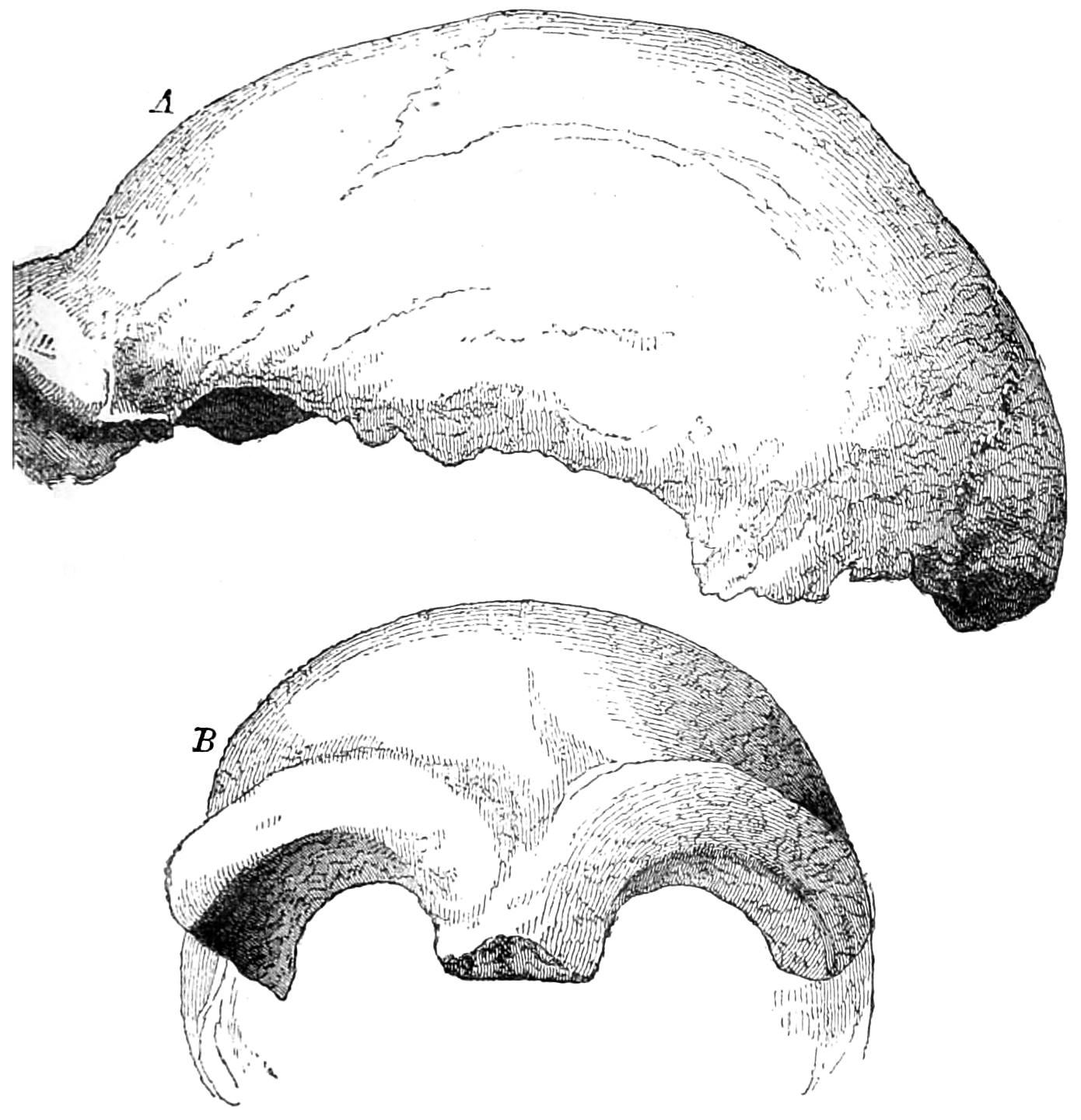
Fig. 35.—The Neanderthal skull. A. In profile. B. A front view.
It may here be observed that our men of science differ widely in opinion respecting the origin and ethnological significance of the Neanderthal skull, of which I will cite only a few instances. Pruner regards it as the skull of an idiot.34 Virchow considers the specimen, and the similar one from Kailykke in the Copenhagen Museum, as an altogether individual formation,35 a typical form modified by disease,36 in other words, a pathological skull.37 King regards the skull as one belonging to one of the primitive races.38 Schaaffhauser has, indeed, endeavoured to make an artistic portrait of such a primitive man. Spengel holds that skulls which are “Neanderthaloid” in form are to be found chiefly in Europe.39 If Huxley says decidedly that the Neanderthal skull can by no means be regarded as the remains of a human being which was a link between man and apes. At most this discovery only proves the existence of a man whose skull reverted in some respects to the simian type, just as a carrier or tumbler pigeon may sometimes display the plumage of their original ancestor, the rock-pigeon (Columbia livia). And although the Neanderthal skull is more like that of the ape than any other human skull with which we are acquainted, yet it is by no means so isolated as it at first appears, but is rather the ultimate expression of a series which may be gradually traced back from the highest and most fully developed type of human skulls. On the one side it approximates to the flattened Australian skulls, from which other Australian forms gradually lead to skulls which rather resemble the type afforded by the Engis skull. On the other side, it is still more closely allied with the skulls of certain ancient races which were either contemporaries or successors of those which dwelt in Denmark during the Stone Age, people whose kitchen middens have been discovered in that country.40
Huxley justly observes that some of the skulls drawn by Busk, and taken from the tumuli of Borrely, resemble the Neanderthal skull, especially in the abruptly retreating forehead. Some other European skulls may, within certain limits, be compared with the Neanderthal skull, as, for instance, those found at Brüx, Staengenaes, Olmo, Louth, Clichy, Bougon, Cro-Magnon, Grenelle, Furfooz, Engisheim, Cannstadt, and Toul. These all present interesting peculiarities of structure—strongly developed supra-orbital arches, a retreating forehead, a flattened crown, etc., although none of them are so remarkable in these particulars as the Neanderthal skull. It has not, however, yet been proved that this skull represents a definite racial type, and it seems more probable that it was simply an individual form.
The skulls of the Australian aborigines are, as Spengel justly observes, distinguished from the Neanderthal skull, and from others of like character, by their pronounced scaphocephalism. On the other hand, they have the prominent supra-orbital arches, the retreating forehead, the skull compressed in the temporal region, the prognathous countenance, relatively shorter than that of Europeans, and in all these respects the skulls of the Australians greatly resemble those of anthropoids. If, for instance, we turn to the illustration given by de Quatrefages and Hamy of a skull procured from Camp-in-Heaven, Arnhem’s Land, North Australia, and also Dr. Schadenburg’s negro skull, the most determined sceptic must be struck by their resemblance to the anthropoid skull.41
Similar characteristics to those which we have already mentioned as distinguishing the structure of the Australian skull, enable us to determine the anthropoid character of the skulls of many individuals belonging to the dark-skinned African races. These consist chiefly in the retreating forehead, the flatness and compression of the coronal arch, the pronounced prognathism, and the obtuse angles of the lower maxillary bones, which may be noted in so many negro skulls. On the other hand, the prominence of the supra-orbital arches is, as a rule, less marked in African races than in anthropoid species. There are specimens, however, as, for instance, the Congo skull given by de Quatrefages and Hamy,42 which give an overwhelming impression of anthropoid characteristics. And we find the same to a surprising degree in the skulls of intelligent, warlike, and light-skinned races of Central and Western Africa, and as the Monbuttre, Haussaua, Bakale, Fan, etc. This character may be discovered in all races of men, and especially among the Papuans and some African negroes.
A mutual approximation of the temporal ridges in the coronal region may be observed in the skulls of various nations. This formation is most frequent in the long-headed negro and Papuan skulls. In these cases it is generally allied with the shortness of the interval between the sides of the skull, taken in its transverse diameter (stenocephalism).
In an adult female chimpanzee, the parietal bones often rise abruptly towards the sagittal suture, and in its vicinity there arises a longitudinal bony prominence, of which the sides pass gradually into the external surface of the parietal bones. The sagittal suture sometimes remains intact, and is sometimes included by this process. This produces a modified development of the so-called keel-shaped skull (scaphocephalus). Such a formation may be often observed in negroes and Papuans, and more rarely in the skulls of other races. The occurrence of a divided malar bone in human skulls, especially in those of the Ainos and Japanese, has been considered to be theromorphic, since it is occasionally observed in the skulls of apes.43 I have myself, in a very few instances, found obscure traces of such a formation among anthropoids.
In 1863 Boucher de Perthes found at Abbeville half of a human lower jaw deposited in a black layer of clay and sand mixed with iron, and lying on the chalk. As far as we can judge from illustrations which are for the most part imperfect, there was nothing remarkable about it except its abruptly retreating ramus (Fig. 36), but the specimen aroused great attention at the time, and it was assigned by many intelligent observers to the primitive men of the diluvial period. Unfortunately it was afterwards proved to be a gigantic imposture.44
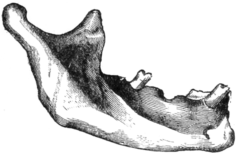
Fig. 36.—Lower jaw of Moulin-Quignon.
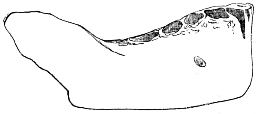
Fig. 37.—Naulette lower jaw.
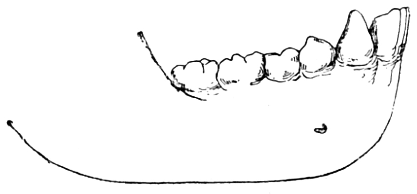
Fig. 38.
This is not the case with the lower jaws of Naulette, Aurignac, and Arcy, which are undoubtedly genuine and of great antiquity. The Naulette jaw is, indeed, very imperfect, yet we can trace the construction of the symphysis of the chin, which provokes comparison with the lower jaws of many anthropoids, especially those of the gorilla and chimpanzee (Fig. 37). The resemblance consists chiefly in the uprightness of the anterior surface, and especially of the body of the maxillary bone. In anthropoids this surface of the bone retreats from the row of teeth backwards and downwards to the lower edge of the body of the maxillary bone (Fig. 38); and in the Naulette specimen, as well as in the lower jaws of some modern Papuan skulls (of New Hebrides and elsewhere), there is a certain approximation to the simian type. A fossil ape (Dryopithecus Fontanii) has been found in the Middle Miocene of Saint-Gaudens, assumed to be one of the higher anthropoids, and in this case the jaw is only slightly retreating. Gaudry considers that the Dryopithecus was about the size of a man. The incisor teeth were small. The cusps of the back molar teeth were less rounded than in Europeans, and more like those of Australians. It has been surmised, although the fact cannot be established, that the last molar teeth were only cut after the canine teeth, as is the case with the human wisdom teeth. Gaudry gives the illustration of the lower jaw of a Tasmanian, from eleven to twelve years old, together with that of Dryopithecus Fontanii. In the human jaw the first molar tooth is larger than in the Dryopithecus, while the canine tooth and the pre-molars are much weaker. This distinction is important, since the smaller size of the front teeth is connected with the slight projection of the face, which is always a sign of human superiority. Although the canine tooth of the Dryopithecus is broken, we can see that it must have been considerably higher than the other teeth, and indeed the canine teeth of the male animal must have been very powerful. There is also a slight prominence in the teeth of this ape, which is absent in those of men. Mesopithecus, from the Miocene of Pikermi, Attica, was an ape less closely resembling the anthropoids. In the structure of the head it resembles the slender ape (Semnopithecus), and in the structure of the limbs it is like a macaca (Macacus). Gaudry believes that Sansan’s Pliopithecus was related to the gibbon. An ape of the size of the orang-utan, which belongs to the slender apes (Semnopithecus sub-himalayanus),45 was found by Baker and Durand in the Miocene of the Sewalik mountains.
In the comparative study of the human organization, and that of anthropoid apes, it is important to examine sections, and especially longitudinal sections, of characteristic skulls.46 Virchow has caused drawings to be made, from specimens in the Berlin Museum, of a gorilla, a chimpanzee, an orang-utan, and an Australian woman. The gorilla’s skull, when compared with the Australian’s, is so narrow that it looks as if compression had been applied to it; and yet the Australian skull is extremely small in comparison with that of men in general, since its cubic space is only 1150 ccm. In the gorilla47—at least in the old male, from which the drawing is taken—the immense size of the frontal sinuses, and the swellings which cover them, together with the strongly developed jaw, increase the impression of size. But, as Virchow observes, “all which adds to the size of the skull is bestial, and not human.” It is much the same in the orang-utan. Only in the chimpanzee the cubic space of the skull may be somewhat more favourably compared with that of the human skull. It approaches in size to that of a microcephalic native of the Rhein-Pfalz (of which an illustration is also given), which ranks a good deal below the Australian skull, and approximates more closely to the simian type. The internal space of the skulls of an adult female gorilla or orang may also be more favourably compared with those of men.
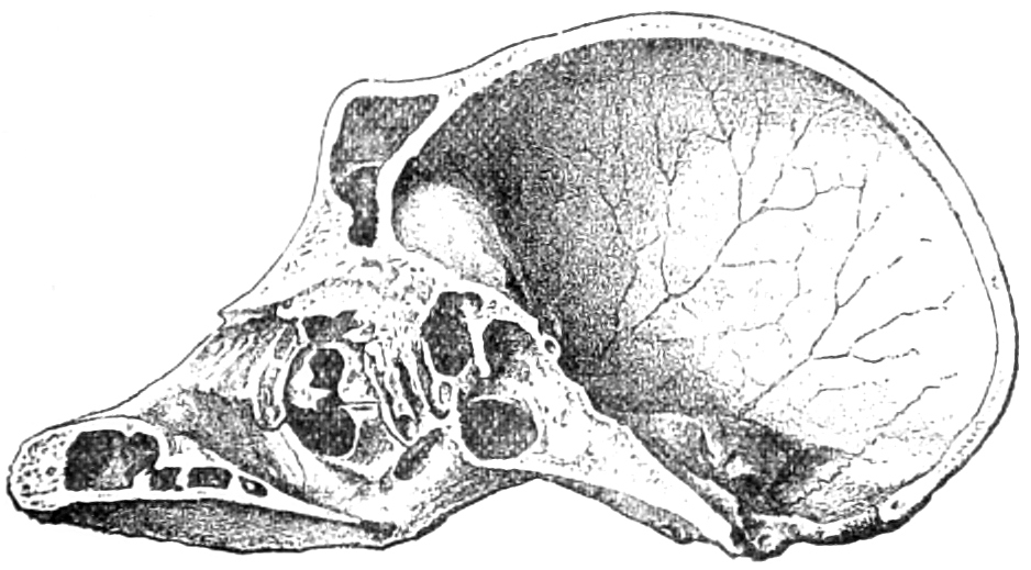
Fig. 39.—Sagittal section through the skull of a bam-chimpanzee.
We have already mentioned the presence of extensive sinuses and cells in the skulls of anthropoids, exceeding those of human skulls, and this is apparent in the accompanying illustration of a longitudinal section of the skull of a chimpanzee carried through its centre (Fig. 39). The length of this skull between the nasal partition and the most prominent part of the occipital bone is 128 mm.; that of the internal space is 108 mm. 10 mm. of this difference is due to the depth of the frontal sinuses, and the rest is owing to the thickness of the bony part of the skull. In an aged male gorilla, the first measurement is 153 mm., the second 115 mm. In another aged gorilla the measurements were respectively 183 mm. and 117 mm. In a still more aged male orang they were respectively 140 mm. and 114 mm. The comparative thinness of the centre of the squamous occipital portion is to be noted in the aged gorilla male. In the adult chimpanzee the large cells of the squamous portion of the temporal bone extend into this bone, and indeed without interruption into the parietal bone adjoining it. For such investigations the thin and light bones of individuals which have lived a wild life are more suitable than the heavy and fat specimens which have died after prolonged confinement.
Zuckerkandl has observed that among Europeans the orbital part of the nose, or that part which is between the orbits, is longer than the infra-orbital or lower part. In anthropoids the infra-orbital portion is considerably the longest, although only in adult animals. There are stages in the period of development in which these animals display the characteristics of an adult European, or indeed of a child. The proportions of the skulls of Malays take a middle place between those of Europeans and of apes. The growth of the infra-orbital part of the nose in the Malay does not equal that of apes, but in many cases it differs essentially from that of Europeans. Zuckerkandl makes a skilful attempt to establish this statement by statistics.
The same inquirer makes some interesting remarks on the comparative height and width of the orbits. He observes that the skulls of adult apes and men differ more in these respects than the young specimens of these organisms. The orbits both of a child and an adult, especially in the case of a European, are much more like those of a young ape than of an aged animal of the same species. In the chimpanzee and the orang-utan the proportions are the same as in men; that is, the width of the orbit exceeds its height. In man, this seems to arise from the exceptionally strong development of the supra-orbital ridge. It is most probable that in very young anthropoids the width of the orbit exceeds its height.48 Zuckerkandl goes on to say that in anthropoids the height of the orbits is greater than their width, and that this difference increases with age. But this is not absolutely correct, for even in aged animals the proportions vary, and the height and width of the orbits sometimes, although rarely, remains the same.
In comparing the vertebral column in men and anthropoids, Rosenberg has sought to show in the embryo, that the first sacral vertebra assumes the form of a lumbar vertebra, and that in a later stage of development it is enclosed by the ilia, and anchylosed with the sacrum. The same author has proposed a theory of the homologous or genetic equivalents of the vertebræ, which we must now consider. According to this theory, as Welcker has observed,49 the twentieth vertebra of an animal A is homologous to the twentieth vertebra of an animal B, the thirtieth vertebra of one animal to the thirtieth of another, although in one case it may be a lumbar vertebra, in another a pelvic vertebra, and in a third a coccygeal vertebra. The dorso-lumbar vertebræ of the lower apes have, in the case of men, their descendants, undergone a threefold metamorphosis, and, after their modification into sacral vertebræ, have assumed their fourth form as coccygeal vertebræ.
Froriep, a follower of Rosenberg, remarks that the lumbo-sacral vertebræ, i.e. those constituents of the vertebral column which form the transition from the lumbar to the sacral vertebræ, are invested with fresh interest by Rosenberg’s hypothesis. According to their position in the vertebral column, they are to be regarded as lumbar vertebræ, introduced too early or too late into the structure of the sacrum. If the twenty-fourth vertebra is assimilated with the sacrum, so as to form an upper promontory or outwork, this variety offers a point of transition to a future formation (?) in which this vertebra normally becomes the first sacral vertebra, and the column will now display twenty-three free vertebræ. If, again, this transition occurs in the twenty-fifth vertebra of the series, which thus becomes the chief sacral vertebra, this is, in Rosenberg’s opinion, a characteristic survival of the racial development, an atavism.50
According to Welcker’s theory, the chief sacral vertebra in one animal corresponds to the same sacral vertebra in another animal, whatever their number may be. The cervical vertebræ of one animal, which may be five, seven, or even eleven in number, correspond to the cervical vertebræ of another animal. The vertebral column of one animal corresponds to the vertebral column of another, taken as a whole, but not to two-thirds or three-fourths of that column. In accordance with the requirements of a given animal, that part of the bone which belongs to the sections of the breast and loins is more or less abundant, and the vertebræ are homologous in accordance with their region, and not with their number.
Holl has asserted that one vertebra is in close connection with the ilium, joined with it throughout its extent, and that this vertebra at the same time always appears to support the pelvis. This vertebra is, in normal cases, the first sacral vertebra, and the twenty-fifth of the series. It may be termed, as Welcker suggests, vertebra fulcralis. Such a main support is found, according to Holl, in every vertebral column, however anomalous its other conditions may be, and the only irregularity consists in its number in the series. This bone serves as a natural starting-point in our division of the vertebral column. The vertebra fulcralis must always be regarded as the first sacral vertebra. It begins the series of sacral vertebra, and, on account of its subsequently important position, it must be regarded as primary. Holl finds that it is followed by four lower vertebræ, which are afterwards included with it in the sacrum. When in its primary condition the vertebra fulcralis is twenty-fifth in the series, the twenty-fifth to the twenty-ninth vertebræ are included in the sacrum. When the fulcralis is the twenty-sixth vertebra, the sacrum includes the thirtieth. Hence it follows that the sacrum is, from the first stages of its development, a formation which begins with the twenty-fifth or twenty-sixth vertebra, and includes four other vertebræ. Holl considers that the lumbo-sacral form of the last lumbar vertebra, which stands between the lumbar and sacral vertebræ, does not indicate a gradual transition into a sacral vertebra, but rather an arrest in its development.51
When we examine a human sacrum we see that its first vertebra, the twenty-fifth of the series, is formed like the lumbar vertebræ in its upper part, setting aside those portions of it which form part of the lateral masses of the sacrum. These lateral masses, which serve as a support to the ilia, owe most of their substance to the first sacral vertebra. Thus, since it has to support the whole weight of the pre-sacral vertebræ, it is in fact a true vertebra fulcralis.
Holl justly says that there are few instances in which the human os sacrum consists of less than five vertebræ, and in no case are there less than four. In such a case the first sacral vertebra defines the pre- and post-sacral segment of the vertebral column.
In anthropoids the lower segment of the lumbar vertebral column is deeply sunk between the high, wide, and flattened ilia, which converge closely towards the vertebral column. In man these bones are not so much higher than the base of the sacrum, and their crests diverge more widely from the vertebral column. In the large apes the lateral masses of the sacrum are comparatively deeply set below their anchylosis with the pelvic bones. In an aged male gorilla, for instance, the transverse processes of the two lower lumbar vertebræ often extend to the hinder borders of the ilia, although the second of the lower lumbar vertebræ is somewhat higher than the top of the crest of the ilium. This is still more remarkably the case in an old male chimpanzee, in which the lowest lumbar vertebra seems to be wedged in between the two ilia. In a young male chimpanzee, and in the adult female, both the lower lumbar vertebræ are almost compressed between the upper segments of the ilia. In the orang the lowest lumbar vertebra is placed between the ilia. Out of the five sacral vertebræ the first and second are articulated with these bones.