Chapter III · THE EXTERNAL AND ANATOMICAL STRUCTURE OF ANTHROPOID APES, COMPARED WITH THE HUMAN STRUCTURE. (continued 2)
Reading edition
In the gorilla the highest sacral vertebra, the twenty-fifth of the series, is the fulcralis. In this animal the first to the third sacral vertebræ form part of the connection with the crests of the ilia. In the chimpanzee the twenty-fifth is also the vertebra fulcralis, and from the first to the third are likewise connected with the ilia, but the third only to a limited extent; and in young males and in old females the connection is generally confined to the first and second sacral vertebræ. In the orang-utan the twenty-fourth vertebra is generally the fulcralis.
In the gibbon the twenty-fifth vertebra is usually the fulcralis. In the siamang I found that the fifth of the five lumbar vertebræ was between the ilia. Out of the five sacral vertebræ the first and second were articulated with the said pelvic bones. In Hylobates agilis the fifth and sixth of the six lumbar vertebræ were between the ilia, and the first and second of the five sacral bones were articulated with these.
In the vertebral columns of the gorilla, the chimpanzee, and the orang we may observe an inconsiderable forward projection between the penultimate cervical and the second and third dorsal vertebræ. In the region below the second lumbar vertebra a similar forward projection may sometimes be observed. The so-called promontory at the entrance of the pelvis, that is, in the region developed between the lumbar and sacral vertebræ, which is remarkable in man, is only faintly apparent in anthropoids. The vertebral column is arched behind, since there is a dorsal curvature (see Figs. 17 and 23).
Aeby observes that the bodies of the vertebræ are tapering in the gorilla, and this is, in fact, the case. In climbing, or when he goes on all fours, the dorsal curvature of an anthropoid maintains its position. This curvature is still more apparent when the animal, in climbing, withdraws his body from the tree, mast, or whatever it may be, and bends forward his head. A similar dorsal curvature of the vertebral column may be observed in men who stiffen their hands and feet to climb up a tree or mast. If an anthropoid holds himself so erect as to be able to place his hands behind his head, the dorsal curvature of his spine is necessarily straightened, and indeed it becomes rather a ventral curvature.
The bony pelvis of anthropoids, with its high, narrow, and projecting ilia, and the lowest lumbar vertebræ deeply embedded between them, together with the sacral and coccygeal vertebræ, which directly remind us of the vertebræ of a rudimentary tail, present the points of unlikeness with the human skeleton in this part of the skeleton of these animals in the strongest light (comp. Figs. 40 and 41).
The bony thorax of anthropoids is distinguished from the human thorax in normal cases by the abrupt way in which it widens outwards. The thorax of the gorilla, and the widely diverging pelvic bones, which enclose the belly and give it a tun-shaped form, contrast with the graceful moulding of the corresponding parts of the human form.
Certain peculiarities in the structure of the bones of the shoulder-girdle and of the extremities of anthropoids, in which they differ from corresponding parts in the human structure, have been already mentioned.
With reference to the humerus of the gorilla, Aeby asserts that the head of the bone forms a cycloid, placed transversely, while in man its shape is that of the segment of a sphere. But I have pointed out in my treatise on the gorilla that there is a not inconsiderable variation in the form of the head of the humerus in these animals, and it is sometimes cycloidal or vertically-cycloidal, sometimes a segment of a true sphere. In the chimpanzee, orang, and gibbon this part of the humerus is always a segment of a sphere, while in man its form is not equally invariable. Aeby further observes that the transverse-cycloidal form of the head of the humerus in the gorilla justifies the inference that this animal, in the use of its fore-limbs, is accustomed to turn them transversely on their axis. But the direct observation of a living anthropoid, as well as the examination of its dead body, make it clear that the action of the ball and socket is remarkably free, and this theoretical surmise is contradicted by the perfection of the natural mechanism.
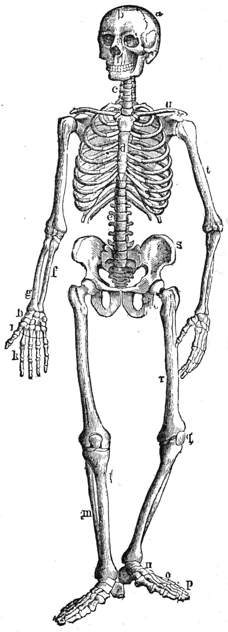
Fig. 40.—Human skeleton.—a, Parietal bone. b, Frontal bone. c, Cervical vertebræ. d, Sternum. e, Lumbar vertebra. f, Ulna. g, Radius. h, Carpus. i, Metacarpus. k, Phalanges. l, Tibia. m, Fibula. n, Tarsus. o, Metatarsal bones. p, Phalanges. q, Patella. r, Femur. s, Os innominatum. t, Humerus. u, Clavicle.
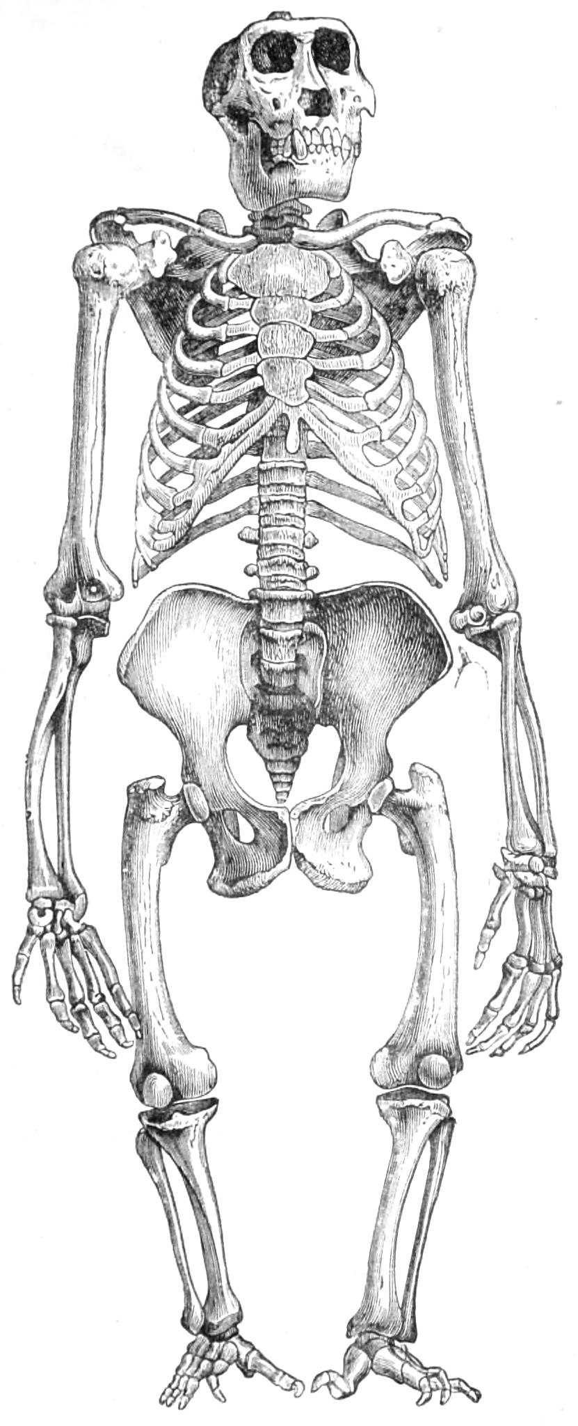
Fig. 41.—Skeleton of an aged male gorilla.
The excessive curvature of the forearm which we notice in the gorilla and the chimpanzee in their natural condition is rare in man, and when it does occur it must be regarded as an abnormal and pathological phenomenon.
The orang-utan always displays a ninth carpal bone, corresponding to de Blainville’s os intermedium and Gegenbaur’s os centrale carpi. In a very young animal I found that this small bone was furnished with a peculiar point of ossification. The bony structure of the wrist is developed in the following succession:—First, the os magnum and unciform bones; second, the scaphoid bone; third, the trapezium; fourth, the semi-lunar bone; fifth, the cuneiform bone; sixth, os centrale carpi; seventh, the trapezoid bone. The pisiform bone and the sesamoid bone, between the trapezium and the scaphoid bone, of which we shall speak presently in their relation to the muscular system, are at first simply cartilaginous.
Up to this time my search for this ninth carpal bone in the gorilla and the chimpanzee has been fruitless, since its occurrence is only exceptional. In the gibbon it is plainly inserted between the scaphoid, semi-lunar, trapezoid, and os magnum. Gegenbaur considers the os centrale to be a true constituent of the wrist, dating from an earlier condition, but he has nothing to suggest as to its subsequent survival. Rosenberg has lately given an incontestable proof of the presence of this bone in the human embryo. It is generally absorbed again, but sometimes it persists, and may be found in an adult as a well-formed ninth carpal bone. Cases of the persistence of the os centrale in man have been chiefly collected and published by the diligence of the Russian anatomist, Gruber. It is now suggested that there may also be indications of os centrale in the carpus of embryos of the gorilla and chimpanzee, but up to this time materials for such researches have been wanting.
I cannot accept the theory that os centrale carpi is merely a detached portion of the scaphoid bone. In a very young chimpanzee this bone is undoubtedly superficially indented with two transverse furrows, but the three segments display only one uniform development of bone. The distinct formation of os centrale, and its occasional appearance in man, testify that it has an independent existence. Rosenberg holds that this bone is not merely the os centrale of mammals, but that it is homologous with the two ossa centralia of the fossil Enaliosauria. It has become abortive in proportion to the reduction in size which has taken place.52 There would be no great difficulty in tracing back this bone to remote types of vertebrate animals, even as far as the Urodela (Wiedersheim) of Eastern Asia.53 The persistence of this bone in man must be regarded as a reversion, not as an arrest, of development.
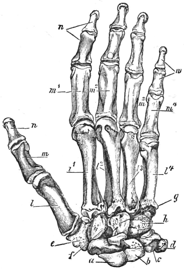
Fig. 42.—Skeleton of human hand, back view. a, Scaphoid bone. b, Semi-lunar bone. c, Cuneiform bone. d, Pisiform bone. e, Trapezium. f, Trapezoid bone. g, Os magnum. h, Unciform bone. l-l′, Metacarpal bones. m-m′ and nn, Phalanges.
On the femur of several mammals, especially in the horse, ass, rhinoceros, and tapir, and more slightly indicated in the carnivora and other families, there is, in addition to the two great and small trochanters, a third, termed by Waldeyer trochanter tertius.54 Such a formation, low, blunt, and generally placed at the top of the outer ridge of the superior bifurcation of the linea aspera, may be observed in human skeletons of all races, but is either absent in anthropoids or only faintly indicated. Virchow justly regards its presence as theromorphic, but not as a characteristic of savage or lower races.55
The human tibia displays in some instances a compression or lateral flattening of its shaft or centre-piece, so that its transverse diameter is quite out of proportion to its depth. Such a tibia is termed sword-bladed, or platycnemic. Bones of this form have been chiefly discovered in ancient deposits, as, for instance, at Gibraltar, at Perthi-Chwareu, in Wiltshire, in Lozère, at Clichy, at Saint-Suzanne (Sarthe), and especially at Cro-Magnon (Fig. 43), Janischwek, etc.
A similar formation has also been observed among men belonging to cultured races, both of ancient and modern times. Virchow, for example, discovered such bones in Transcaucasia (of the third and fourth century of the Christian era) and at Hanai-Tepe in Troas. All the large schools of anatomy in Europe contain specimens of tibiæ, which are to some extent platycnemic. These are also observed in the skeletons of primitive peoples of our time, as for example in the Negritos, Kanakas, and other African races. While some scientific men regard these bones as the result of an unhealthy condition, and the effect of rachitis, others more justly ascribe them to a vigorous exercise of the muscles in a one-sided direction. The idea expressed by Busk and others, that the platycnemic tibiæ discovered in ancient sites of Europe have belonged to a degraded race diffused over the whole continent, is contradicted by the wide diffusion of this characteristic, even in modern times. And it is doubtful whether platycnemy is absolutely restricted to the lower races. At Janischewek, Virchow found an extremely platycnemic tibia, exhumed from a kujawish grave of the Stone Age, which belonged to a skull remarkable for its unusual beauty and size, so that, taken by itself, the impression which it gave to an anatomist was that of a highly organized race.56
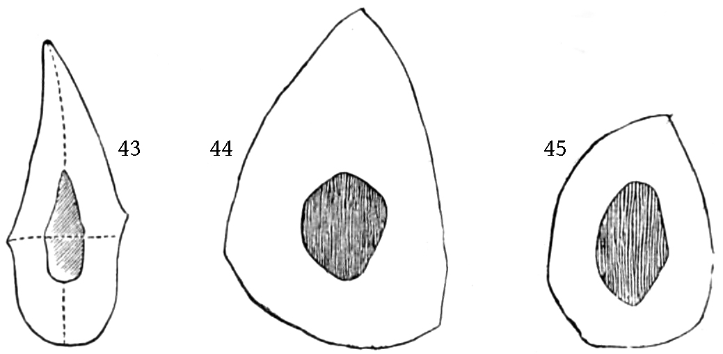
Fig. 43.
Section through a platycnemic tibia from Cro-Magnon.
Fig. 44.
Section through the tibia of a male gorilla.
Fig. 45.
Section through the tibia of a male chimpanzee.
It is important to remark that platycnemy has been regarded as a pithecoid structure, and for this reason the attempt has been made to establish the degraded position of those peoples which are most remarkable for platycnemy. But, as Boyd-Dawkins has already observed, although the tibiæ of the gorilla and the chimpanzee are to some extent platycnemic, they are much less so than the platycnemic bones of the human skeleton. The tibia of a male gorilla in the College of Surgeons Museum has an index width of 68·1, that of a female of 65·0, while the index of the chimpanzee’s tibia is 61·1, which is about the average of the tibias of Perthichwareu. It is unnecessary to indicate the other marked distinctions between the tibiæ of men and apes; if platycnemy is to be regarded as genetic, it must be admitted that man has in this particular far exceeded apes.57 Neither the gorilla, the chimpanzee, the orang-utan, nor even the baboon possesses a tibia which is flattened in its upper or middle part. In all these apes the middle of the bone is more or less rounded, almost as if it had been rounded by a turning-lathe. According to my experience, the degree of platycnemy in anthropoids is subject to certain variations. It appears to me to be least marked in the aged male gorilla (Fig. 41), and in the gibbon (Hylobates agilis, syndactylus), in which latter animal the transverse section of the tibia represents an almost equilateral triangle. The platycnemy was more marked in an almost adult female gorilla, still more decided in an aged male chimpanzee, which came from the river Kiulu, and again in an aged female chimpanzee. On the other hand, the centre of the shaft of the tibia in another aged male chimpanzee which came from Loango, was rounded, and not platycnemic. In the tibia of an adult orang-utan which I examined, the platycnemy was very marked. But I agree with Boyd-Dawkins in never having met with an anthropoid in which the platycnemy is so considerable as it is, for instance, in the Cro-Magnon tibia, and in another found at Troy.
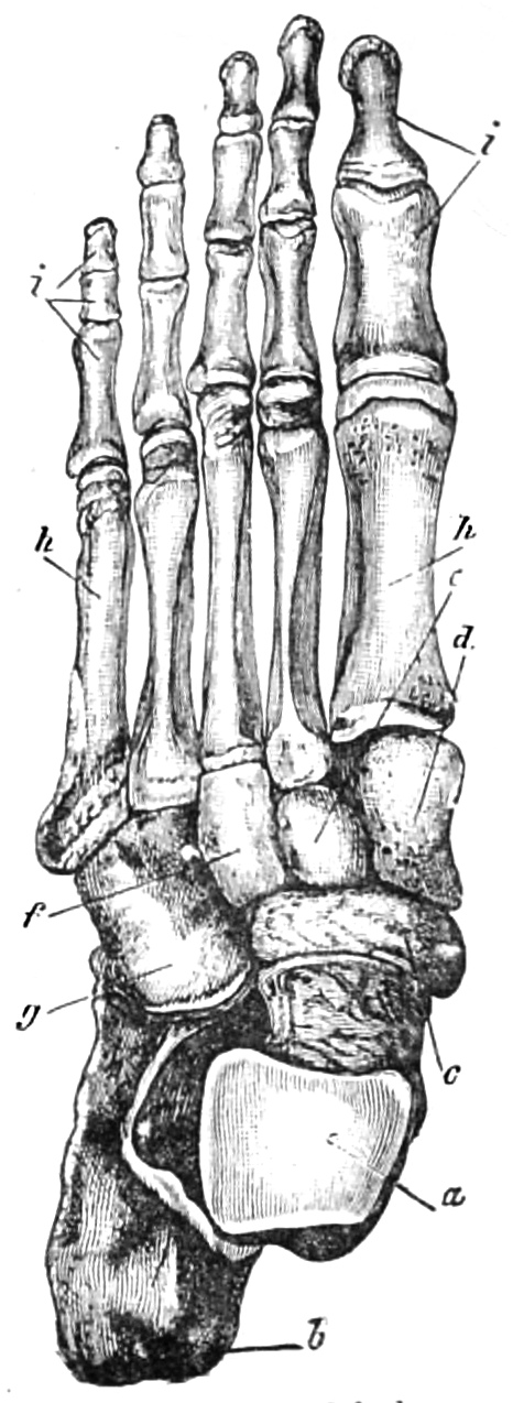
Fig. 46.—Skeleton of the human foot, seen from above. a, Astragalus. b, Os calcis. c, Scaphoid bone. d, e, f, Cuneiform bones. g, Cuboid bone. h, Metatarsal bones. ii, Phalanges.
If we give a cursory glance at the lower limbs of apes, we see that all the same characteristics are present in their tarsus that we find in the human tarsus. In each case there is an astragalus, an os calcis, a scaphoid bone, three cuneiform bones, and a cuboid bone. There are undoubtedly several peculiarities in which the tarsus differs from the corresponding part of the human foot. The first metatarsal bone is joined to the first cuneiform bone by an articular facet which extends from the back to the sole of the foot. This joint plays a part resembling that of the thumb of the human hand (see Figs. 20 and 46).
In Huxley’s opinion, the hinder limbs of the gorilla terminate in a true foot, with a very movable great toe. It is undoubtedly a prehensile foot, but in no sense a hand. It is a foot which does not differ from the human foot in any essential characteristics, but only in relative circumstances, in the degree of flexibility, and in the subordinate arrangements of its parts. Huxley adds that it must not be supposed that he wishes to undervalue differences which, however, he does not regard as fundamental. They are important enough of their kind, since in any case the structure of the foot is in close correlation with the other parts of the organism. Although it cannot be doubted that the increased division of labour in man, which relegates the function of support entirely to the legs and feet, is a significant advance in structure; yet, regarded as a whole from the anatomical point of view, the points of agreement between the human foot and that of the gorilla are much more striking and significant than their differences.
The differences in the foot of the orang are still greater; in the very long toes and short tarsus, the short great toe and the removal of the heel from the ground, in the great obliquity of the joints which connect the foot with the shank-bones, and in the absence of a long flexor muscle to move the great toe, the orang’s foot differs still more from that of the gorilla than the latter differs from the human foot. In some of the lower apes the hands and feet are still further removed from those of the gorilla than in the case of the orang. In the American apes the thumb can no longer be opposed; in the ateles it is reduced to a mere rudiment, covered with skin; in the sahius it is bent forwards and provided with a curved claw like the other fingers. In all these cases there is no doubt that the hand differs more from that of the gorilla than the gorilla’s hand differs from that of man.58
Flower remarks that the chief distinction between the foot of a man and an ape consists in the fact that the latter is transformed into a prehensile organ. The tarsal and metatarsal bones, and the phalanges are of the same number in both orders, and in the same relative position, only in the foot of the ape the facet for articulation of the first cuneiform bone with the great toe is saddle-shaped, and obliquely directed towards the inner or tibial side of the foot. Thus, the great toe is separated from the others, and so placed, that when it is bent, it is directed downwards towards the sole, and is opposed to the other toes, much more opposed to them than is the case with the thumb of the human hand.59 Owen also speaks of the characteristic transformation of the great toe of an ape’s foot into a thumb, opposed to the other toes, and adapted for grasping.60
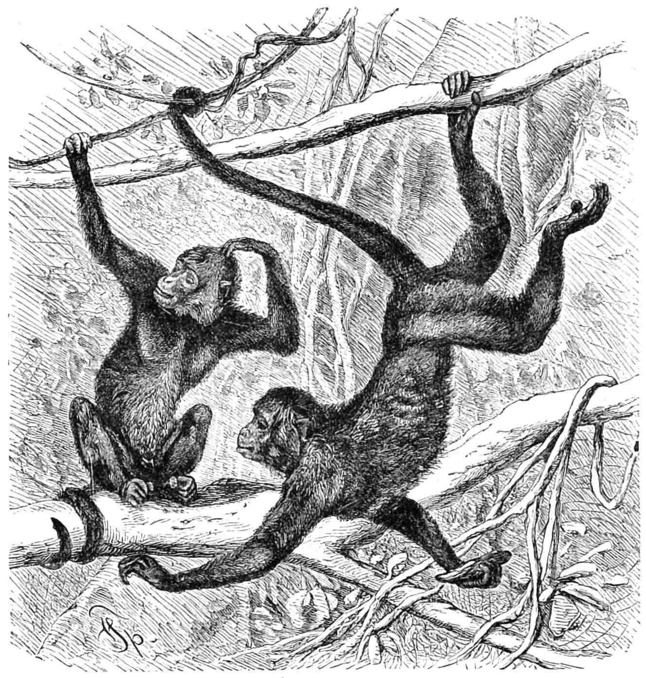
Fig. 47.—Coaita (
Ateles paniscus
).
K. E. von Bär does not agree with Huxley in considering that there is less difference between man and the gorilla than that which exists between different species of apes. “There are,” Von Bär remarks, “differences of various kinds among apes. In some the thumb is only a stump; in others, as in the orang-utan, the fingers of the hinder extremities are so long and curved that they cannot be extended on flat ground; in many of the smaller apes this member is still more like a hand than in the larger species, and the fingers can be easily spread out on the ground. In this case the foot is of a much blunter form, and is more flexible, so that the sole, which is properly turned inward, can lie flat on the ground. The heavier the body of the animal, the more sharply cut the structure of the foot must be, so that it does not admit of the free movements which are possible in the hand. But all these are only modifications of a climbing foot, or prehensile member—that is, of a hand, not modifications of a foot resting firmly on the ground and supporting the whole weight of the body.
“It must not be forgotten that the structure of the skeleton is subject to mechanical laws, which may be traced through the whole series of the animal world. This is readily apparent when we turn to the human structure.
“The human foot rests for the greater part of its length on the ground, that is to say, with the heel and centre of the foot, which form together a firm arch. The tarsus consists of the astragalus, and also of the os calcis, which in man form a very prominent part, taking a backward and downward direction, and of five other bones. The metatarsus consists of five bones, on which the five toes are inserted. In man these metatarsal bones are considerably longer than the separate phalanges. Thus, the arch on which man is supported in an erect position extends from the heel to the extremities of the metatarsal bones. The several bones are slightly movable, but they are so firmly connected that they can diverge but little from each other, unless muscular power is exerted. In order to press the toes upon the ground, it is again necessary to exert the muscles. The arched instep has this advantage, that the foot can take a better hold of the slight inequalities of the ground. In a profile view of the skeleton of a human foot, the shortness of the toes, in comparison with the length of the arched instep, is very apparent. In any natural position, even when man is not walking or standing, the sole of the foot is not turned inwards, but downwards.... The toes of the gorilla take the form of a hand, since the great toe stands separate like a thumb, while the other toes are turned outwards. In the gorilla the tarsus is short, and the heel is bent inwards. The several bones of the human foot are undoubtedly present in the hind hand of a gorilla, but the organ is changed into a prehensile organ or hand. The conditions are the same as in the parts of the mouth in insects which in some cases form movable mandibles, while in others they are attenuated into a proboscis. When it is asserted that apes are not quadrumanous, it is as if we were to say that flies have no proboscis, but attenuated mandibles.”61
All apes, including anthropoids, occasionally make use of their hinder extremities in order to snatch at objects. They also grasp with them in climbing. On such occasions, when they wish to secure the fruit they have seized from the voracity of their fellows, they take it between the toes of one hinder extremity, in order to be able to get away more quickly by means of the other, and by the use of both hands.
From what we have said, it will be seen how difficult it is to reconcile the views of different observers with respect to the fitting term to be given to the hinder extremities of apes. Against those who uphold the designation of hind hands we must oppose the anatomical structure, and also the fact that a true hand ought to possess the power of rotation in a degree which exists in the fore, but not in the hind, extremities of apes. On this account I have already adopted, as more suitable and equally distinctive, the term of prehensile foot for this member.62 I agree with Haeckel in rejecting the common designation of apes as four-handed or quadrumanous.
The bands or ligaments which connect the different parts of the anthropoid skeleton together, and convert the detached elements into a movable machinery, do not on the whole differ much from the same structure in man. A detailed account of these ligaments would, for several reasons, be out of place in this work, and I shall only mention a few special and more interesting distinctions. Such, for example, is the uncommon strength of the ligamentum nuchæ in the gorilla, which is quite in harmony with the great development of the spinous processes of the upper cervical vertebræ, and with the flattening of the squamous occipital portion. Since the sacral vertebræ are deeply inserted between the high ilia, the ilio-lumbar ligaments (ligamenta iliolumbalia) and the sacro-iliac ligaments (ligamenta iliosacralia) are of considerable size. In agreement with the projection in a downward direction of the high, narrow ischial bones, the sacro-sciatic ligaments which extend between these and the sacrum are very long in the chimpanzee. Although in this case the ischial spine is only represented by a roughness of the bone, yet there is on either side between this and the sacrum a powerful lesser sacro-sciatic ligament (ligamentum spinoso-sacrum).
The well-known anatomist, J. F. Meckel, has asserted that the depression in the head of the femur (fovea capitis), which serves for the insertion of the round ligament (ligamentum teres), is absent in the chimpanzee and orang, and he adds that it is also absent in the gibbon. In a skeleton of a young chimpanzee which had not shed its milk-teeth, and of which the ligaments were also preserved, Welcker found a fully developed round ligament inserted almost in the centre of the head of the femur. This agrees in every particular with the same formation in man. On the other hand, no trace of a round ligament was to be found in the hip-joint of a young orang-utan. The cartilaginous envelope of the head of the femur was smooth throughout, without any indication of a place for inserting the ligament. Welcker again found no such depression in the femur of an aged male orang-utan, nor was there any trace of it in another aged male orang, designated as Simia Morio. Welcker believes that he has established the fact that the round ligament is wanting in the orang-utan, but that it is present in the gorilla, chimpanzee, and gibbon. The same naturalist remarks that, although we may certainly assume that the round ligament is absent wherever there is no depression in the head of the femur, yet the existence of such a depression in the acetabulum (fovea acetabuli) is not enough to prove that a round ligament was inserted in it. The innominate bones of an adult orang-utan were examined by Welcker, and displayed a small, but well-defined depression, as if destined for a receptacle for this ligament,63 running from the cotyloid notch down to the bottom of the acetabulum, between the two horns of the semilunar-shaped articular cartilage.
In a subsequent paper, Welcker states that the absence of the round ligament in the orang-utan, and its presence in the chimpanzee, had been previously established by Camper and Owen.64 In three specimens of orangs which he had obtained immediately after death, Owen found that the round ligament was imperfectly developed on both sides. The chimpanzee differs from the orang in possessing a depression on the head of the femur. In the gorilla, as Owen observes, this depression has almost the same depth and relative position as in man. At Welcker’s request, Professor Dippel ascertained the presence of the depression in the femur of a gorilla skeleton which is preserved in the natural history collection at Darmstadt. St. George Mivart saw the skeleton of an orang in which the femur was marked with a slight but plainly indicated depression, just where the round ligament is usually attached. Welcker thinks it probable that in some specimens of the gorilla the round ligament is only slightly developed, and that in others it is altogether wanting. On several femurs of gorillas, this naturalist observed only doubtful traces of the depression in question. Duvernoy found the round ligament fully developed in the gorilla and chimpanzee. Vrolik failed to find it in the orang-utan, but ascertained its presence in the chimpanzee. Gratiolet and Alix saw that it was fully developed in Troglodytes Aubryi.
In addition to these somewhat conflicting assertions, I have myself observed, in the gorilla innominate and femur bones examined by me, more or less distinct indications of the depression which receives the round ligament. The ligament itself has been preserved with the body of a gorilla. The same remark applies to the skeletons and bodies of chimpanzees. In the case of the skeleton of an orang, slight indications of a depression were observed on the head of the left femur, and these indications were absent in the femurs of other specimens. In a large orang-utan which died in the Berlin Aquarium, only short, filamentous tufts of streaky fibres were apparent in the right acetabulum, and these were intermingled singly or in groups with the cartilaginous cells, somewhat resembling the cartilaginous corpuscles of the synovial membrane. From these facts we may conclude that the round ligament is generally but not invariably present in the gorilla and chimpanzee, and that it is altogether absent in the orang-utan. In the gibbon it is present in the majority of cases. I have myself observed it in Hylobates agilis, leuciscus, and syndactylus. Owen asserts that the unsteady gait of the orang is partly due to the absence of this ligament, but the truth of this surmise is rendered doubtful by the fact that the ligament is not unfrequently absent in other anthropoids. Moreover, the gait of all these arboreal and climbing animals is extremely ungainly.
The muscular system of anthropoid apes is very interesting. I must necessarily refrain from giving a detailed account of it, and will only mention some points in connection with this organic system, and their relation to corresponding points in the muscular system of man. I rely partly on the researches of others, and partly on my own. The amount of material which has been collected up to this time is, unfortunately, too scanty to enable us to draw satisfactory conclusions in all cases. We are often unable to decide whether the conditions presented to us in the case of anthropoids are normal or exceptional. Nor are the statistics of muscular variations in the human subject by any means firmly established. My own labours in this direction are not yet concluded. The assertions on the subject which have been published to the world and accepted as authoritative have already been shown to be to some extent untrustworthy. Even the little which I am now able to produce may not altogether stand the test of subsequent research. Brühl justly remarks that in no department of anatomy more than in that which treats of the muscles, is it more essential that we should not decide whether a form is normal or exceptional until it has been repeatedly examined.65
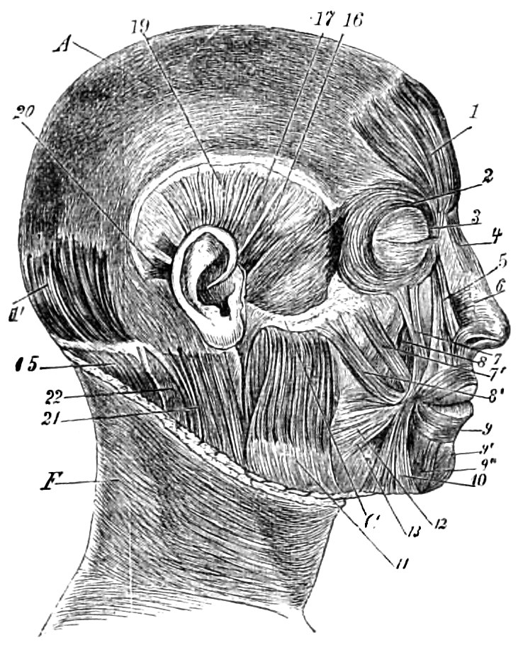
Fig. 48.—Muscles of the head and face of a European. 1, 1′, Occipito-frontalis. 2, 3, Orbicularis palpebrarum. 4, Pyramidalis nasi. 5, Levator labii superioris alæque nasi. 6, Compressor naris. 7, Levator labii superioris. 7′, Zygomaticus minor. 8, Levator anguli oris. 8′, Zygomaticus major. 9, Orbicularis oris. 9′, Levator menti. 9″, Depressor labii inferioris. 10, Depressor anguli oris. 11, Masseter. 12, 13, Risorius and the buccinator by which it is covered. 15, Trapezius. 16, Attrahens. 17, 19, Attollens. 20, Retrahens aurem. 21, Sterno-mastoid. 22, Splenius. A. Tendinous aponeurosis. C. Malar bone (the parotis is removed). F. Skin of neck.
The cranial muscles of anthropoids are formed like those of men, except in a few unimportant particulars (comp. Figs. 48 and 50). I have not observed in anthropoids the muscular fibres which in man branch out from the orbicular muscle of the eye, and overlap the cheeks and temples, and which are considerably developed in the head of a Monjalo negro which was dissected by me (Fig. 49, 3, 3′). In apes that portion of the orbicular muscle which covers the supra-orbital ridge is very marked. There is generally a considerable layer of muscle on the nose and upper lip. I have dissected it in detail in anthropoid and other apes, including those of America; i.e. the zygomatic muscles, the levator labii superioris, and the levator labii superioris alæque nasi. This has also been done by Duvernoy, Alix, and Gratiolet, in the case of anthropoids dissected by them, as well as by Macalister and Bischoff.
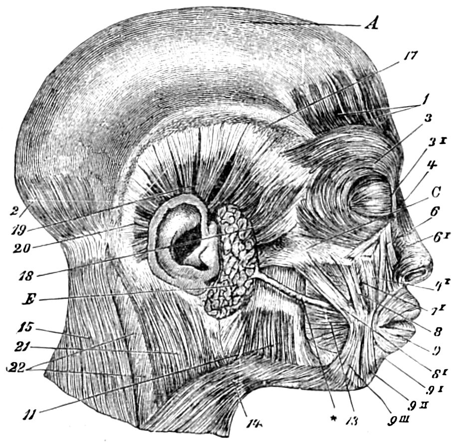
Fig. 49.—Head-muscles of a Monjalese negro. 1, 2, Occipito-frontalis. 3, 3′, Orbicularis palpebrarum. 4, Pyramidalis nasi. 4′, Levator labii superioris. 6, Levator labii superioris alæque nasi. 6′, Compressor naris. 7′, Levator anguli oris. 8, 8′, Zygomatici major et minor. 9, Orbicularis oris. 9′, Levator menti. 9″, Depressor labii inferioris. 9‴, Depressor anguli oris. 11, Masseter. 13, Buccinator. 14, Platysma. 15, Trapezius. 17, 18, Attollens and attrahens aurem. 19, Embedded temporal muscle. 20, Retrahens aurem. 21, Sterno-mastoid. 22, Deeply set muscles of neck. A, Tendinous aponeurosis. C, Zygoma. E, Parotis. *, Stensonian duct.
Bischoff was only able to identify a wide zygomatic muscle in the orang with the small zygomatic in man. In the orang, the gibbon, and the baboon, as well as in Innus sinicus and Ateles, I myself was quite able to trace a division into a large and small zygomatic. In the gorilla dissected by me the levator labii superioris alæque nasi was very wide (Fig. 50, 6). In the case of a gorilla, Ehlers dissected the small zygomatic muscle, together with the levator labii superioris alæque nasi, in the manner introduced by Henle as a single square muscle of the upper lip (Musculus quadratus labii superioris). In the gorilla I observed a levator alæque nasi, together with the already mentioned levator labii superioris; but I failed to find any separate levator labii superioris. The very wide cartilage of the nose is occupied by a considerable amount of muscular tissue. All these muscles are present in the orang, but they are of small size and separated into detached bundles. The pyramidalis nasi may be traced in every instance, especially in the gorilla (Fig. 50, 4) and in the orang. It is not so strongly developed in the chimpanzee and gibbon, but is not absent in these apes, nor in those which are not anthropoid, such as the baboon, and ateles, or climbing ape.
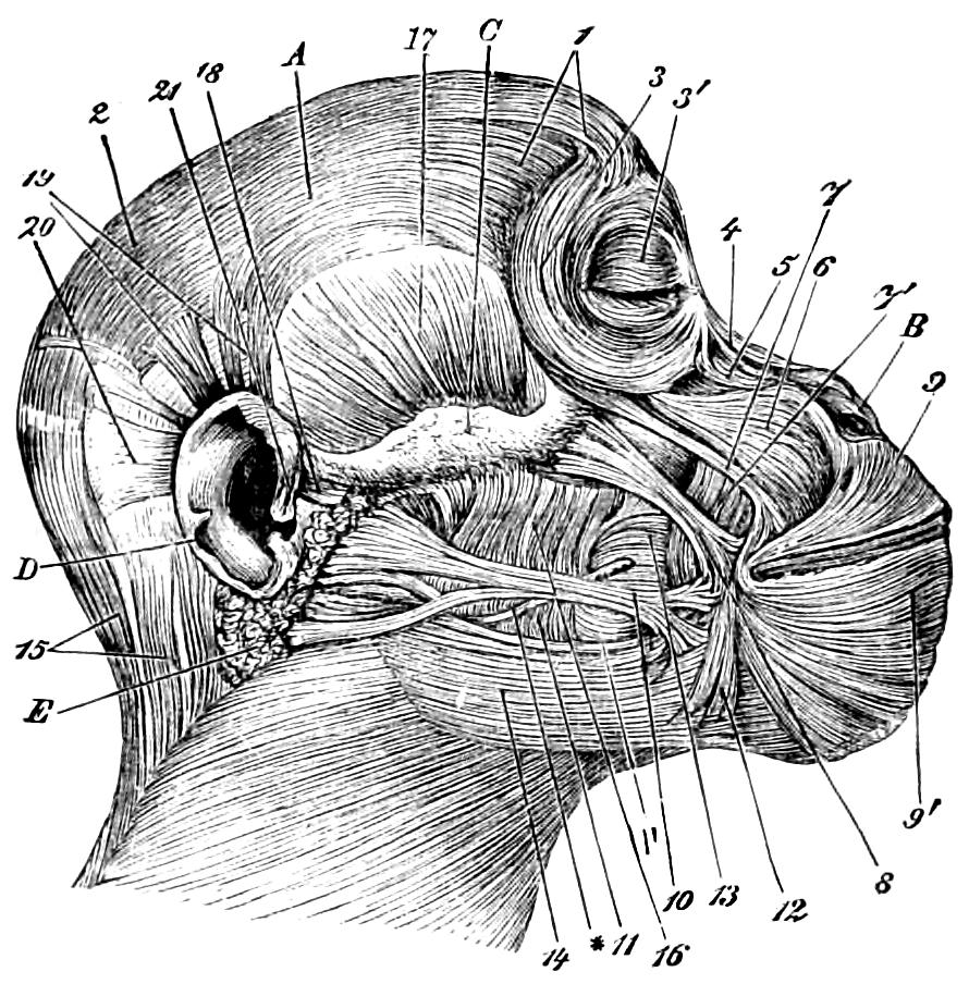
Fig. 50.—Head-muscles of gorilla presented in Fig. 3. 1, 2, Occipito-frontalis. 3, 3′, Orbicularis palpebrarum. 4, Pyramidalis nasi. 5, Levator alæ nasi. 6, Levator labii superioris alæque nasi. 7, Zygomaticus minor. 7′, Levator anguli oris. 8, Zygomaticus major. 9, 9′, Orbicularis oris. 10, Risorius. 11, 16, Masseter. 1′, Buccinator. 12, Depressor anguli oris. 13, Buccinator. 14, Platysma. 15, Trapezius. 17, Temporal. 18, 19, 20, Attrahens, attollens, and retrahens aurem. 21, Lesser muscle of helix. A, Tendinous aponeurosis. B, Cartilage of nostril. C, Zygoma. D, External ear. *, Stensonian duct.
I myself follow the original division of the muscles into those which belong to the nostril and upper lip, in accordance with the principles of Duchenne, Darwin, Gamba,66 and others, and I do so the more readily, since it is impossible not to perceive the manifold and lively mimetic action which takes place in this particular region of an ape’s head. The distinct action of the levator labii superioris alæque nasi, the dilation of the nostrils, the function of a strongly developed levator anguli oris, are especially characteristic of the gorilla; but they are also perceptible in the chimpanzee and gibbon. The orang’s face is the least mobile. I observed that in the gorilla the risorius was very long, branching slightly in the fore-part of the corner of the mouth, and behind into three distinct wide bundles. The lowest bundle covered the platysma myoides, but could not be regarded as part of the latter. In one chimpanzee I found that the risorius was slightly developed, and in other animals of that species I failed to trace it at all. Alix and Gratiolet represent the Aubry chimpanzee (Plate ix. Fig. 1, 15) with the risorius strongly developed. I have not observed this formation either in the orang or the gibbon, but it was apparent in one of the ateles (Ateles leucophthalmos). In this case the muscle covered the platysma myoides and Stenson’s duct, i.e. the duct leading out of the parotid gland (Fig. 50, *).
For some time I was disposed to regard the risorius of this ape as only a radiation of the platysma myoides, but my opinion upon this point is again shaken.
In the gorilla a faint depressor anguli oris and an equally faint depressor labii inferioris may be observed, the latter partly covered by the large and predominant orbicularis oris (Fig. 50). In the chimpanzee the two depressors are plainly apparent, and in the gibbon the one first named was at any rate developed. The platysma myoides, the depressors just mentioned, and the crescent-shaped orbiculares are in this animal in close connection with each other. Froriep’s suggestion becomes ever more probable, that these muscles of the lower lip owe their origin to the intersection of the opposite portions of the skin-muscles of the neck which overlap the face. The buccinator muscle in anthropoids resembles on the whole that of man, and in both cases is pierced by Stenson’s duct (Fig. 50). The form of the masseter muscle is common to both (see Fig. 50, 11, 16). In the external ear of anthropoids there is an attrahens, attollens, retrahens (Fig. 50). Compared with that of a white man, and still more with that of a negro (see Figs. 48, 19, and 49, 17), the attollens is only slightly developed. The muscles attached to the cartilages of the ear are extremely scanty or partially wanting, which is also sometimes the case with man. The muscles of the helix are most strongly marked in the gorilla (see, for example, Fig. 50, 21). Tiedemann, Bischoff’s brother-in-law, carefully observed two living chimpanzees in Philadelphia for six months without detecting any movement of the ears. My own observation confirms his assertion and the remarks of Darwin, which I have already quoted, to the effect that anthropoids are incapable of moving their ears. I know of no individual exceptions. This is the more remarkable since some men have retained the power of voluntarily moving their ears, and the same power is also found in some species of apes, such as the sea-cats, baboons, macacas, and magots.
It will not here be out of place to say something of the characteristics, previously mentioned, of the physiognomical expression of anthropoid apes. Thus, for example, when the gorilla is agitated, he can move the skin of his head and bristle the hair which covers this region. The chimpanzee can also move the skin of the head, but with no very apparent bristling of the hair. The large male orang, which was in the Berlin Aquarium in 1876, bristled his hair and the skin of his head when he was much enraged. It is known that in some instances man also possesses this power.
I have already spoken of the expression of the eyes of these animals. I will only add that when anthropoids of every species are in great pain or seriously ill, the expression of their eyes is often most affecting.
The forehead of these animals is frequently marked by transverse furrows, and especially, as Darwin justly observes, when they raise their eyebrows. The same great observer considers that the countenances of anthropoids are, in comparison with those of men, generally inexpressive, and indeed, chiefly in consequence of the fact that they do not wrinkle the forehead when they are excited. The wrinkling of the forehead, which is one of the most significant forms of expression in man, is due to the action of the corrugatores supercilii, by which the eyebrows are drawn down and closer to each other, so as to form vertical folds on the forehead. It has been asserted that the orang and chimpanzee possess these muscles, but they seem to be rarely exercised—at any rate, to any remarkable extent.67 When Darwin brought a chimpanzee out of his dark chamber into bright sunshine, he only once observed a slight wrinkling of the forehead. When the same observer tickled the nose of a chimpanzee with a straw, its face was slightly wrinkled, and faint vertical furrows appeared between the eyebrows.68 Darwin never observed any wrinkling of the forehead in an orang. I myself have observed a contraction of that region of the brows which is covered with bristly hairs, and a wrinkling of the skin which covers the bridge of the nose in the gorilla and the chimpanzee, and have illustrated this expression by a drawing.
Darwin goes on to say that when a young chimpanzee is tickled, to which, as in the case of children, their armpits are peculiarly sensitive, he generally utters a chuckling or laughing sound, although sometimes the laugh is silent. The corners of the mouth are then drawn back, and this sometimes causes the eyelids to be slightly wrinkled. This wrinkling, which is so characteristic of the human laugh, is still more apparent in some of the other apes. In the chimpanzee the teeth of the upper jaw are not exposed when he utters this laughing sound, and in this respect he differs from man. Darwin further observes that when the tickled young orang ceases to laugh, an expression passes over his face, which, according to Wallace, may be called a smile. Darwin has observed something similar in the chimpanzee.69
My own observation confirms what has been said of the chuckling of a tickled chimpanzee. When Dr. Hermes, the director of the Berlin Aquarium, played with the chimpanzee which was kept in that establishment, a contortion of the corner of the mouth, resembling a somewhat sardonic smile, at once appeared. No specimen displayed this smile with so much effect as the lively Augustus, who delighted visitors by his inexhaustible humour in 1879. The gorilla, of which an illustration is given in Fig. 3, also drew down the corner of his mouth when he was pleased, by means of the muscular system which we have just described.
When the gorilla is provoked, he displays both rows of teeth, and opens his mouth to utter sounds of fury, while making ready to fight. It is well known that anthropoids are able to pout and project their lips; and Darwin says that they do this, not only when they are slightly teased, and are sullen or disappointed, but also when anything occurs to make them uneasy.
I have often observed in chimpanzees a slight wrinkling of the region of the nasal cartilage, and even a vibration in a lateral and upward direction. In any case, the muscles which we have described as acting on the nose and upper lip are exercised.
The platysma myoides, which extends in man from the lower row of teeth to just below the clavicle, occupies about the same area in the gibbon and in other apes (Fig. 50). In the chimpanzee, however, this muscle extends as high as the zygomatic arch, or even higher. In the gorilla also I observed that this part extends comparatively high on the face. In chimpanzees, orangs, and gibbons the upper fibres of this muscle seem to form the risorius. In one case the platysma myoides sent forth a fasciculus, about 18 mm. in width, to the beginning of the lower temporal ridges. In the gorilla I saw that the uppermost fibres of the platysma myoides were partly covered by the risorius (Fig. 50, 10).
From the corresponding muscle in the orang the lower fibres tend far backward, and are in connection with the deltoid muscle covering a segment of the capsular ligament. This muscle wrinkles the skin of the neck, and helps to draw down the lower jaw. In cases in which it extends far in an upward direction, as in those we have cited, it affects the lateral extension of the middle and lower skin on the faces of these animals, as well as the grinning contortion of the corner of the mouth. It may also have to do with the grumbling sound issuing from the throat-pouch, which is uttered by the animal when agitated, as he rapidly opens and closes his mouth.
The strong sterno-cleido-mastoid muscle found in these animals, and especially in the orang and gibbon, can be divided without difficulty into a sternal and clavicular portion. The two portions diverge from each other in a downward direction. As Bischoff justly states, a muscle not hitherto observed in man may be traced in all four species of anthropoids, a muscle which extends from the external part of the clavicle to the transverse process of the first cervical vertebra. Bischoff has called it the musculus omocervicalis. It is found in other apes, although the site of its origin varies, sometimes occurring on the spine of the scapula. Our Munich anatomist differs from Huxley in regarding this muscle as “a brilliant proof of the relation of all apes with each other.” I give this assertion without further comment.
The muscles which extend between the head, sternum, and clavicle, together with the muscles of the acromion process of the scapula, make an external covering to the throat-pouch, which I shall describe presently. The pectoralis major of the gorilla, as well as that of man, divides into two portions, one attached to the clavicle, the other to the cartilages of the true ribs. The former is divided from the deltoid by a wide interval, filled with connective tissue and fat. But both portions of the pectoralis major are divided by a tolerably wide space, into which, in Bischoff’s opinion, the throat-pouch is inserted. This, however, I do not believe, since that organ would be compressed and strangulated between the two portions of muscle whenever they were exercised. It may, however, be supposed that room for an enlargement of the throat-pouch when the animal is bellowing is afforded by the existence of these spaces. Bischoff is right in the assertion that the clavicular portion of the pectoralis major is wanting in the orang-utan. The upper part of this muscle springs directly from the sternum. The lower sternal ribs give origin to the pectoralis minor. The chimpanzee and gibbon display clearly in this muscle the separation we have mentioned into a clavicular and a sternal portion.
The structure of the pectoralis minor in these apes is full of interest. In the gorilla it divides into an upper portion of firmer tissue, less easily separable into digitations, which arises from the third to the fifth ribs, and a lower portion, separable into three digitations, of which the upper segment laps considerably over the lower segment of the upper portion. In the chimpanzee an upper portion of less firm texture extends from the second to the fourth, and a lower with three digitations from the fourth to the seventh ribs. This second lower portion is sometimes absent. I have seen the upper portion attached to the coracoid process of the scapula, and the lower portion to the ridge of the greater tuberosity of the humerus. In the orang an upper portion, separable into three digitations, extends from the second to the fifth ribs, and is attached to the coracoid process. A lower portion, also separable into three digitations, extends from the fifth to the seventh ribs, and is also attached either to the greater tuberosity of the humerus or to its edge; this latter portion projects below over the pectoralis major. In the gibbon (Hylobates albimanus), the upper portion starts from the second, the lower from the third to the fifth ribs. It may here be remarked that the pectoralis minor is in man also sometimes separable into digitations, which may be connected both with the coracoid process and with the capsular ligament of the shoulder-joint. In anthropoids the tendon of insertion of this muscle is remarkably slender.
According to Duvernoy, in the gorilla a fibrous, hood-like fascia covers the whole region of the occiput and neck. In adult males this fascia is 20 mm. in thickness. In a female dissected by me the rudiments of a similar hood-like cervical fascia were present. Duvernoy is justified in supposing that this is not yet developed in the young gorilla, and that a layer of connective tissue and fat is substituted for it. In a young gorilla I saw the trapezius divided into distinct bundles of flesh by layers of fat (Fig. 50, 15). The fascia corresponds to the great development of the trapezius, and the same characteristic development exists in other anthropoids. The adult male gorilla displays a powerful ligamentum nuchæ in connection with the long spinous processes of the cervical vertebræ, as well as powerful inter-spinales muscles, spinales colli, and semi-spinales colli and dorsi. The great development of the spinous processes of the dorsal vertebræ of gorillas (Fig. 17), and also chimpanzees and orangs, involve the development of powerful semi-spinales, as well as of strong, fourfold spinales and inter-spinales muscles. The whole of the fleshy formation of the neck of an adult male gorilla which is covered by the trapezius is very voluminous, and especially the splenius capitis and colli, the long cervical muscle (Musculus longissimus cervicis), and the long head-muscle (Musculus longissimus capitis), which have also been regarded by me as parts of the long spinal extensor, and finally the oblique and vertical muscles at the back of the head. With Chappuy, I am disposed to regard the latter as modifications of the spinales and inter-spinales.
The levator anguli scapulæ is divided in anthropoids as in man. The subclavius is slender, except in the gorilla, and in the latter animal it sends a tendon obliquely to the coracoid process.
In all anthropoids the deltoid is strongly developed. In the gorilla it projects forwards and outwards in order to attach itself to the humerus, almost in its centre. Here it is separated from the brachialis anticus in a manner with which we are only imperfectly acquainted. It extends nearly as far in the gibbon and orang, while in the chimpanzee its attachment is higher up. Bischoff observes, and it was previously suggested by Vrolik, that in the chimpanzee the coraco-brachialis muscle possesses at its origin a moderately large second portion, which tends downwards over the lesser tuberosity of the humerus, and adheres to its edge. But I have seen both portions of the muscle in question attached to the coracoid process of the scapula in apes of this species. In the gorilla, orang, and gibbon the position of this muscle corresponds to that in man.
Chapman and Bischoff speak of a muscle common to all apes which starts from the tendinous attachment of the latissimus dorsi on the edge of the lesser tuberosity of the humerus, and tends downwards on the inner side of the humerus, and to this muscle they give the name latissimo-condyloideus. Bischoff goes on to say that this muscle goes in some cases into the fascia which covers the biceps; and in others, as in the baboon, it is attached to the inner inter-muscular septum and to the internal condyle of the humerus. In the gibbon it only extends as far as the centre of the humerus, but in the orang it reaches to the condyle, where it is pierced by the ulnar nerve. Bischoff adds that this formation is wanting in man.
This structure is indeed remarkable in anthropoids. The muscle starts in a lateral direction from the insertion point of the latissimus dorsi. In the gorilla alone I observed that it started from the coracoid process of the scapula, together with the two portions of the pectoralis minor; it was connected for a space with the coraco-brachialis, and finally it was attached, in the upper part of the lower third of the humerus, to the inter-muscular septum which is found between the brachialis anticus and the triceps. In the chimpanzee, on the other hand, it has its origin in the latissimus dorsi, and divides into an anterior and posterior portion; the former is attached to the inner condyle of the humerus, while the latter is connected either with the middle or inner head of the triceps. In the orang the same division of this muscle may occur. In one of these animals I observed an anterior portion, very thin and semi-membranous, attached by an extremely slight tendon to the coracoid process of the shoulder-blade, while the hind portion issued from the latissimus dorsi. They were both in connection with the triceps and brachialis anticus. In other instances the muscle consisted only of the posterior portion, issuing from the latissimus dorsi. In the white-handed gibbon, the muscle issued from the region in which the tendons of the latissimus dorsi and of the teres major are united, and was inserted into the fascia which is found between the bicipital and the brachialis anterior. This attachment may also occur in the centre of the shaft of the humerus. Chapman and Chudzinsky have observed anomalous instances of this formation in coloured races.70
It is well known that in man the biceps is inserted into the tuberosity of the radius by means of a flattened round tendon. This tendon, however, opposite the bend of the elbow, gives off a broad expansion, which passes into the fascia of the forearm, and is termed Aponeurosis bicipitis. In the gorilla this aponeurosis is carried on as strong fibrous bundles of the fascia of the forearm into the palmar fascia. In the gibbon the short head of the muscle does not always start from the lesser tuberosity of the humerus, nor from the tendon of the pectoralis major (Huxley), but sometimes from the edge of the lesser tuberosity, which is here connected with the latissimus dorsi, as well as with the sub-scapularis, the brachialis anticus, which is more to the side, and with the triceps. In the gibbon, as Bischoff justly observes, the supinator longus only reaches as far as the centre of the radius, instead of extending to the styloid process of that bone, as it does in other anthropoids, and in man.
The palmaris longus is wanting in the gorilla, but not in other anthropoids. The long flexor muscles of the fingers and the lumbricales resemble those of man (Figs. 51, 52). The flexor longus pollicis is absent in the gorilla. Duvernoy considers that it is replaced by a tendon of the long flexor of the fore-finger, but I have been unable to verify the existence of this tendon. The same muscle is also absent in the chimpanzee and the orang, but it may be traced in Hylobates albimanus. Chapman states that in the gorilla the pronator radii teres only sends forth one head,71 but I have found it to be bicipital in animals of this species. The lower or hinder head issues, as in man, from the coronoid process of the ulna. Both in the gorilla and in the chimpanzee it extends far in a downwards direction on the radius (Fig. 52). The flexor carpi radialis starts in the chimpanzee with one head from the inner condyle of the humerus, and with the other from the radius. Bischoff describes the structure of the long abductor of the thumb in the orang, the baboon, the pithecia, and the hapale as resembling that of man. But in the gorilla, the chimpanzee, and the macaca the tendon divides into two parts. Nor does one tendon belong, as in man, to a short extensor of the thumb, but the latter is wholly absent, and the division of the tendon only implies a continued division of the attachment to the trapezium, as well as to the metacarpal bones of the thumb. This division of the tendon also occurs in the gorilla, which likewise possesses a short extensor of the thumb. In this point, again, apes display a greater likeness to one another than to man.
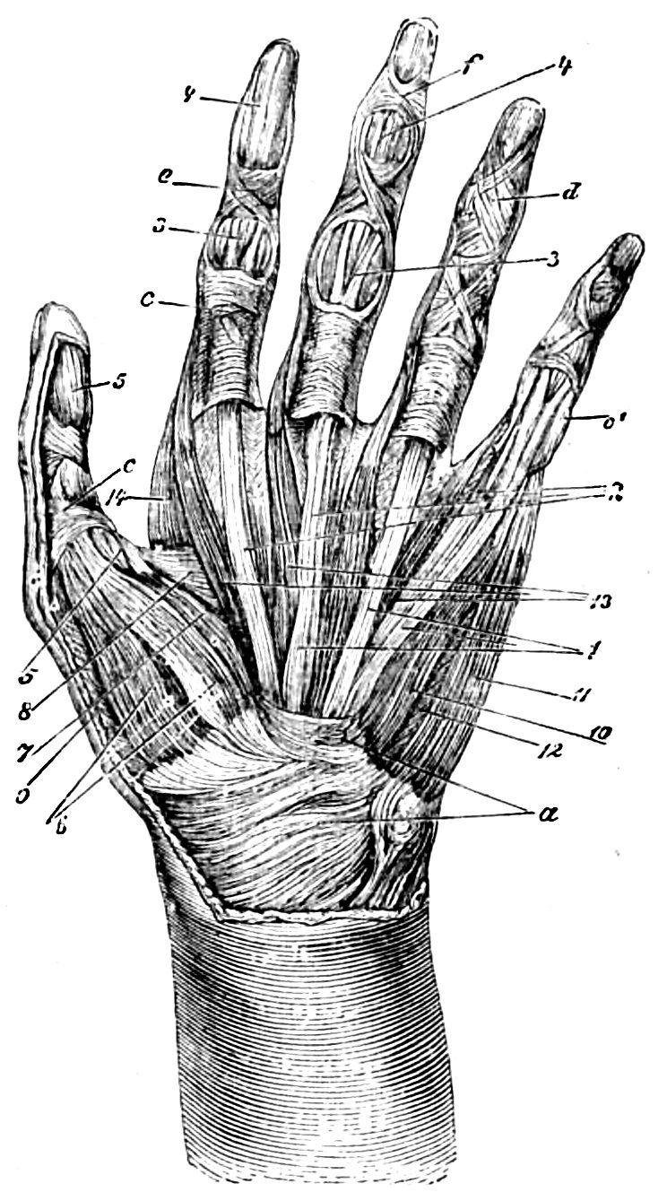
Fig. 51.—Palmar muscles of man. a, Ligaments of wrist, especially the anterior ligament. c, c′, Sheathing ligaments. d, e, f, Oblique fibres of the ligaments of the sheath of the flexor tendons. 1, 2, Tendons flexor sublimis, and of the flexor profundus muscles of the fingers. 3, The reciprocal perforation of these tendons. 4, Continuation of the tendons of the flexor profundus of the fingers. 5, Tendon of the flexor longus pollicis. 6, Abductor pollicis. 7, 8, 9, Flexor brevis, adductor, and opponens pollicis. 10, 11, 12, Flexor brevis, abductor, and opponens minimi digiti. 13, Lumbricales. 14, First dorsal inter-osseous muscle.
According to my own researches, the long abductor of the thumb in anthropoids forms a muscle not more considerable than one in proximity with it, of which the origin and more central direction recall the short extensor of the human thumb. In all four species I found that the abductor had two tendons, and was attached to the trapezium. The muscle in its vicinity is inserted above the base of the first metacarpal bone. I have not been able to discover an extra extensor of the thumb in the gorilla. The question now arises what we should think of the second muscle, which is found in these animals in the vicinity of the abductor. In my opinion, it may be confidently accepted as a short extensor of the thumb, since it always effects an extension of the metacarpal bone of that member, and in this act of extension it is supported by the long extensor which acts upon the phalanges. It must be remembered that the comparatively short thumbs of anthropoids have not to be employed in so many different ways as the human thumb, and that we cannot therefore be surprised that the development of the short extensor is less complete. A special extensor muscle of the index finger is either altogether absent in the gorilla or very slightly developed, while it is very apparent in Hylobates albimanus (6, Fig. 53). In the chimpanzee this muscle sends a tendon to the middle finger. In the orang there is one extensor common to the four fingers. In the gibbon’s hand, this, as well as the other extensor and flexor muscles, is remarkable for its excessive slenderness. The manifold connections of the extensor tendons with each other are an interesting peculiarity (Fig. 53).
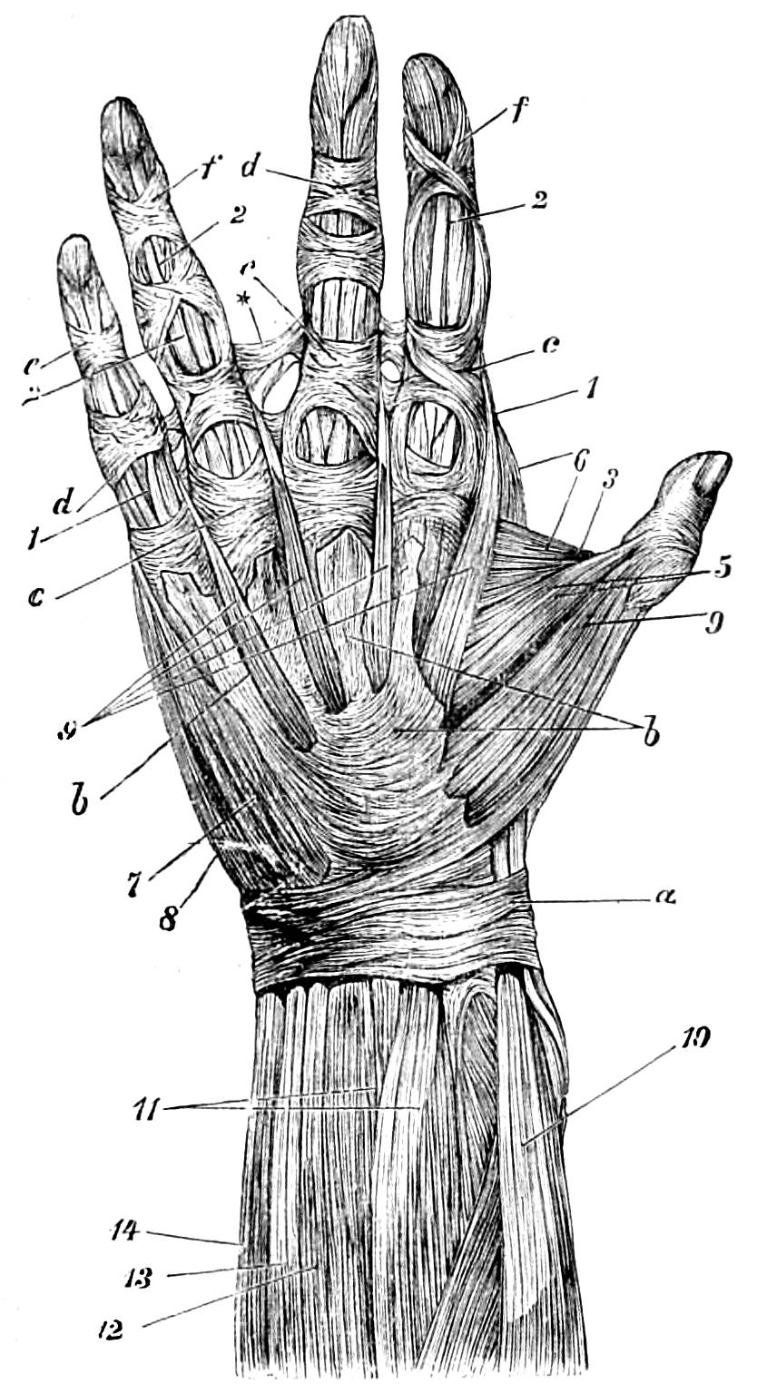
Fig. 52.—Palmar muscles of gorilla. a, Anterior ligament. b, Remains of the skin of palm, here covered with a very sinewy skin. c, f, Oblique fibres of the ligaments of the sheath of the flexor tendons of fingers. 1, 2, Flexor tendons. 3, Spaces between the heads of the flexor brevis pollicis, whence in man the tendon of the flexor longus pollicis issues (comp. Fig. 51, 5). 4, 3, 3′, 5, Abductor, flexor brevis, abductor pollicis. 6, 7, 8, Opponens, flexor brevis, abductor, minimi digiti. 9, Dumbricales. 10, Supinator longus. 12, Flexor sublimis digitorum. 13, Flexor minimi digiti. 14, Flexor carpi ulnaris.
In the chimpanzee I observed a superficial flexor, common to the fingers, and enlarged in the region of the third and little fingers. A superficial flexor, belonging to the index finger, started from the inner condyle of the humerus, and from the back of the inter-muscular septum. The deep finger-flexor was attached to the four fingers. In the orang the first of these flexors forms a two-tendoned belly for the index finger, as well as one for the other three fingers. The deep flexor only displayed two bellies. In the gibbon, on the other hand, the superficial flexor displays four bellies.
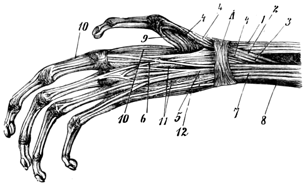
Fig. 53.—Muscular system of the back of a gibbon’s hand. 1, The extensor carpi radialis longior and brevior. 2, Abductor longus pollicis. 3, Extensor primi internodii pollicis. 4, Extensor secundi internodii pollicis. 5, Extensor communis digitorum. 6, Extensor indicis. 7, Extensor minimi digiti. 8, Extensor carpi ulnaris. 9, First dorsal inter-osseous muscle. 10, Continuation of the same to index finger. 11, 12, The other inter-osseous muscles of this region. A, The posterior annular ligament.
In the carpus of the chimpanzee there is, so far at least as my experience goes, a so-called sesamoid bone. It is in this instance in connection with the scaphoid and trapezium bones, just where the fibres of the anterior and posterior ligaments of the wrist pass into each other. In the chimpanzee the tendon of the long abductor muscle of the thumb sends some fibres into this sesamoid bone, while the other fibres of the tendon of this muscle, which divides into several strips, are inserted in the trapezium bone, and a few also in the base of the first metacarpal bone.
The short flexor muscle of the thumb, of which Bischoff has denied the existence, is certainly present in these animals. In the chimpanzee the lower fibres of the short abductor muscle of the thumb have their origin in the sesamoid bone. The middle fibres, of the same muscle issue from the strips of ligament attached to the sesamoid bone. On the other hand, the upper part of the muscle has its origin in the anterior annular ligament. In the orang, the lower fibres of the short abductor of the thumb likewise have their origin in the sesamoid bone, while the central fibres again start from the anterior annular ligament. The upper fibres are strong, and are inserted into the base of the first metacarpal bone. In a dissection of the orang the flexor longus pollicis sent a thin, tendinous expansion on to the bone. This sesamoid bone is also found in the gorilla, although Duvernoy and Rosenberg do not appear to be aware of its existence.72
In the palm of the gorilla’s hand there is a short abductor, a short bicipital flexor, an opponens, and an abductor of the thumb. The longer belly of the short flexor extending in a more radial direction, and in connection with the opponens, is only slightly developed. In the muscular system of a gorilla’s little finger we may observe an abductor, a short flexor, and an opponens. The palm of the chimpanzee displays a short abductor, an opponens, a short bicipital flexor, and an adductor of the thumb; also an abductor, a short flexor, and an opponens of the little finger. In the orang I observed a short abductor, a short flexor with two bellies, an opponens, and an adductor of the thumb. In addition to the short flexor of the thumb, Langer and Bischoff describe another short, independent muscle, representing the long flexor, and attached to the second phalanx, but I have not myself ascertained the existence of this muscle. The same anatomists mention an adductor between the third metacarpal bone and the first joint of the thumb, and another between the second metacarpal bone and the second joint of the thumb, passing on into the extensor tendon. I am myself convinced of the existence of a twofold adductor, but not of the fact that the tendon of one of the muscles (termed by Langer the second opponens) passes on into the extensor tendon. In the little finger of the orang there is an abductor, a short flexor, and an opponens. In the gibbon there is a short abductor, a faintly indicated opponens, a short bicipital flexor, and an adductor of the thumb. In Hylobates albimanus this adductor divides into four or five portions, which are attached to the whole of the first metacarpal bone. In the little finger there is an abductor, a short flexor, and an opponens. In the same animal the first inter-osseous muscle is attached by one portion to the second metacarpal bone, by the other to the base of the second phalanx of the index finger (Fig. 53, 9, 10).
Bischoff has described the muscles which Halford terms Contrahentes digitorum (contractors of the digits), which lie deep in the palm of the hands and feet of the chimpanzee and gibbon, the mandril, baboon, and other apes.73 They rest upon the inter-osseous muscles, and are covered by the tendons of the long flexors of the digits, as well as by the lumbricales muscles. I have been unable to trace these Musculi contrahentes in the gorilla. In a female chimpanzee I observed a Musculus contrahens for the fourth, and another for the fifth finger, and the same for the fourth and fifth toes. In the orang I observed a Musculus contrahens for the fourth, and one for the fifth fingers, and two faintly indicated Contrahentes for the fourth and fifth toes. Similar muscles of the second, fourth, and fifth fingers, and of the fourth and fifth toes, may be observed in the white-handed gibbon.
In correspondence with the height of the pelvic bones, the gluteus maximus of these animals only displays a moderate width in comparison with its length. The tendon which attaches it to the femur extends low down, almost as far as the knee-joint. The gluteus medius and minimus are also long, in correspondence with this structure of the pelvis, although they are attached to the large trochanter, and to the posterior inter-trochanteric line. The climbing muscle (Musculus scansorius), which extends between the hip-bone and the condyles of the femur, was discovered by Troill in the chimpanzee, and by Bischoff in the orang, and is described by them as strongly made; it appears to be absent in the gorilla and the gibbon. The pyriformis generally forms portions of the neighbouring muscles. The tensor vaginæ femoris, which is strong and wide in most anthropoids, is either greatly reduced or altogether absent in the orang. The sartorius is not, as in man, attached to the inner surface of the tibia, just below the internal tuberosity, but it is inserted much lower down on this surface. In the gorilla it has a tricipital attachment, one to the deep fascia of the thigh, and two others to the internal border of the tibia. In the chimpanzee and the gibbon the muscle extends equally low down. In the orang it does not go so far, but the gracilis and semi-tendinosus are in the same relative position. The biceps of the femur is very apparent in the orang; its long head divides in two parts, of which the lower is inserted in the fibula, and is here united with the short head.
Bischoff at first denied the existence of the plantaris in the chimpanzee, and Brühl had previously done the same, but it is as normally present in that animal as in man, in whom also it is sometimes absent. I, however, as well as other observers, have failed to discover it in the gorilla, orang, and gibbon. The popliteus is developed in every instance. The tibio-fibular muscle (Musculus peroneotibialis), covered by the popliteus, of which the existence was ascertained by Gruber, has not been observed by me in any of the anthropoids, with the exception of the chimpanzee. But it was very apparent in a red sea-cat monkey (Cercopithecus ruber).
The gastrocnemius, which is easily separable into two heads, and the peroneal muscles have not the same relative width in anthropoids and man, since in the former case the calf of the lower limb is small, and it lacks the pleasing roundness which characterizes this part of the human structure. These muscles, especially in the orang and gibbon, appear to take a lateral direction. The Tendo Achillis is present, but it has not the prominent development in height and width which we observe in man. The long extensor, flexor, and tibial muscles are in all cases fully developed. The peroneus tertius, as it is termed, although it should only be regarded as a part of the extensor longus digitorum, is absent in anthropoids.74 I myself am not disposed, with Huxley, Bischoff, and others, to regard this muscle as an abductor. Brühl perceived in a chimpanzee a fourth rudimentary peroneal muscle (Musculus peroneus intermedius), extending between the peroneus and the little toe, a muscle sometimes found in man, and which I have myself only observed in one adult chimpanzee. In the gorilla and the chimpanzee the extensor longus digitorum passes through a remarkably strong transverse ligament, formed of fibrous cartilage, which covers the tarsus. It acts upon the four outer toes (Fig. 55). Brühl has described the characteristic contraction and extension of the tendons of the long and short extensors of the toes in the chimpanzee, but I have myself found some difficulty in producing this action. In Fig. 55 I have endeavoured to represent this condition in the most natural way. The extensor proprius pollicis is in all cases developed. The extensor brevis digitorum produces a large, oblique belly for the great toe (Fig. 55). In the gorilla there is for the great toe an abductor, a bicipital flexor, an adductor, and an opponens (comp. Fig. 54).
From the extensor brevis digitorum the belly for the great toe rises with a certain independence. On the right foot of a chimpanzee I observed a fifth belly of this muscle, going to the little toe (Fig. 55). As my illustration is taken from this specimen, I have represented the foot with, or in spite of, this interesting anomaly, which, as we know, sometimes occurs in man.
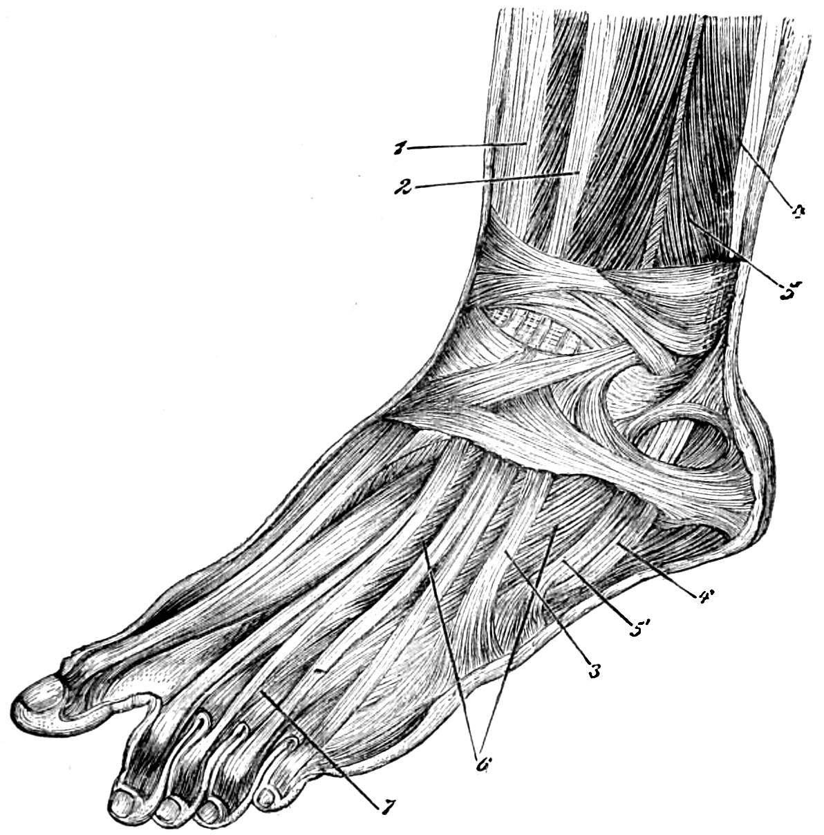
Fig. 54.—Muscular system of the human foot. 1, Tibialis anticus and extensor proprius pollicis. 2, Extensor longus digitorum. 3, Tendon of peroneus tertius. 4, 5, Peroneus longus and peroneus brevis. 4′, 5′, Tendons of the same. 6, 7, Tendons of the extensor longus and extensor brevis digitorum.
The flexor brevis digitorum displays perforated tendons, belonging to the second and third toes. The flexor longus digitorum displays perforated tendons for the fourth and fifth toes. The flexor longus pollicis divides into two tendons, one of which goes into the toe itself, while the other is connected with the flexor longus digitorum, and displays perforated tendons for the third and fourth toes, while the perforated tendons of the second and fifth toes have their origin in the other flexor.
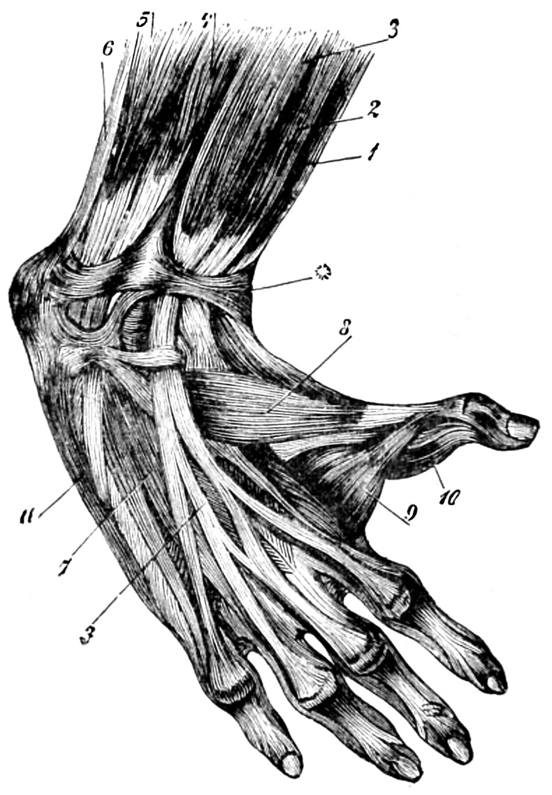
Fig. 55.—Muscles on the upper side of chimpanzee’s foot. 1. Tibialis anticus muscle. 2, Extensor proprius pollicis. 3, Extensor communis digitorum. 4, 5, Peroneus brevis and peroneus longus. 6, Tendon Achilles. 7, Extensor brevis digitorum. 8, Slip of the same for great toe. 9, First dorsal inter-osseous muscle. 10, Adductor pollicis. 11, Abductor minimi digiti.
In the gorilla the lumbricales muscles of the foot are powerful. The first inter-osseous muscle is likewise well developed and bicipital. There is a short flexor and an abductor for the little toe. I have not yet been able to assure myself of the existence of an opponens for that toe. In the chimpanzee the muscular system of the great and little toe does not essentially differ from that which we have described in the gorilla. The flexor brevis digitorum forms the perforated tendons of the second and third toes. The flexor longus digitorum provides the fourth and fifth toes with perforated, and the second and fifth toes with perforating, tendons, while those which belong to the third and fourth toes have their origin in the flexor longus pollicis. As in the gorilla, the latter muscle produces a fibrous investment for the tendons of the flexor longus digitorum. In the orang there is an abductor of the great toe, a very slightly developed opponens, a short bicipital flexor, and an adductor. One of the long flexors of the toes appears to represent the flexor longus pollicis in man. It provides the second and fifth toes with perforating tendons, while those of the third and fourth toes have their origin in the other flexor longus digitorum. There is no long flexor tendon on the great toe. The perforated tendons in this case generally belong to the short flexor muscle. In addition to the perforated tendons of the fourth toe, there is the long flexor already described.
In a gibbon’s great toe I observed an abductor, a short bicipital flexor, and a slightly developed opponens, to which a wide fan-shaped adductor is attached. The first dorsal inter-osseous muscle is, as in the same animal’s hand (Fig. 53), attached to the first phalanx of the second toe. The flexor longus digitorum provides the third and fourth toes with perforating tendons, and also gives off a tendon for the great toe. On the little toe there is a remarkably slender perforating tendon. While the first of the two long flexors represents the human flexor longus pollicis, the flexor longus digitorum is in this instance limited to the little toe. In the gibbon, as well as in the orang, the gorilla, and the chimpanzee, the two muscles are connected together by an aponeurosis. It may be here mentioned that in the human foot the flexor longus pollicis occasionally gives off a flexor for the second and even for the third toes. In the gibbon, as Bischoff justly observes, a muscle covers the flexor longus digitorum, which is still undivided, but already enlarged. From this muscle perforated tendons issue for the third and fourth toes. The second toe is provided with such a tendon from the flexor brevis digitorum. The muscle we have mentioned seems to represent the Quadratus plantæ, which is often developed in the other anthropoids, although only to a slight extent. With respect to the muscles of the small toe of the orang and gibbon, I need only say that in the latter species the opponens seems to be absent (Fig. 55).
It will be seen from the foregoing account that, in spite of several apparently important peculiarities, in spite of great and manifold variations which are established, even although our authorities do not always agree together, the muscular system of anthropoids is on the whole very like that of man. It displays, especially in the lower limbs, peculiarities of structure which render them capable of walking in an upright position, and others again which they have in common with the lower animals, but on the whole the anthropoid characteristics of the muscular system of these animals are predominant.
The digestive system of anthropoids likewise affords interesting points of comparison. The cavity of the mouth is, as we have seen, bordered by large and flexible lips. The mucous membrane of the mouth and the gums are flesh-coloured; they assume a darker colour in older animals, and are then sometimes marked with spots of a bluish or brownish grey. Ehlers describes, as a peculiarity in the mucous membrane of the mouths of the gorilla and chimpanzee, that there are what he calls buccal folds, which pass on both sides from the fore surface of the upper and lower jaw into the mucous membrane of the cheeks, and are of the height of the canine teeth.75 I have myself only observed these folds in the gorilla, of which an illustration is given in Fig. 3, and not in any other specimen. I have observed scarcely any indications of these folds in other anthropoids, and then only of such a doubtful nature that I am not disposed to regard the circumstance as of any special significance. A small band on the upper and lower lips, sometimes only slightly developed, but always perceptible, is present in all anthropoids.
The tongue is small, and not provided at its base with several great concave follicles as in man; these are at least only faintly represented, and not easy to observe. Around them there rise pock-like, tufted warts, very close together, which in an aged gorilla are apt to become hard and horny. These are also prominent between the follicles of the tonsils. The circumvallate papillæ of the tongue are less numerous than in man, and often, especially in the chimpanzee, they take the form of a cross, or of the letter T, or in the gorilla of a V.
The uvula and palate present no special variation from the human type. On the hard palate there are a number of folds, or rather swellings, which extend laterally from the central suture of the palate, towards the row of teeth in the upper jaw; these are sometimes simple, sometimes complex, and vary in their details in individual cases. They are particularly marked in the adult chimpanzee, and are also very apparent in the gibbon, and they are arranged with a somewhat ornamental regularity. These inequalities are not altogether insignificant in the human palate, but this subject has not been much studied since Gegenbaur directed the attention of scientific men to them, and special light has been thrown upon it by Bischoff and Ehlers, as far as anthropoids are concerned.
The teeth afford us important material for comparison. In the case of anthropoids the formula for the teeth of the slender-nosed or Old-World apes (Catarrhina) will generally apply: i 2/2 c 1/1 p 2/2 m 3/3. The following is the formula for the milk-teeth: i 2/2 c 1/1 m 2/2. Magitot and Giglioli76 have shown that the milk-teeth are cut in the same order as those of man—first, the lower; second, the upper incisor teeth; third, the front pre-molars; fourth, the back pre-molars; fifth, the canine teeth. According to the same authors, the permanent teeth are cut in the following order:—first, the first molar teeth; second, the lower, and then the upper incisor teeth; third, the pre-molars; fourth, the canine teeth; fifth, the second molar teeth; sixth, the third molar teeth. In the skull of a male gorilla, Giglioli found that the permanent canine teeth were cut almost simultaneously with the third molar teeth, and after the appearance of the second molar teeth. The cutting of the canine teeth appears to be a longer process than that of the other teeth.
In anthropoids the structure of the permanent teeth varies with the species, and even with the sex. In the gorilla the two upper central incisor teeth are wide, chisel-shaped, and much larger than the pair of lateral incisors. The four lower incisor teeth are of about the size of the upper lateral incisors, and, like these, are chisel-shaped, but not so wide. The powerful upper canine teeth of an aged male are curved in their lower part, both outwards and inwards. Their form is that of a three-sided, cuneiform pyramid. The anterior surface is rounded, and near its inner edge a deep furrow may be observed, extending from the neck of the tooth almost to its point. The outer and inner sides of the tooth meet in a sharp angle, somewhat convex in front, and level or slightly concave behind. The inner side is concave, and furnished, nearly in the centre, with a deep longitudinal farrow. The lower canine teeth of an aged male are shorter than the upper, curved on their upper and outer surface, and somewhat behind. Their form is also that of a three-sided pyramid, rounded in front. The longitudinal furrow which traverses their inner segment is much shorter than that on the upper tooth. The outer side is somewhat convex, and at the same time somewhat retreating, and is provided on its posterior segment with two longitudinal furrows, or more rarely with one, reaching from the neck to about the centre of the tooth. The inner side, like that of the upper teeth, is somewhat concave. The lower canine teeth project like pillars over the upper ones (Figs. 15, 16). The canine teeth of a young male gorilla are less sharp in their angles, although they already present the form of a three-sided pyramid. The canine teeth of the adult female gorilla are much smaller than those of the adult male, and are laterally more compressed. The three-sided pyramidal form is only slightly marked. The outer surface is convex and furnished with a scarcely apparent central longitudinal ridge. On the inner surface, or that which is turned to the cavity of the mouth, there are from two to three longitudinal furrows reaching from the neck to the centre of the tooth. The lower teeth are of a three-sided, pyramidal form, presenting an interior, posterior, and inner superficies.
The pre-molars of an aged male gorilla are wide, and are furnished with a large outer, and a smaller inner, cusp. The three four-cusped upper molars display a more regular and symmetrical arrangement of their cusps than is the case with the female, in which the position of the cusps is rather variable. Except for the difference of size, the relative conditions of these teeth are the same in male and female. The first pointed lower pre-molars are in the male of the form of a four-sided pyramid, convex on the anterior and outer surface, flat on the side directed to the cavity of the mouth, and marked with furrows on the posterior surface. The small second and lower pre-molars have two anterior and one posterior cusp. The last is generally worn away at an early age. Each molar tooth has two outer and two inner cusps, opposite to each other, and one posterior cusp. We cannot here fail to notice the likeness to the conditions of the human teeth, a likeness which is still more striking in the female.
In the chimpanzee, also, the upper central incisor teeth are broadly chisel-shaped, while the upper and lower lateral incisors are smaller. In the male there is often a considerable gap between these and the canine teeth. The latter present the form of a three-sided pyramid, of which the anterior edge is blunt and tends outwards, while the posterior angle is sharp, scooped out in its upper third, and terminating at the base of the crown in a posterior cusp. The pre-molars have an external and an inner cusp; the molars have two external and two inner cusps, connected with each other by their enamel. The lower canine teeth of these animals are likewise of the shape of a three-sided pyramid, of which the anterior angle is very blunt, while the inner and posterior angles are sharply cut. The anterior surface is not grooved like the upper canine teeth. The lateral angle is much rounded. The back teeth plainly display the posterior fifth cusp, which may also be observed in man. In the orang-utan the characteristics of the upper incisors are such as we have described in the case of other anthropoids. The upper canine teeth are shaped like a three-sided pyramid, and are furnished with a longitudinal furrow on the anterior side. A similar furrow is found on the posterior superficies of the lower canine teeth. The back teeth display no special characteristics when compared with those of other anthropoids.
The canine teeth of these anthropoids are much worn down by age on their posterior surface. Deep transverse grooves of varying size characterize the teeth of anthropoids, owing to the unequal distribution of the coating of enamel. These are developed with their advancing growth. In addition to these incised furrows, longitudinal marks, with raised edges, also appear, and especially on the anterior surface of the incisor teeth.
In the gibbon the anterior surface of the incisor teeth is smooth; in this animal the upper central incisor teeth are the largest, while the lower central incisors are the smallest. The long and strong upper canine teeth, which are laterally compressed, display a sharp posterior angle, and an anterior and inner longitudinal furrow.
It has sometimes been said that the grooves found on the external contour of the back teeth of anthropoids, extending to their roots, constitute a not unimportant distinction between their structure and that of the human teeth, in which the grooves do not extend to the roots. But the corresponding human teeth do sometimes exhibit very deep and extensive furrows. I cannot, therefore, ascribe any peculiar significance to this assumed distinction. The development of the canine teeth, like those of beasts of prey, seems to me much more important. A supernumerary back tooth may sometimes be observed both in man and in anthropoids, including also the gibbon.77
The stomach and intestines of these animals present only a few striking differences from the same organs in man. The length of the intestines varies in man as well as in anthropoids. I have only observed the valvulæ conniventes to be somewhat clearly developed in the gorilla and the orang. The cæcum of these apes is long, broad, placed with the power of free movement in the peritoneum, and furnished, especially in the case of the orang, with a large, very long, and spirally coiled vermiform appendix.
The liver is divided into two principal lobes, but in the orang this division is not very clearly marked. I have not myself observed a subdivision of these lobes, occurring on their edges, which is mentioned by Bolau and Auzoux in the case of the gorilla. Bischoff notices in the gorilla the absence of the H-shaped arrangement of the fissures on the under surface of the liver, so noticeable in man; and the same remark applies to other species of anthropoids. Moreover, the fissures on this part of the liver are not incised on the substance with the same uniform depth. The gall-bladder of the gorilla and the orang is not remarkable for its size; in the chimpanzee I found that this organ is large and twisted, and it is also large in the gibbon.
The spleen is elongated in the gorilla, chimpanzee, and gibbon, shorter and wider in the orang. On its left contour it is uniformly bevelled off. There is nothing in the pancreas which calls for remark.
The larynx of anthropoids possesses on the whole a structure resembling that of man. This is especially the case at the entrance to that organ. The anterior and specially vocal portion of the glottis is short, about as long as the respiratory portion. In the chimpanzee there is a deep cavity in the body of the hyoid bone. In the gorilla, chimpanzee, and orang the throat-pouches or air-sacs correspond to Morgagni’s sacs. These are the thin-skinned elastic sacs, closely united with their surroundings by connective tissue. The right laryngeal sac appears to be of larger diameter than the left. According to Duvernoy’s and Ehlers’ accurate account only the upper portion of this organ occurs in the gorilla. In that animal, and in the orang, a lower projection is displayed, extending behind the sterno-mastoid as far as the shoulder, and another extending to the pectoralis major muscle. In the chimpanzee only the posterior segment is developed. It has been asserted that in several cases there is found a single, irregular laryngeal sac, communicating with the two Morgagni sacs, but I agree with Ehlers in thinking this improbable. In such instances it seems likely that, owing to the great want of symmetry in this organ, one of the sacs has been overlooked. In an aged orang the throat-pouches, fastened together by connective tissue, and covered by the external skin of the throat, hang down slackly and heavily over the middle of the breast (see Fig. 9). According to Sandifort, the siamang is the only one of the gibbons which displays a single throat-pouch; while Broca asserts that it has two detached sacs, placed close to the larynx.78 The halves of the thyroid cartilage are generally connected with each other by an intermediate piece.
The trachea of anthropoids generally includes from sixteen to eighteen cartilaginous rings, but in the siamang there are twenty-one. They ramify into branches which are, as a rule, wider on the right than on the left side.79 There is a further lateral ramification on the right side, situated above the artery. Huxley and Ehlers hold that the lungs of a gorilla are cleft like those of the human organism, the right divided into three, and the left into two lobes. I have myself observed this type, and in one instance I found three lobes on the left. In the chimpanzee I saw that the right lung was divided into three, and the left into two lobes. Bischoff observed an instance of a chimpanzee which had four lobes on the right and two on the left side. In an orang dissected by me I found only one lobe on each side, with thin, slightly indented notches on the anterior edges of the right lobe, and two on the left, and there was at the same time a strongly marked indentation between the lobes. The lungs of a gibbon are described as having four lobes on the right, and only one or two on the left. I myself have examined a gibbon in which there were three lobes on the right, and two on the left. It appears that there are not unimportant individual variations of this structure in every species of anthropoids; and indeed, human lungs are by no means exempt from them.
The male sexual organs correspond on the whole with the form and arrangement of these organs in man. I must not omit to mention that the penis of the swine-snouted baboon, and of other dog-headed apes, is much more like the penis in man than is the case with anthropoids, with the exception of the gorilla. In the last-named animal the scrotum is short and tightly stretched. The right testicle is a little higher than the left, and is divided from it by a wide raphé. The internal female organs are also like those of the human organism, with only slight variations. Bischoff is correct in the assertion that the external lips of the pudendum and the mons veneris are almost wholly absent. Bolau, Ehlers, and Hermes have ascertained that there is a menstruation which occurs periodically, at any rate in the case of the chimpanzee, and the other species cannot be exempt from the process. At such times there is a blush and enlargement of the external parts, and a profusion of the external lips of the pudendum, which are at other times scarcely apparent. The nymphæ and the clitoris are of considerable size and importance. There is often an excessive enlargement and reddening of these parts, as well as of the posterior callosities in the chimpanzee, and also in the baboon and macaca, during the period of sexual excitement.
Fig. 56.—The brain of an orang, seen from the side (Vogt, from Gratiolet). F, Frontal lobe. P, Parietal lobe. O, Occipital lobe. R, Fissure of Rolando. S, Fissure of Sylvius. C, Cerebellum.
Nervous system.—In this part of the organism we are especially interested in the structure of the brain. Bastian justly remarks, with reference to the brain of apes, that this family possesses many cerebral characteristics in common, by which their close connection with each other may be verified. Distinct stages of development have been observed, which, however, cannot be classified in a consecutive series. Starting from the brains of lemurs, which do not greatly differ from those of rodents, we can advance by means of very distinct transition forms to the more highly developed cerebral hemispheres of the large anthropoid apes, the chimpanzee, the gorilla, and orang-utan.80
Fig. 57.—Brain of the chimpanzee, seen from above. The upper part of the right hemisphere is removed so as to lay bare the lateral ventricle (Vogt, from Marshall). L, Longitudinal fissure (other indications the same as in Fig. 56). c s, The corpus striatum in anterior cornu of the ventricle. c a, Hippocampus major in descending cornu. h m, Hippocampus minor in posterior cornu.
Very opposite views prevail among anatomists with regard to the question which species of anthropoids possesses the most highly developed brain. Some regard the chimpanzee’s brain as the simplest, and that of the orang as the most highly developed. In all these apes the lateral halves of the cerebrum, always divided from each other by a deep longitudinal fissure, overlap the cerebellum as far as a minute posterior segment. In this respect I find the brain of the gorilla a little behind the other anthropoids. Up to this time, I have only observed the projection of the cerebellum through the cerebrum in the case of an orang81 (see also Fig. 56). Retzius asserts that the cerebellum of Lapps is incompletely covered, while the covering is generally complete in the case of Slav and Tartar races. In German and Latin races the cerebrum overlaps the cerebellum. In Mongolian, Indian, and Negro races the covering appears to be generally imperfect.
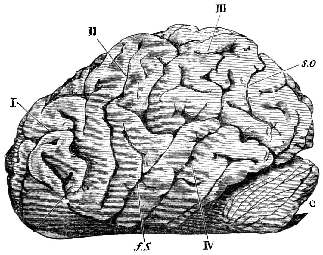
Fig. 58.—Brain of gorilla, side view (from Bolau and Pansch). I., Frontal lobe. II., Fissure of Rolando. III., Parietal lobe. IV., Temporal lobe. C, Cerebellum. f s, Fissure of Sylvius. s c, External fissure parieto-occipital.
While the ground form of the gorilla brain approximates to a long oval, and in this respect resembles the human brain, the brain of chimpanzees and orangs is of a round-oval form. This is especially the case with the chimpanzee (Fig. 57). In my opinion, the gorilla brain is distinguished from that of the chimpanzee, but not from that of the orang, by its very complex convolutions (Fig. 56).
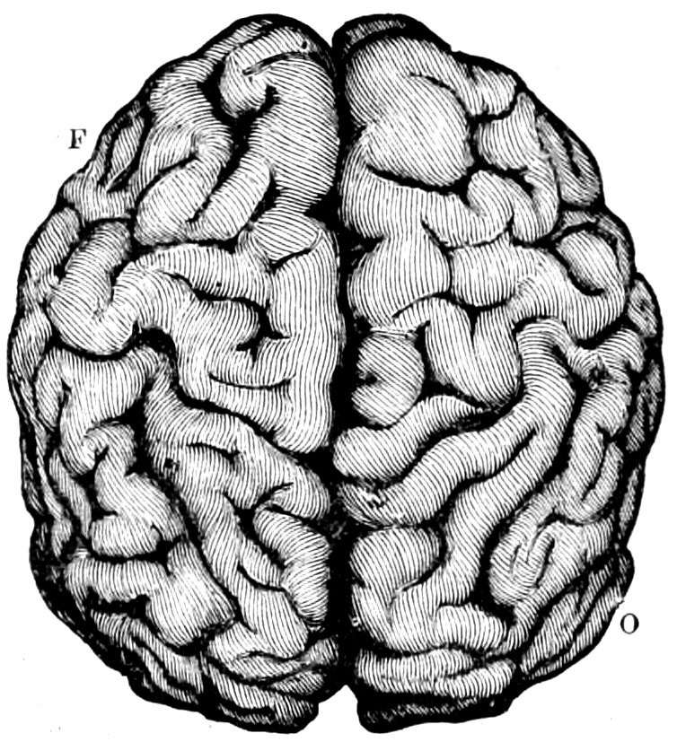
Fig. 59.—Brain of orang, seen from above (Duncan, from a specimen in the Museum of Royal College of Surgeons). F, frontal lobe. O, Occipital lobe.
In the gorilla, chimpanzee, and orang, the island of Reil in the fissure of Sylvius is generally—at least, according to my experience—overlapped by the operculum, although there are instances in which this is not the case. In these three anthropoids, as Bastian justly observes, the fissure of Sylvius is much less horizontal than in man, and occupies a position more like that which it takes in the black sea-cat monkey, the wanderers, and other macacas. In the gorilla its direction is more horizontal than in the two other species of anthropoids. The central fissure, termed fissure of Rolando, is very marked, especially in the chimpanzee (Fig. 57 R); but it may also be easily traced in other species of anthropoids (Fig. 58, II., 56, R). The so-called simian fissure between the parietal and occipital lobes of the cerebrum (Meynart’s elongated external occipital fissure), presented in Fig. 58 s c, is very marked in the chimpanzee (Fig. 57, d). The frontal lobes of the gorilla brain are high, while those of the chimpanzee are short and low. It is said that those of the orang, which are high and short, terminate in a beak-shaped curvature, but this is not invariably the case.
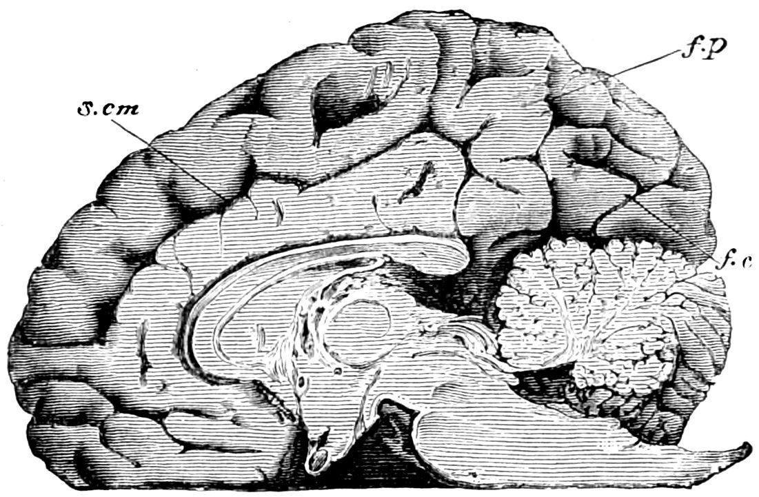
Fig. 60.—Longitudinal section of a gorilla’s brain (Bola and Pansch). s.cm, Colloso marginal fissure. f, p, Internal parieto-occipital fissure. f, c, Calcarine fissure, the posterior part of the hippocampal fissure.
In the anthropoids we have been considering, and also in several of the lower species of apes, there are three other fissures of less importance in addition to those we have mentioned, namely, the fissure parallel to the fissure of Sylvius, and placed behind it, the corpus callosum fissure, placed immediately above the corpus callosum on the inner side of the hemisphere of the cerebrum, and the calcarine fissure (Fissura calcarina) (Fig. 60). The latter ends near the point of junction of the inner and lower surfaces of the posterior division of the hemisphere. The upper temporal convolution, termed by several anatomists Gyrus supramarginalis, is said by Gratiolet to be absent in anthropoids; but Rolleston, Bastian, and myself have all found it well developed82 (Fig. 56, orang, and Fig. 58, gorilla).
Bischoff asserts that the third frontal convolution (Broca’s convolution) is very slightly developed in the chimpanzee, orang, and gibbon. “Its great development in men,” Gewährsmann writes, “constitutes one of the most marked distinctions between the brains of apes and of men.”83 In most of the other species of apes this convolution is altogether absent, but Pansch is justified in the assertion that it is fully developed in anthropoids. I cannot wholly agree with Pansch in his analysis; but I must accept his statement on this point (see the orang, Fig. 59). Gratiolet remarks that the so-called annectant gyri (plis de passage) which serve as a covering or operculum for the posterior lobes in apes, are only superficially apparent in man. In the chimpanzee the upper of those convolutions is absent, while it is large in the orang, and likewise large and undulated in man. In the orang the second annectant gyrus is covered, but this covering is absent in man.84
In considering the inner structure of the brain of these animals, we are first struck by the shortness of the corpus callosum. The soft and thick anterior commissure of the third cerebral ventricle and the thin posterior commissure have also been justly noted. In the lateral ventricles more of the characteristics described in the human brain are absent. The four eminences resemble those of man; nor does the fourth cerebral ventricle present any remarkable differences of form. Neither does the base or lower surface of the brain display any important deviation from the human type. The transverse section of the nerves at their intersection appears to me, however, to be somewhat more oval than is the case in man.
There has recently been an attempt to recognize a pithecoid character, or atavism, in microcephalic men, the smallness of whose heads is allied with a greater or less degree of idiocy. A pithecoid structure of the brain has also been traced in several individuals who are not microcephalous, but subject to pathological affections. We will first consider those who belong to the latter category. Krause examined the brain of an ape-like boy aged seven years and a half, which, as the author remarks, approximated in structure to the pithecoid type, although without displaying microcephalic characteristics. The two cerebral hemispheres were wanting in symmetry; they diverged from each other in the region where the parieto-occipital fissure occurs on the left cerebral hemisphere, and they formed an edge which curved outward and backward so that the cerebellum remained uncovered. On the lower surface of the frontal lobes there was a strongly marked ethmoidal prominence. Neither of the fissures of Sylvius were closed, the left less so than the right; the operculum was only slightly developed; and the island of Reil and its fissures were almost uncovered. This formation is almost the same as that of the brain of anthropoids. The two central fissures of Rolando were close together, or less deeply impressed on the edge of the hemispheres than is normally the case, and forming no joint angle. Large and deeply marked pre-central fissures seemed to represent the central fissures. The intra-parietal fissures, diverging outwardly further than in man, received the parieto-occipital fissure, a structure in conformity with the typical brain of apes. The transverse occipital fissure became in this case a deep fissure like the simian fissure, crossing the occipital lobes, and almost completely dividing them from the parietal lobes. The so-called Fissura calcarina, to which we have referred above, had its origin on the upper surface of the occipital lobe, then joined the parieto-occipital fissure, and went directly into the hippocampal fissure (Fissura hippocampi) on its right side. This abnormal structure is also in conformity with the typical brain of apes. The first occipital convolution is divided from the upper parietal lobes by the parieto-occipital fissure. Gratiolet asserts that this formation occurs in many species of apes. The upper temporal convolution was remarkably reduced on both sides, possessing only an average width of 5 mm. This characteristic reminded Krause of the brain of the chimpanzee. In that animal the upper temporal convolution is always reduced. Krause therefore asks whether some human brains may not possess the typical structure of apes without being microcephalic. The brain we have described scarcely differed from the normal weight; it possessed all the convolutions and fissures, and indeed, the convolutions were perhaps more numerous than in the normal structure, yet it was different in every respect, and approximated in its whole structure to the simian rather than to the human type. Krause adds that if the brain had been placed before him without any intimation of its origin, he should have been quite justified in concluding that it belonged to an anthropoid ape, which stood somewhat nearer to man than the chimpanzee.