FOSSIL FAUNA.
Reading edition
Plates XXXIV. to LXXIV. inclusive.
Plate XXXIV.
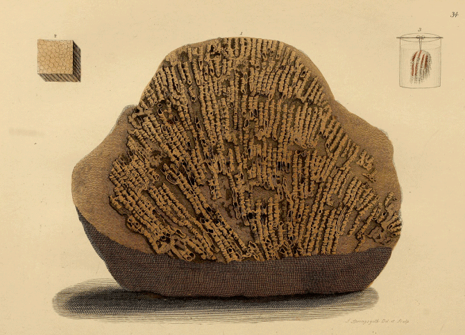
PLATE XXXIV.
(Plates XXXIV. to LXXIV. inclusive, are from Parkinson's Organic Remains.)
Fossil Tubipore, from Derbyshire.
(Syringopora geniculata, of Phillips, from the Mountain Limestone, Derbyshire.)
The specimen figured is a mass of limestone, on the surface of which is spread out in high relief a delicate tubiporite, or fossil coral, allied to the Tubipora, or "Organ-pipe coral," so generally preserved in cabinets of natural curiosities, from the beauty and elegance of its crimson tubes. The fossil, however, though somewhat resembling the recent coral in its general form, belongs to an extinct genus.
This Syringopora appears to have been very abundant in the sea in which the strata of mountain or carboniferous limestone were deposited, for it forms entire beds of great extent. A beautifully figured marble results from this coral, when the interstices of its tubes have been filled up with compact calcareous matter. A small polished slab is represented in fig. 2. At Matlock, vases, and other ornamental articles, are made of it; and the sections of the coral tubes impart considerable variety of figures.[25]
Some slabs of this fossil coral are of a dull red hue, which there is every reason to conclude is due to the colour of the original; and not only are traces of the natural tints of the living zoophyte preserved, but even the animal membrane of the coral; and this may be exposed by immersing a fragment of the marble in dilute muriatic (hydrochloric) acid. Mr. Parkinson thus describes the result of his first experiment:—
"A fragment of the marble (Plate XXXIV. fig. 2) was exposed to the action of muriatic acid in a very dilute state. As the calcareous earth was dissolved, and the carbonic acid escaped, I was delighted to observe the membranaceous substance appear, depending from the stone in light, flocculent, elastic flakes. Many of these retained a deep red colour, and appeared in a beautiful and distinct manner, although not absolutely retaining the form of the tubipore. A faithful representation of this appearance is given in fig. 3."
This experiment of Mr. Parkinson was highly important, as proving the previously almost incredible fact, that animal membrane, when hermetically sealed, as it were, in the solid stone, was as indestructible as the rock itself. It suggested, too, the probability that vestiges of other animal tissues might be traced in organic remains, and encouraged subsequent observers to seek for evidence of the soft parts of animal bodies entombed in the strata. It was the first step in the right direction, and led to the detection of many highly interesting phenomena. In Dr. Buckland's Bridgewater Essay will be found figures and descriptions of the eyes of crustacea: of the wings, elytra or wing-covers, and the integuments of the body of insects; of the skin of reptiles; and, in the "Wonders of Geology," and "Medals of Creation," of the membranes of the air-bladder, and of the capsule of the eye of fishes; of the soft parts of the animalcules called foraminifera, &c. The bodies of mollusca, or shell-fish, converted into a dark brown mass (mollushite), occur in such abundance in some deposits, as to yield a rich manure from the quantity of phosphate of lime. The excrementitious substances termed by Dr. Buckland "Coprolites," are also used for agricultural purposes.
Plate XXXV.
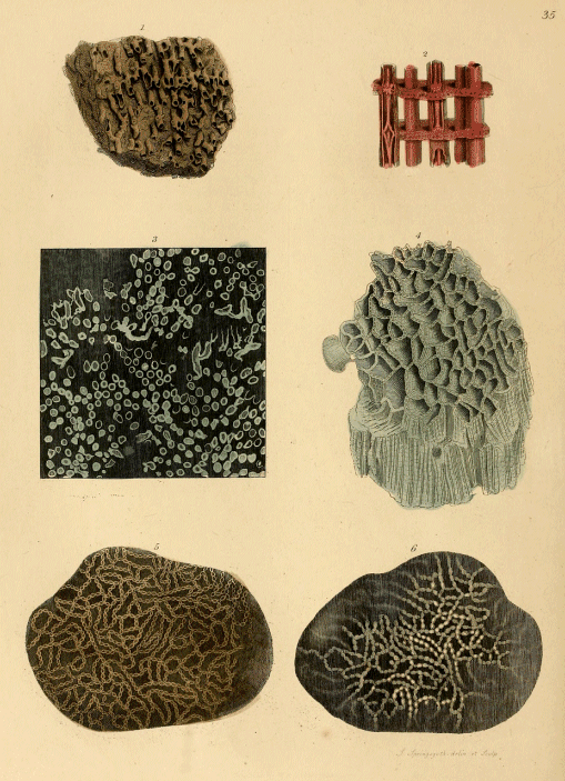
PLATE XXXV.
The subjects here figured are Fossil Corals.
Fig. 1. (Syringopora ramulosa.) A fragment of another species of the coral previously described; from the mountain limestone.
Fig. 2, represents four connected tubes of the recent organ-pipe coral (Sarcinula musica) of New Holland, to show the structure of this type of Zoophytes. Coloured figures of the live polypes of this coral are given in Wonders of Geology, sixth edition, vol. ii. plate vi.
Fig. 3. A polished slab of marble, the white markings in which are produced by sections of the tubes of the same species of coral as that represented in fig. 1.
Fig. 4. (Catenipora escharoides.) The fossil here delineated is well known to collectors by the name of "chain-coral" derived from the elegant cateniform markings produced by transverse sections of the parallel tubes, which being of an oval form, and in close apposition, give rise to chain-like figures, as shown in figs. 5 and 6. From Dudley.
This fossil coral abounds in that division of the Silurian formation termed the Wenlock or Dudley limestones, wherever these deposits occur. The most exquisite specimens are obtained from the Falls of the Ohio, at Louisville, in the United States of America. A coral reef of the Silurian epoch here exists in the bed of the mighty stream of fresh water, almost as perfect as when growing in its native sea! The river dashes over the entire mass in the season of high water; but in those periods when the stream is low, the ridge of coral is exposed, and its surface then presents the most extraordinary display of Silurian corals, of numerous species and genera, standing in relief on the more compact masses of the rock. The substance of the corals, being siliceous, resists the action of the cataract, while the softer calcareous matter which filled up the interstices of the tubes, lamellæ, &c. of the zoophytes, is washed away atom by atom; and natural dissections are formed, which art would in vain attempt to imitate. Dr. Yandell, of the Medical College, Louisville, and Dr. Clapp, of New Albany, have splendid collections from the Falls, which every geologist and intelligent traveller who visits Kentucky should not fail to examine: the masses of Astreæ, Madrepores, &c. are so fresh in their aspect, as not to be readily distinguished from the recent specimens of the same genera which are placed beside them.[26]
Fig. 5, is a transverse section of a mass of chain-coral from Dudley.
Fig. 6. The same, as seen by transmitted light.
Plate XXXVI.
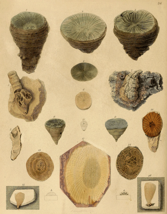
PLATE XXXVI.
Various Fossil Corals from different Formations.
Figs. 1, 2, 3. (Cyathophyllum turbinatum, of Goldfuss.) These three turbinated or top-shaped corals are referable to a genus of which many species are exceedingly abundant in the Wenlock or Dudley limestone of the Silurian System. They belong to the Anthozoa, or flower-like corals. The living animal, of which the coral is but the durable earthy fabric or skeleton, bore a close analogy to the sea-anemone, or animal flower (Actinia), of our coasts. Each of these specimens belonged but to a single animal: the Cyathophylla are not, like the tubipores previously described, an aggregation of numerous individual polypes.[27]
Fig. 4. A small coral (Fungia) from Dudley.
Fig. 5. On this block of mountain limestone there are the remains of two different kinds of corals. The upper cylindrical part is a fragment of Cyathophyllum, to the lower part of which is attached a species of another genus (Michelinia).
Fig. 6, is a small coral (Fungia numismalis, of Goldfuss), common in the Oolite.
Fig. 7. A piece of encrinital limestone, from Derbyshire, having a conical cast—that is, the stone has been moulded in the interior or cavity—of a turbinated coral (Turbinolia).
Fig. 8. A longitudinal section, showing the transverse cells and lamellæ of the same kind of coral (Cyathophyllum) as figs. 1, 2, 3.
Fig. 9. A species of Turbinolia (Turbinolia complanata, of Goldfuss).
Fig. 10. A small turbinated coral (Turbinolia mitrata, of Hesinger), from the Silurian strata of Gothland.
Fig. 11. a Turbinolia from the Silurian deposits of Sweden.
Fig. 12. A remarkable coral (Petraia, of Munster), from the Devonian strata.
Figs. 13 & 14, are sections of Cyathophylla, like figs. 1, 2, 3, to exhibit the internal structure.
Figs. 15 & 16. Two elegant simple corals (Caryophyllia centralis, of Mantell), from the chalk of Kent. The form and disposition of the lamellæ of the cavity, as seen at the upper part of the specimens, are shown at a and b.
Fig. 17. A transverse and polished section of a species of Cyathophyllum, from the Devonian strata, at Blackenberg on the Rhine.
Plate XXXVII.
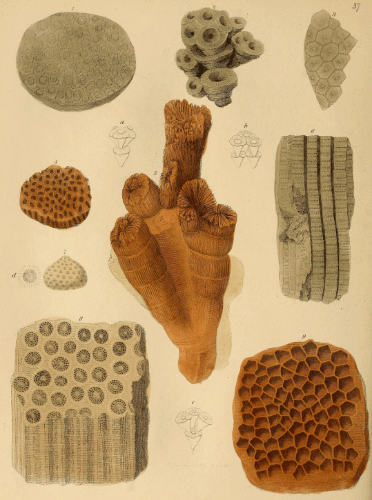
PLATE XXXVII.
Various Fossil Compound Corals.
Fig. 1. A beautiful specimen of Star-coral (Astrea ananas, of Goldfuss), from the Silurian strata of Sweden. At a, is shown "the mode in which, as in proliferous flowers, new polypes bud from the centre of the parent disk. At b, is represented the growth in the recent Madrepora stellaris of Linnæus."—Mr. Parkinson.
Fig. 2. An elegant Cyathophyllum (C. dianthus, of Goldfuss), from the Silurian formation of Sweden. At c, (the lower part of the plate,) is shown its probable mode of increase.
Figs. 3 & 6. A columnar compound coral (Lithostrotion striatum, of Lhwyd), from the mountain limestone of Derbyshire; fig. 3, is a transverse section of fig. 6, showing the basaltiform arrangement of the columns.
Fig. 4. "A fossil madrepore, from Lincolnshire."—Mr. Parkinson.
Fig. 5. A very elegant and abundant coral (Caryophyllia annularis, of Parkinson), in the bed termed "Coral Rag," of the oolite of Wiltshire, Berkshire, &c. Large conglomerated masses of this branched species form a considerable proportion of the fossil coral-reef which traverses some parts of the oolite: and when this bed is worked for road materials, blocks of this coral, more or less changed into calcareous spar, may be seen lying on the way-side. Near Faringdon, in Berkshire, a quarry in the Coral-rag has yielded many beautiful examples.
Fig. 7. Called "Spider-stone," by Mr. Parkinson. It is a species of Astrea: d, is an enlarged view of one of the polype-cells.
Fig. 8. A beautiful fossil coral, from Transylvania (apparently a species of Lithostrotion?).
Fig. 9. The specimen figured is from the mountain limestone of the Mendip Hills. (It is the Michelinia tenuisepta, of Phillips; Manon favosum, of Goldfuss?) It is described by Mr. Parkinson as "bearing somewhat of a honeycomb appearance."
Plate XXXVIII.
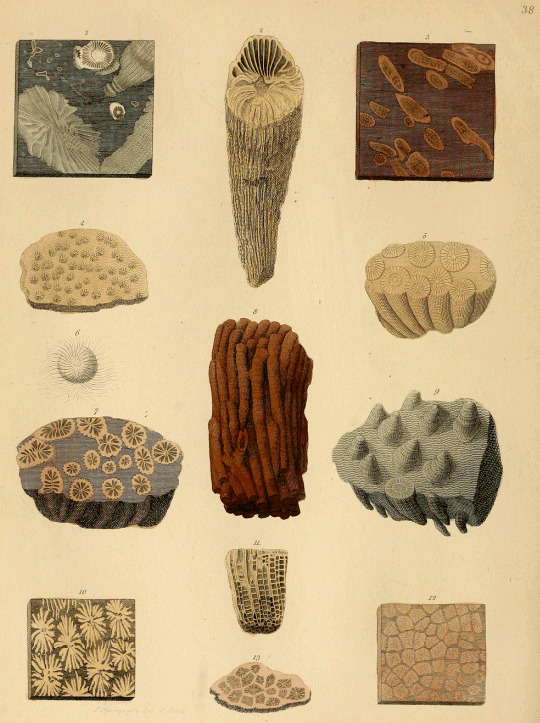
PLATE XXXVIII.
Fossil Corals, and Coral Marbles.
Fig. 1, is a polished slab of the carboniferous limestone, well known as the Kilkenny marble, and much used for chimney-pieces. The figures exposed on the surface are produced by sections of enclosed corals (some species of Cyathophyllum), which are transmuted into white calcareous spar.
Fig. 2. A coral of the same kind (Cyathophyllum turbinatum), from the mountain limestone of Derbyshire.
Fig. 3. A polished slice of Derbyshire marble, the markings on which are derived from sections of enclosed branches of corals (Syringopora), resembling that figured in Pl. XXXIV.
Fig. 4. An elegant compound coral, called "Spider-stone" by collectors (Astrea arachnoides, of Dr. Fleming); from Wiltshire: the geological habitat uncertain; probably the Oolite.
Fig. 5. This specimen appears to be a cluster of corals belonging to the genus Cyathophyllum.
Fig. 6. A magnified sketch of one of the cells of fig. 4.
Fig. 7. A polished transverse section of a coral; the precise relation of this species is not certain.
Fig. 8. This is a very abundant coral in some of the beds of mountain limestone, (Lithodendron fasciculatum, of Phillips.) The specimen figured is from Clifton, near Bristol. The marble cups, and other ornaments, manufactured from the rocks near that place, often exhibit sections of this species.
Fig. 9. A mass of coral from Ingleborough, (Cyathophyllum fungites.)
Fig. 10. A polished slice of a beautiful marble richly marked by the sections of the enclosed corals (Astrea undulata, of Dr. Fleming); from Switzerland: probably from the Oolitic or Jurassic formation.
Fig. 11. Vertical section of a fossil coral, showing the transverse arrangement of the internal cells.
Figs. 12 & 13. These specimens are polished sections of a very beautiful compound coral (Astrea Tisburiensis, of Miss Benett), which occurs in a silicified state in the Portland beds that are quarried at Tisbury, in Wiltshire. Masses of chert (a kind of coarse silex or flint), wholly made up of this coral, are often met with, and when sliced and polished are extremely beautiful and interesting; the originally calcareous fabric of the zoophytes being perfectly transmuted into silex, and the interstices filled up with a similar substance, but of a different colour.[28]
Plate XXXIX.
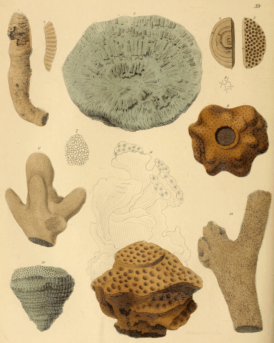
PLATE XXXIX.
Various Fossil Corals and Sponges, or Amorphozoa.
Fig. 1. A coral from the Dudley limestone. (Favosites?)
Fig. 2, is a vertical section of figs. 4 and 5, to show the internal arrangement of the cells.
Fig. 3. The under surface of a very common species (Favosites Gothlandica, of Goldfuss); from the Wenlock limestone of Dudley. A magnified view of part of the surface, to show the honeycomb structure, is given in fig. 7.
Fig. 4, the under, and fig. 5, the upper surface, of a small coral (Cyclolites ?) from the Oolite.
Fig. 6. A silicified branched sponge, (Spongites lobatus, of Dr. Fleming,) from the chalk of Berkshire.
Fig. 9, is a beautiful silicified, lobate, spongoid body, (Siphonia,) probably from the greensand. Siliceous cruciform spicula obtained from this fossil are represented in fig. 8.
Zoophytes of this kind, like many of the sponges, have their tissues strengthened by, and largely composed of spicula, which vary in form and size in the different species and genera. Many sponges and Siphoniæ in flint, and in the chert of the greensand, consist almost entirely of spicula, which may be easily detected by a slightly magnifying power.
Fig. 10. Another common Dudley Coral. (Porites pyriformis, of Mr. Lonsdale.)
Fig. 11. A beautiful coral (Explanaria flexuosa, of Dr. Fleming), from the Coral Rag of Steeple Ashton, Wilts. The outline indicates the mode of increase, according to Mr. Parkinson, of this form of zoophyte.
Fig. 12. This is a portion of a delicate ramose sponge (Spongites ramosus, of Mantell), whose remains are abundant in the chalk-flints, and have given rise to the irregularly branched siliceous nodules. A specimen nine inches long, with seven branches, is figured in Fossils of the South Downs, Pl. XV. fig. 11. Siliceous spicula are thickly interspersed throughout the mass.
Plate XL.
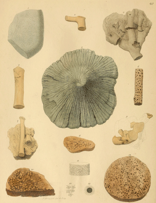
PLATE XL.
Fossil Corals, &c.
Fig. 1. The shells of Oysters, and other mollusca, are subjected to the ravages of a parasitical sponge, (Cliona, of Dr. Grant,) which is beset with minute siliceous spines or spicula, and inhabits hollows formed in the substance of the shell. Shells thus honeycombed, as it were, may often be found on the sea-shore with the excavated parts filled up by sponge. I have shells collected by my eldest son on the shores of New Zealand, that are hollowed out in a similar manner, and occupied by sponge. Whether these cavities are produced by mechanical means, or are the result of the decay and absorption of the shell induced by the growth of the parasite, are questions still undetermined. There are several kinds of shells found fossil, which were infested with a similar parasitical sponge; and when the cavities thus produced have been filled up by flint, and the shell has subsequently decomposed, or been worn away, the surface of the flint is studded with the casts of the cells, in the form of small irregular globular bodies, connected by filaments or strings of flint. The fossil, fig. 1, is a fossil of this kind, described by Mr. Parkinson as being "covered with minute round bodies, the nature of which is unknown;" fig. 12, is an enlarged view of five of these globular casts connected by filaments.
The origin of these fossils was first pointed out by the Rev. W. Conybeare.[29] The fibrous shells of a fossil genus of bivalves named Inoceramus, of which several species abound in the Chalk, appear to have been particularly subjected to depredations of this kind. Hence among partially water-worn flints, specimens of the siliceous casts are common; figs. 8, and 10, are examples from the Hackney gravel-pits.
Mr. Morris has named these fossils, Clionites; fig. 1, is C. Parkinsoni.
Figs. 2, 4, 7, are portions of a recent species of jointed zoophyte (Isis), from a modern concretionary deposit on the shores of the Mediterranean, Sicily.
Fig. 3. A branched fossil coral (Millepora ramosa, of Dr. Fleming), imbedded in compact oolitic limestone from Wiltshire. A portion of the surface magnified is represented in fig. 11.
Fig. 5, appears to be a fungiform Spongite; its locality is not mentioned.
Fig. 6. Portion of a fossil coral (Ceriopora), from Switzerland.
Figs. 8, & 10. These pebbles have the surface covered with casts of Clionites (Clionites Conybeari, of Mr. Morris.[30])
Fig. 9. Fragments of the radicle processes of attachment of some Apiocrinite or Lily-shaped animal in chalk; see description of Plate LI.
Fig. 14. A section of a siliceous nodule; probably the cellular appearance is inorganic: fig. 13, is a magnified section of the cells.
Plate XLI.
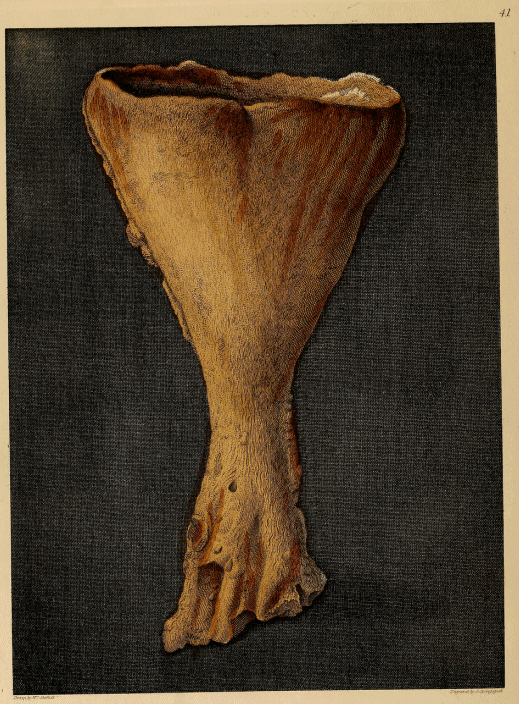
PLATE XLI.
A Silicified cup-shaped Sponge, from Touraine.
(Chenendopora Parkinsoni, of Michelin. Spongites Townsendi, of Mantell.)
This beautiful plate of a petrified zoophyte allied to the Spongia, formed the frontispiece to Mr. Parkinson's second volume. The fossil delineated is from Touraine in France, and is one of the most perfect examples of this kind hitherto observed. It belongs to a group of cup-shaped Amorphozoa, (as these organisms are now named by naturalists, from the great irregularity of shape which they assume,) termed Chenendopora. The original organic substance is transmuted into silex, and the interstices are filled up with carbonate of lime. The same species occurs in the greensand in the Vale of Pewsey in Wiltshire, and, I believe, also in the white-chalk; for many cyathiform flints from the South Downs appear to have the same internal structure.
In the so-called "gravel-pits," near Faringdon, in Berkshire,—which are quarries of a loosely-aggregated grit of the greensand, almost wholly made up of the relics of shells, corals, amorphozoa, &c.—numerous sponges of this genus are met with. One beautiful species (Chenendopora fungiformis) has acquired, from its cup-like form, the local name of "petrified salt-cellar."[31]
Plate XLII.
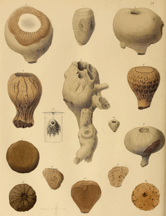
PLATE XLII.
The Fossils represented in this Plate are chiefly Zoophytes in Flint.
Fig. 1. A flint from the gravel-pits at Hackney. Its form is derived from the enclosed zoophyte, part of whose structure is exposed in the upper portion of the figure. This fossil zoophyte (Choanites Königi, of Mantell) is very abundant in some of the chalk strata, and many of the most beautifully marked pebbles cut and polished for brooches by the lapidaries of Brighton, Bognor, and the Isle of Wight, are the silicified soft parts of this animal. The original was of a subglobular form, and probably of a soft fleshy consistence; it had a deep central cavity, whence numerous tubes diverged, and ramified throughout the mass; it was fixed at the base by radicle or root-like processes.[32]
Fig. 2. This is another characteristic and abundant fossil zoophyte of the chalk and flint. The specimen figured is a water-worn pebble, and therefore gives but obscure indications of the form and structure of the original. The fungiform flints—called in Sussex petrified mushrooms—belong to the same genus (Ventriculites, of Mantell): and highly interesting specimens occur in which some part of the zoophyte is invested with flint, and the other part expanded in the chalk. The original was probably a polyparium—that is, the skeleton or support of an aggregation of coral-polypes—of a funnel shape, the polype-shells being situated on the inner surface: the base was attached by root-like fibres.[33] The polype-cells are cylindrical and regular, and clusters of beautiful casts of them often occur on flints.
Fig. 3. This specimen is described by Mr. Parkinson as "a pear-shaped alcyonite from Switzerland." It is probably one of those fossil zoophytes allied to the sponges (called Siphonia), in which the upper part is of a bulbous or pear-like form, and is supported by a stem with root-like processes at the base. The bulb has a central cavity studded with irregular pores, that communicates with the parallel longitudinal tubes of which the stem is composed: a structure admitting of that ready ingress and egress of the sea-water, which this class of organisms requires. There are numerous species in the greensand of the chalk formation.[34]
Fig. 4. A variety of Siphonia (Jerea excavata, of Michelin), from the greensand of Wiltshire.
Fig. 5. A silicified Siphonia from Saumur.
Fig. 6. A Ventriculite from a gravel-pit; the markings are produced by the exposed and partially abraded outer integument, which in perfect examples consists of a regular net-work of sub-cylindrical fibres.
Fig. 7, is a transverse section of a Siphonia (Siphonia pyriformis of Goldfuss).
Fig. 8. A nearly perfect specimen of a similar fossil. In fig. 7, are shown sections of tubes passing from the periphery to the centre; in fig. 8, the central aperture of the cavity of the bulb, and part of the stem, are displayed.
Figs. 9, & 10, are imperfect specimens of Choanites: fig. 10, is a vertical section showing the central cavity and the connected tubes.
Fig. 11, is another example of Siphonia pyriformis.
Fig. 12, a vertical, and fig. 13, a transverse section, of the same species of Siphonia.
Fig. 14. A small turbinated calcareous spongite from Switzerland.
Fig. 15. The appearance of the animal membrane exposed by immersion of the fossil (fig. 14), in diluted hydrochloric acid.
Plate XLIII.
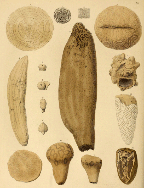
PLATE XLIII.
Fossil Corals, and other Zoophytes.
Figs. 1, 2, 3, & 4, are representations of different aspects of a simple coral (Fungia polymorpha, of Goldfuss); the locality is uncertain. Fig. 1, the base; fig. 2, a magnified representation of part of the same; fig. 3, magnified view of part of the lamellated surface of fig. 4.
Fig. 5. The nature of this fossil is not obvious; it may be a rolled Siphonia.
Fig. 6, is a fine specimen of a Siphonia (Jerea pyriformis, of Lamouroux). At both extremities the apertures of the numerous tubuli are seen.
Figs. 7, 8, & 9, are varieties of the same species of fossil sponge (Scyphia articulata, of Goldfuss), from Switzerland.
Fig. 10. A spongite of a very peculiar form.
Fig. 11. A spongite investing a fossil shell (Nerita), from Faringdon.
Fig. 12, is an imperfect specimen of a Ventriculite (Ventriculites alcyonoides, of Mantell), from the chalk of Wiltshire.
Fig. 13. A calcareous spongite which has been immersed in dilute hydrochloric acid to show its structure.
Fig. 14. A pebble deriving its shape from a zoophyte apparently related to the Ventriculites (Spongites labyrinthicus, of Mantell). The aperture at the base has arisen from the decomposition of the process of attachment.
Fig. 15. A pebble enclosing part of the base of a Ventriculite; the circular spots on the large end are sections of the ramifications of the stirps or base of the zoophyte; for this figure and the following are drawn in an inverted position.
Fig. 16, is a similar fossil, split vertically, and showing the enclosed stem of the Ventriculite.
Plate XLIV.
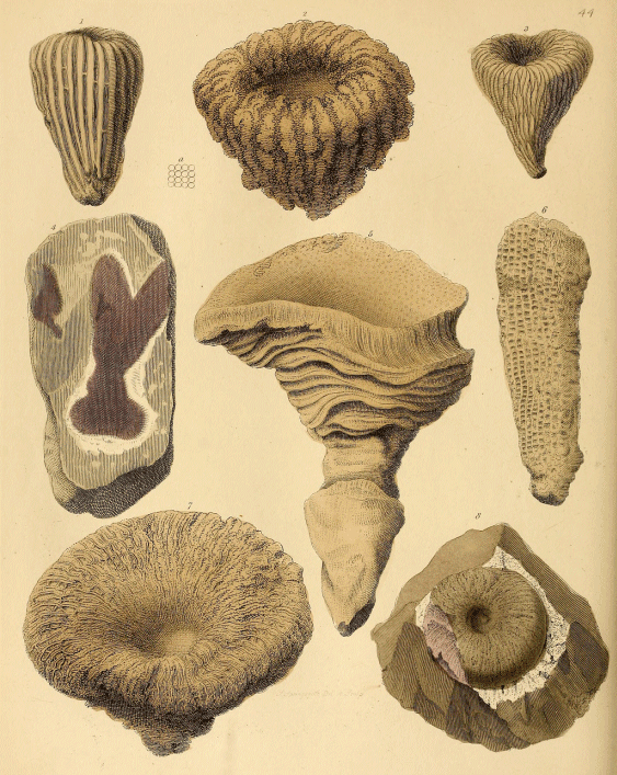
PLATE XLIV.
Fossil Zoophytes.
Fig. 1. A spongite (Scyphia costata, of Goldfuss), from Switzerland. The fossil spongeous bodies named Scyphia, are characterized by the "mass or body being either cylindrical, simple or branched; fistulous, and terminating in a rounded pit; entirely composed of a firm reticulated tissue."[35] Like the other bodies comprised in the group of Amorphozoa, the form in this genus is exceedingly diversified, and as the structure is often but obscurely shown, the determination of these fossils is oftentimes impossible. It is however convenient, in the present state of our knowledge, to distinguish the principal kinds by names which may be modified or abandoned, when the structure and natural affinities of the original organisms are more accurately determined.
Fig. 2. Another species of Scyphia from Switzerland; a small portion of the surface magnified is seen at a.
Fig. 3. The peculiar form and tissue of another genus of Amorphozoa (Cnemidium rimulosum, of Goldfuss), are shown in this beautiful specimen.
Fig. 4, is a section of a chalk flint, from Wycombe Heath; the purple body, partially invested by a white border, is evidently a mass of the soft parts of some zoophyte, which served as a nucleus to the siliceous nodule. A purple or pink hue often prevails in the sections of zoophytes immersed in flint, and doubtless depends on the original colour of the living animal.
Fig. 5. A very fine spongite (Chenendopora fungiformis, of Michelin), from France.
Fig. 6. This is evidently a fossil zoophyte, but the structure exposed is not sufficiently characteristic to determine the genus.
Fig. 7. A beautiful fungiform Scyphia.
Fig. 8. This elegant specimen, which Mr. Parkinson highly valued, is evidently a Choanite imbedded in flint. The body retains a pink colour, and is surrounded by a white band, which is probably the remains of the cortical or external tissue of the original zoophyte. I have seen many transverse sections in which the central mass was either of a pink or purple colour, and encircled by a white zone, in the squared flints of the walls of churches and other ancient edifices in Sussex.[36]
Plate XLV.
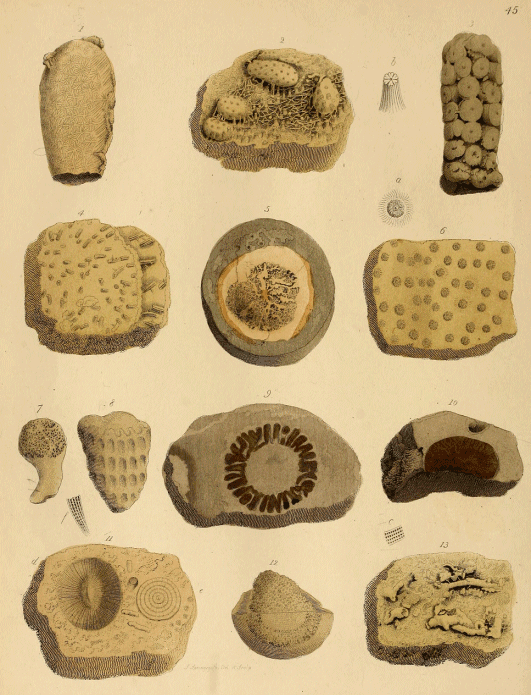
PLATE XLV.
Fossil Corals and other Zoophytes.
Fig. 1. "A fossil body, from near Bath, the surface of which is covered by stelliform markings, which seem to have been formed by a coralloid."—Parkinson. This fossil is supposed by Mr. Morris to be the cast of one of those mollusca which form and inhabit hollows in stone, coral, &c. (hence termed Lithodomi). In the present instance, the mollusk had bored into a mass of coral, the imprints of the stellular polype-cells of which remain on the surface of the cast. It closely resembles fig. 3, Plate XXXVI. of Faujas St. Fond, Hist. Mont. St. Pierre, which is described as a coral; it is the Astrea geometrica, of Goldfuss.
Fig. 2. A fossil coral from Maestricht. At b, is shown an enlarged view of one of the stars.
Fig. 3. "A siliceous fossil from Essex."—Mr. Parkinson. (Ventriculites racemosus, of Mr. Toulmin Smith.) I must confess myself unable to determine the nature of this specimen.
Figs. 4, & 6. Corals from the cretaceous strata of St. Peter's Mountain, Maestricht (Gorgonia bacillaris ? of Goldfuss). At a, is shown one of the cells in fig. 6, magnified.
Fig. 5. A pebble, split asunder, exposing the remains of a spongite, which formed the nucleus of the flint.[37]
Fig. 7. Another spongite in a pebble; from Sewardstone, Essex.
Fig. 8. A water-worn, silicified, or rather chalcedonic Ventriculite, from France.
Fig. 9. A very beautiful transverse section of the stem of a Ventriculite in a flint; the colour of the original being retained. This was another precious gem in the estimation of the amiable author of "The Organic Remains of a Former World."
Fig. 10. A portion of a Choanite in flint; from gravel, Islington.
Fig. 11. A perfect specimen of a small simple coral (Fungia), from Maestricht.
Fig. 12. A spongite in a pebble; similar to fig. 5. Such specimens are very common in the shingle along the sea-shore at Brighton, Dover, &c.
Fig. 13. A fossil coral in limestone, from Maestricht. It is too imperfectly defined to determine the species or genus; an enlarged sketch of the structure is given at c.
Plate XLVI.
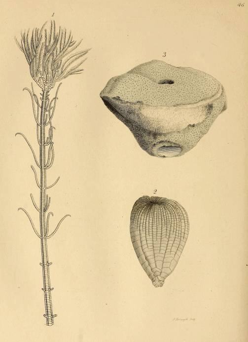
PLATE XLVI.
Pentacrinus.
Fig. 1. Specimen of a recent Pentacrinus Caput Medusæ, from the Caribbean Sea.
The Lily-shaped animals (Crinoidea), so named from a fancied resemblance of some species when in a state of repose to a closed lily, may be compared to a Feather-star (Comatula) fixed to a jointed column, with its mouth upwards; the base of the stem being attached to the rock by root-like processes. The only known living genus inhabits the seas of the West Indies, and the specimen figured represents the body (or upper part of the animal), with a considerable portion of the stem remaining attached. The Crinoidea are divided into two groups; Encrinites, having the ossicula (little bones) of the stem rounded, and Pentacrinites, in which the ossicula of the column are pentagonal, or angular. The Crinoidea are characterized by having a fixed base, a column or stem composed of numerous separate articulated pieces of a solid calcareous substance, supporting on its summit a vase, or receptacle, formed by a series of closely adjusted plates, which contain the body, or viscera. The upper part of the receptacle is covered by a plated integument, on one side of which an aperture or mouth is placed. From the upper margin proceed five articulated tentacula or arms, which subdivide into branches that in some species are very numerous and of extreme tenuity. On the inside, the arms are beset with articulated cirri or feelers. The joints composing the column are perforated by a central opening; there are also side-arms, that radiate from the column in groups of five at different points. When the animal is alive, the skeleton is covered by a soft integument, as in the star-fishes, and the arms spread out and expand, forming a net, by which living prey is captured and conveyed to the mouth by the tentacula, in the same manner as in the fresh-water polype or Hydra.
The fossil remains of Crinoidea consist of the ossicula of the column, arms, and tentacula; of the plates of the vase, or receptacle; and of the peduncle, or base of attachment. This family of Radiaria, though now of such excessive rarity, swarmed in the seas that deposited the ancient secondary strata; whole mountain chains and extensive tracts of country are composed of strata almost entirely made up of their fossil remains.[38] The number and species of genera is very great.
Fig. 2, is a remarkably beautiful specimen of the receptacle of a Pentacrinite from Gloucestershire, showing the arms introverted, as if the animal had suddenly perished while in the act of closing over its prey; the stem is wanting.
Fig. 3. A spongite (Chenendopora subplana, of Michelin) from the greensand of the Vale of Pewsey, in Wiltshire.
Plate XLVII.
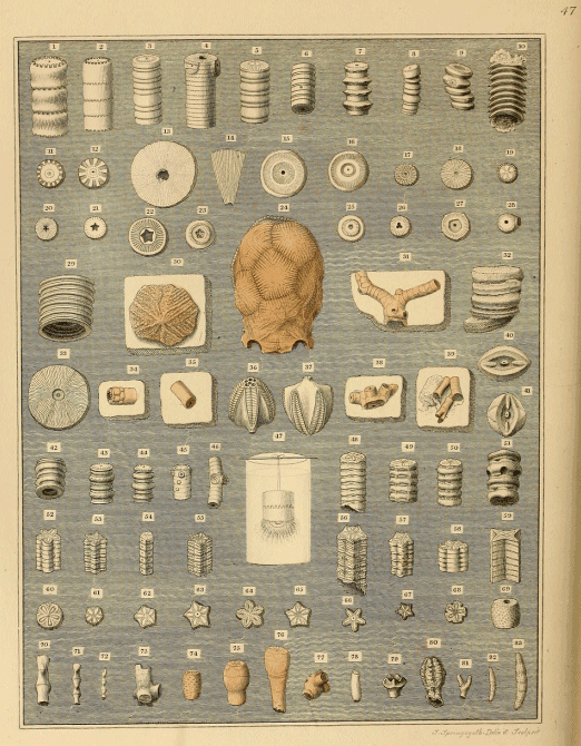
PLATE XLVII.
Fossil Remains of Crinoidea.
In this beautiful plate Mr. Parkinson has figured a great variety of ossicula and portions of stems belonging to many species and genera of Crinoidea; the markings or sculpturing on the articulating surfaces of the columnar ossicula are represented with great accuracy. It is not within the plan of this work to give detailed descriptions of these numerous detached parts; a few of the most interesting objects only will be particularized.
The specimens figured in the upper part of the plate, figs. 1 to 28, are cylindrical ossicula, and portions of stems of Encrinites: those in the lower division are for the most part pentagonal, and therefore belong to Pentacrinites.
Fig. 24. The "Tortoise Encrinite," of Mr. Parkinson, (Marsupites Milleri, of Mantell,) from the chalk of Kent. The specimen figured is the receptacle or body of a very remarkable crinoideal animal which forms the link that unites the Lily-shaped animals with the Star-fishes; like the former, the receptacle is composed of articulated plates, closed at the top by a tessellated plate-work with a buccal aperture, and surrounded by five flexible arms; but the original animal, like the Star-fishes, was destitute of a stem, and could float through the water at pleasure. Its true structure was first pointed out by me in 1822;[39] the name Marsupite was suggested by the purse-like form. In the figure, the base of the receptacle is uppermost. Fig. 30, is a single plate of a Marsupite attached to a piece of chalk.
Figs. 31, 35, 38, 39, 40, 41, 74, 75, 76, 77. These are portions of a small species of Encrinite (Apiocrinus ellipticus) peculiar to the white chalk, in some localities of which the detached ossicula and peduncles are abundant. At Northfleet, near Gravesend, these fossils are often met with. Figs. 75, and 76, are portions of the receptacle with part of the column; figs. 31, 38, and 39, are parts of the processes of attachment. I have never seen any specimen with the arms.[40]
Fig. 34. This is part of the receptacle and stem of another small chalk Encrinite (Bourgeticrinus, of D'Orbigny) from Kent; it is remarkable for the very slight increase in bulk of the receptacle, and the peculiar form of the plates of which it is composed.
Figs. 36 & 37. Two views of the receptacle of a very remarkable crinoidean animal (Pentremites florealis, of Say), from the cherty carboniferous limestone of Kentucky. This zoophyte, though resembling the Crinoidea in having a plated receptacle supported by an articulated stem, has a remarkable affinity to the Sea-urchins (Echinidæ) in the porous bands and pentagonal aperture, and in being destitute of arms or tentacula. Some of the Kentucky limestone beds swarm with the remains of these zoophytes.[41]
Fig. 47. "Two ossicula of the Lily Encrinite immersed in diluted muriatic acid, by which the animal membrane was exposed, and is seen hanging in flocculæ from the bottom of the fossil,"—Mr. Parkinson.
Figs. 57, 64, 66. Part of the stem, and the articulating surfaces of two ossicles of a very elegant pentacrinite (Pentacrinus scalaris, of Goldfuss), from the Lias of Lyme Regis.
Figs. 53, 56, 59, 61, 62, 63, 65, 67. Portions of stems, and the various modifications of the ossicula of another Lias Pentacrinite (Pentacrinus basaltiformis, of Goldfuss).
Fig. 79. This elegant little crinoidean receptacle was named the "Clove Encrinite," by Mr. Parkinson, from its form; (Eugeniacrinus caryophyllatus, of Goldfuss). It is from the Oolite of Mount Randen, in Switzerland.[42]
Figs. 80, 81, 82, & 83. Appear to be fossil corals of the genus Ceriopora.
Plate XLVIII.
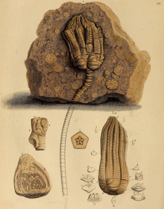
PLATE XLVIII.
The Lily Encrinite (Encrinites monileformis).
This exquisite species of the extinct Crinoideans which swarmed in the seas of the secondary ages of Geology, is equally interesting and attractive to the amateur collector and the scientific observer. The specimen figured is a charming example of the "Stone Lily" partly expanded, attached to a block of limestone studded with encrinal ossicula. Mr. Parkinson informed me that it was formerly in the collection of Mr. Jacob Forster, and cost him twenty guineas; from five to ten guineas is now the usual price for a specimen in a good state of preservation, with any part of the column attached. This Encrinite is not known to occur in England. The specimens seen in collections are for the most part from Lower Saxony: this species has only been found in the limestone strata called "Muschelkalk" one of the subdivisions of the Trias, or New Red Sandstone formation, of Germany.[43] The most celebrated locality of these fossils is in Brunswick, near the village of Erkerode, about two miles from the town bearing the same name. The bed in which they are found is a soft argillaceous cream-coloured limestone, about one foot and a half in thickness; and the stone is composed chiefly of trochites, or detached ossicula of the stems, and a few fragile shells and corals.
An elaborate account of the structure of the skeleton of the Lily Encrinite is given by Mr. Miller, in his valuable work, "The Natural History of the Lily-shaped Animals," (1 vol. 4to. 1821.) Mr. Parkinson had previously carefully investigated the different parts which enter into the composition of the receptacle and column, and had given them names analogous to those employed to designate the bones of the skeleton in vertebrated animals. This nomenclature has very properly been abandoned; but I subjoin Mr. Parkinson's description of the figures, to record his ingenuity and skill in dissecting organic remains:—
"Fig. 1, The Lily Encrinite, with part of its vertebral column attached. In this specimen is seen the extensive capacity for motion yielded by the peculiar form of the vertebra; in the superior part of the column; and by the fortunate removal of a portion of the fingers, a fair view is given of the natural arrangement of the tentacula.
Fig. 2. The pentagonal base, composed of the ossa innominata, and forming with the scapulæ and clavicles, the pelvis, in which were contained the organs of digestion, &c.
Fig. 3. The Lily Encrinite, detached from its vertebral column.
a, the centre of its base, formed by five cuneiform ossicula, or ossa innominata.
a, one of the ossa innominata detached.
b, the ribs, or articuli trapezoides; forming, with the preceding bones, the pentagonal base.
b, one of the ribs detached, showing its internal surface.
c, the clavicles.
c 1, the interior surface.
c 2, the superior surface.
d, the scapulæ.
d 1, the inferior surface.
d 2, the superior surface.
e, the arms.
f, the two first bones of the arms united.
g, h, i, k, l, m, the bones of the fingers gradually diminishing.
Fig. 4. Part of the supposed base, or organ of attachment, of the Lily Encrinite.
Fig. 5. The supposed base, or organ of attachment, of the 'Cap Encrinite.'"
Plate XLIX.
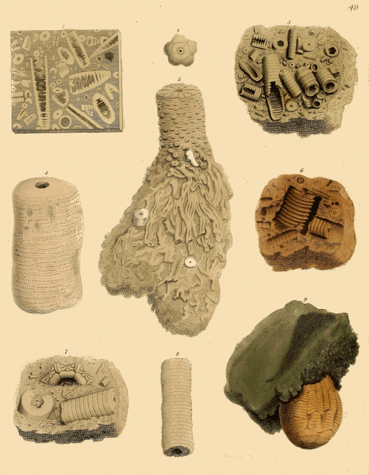
PLATE XLIX.
Remains of Encrinites.
Fig. 1. A polished slab of limestone formed of portions of the stems of encrinites; the white figures are produced by sections of the calcareous spar into which the ossicula are transmuted. The dark spots are the cavities of the entrochites, filled with mineral matter of a different colour.
Fig. 2, is the pentagonal base of the receptacle of the Derbyshire Encrinite.
Fig. 3. A mass of Derbyshire encrinal marble, with numerous portions of stems lying in relief.
The Derbyshire encrinal marble is so extensively employed in the manufacture of tables, chimney-pieces, vases, &c., that it must be familiar to every reader; and yet probably but few are aware of its origin, or of the nature of the fossil remains of which it is composed, and that give rise to the elegant figures in which its beauty consists. On Middleton Moor, near Matlock, extensive quarries of this marble are worked, and good specimens of the ossicula and stems may be easily obtained.[44]
Fig. 4. Part of the stem of a large Encrinite, (Cyathocrinus rugosus, of Miller,) from the Wenlock limestone, Dudley.
Fig. 5. A fine specimen of the lower part of the stem, and the root-like processes of attachment of the base, of the same species as fig. 4: from Dudley.
Fig. 6, is called the "Screw or Pulley-stone" of Derbyshire. These curious fossils are found in the chert (a kind of flint) which occurs in veins and layers in some of the limestone strata: they are siliceous casts of the interior cavities of the stems, and small branches of ossicula, of Encrinites. Plate XL VII. fig. 10, is a detached specimen of this kind.
Fig. 7, is described by Mr. Parkinson as "a piece of marble from Shropshire, in which is discovered a part of the pentagonal base of the Turban or Shropshire Encrinite."
Fig. 8, is part of the column of the same species. These specimens belong to the Rose Encrinite (Rhodocrinus verus, of Miller).
Fig. 9. The receptacle of a very remarkable form of Encrinite, called by Mr. Parkinson "the Cap Encrinite of Derbyshire." I can find no notice of this beautiful and unique specimen in the work of Miller or of subsequent authors; neither am I aware of any data by which a relation can be established between this receptacle and the ossicula and stems, so abundant in the carboniferous limestone of Derbyshire.
Plate L.
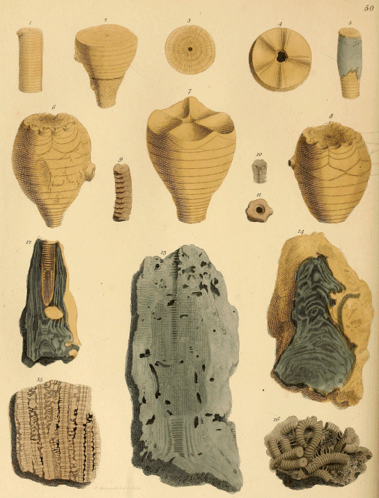
PLATE L.
Encrinites and Pentacrinites.
The Pear Encrinite of Bradford; Mr. Parkinson.
(Apiocrinus rotundus, of Miller. —— Parkinsoni, of Bronn.)
The most generally known of the British Crinoidea, from its size, and abundance in one particular locality, is the "Pear Encrinite" of Bradford in Wiltshire, some of the quarries of the oolite on the heights above that picturesquely-situated town, yielding not only immense quantities of detached plates and ossicula, but also numerous examples of the receptacle, and occasionally the entire skeleton from the peduncle of the base to the extremities of the arms. The lamented Mr. Channing Pearce, and his father (now of Percy Place, Grosvenor, Bath), when resident at Bradford, paid such unremitting attention to the collection of these fossils, that perfect specimens were obtained, exhibiting the entire structure of the originals; of these some fine examples are preserved in the British Museum. Sir Charles Lyell mentions a very interesting fact relating to the occurrence of these Crinoidea in the strata. He states that the upper surface of a bed of limestone at Bradford is incrusted with a continuous pavement formed by the stony roots of the Apiocrinites; and upon this is a layer of clay in which are the stems and bodies (receptacles) of innumerable examples; some erect, others lying prostrate; while throughout the clay are scattered detached arms, stems, and receptacles. This submarine forest of Crinoideans must therefore have flourished in the clear sea-water till invaded by a current loaded with mud, which overwhelmed the living zoophytes, and entombed them in the argillaceous deposit in which their remains are now imbedded.[45]
The receptacle of this Apiocrinite is pyriform and very smooth, the plates are large and thin, with radiating articulated surfaces; the stem is short, smooth, and strong, the arms are simple, and like those of the Marsupite; the peduncle spreads out into an expanded base, which is firmly attached to the rock; sections of this part are generally of a purple colour.
Fig. 1. Part of the column of the Bradford Encrinite. 2. Part of the receptacle; a minute incrusting coral (Bryozoa) is attached to the lower part, giving the stem a rough appearance.
Figs. 3, & 4. Surface of detached plates of the receptacle.
Fig. 5. Portion of the column partly covered with a cortical covering of a purple colour possibly the original investing membrane.
Fig. 6. A receptacle, in which a few of the ossicula of the arms remain attached to the margin.
Fig. 7. Another receptacle, in which the plates called by Mr. Parkinson "clavicles and scapulæ," are retained in their natural positions.
Fig. 8. A receptacle, in which the principal plates are well defined: these are named by Mr. Parkinson as follow: a, clavicle; b, scapula; c, ossicula of the arms; d, the last series of the same. The ossicles forming the elongated tentacula, Mr. P. termed "bones of the fingers."
Fig. 9. Portion of an encrinital stem with digitated processes: the nature of this fossil is unknown to me.
Fig. 10. Three united ossicula of a Pentacrinite with depressions for side-arms: from the Lias of Lyme Regis.
Fig. 11. A distorted pentacrinal ossicle; said to be from Africa.
Figs. 12, & 14, are vertical polished sections of the peduncle, or base of the stem, of the Bradford Encrinite.
Fig. 13. Vertical section of the peduncle of a Pentacrinite from Soissons.
Fig. 15. A polished slab of pentacrinal marble from Charmouth, Dorsetshire.
Fig. 16. Variously contorted pentacrinal stems with numerous side-arms, from Charmouth.
Plate LI.
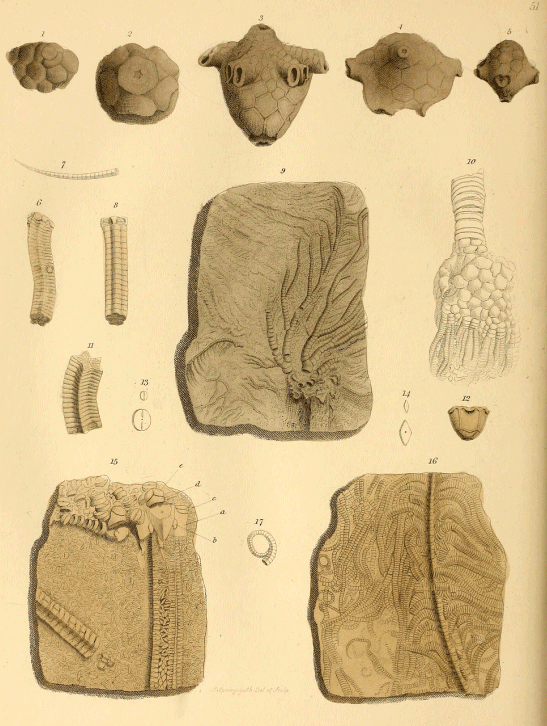
PLATE LI.
Fossil Crinoidea, or Lily-shaped Animals.
Fig. 1. Part of the receptacle of the "Nave Encrinite" of Mr. Parkinson (Actinocrinus, of Miller). Mountain limestone.
Fig. 2. A portion of the receptacle of a "Rose Encrinite" (Rhodocrinus), viewed from the base.
Fig. 3. The "Nave Encrinite" (Actinocrinus triacontadactylus, or thirty-fingered, of Miller), from the mountain limestone. This is a good example of the structure of the receptacle in this group of Crinoideans, which is distinguished by the arms passing off at right angles from the periphery of the receptacle, like the spokes of a wheel; whence the name, Nave Encrinite. The upper part is covered by closely adapted plates, and the buccal aperture or mouth is situated at the side. The stem of this group is thickly beset with side-arms. (Fig. 7 is a very small detached one.) The arms are numerous (amounting to thirty in the species figured), and of great length; these subdivide into jointed filaments of extreme minuteness. Slabs of limestone are often entirely covered with them, and many layers are wholly made up of their aggregated remains. The plates of the receptacle are generally highly ornamented: in one species the sculpturing so closely resembles that of the Marsupites ornatus of the chalk, that it was with difficulty I convinced Mr. Parkinson that the latter did not possess a stem, and therefore was not an Actinocrinite.[46]
Figs. 4, & 5. Portions of receptacles of Actinocrinites.
Figs. 6, & 8. Fragments of stems of a Pentacrinite (Pentacrinus scalaris, of Goldfuss); from Gloucestershire.
Fig. 9. A Pentacrinite expanded on a slab of Lias-shale. Gloucestershire.
Fig. 10. Stem, receptacle, and arms of a Crinoidean (probably a Cyathocrinite); it is drawn in an inverted position. The figure is stated by Mr. Parkinson to be copied "from a plate by Dr. Capeller." Neither the locality, nor the stratum from which it was obtained, is mentioned.
Fig. 11. Part of the stem of a Pentacrinite (P. basaltiformis, of Miller); from the Lias. Gloucestershire.
Fig. 12. The receptacle of a Crinoidean (Platycrinus lævis, of Miller); from the mountain limestone, Ireland. Fig. 13, ossicles of the arms; and fig. 14, joints of the stems, slightly magnified.
Fig. 15. "The superior part of the Briaræan pentacrinite."—Mr. Parkinson. (Pentacrinus Briareus, of Miller.) The specimen is a slab of Lias, almost wholly made up of crinoideal remains. In relief on the surface are the stems and dislocated ossicles of the receptacle; the latter are thus enumerated by Mr. Parkinson;—a, scapula; b, clavicle; c, first bone of the arm; d, second arm-bone; e, commencement of the two series of bones forming the fingers.
Fig. 16, is another slab of pentacrinal Lias limestone, with portions of a stem and numerous side-arms: these are generally electrotyped, as it were, with a brilliant pyrites, giving a rich metallic lustre to the animal remains. In the British Museum there are many splendid specimens of this highly interesting family of Radiaria. I would especially direct the intelligent visitor's attention to a slab of stone, many feet in height and breadth, on which a group of Pentacrinites is displayed, as palpable and perfect as if the animals were sporting in their native element. This matchless specimen is from Germany.
Fig. 17. One of the small auxiliary lateral tentacles of a Pentacrinite.
Plate LII.
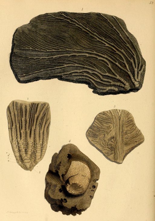
PLATE LII.
Pentacrinites.
Fig. 1. This specimen displays the usual appearance of the mode in which the arms of Pentacrinites are spread out in relief on the pyritous lias limestone of Charmouth.
Fig. 2. The arms, from the upper part of the receptacle to their third or fourth subdivision of the Briaræan Pentacrinite. Charmouth.
Fig. 3. A small specimen, showing the ramifications and delicacy of the extremities of the arms or tentacula.
Fig. 4. "A fossil body, supposed to be a species of oval encrinite."—Mr. Parkinson. This fossil is certainly a coral, probably some species of Turbinolia, from the Devonian formation.
Plate LIII.
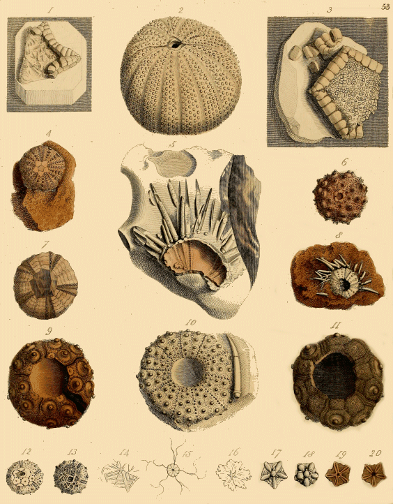
PLATE LIII.
Fossil Star-Fishes and Echini.
The radiated animals popularly called Star-fishes, from their stellular figure, are so abundant along our sea shores, that the nature of the common five-rayed species (Asterias, or Uraster rubens)[47] must be familiar to most of my readers. This species belongs to the group in which the rays are elongated, and far exceed in length the diameter of the disk; in another subdivision (the Goniaster, or Cushion-star), the body is angular, and the lobes or rays are short, and do not exceed in length the diameter of the body. In another group (the Comatula, or Feather-star), the rays are fringed with long jointed tentacula, which divide and subdivide like those of the Crinoidea; and these star-fishes may, in fact, be regarded as free Lily-shaped animals.[48] There is another tribe in which the arms are elongated into slender rays, without grooves or tentacula; these are called the Serpent Star-fishes (Ophiura). Species of all these groups occur in a fossil state.[49]
Fig. 1. "Part of a fossil lunated star from the chalk of Kent."—Mr. Parkinson. (Goniaster semilunata, of Parkinson; Goniaster Parkinsoni, of Prof. E. Forbes). Remains of Star-fishes are by no means rare in the chalk strata of Kent; in those of Sussex they are far less common. When the "Fossils of the South Downs" was published, in 1822, a few fragments only had been discovered. Of late years, some beautiful examples have been obtained from the chalk-pits near Arundel and Worthing, by Mr. Dixon, Mr. Coombe, Mrs. Smith, of Tunbridge Wells, and other collectors. The cabinet of the Marquess of Northampton is very rich in this class of fossils. Several unique examples of new species have been obtained from the chalk near Maidstone.
Fig. 2. "An echinite, from France."—Mr. Parkinson. The locality of this fossil is uncertain; no similar specimen is known either to Mr. Morris, or the other eminent palæontologists I have consulted; and the original cannot be discovered. I have reason to believe it was purchased, after Mr. Parkinson's death, together with the greater number of the fossils already described, by an American gentleman, and taken to the United States.
Fig. 3. "Part of a stellite or fossil star-fish, resembling Pentagonaster regularis."—Mr. Parkinson. This well-known chalk species (Goniaster Mantelli, of Prof. E. Forbes), occurs frequently in an imperfect state in the quarries near Gravesend. The collection of the Marquess of Northampton contains a perfect and exquisite specimen attached to a flint, from that locality.
Fig. 4. A beautiful example of the Turban Echinite (Cidaris Parkinsoni, of Dr. Fleming), from Wiltshire.
The Cidaris, or Turban Echinite, belongs to the family of radiated animals, of which the recent Sea-urchin (Echinus sphæra) is a well-known example. The globular shell or envelope of these animals is composed of numerous calcareous polygonal plates, arranged in regular and elegant patterns, like the lines of the meridian on a globe. These plates are externally covered with papillæ of various sizes, to which spines of corresponding magnitude are articulated. In some of the Cidares the principal tubercles are very large, and their spines several inches in length. The number and variety of the animals of this family that occur in a fossil state are so great, that a work expressly devoted to the subject would be required to thoroughly investigate the characters and relations of the known species. An elementary knowledge of this class of fossil remains may be obtained by reference to "Medals of Creation," chap. xi. p. 240.
Fig. 5. Part of the case of a Cidaris attached to a flint by its outer surface, surrounded by upwards of twenty spines; the interior of the shell, of a light pink colour, is exposed. This exquisite fossil is now in the cabinet of the Marquess of Northampton. It was purchased by Mr. Parkinson for the sum of twenty guineas; but this was in the palmy days of the study of organic remains, before the terms Geology and Palæontology were invented, and when a choice relic of "a former world" was cheap at any price, in the opinion of the enthusiastic collector.
Fig. 6. A Turban Echinite (Cidaris (Hemicidaris, of Agassiz) crenularis, of Lamarck): from the Coral Rag of Wiltshire.
Fig. 7. A siliceous cast—that is, a flint that has been moulded in the interior of the shell, and received the impress of the internal structure—of "Cidaris corollaris," of Parkinson; (Cyphosoma correlare, of Agassiz): from Sussex.
Fig. 8. Cidaris with spines, from the Oolite of Stonesfield.
Fig. 9. A specimen of one of the Cidares with large tubercles (Cidaris coronatus, of Goldfuss); from the Coral Rag, Oxfordshire.
Fig. 10. An elegant chalk echinus (Cidaris Königi, of Mantell;[50] Cyphosoma Milleri, of Agassiz; C. granulosus, of Goldfuss): from Kent.
Fig. 11. A fine species from the chalk at Gravesend (Cidaris vesiculosus, of Goldfuss).
Fig. 12. A peculiar type of Cidaris (Salenia scutigera, of Goldfuss), from the freestone or upper greensand of Warminster, Wilts.
Fig. 13. Another species of the same genus (Salenia stellulata, of Agassiz); from Warminster.
Fig. 14. An enlarged view of part of the structure around the vertex of fig. 13.
Fig. 15. A species of Feather-star (Comatula pectinata, of Goldfuss); from Solenhofen.
Figs. 17, 18, 19, 20. "Minute Stellitæ (that is, fossil Star-fish); from Verona."—Mr. Parkinson. These are probably the bodies or disks of Ophiuræ deprived of their arms.
Fig. 16. The nature of the specimen figured is unknown to me.
Plate LIV.
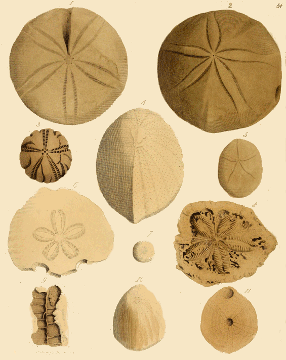
PLATE LIV.
Various Species of Fossil Sea-Urchins.
Fig. 1. A large, discoidal echinite, of the type called Clypeus or Shield-echinus, (Clypeus sinuatus, of Leske,) from the Coral Rag of Oxfordshire. This species abounds in the beds of this division of the Oolite in Berkshire, Wiltshire, Gloustershire, &c.
Fig. 2. "Echinanthites orbicularis (Pygurus) of Leske."—Mr. Parkinson.
Fig. 3. An imperfect flint cast of an echinus (Discoidea), from the South Downs.
Fig. 4. The Helmet Echinite, (Ananchytes ovatus, of Lamarck,) from the Chalk of Kent. This is a characteristic species of the white chalk, and abounds in the strata of the North and South Downs. At Northfleet, near Gravesend, the quarry-men find beautiful specimens.
Fig. 5. An oval echinite (Nucleolites,) from Verona.
Fig. 6. A portion of a very flat echinite, in which the rays or ambulacra are in a floriform arrangement, (Echinodiscus bisperforatus, of Parkinson; Lobophora biperforata, of Desor,) from Tertiary Strata, Verona.
Fig. 7. A small discoidal echinite (Discoidea subuculus, of Leske,) from the upper greensand of Warminster.
Fig. 8. The floriform radiated part of the shell of an echinite (Clypeaster), from the tertiary strata of Malta.
Fig. 9. A cast in flint of part of the interior of the case or shell of an echinite.
Fig. 10. An elegant conical echinite (Conulus albogalerus, of Leske; Galerites, of Agassiz), common in the chalk of Kent and Sussex.
Fig. 11. View of the base of fig. 10, showing the situation of the two apertures of the shell.
Plate LV.
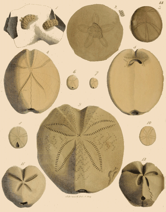
PLATE LV.
Fossil Sea-Urchins, or Echinites.
Fig. 1. The shell of a Turban Echinite (Cidaris saxatilis, of Parkinson), broken in two, and each piece imbedded in the same fragment of flint. From Kent.
Fig. 2. A round Buckler Echinite (Echinodiscus (Clypeaster) subrotundus, of Parkinson), from Italy.
Fig. 3. The upper surface of an Echinite (Spatangites (Disaster, of Agassiz) ovalis, of Parkinson); from Scarborough.
Fig. 4. View of the upper, and fig. 5, of the lower surface of an Echinite, (Spatangus (Hemipneaster, of Agassiz) radiatus, of Parkinson,) from the cretaceous strata of St. Peter's Mountain, Maestricht.
Fig. 6. A small Echinite (Echinites (Nucleolites, of Leske) pyriformis, of Parkinson), from the cretaceous strata of Maestricht.
Fig. 7. A small Echinite of a different genus (Echinites (Cassidulus, of Lamarck,) Lapis cancri, of Parkinson), from Maestricht.
Fig. 8. An Echinite (Spatangites (Nucleolites) brissoides ovalis, of Parkinson). Locality unknown.
Fig. 9. A beautiful specimen of a large heart-shaped Echinite of a recent species (Spatangus purpureus), from a modern tertiary deposit, Malta.
Fig. 10. An Echinite (Echinodiscus (Clypeaster) laganum, of Parkinson), from a tertiary deposit, Verona.
Fig. 11. This Is a very abundant Spatangus or heart-shaped echinite, (Spatangus cor marinum, of Parkinson; Cor testudinarium, of Goldfuss; Micraster cor anguinum, of Agassiz,) in the chalk of Kent, and some parts of Sussex. Siliceous casts, forming cordiform flints, with deep imprints of the pentapetalous rays on the vertex, are common among the stones of the ploughed fields of the Downs.
Fig. 12. A Spatangite, (Spatangus (Micraster, of Agassiz) lacunosus, of Parkinson), from tertiary strata, Malta,
Plate LVI.
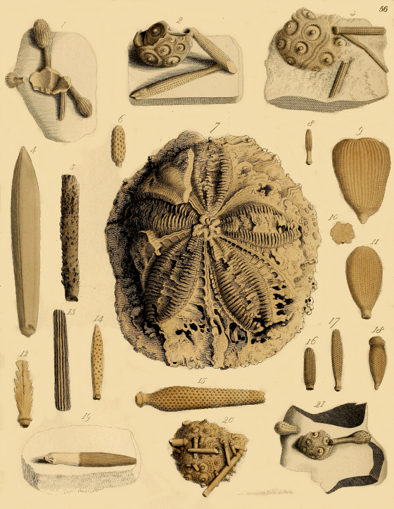
PLATE LVI.
Echinites and Echinital Spines.
Fig. 1. A fragment of the shell of a Turban Echinite, with three clavated or club-shaped spines attached, on chalk, from Kent (Cidaris claviger, of König). The inner surface of the fragment of shell is exposed.
Fig. 2. A crushed shell of an elegant species of Turban Echinite (Cidaris sceptrifera, of Mantell), on a block of chalk; with two displaced spines near it. The sceptre-like form of the spines suggested the specific name. The chalk has been carefully cut away so as to display the shell and spines as much as possible without detaching them. From Sussex; common in the chalk near Gravesend.
Fig. 3. Part of the shell, with two spines of another species (Cidaris vesiculosus, of Goldfuss), from Kent.
Fig. 4. "A fossil echinital spine resembling a belemnite."—Mr. Parkinson. I am unable to determine either the species or locality of this fossil: it is indeed doubtful whether it is a spine of an echinus.
Figs. 5 to 19, represent various kinds of echinital spines of Turban Echinites or Cidarites.
Fig. 5. "A fossil spine named 'Bacolo di Santo Paulo,' by Scilla."—Mr. Parkinson. From Verona.
Figs. 6, 8, 9, 10, 11, 14, 15, 16, 17, & 18, are, I believe, referable to various species of Cidaris that occur in the Oolite or Jurassic deposits.
Figs. 9 and 11. Species of Cidaris glandiferus, of Goldfuss.
Fig. 15, is a well known form, which occurs in thousands in the Oolite Limestone, the Coral Rag, of Caen, and other localities in Wiltshire; it belongs to a beautiful Cidarite (Cidaris Blumenbachii[51]), which is occasionally found with similar spines attached.
Fig. 12. "A flat serrated spine from Verona."—Mr. Parkinson. It belongs to the Cidaris Schmidelii, of Goldfuss.
Fig. 7. The interior of the upper part or vertex of a large Echinus, from the tertiary strata of Malta. The greater portion of the shell is broken away, but a small fragment showing the outer surface remains on the upper left hand of the specimen. The five large petalous ambulacra are beautifully seen. Perfect examples of this echinite (Echinanthus Clypeaster altus, of Parkinson), are not uncommon.
Fig. 19. A spine of Cidaris sceptrifera, from the chalk of Kent.
Fig. 20. An elegant Turban Echinite, (Hemicidaris crenularis, of Lamarck,) common in the Coral Rag of Wiltshire. Groups of this beautiful echinoderm, with numerous spines attached, are found at Caen. I have seen on one slab of limestone, upwards of twenty individuals with the spines radiating round the shell, as if the animals were alive on a mud bank in shallow water.
Fig. 21. A fragment of the shell with two spines (Cidaris claviger), attached to a flint; from Kent.
Plate LVII.
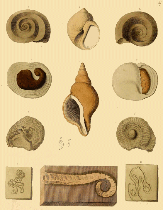
PLATE LVII.
Fossil Shells.
Figs. 1, & 3. Upper and under view of a discoidal spiral univalve shell (Euomphalus pentangulatus, of Sowerby), from the mountain limestone of Derbyshire. The extinct genus Euomphalus, a name suggestive of the deeply excavated disk, comprises many species which occur in the Silurian, Devonian, and Carboniferous formations. The shell has chambers, or rather obsolete cavities sealed up by a shelly partition, in the abandoned part of the spire.[52]
Fig. 2. An elegant univalve shell, completely silicified or transmuted into flint (Natica canrena, of Parkinson, Natica Gentii, of Sowerby), from the upper greensand of Blackdown.
Figs. 4, & 6. Two views of the same specimen; a univalve (Nerita conoidea, of Lamarck), in which the apex or upper part is destroyed, and the interior of the shell is filled with yellowish brown chalcedony; in fig. 4, a cast of the spire is seen, and in fig. 6, the mouth of the shell, with the chalcedony partially filling up the interior. From tertiary strata near Paris.
Fig. 5. A beautiful fossil univalve shell, from the "Red Crag" of Suffolk, known to collectors as the "Essex reversed whelk," from the spire being coiled in the opposite direction to the common mode; the mouth is consequently situated to the left of the observer; the same species occurs with the spire in the usual direction. This shell is the Murex (Fusus) contrarius, of Parkinson.
Figs. 7, & 8. Under and upper view of another species of Euomphalus (E. rugosus, of Sowerby), from the Wenlock limestone, Dudley.
Fig. 9. An enlarged view of fig. 10. "A shell of the genus Sigaretus."—Mr. Parkinson. Mr. Morris thinks it is merely an operculum of a small univalve.
Fig. 11. A chambered cephalopodous shell (Lituites lituus, of Hisinger), from Silurian strata, Sweden.
Figs. 12, & 13. These curious contorted bodies are named "Vermiculitæ" by Mr. Parkinson. They occur in the cream-coloured limestone of Pappenheim and Solenhofen. They are termed "Lumbricaria colon" by Goldfuss; and "Cololites" by M. Agassiz; the last-named eminent naturalist has demonstrated that they are the fossilized intestines of fishes.[53]
Plate LVIII.
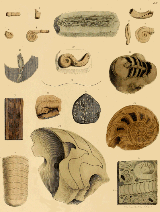
PLATE LVIII.
Fossil Shells.
Fig. 1. "Part of a hexahedral Serpulite."—Mr. Parkinson.
Fig. 2. A silicified mass of delicate filiform serpulæ, from the upper greensand of Devonshire (Serpula filiformis, of Sowerby).
Fig. 3. Portion of a species of Siliquaria, from tertiary strata, France. It is the shell of an Annelide related to Dentalium.
Fig. 5. A spiral Serpulite (it resembles the Serpula conica); probably from the cretaceous beds of the Isle of Rugen.
Fig. 6, is a piece of polished sandstone, from the upper greensand of Wiltshire, "the markings on which are produced by sections of a species of Serpula (Vermetus concavus, of Sowerby)."—Mr. Morris.
Fig. 7. A species of Vermetus; from Bayonne?
Figs. 8, & 9. A species of Vermetus which abounds in the coarse arenaceous limestone of Bognor Rocks, in Sussex (Vermetus Bognoriensis, of Sowerby).
Fig. 10. "A section of the shell of a Nautilus, to show that the siphuncle sometimes suffered distension."—Mr. Parkinson.
Fig. 11. A species of Serpula (Serpula ampullacea, of Sowerby), from the chalk of Kent.
Fig. 12. A fragment of the back or dorsal part of the shell of a fossil Nautilus (Nautilus centralis, of Sowerby), from the London clay, Brentford. The outer shell is broken away, and the siphuncle, traversing five of the septa of the chambers, is exposed.
Fig. 13. "The outline of the back of a Nautilus."—Mr. Parkinson.
Fig. 14. An Orthoceratite (Orthoceras annulatum, of Sowerby; O. undulatum, of Kissinger), from the Wenlock Limestone, Dudley.
Fig. 15. A fragment of a fossil Nautilus (Nautilus Parkinsoni, of Mr. Edwards), from the London clay of Harwich. It shows the situation of the siphuncle and the form of the septa, as indicated by the sinuous transverse lines.
Fig. 16. A polished section of a Nautilus (N. truncatus, of Sowerby), from the Inferior Oolite of Yeovil, Somersetshire. The chambers are filled up with crystalline limestone, with the exception of the six outermost cells, in which are left hollows that are lined with calcareous spar.
Fig. 17. Polished section of an Orthoceratite, from the Silurian strata of Oëland, Sweden.
Fig. 18. The discoidal part of a Lituite from the same locality as fig. 17.
Fig. 19. a polished slab of grey marble, from the Devonian formation of the Rhine. The figures are sections of Orthoceratites, a; and Lituites, b.
Plate LIX.
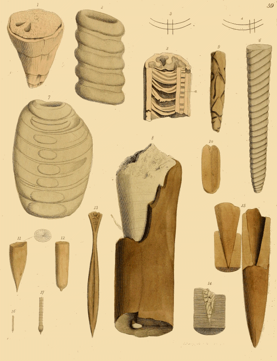
PLATE LIX.
Fossil Cephalopoda, &c.
Fig. 1. A fossil shell named Hippurite (Hippurites bioculatus, of D'Orbigny), from the south of France. This shell belongs to a family termed Rudistes, whose characters are somewhat problematical,—some naturalists referring them to the bivalves, and others to the univalves. The Hippurite is generally of an elongated conical form, and has internally two obtuse longitudinal ridges; the base is sometimes partitioned by transverse septa.
Fig. 5, is a longitudinal section of a specimen in which septa are displayed. The aperture is closed by a moveable operculum, or upper valve, as in the specimen fig. 1. The substance of the shell is cellular and very thick, and when fractured, resembles that of the lamelliferous corals. Some kinds attain a large size, and are called "petrified horns" by the inhabitants of the districts in the Pyrenees where they abound. Though Hippurites are abundant in the chalk of the south of France, and in Spain and Portugal, none have been found in England. The Spherulite, a nearly allied genus, which has no internal longitudinal ridges, occurs in the chalk of Sussex: it was first discovered near Lewes. (Spherulites Mortoni, of Mantell.)[54]
Fig. 2. The siphuncle of a very large Orthoceratite ("related to the genus Ormoceras," Mr. Morris), from the Rhine.
Figs. 3 & 4, "show the direction in which the siphuncle in Orthoceratites intersects the septa."
Fig. 6. Siphuncle of an orthoceratite (related to Orthoceras duplex, of Kissinger), from the Silurian strata, Sweden.
Fig. 7. An Orthoceratite (O. pyriforme, of Sowerby), from the Silurian strata, Dudley.
Figs. 8-15. Various kinds of Belemnites.
In the "Supplementary Notes" I have, under the head, "Belemnites," explained somewhat fully the nature of those fossils which, by the name of "thunderbolts," have for so many centuries excited the interest and perplexed the ingenuity of collectors of fossil remains. Referring the reader to that note, I shall therefore in this place merely give such specific names of the specimens figured by Mr. Parkinson as I have been able to determine.
Fig. 8. "A Belemnite of large size," Mr. Parkinson. This specimen is part of the phragmocone from near the lower apical portion, partially invested with the fibrous rostrum or guard. It is the species named Belemnites giganteus by M. D'Orbigny; from the Oxford clay of Wiltshire.
Fig. 9. The guard of a Belemnite, eroded by some Annelide.
Fig. 10, is a vertical section of a fragment of a Belemnite, showing the alveolus or cavity for the reception of the apex of the phragmocone in the upper part.
Fig. 11. The distal or apical part of the rostrum or guard of a Belemnite. The annexed outline of a transverse section exhibits the radiated structure.
Fig. 12. The distal part of the guard of a chalk Belemnite (Belemnitella mucronata); from Norwich. Siliceous casts of the phragmocone of Belemnitella are occasionally met with in the flints of the South Downs. This phragmocone has a longitudinal flat band or ridge, extending down the dorsal aspect: the chambers are very numerous; the slit or fissure in the ventral aspect of the guard, is occupied by a thin expansion of the phragmocone.
Fig. 13. A Belemnite from the great oolite of Stonesfield (Belemnites fusiformis, of Parkinson). The upper part shows the alveolus for the reception of the apex of the phragmocone.
Fig. 14. A fragment of a guard split vertically, the flat surface showing a section of the alveolus filled with spar. This specimen belongs to the Belemnites cylindriformis, of Parkinson.
Fig. 15. a Belemnite (Belemnites coniformis, of Parkinson), having part of the guard broken off, to show the alveolus or hollow in which the apical part of the phragmocone is received. The removed portion has the cast of the alveolus attached to it.
Fig. 16, of which fig. 17, is an enlarged view, is a species of chambered foraminiferous shell, called Nodosaria (N. raphanistrum, of Lamarck); from Sienna. See description of Plate LXII.
Plate LX.
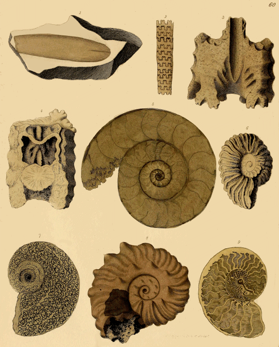
PLATE LX.
Ammonites.
Fig. 1. A Belemnite (Belemnitella mucronata) attached to a flint. Kent.
Fig. 2. Cast of part of a straight-chambered shell (Baculites Fraujasii, of Lamarck), in which the septa, or partitions, are deeply and regularly sinuated. In fossils of this kind, the cast of each chamber is distinct from the others; but the series is held together by the flexuosities of the septa. From Maestricht.
Fig. 3. A limestone cast of the chamber of an Ammonite: from Bath. The elongated channel in the middle indicates the position of the siphuncle.
Fig. 4. Fragment of an Ammonite, showing cavities of two chambers, and the canal of the siphuncle, partly lined with calcareous spar.
Fig. 5. Polished sections of an Ammonite (Ammonites Walcotii) from the Lias, Whitby. The chambers are filled with semi-transparent spar. The siphunculus is seen running along the dorsal, or outer margins of the volutions. The dark appearances observable in several parts of the siphuncle result from the carbonization of the animal membrane with which the tube was lined in the living state.
Fig. 6. "An Oval Ammonite."—Mr. Parkinson. This is evidently the cast of a discoidal shell pressed into an elliptical form. In the Chalk-marl, casts of Ammonites, Nautilites, &c. are very commonly more or less distorted by compression. The marl appears to have remained in a plastic state after the decomposition of the shell in which it was moulded, and to have admitted of being squeezed into close contact with the surrounding matrix; when the stratum became consolidated the cast retained its accidental shape, and adhering but slightly to the investing marl, was separable by a properly directed blow. This explains the otherwise unintelligible fact of a cast being closely invested by the rock, and all traces of the shell in which it was formed absent. When both the cast and the matrix became solid and uncompressible before the shell was decomposed, then loose casts were formed; as is common in the Portland stone, &c. The fossil figured appears to be an indifferent example of a common chalk-marl species (Ammonites Mantelli, of Sowerby).
Fig. 7. A beautiful cast of an Ammonite, in which the foliaceous septa transmuted into pyrites (sulphuret of iron, or marcasite), are exquisitely shown.
Fig. 8. A very fine specimen of an Ammonite (Ammonites latus, of Sowerby), from the "Galt;" a subdivision of the Lower chalk, in which Ammonites, with their pearly shells beautifully preserved, are abundant. From Folkstone, in Kent; a celebrated locality for these and other fossils of the same cretaceous deposits.
Fig. 9. Sections of a pyritous cast of an Ammonite, showing the sinuous edges of the septa.
Plate LXI.
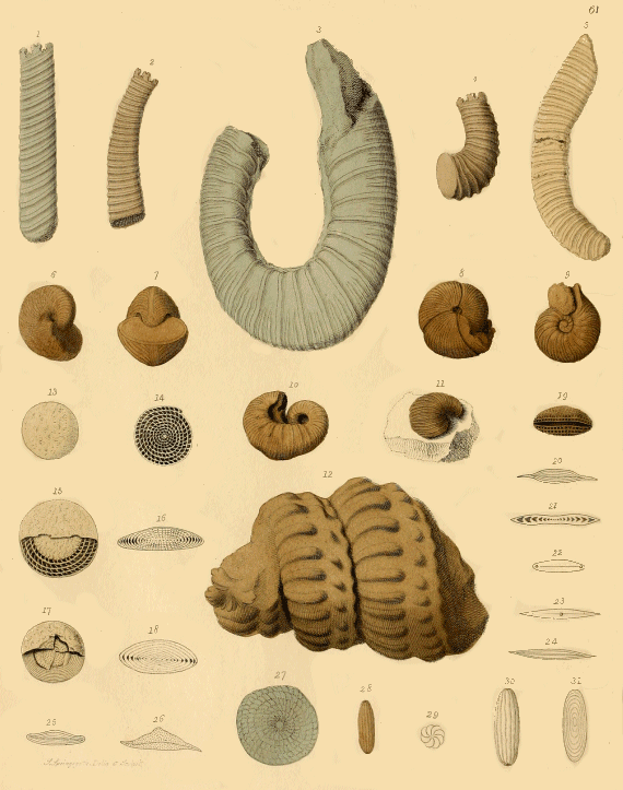
PLATE LXI.
Fossil Cephalopoda, &c.
Fig. 1. Part of the cast of a species of Hamite (Hamites intermedius, of Sowerby), from the Gait of Folkstone. The name Hamites was employed by Mr. Parkinson to designate a genus of chambered shells, in which the direction of the spire, instead of being straight, as in Baculites, or discoidal, as in Ammonites, was bent like a hook beyond the inner reflected part. All the specimens here figured are but fragments.[55]
Figs. 2, & 5. Portions of Hamites intermedius, of Sowerby.
Fig. 3. Hamites plicatilis, of Sowerby.
Fig. 4. A fragment of Hamites rotundus, of Sowerby.
Figs. 6, & 7. Two views of a species of an extinct genus, the shells of which, though not chambered, are supposed to have been inhabited by Cephalopoda, like the recent Argonaut. The specimen (Bellerophon costatus, of Sowerby) is from the Mountain limestone of Derbyshire.[56]
Figs. 8, & 9. An Ammonite with a contracted aperture, and three deep constrictions across the disk. From the Inferior oolite of Normandy.
Figs. 10, & 11. Two specimens of "Scaphites, or Boat-like Ammonite," of Mr. Parkinson. A remarkable cretaceous genus of extinct cephalopoda. The specimens figured are from the Lower chalk of Sussex (Scaphites costatus, of Mantell; S. equalis, of Sowerby).
Fig. 12. Cast of a spiral chambered shell, called Turrilite, of which many species occur in the lower cretaceous strata (Turrilites costatus, of Langius). The quarries of lower chalk at St. Catharine's Mount, near Rouen, in Normandy, have long been celebrated for the number and perfection of specimens of this elegant type of cephalopodous shells. The first known English examples of this genus, as well as of Scaphites, were discovered by me in the chalk marl, at Hamsey, near Lewes, in Sussex, in 1810. Several very fine specimens of a large species (Turrilites tuberculatus), some of which are more than two feet in length, have been obtained from the same strata. The tubercles on the casts of this species are the bases of strong spines. The siphunculus, in the state of a pyritous cast, is preserved in some examples.
Figs. 13 to 27. These figures all refer to a very curious group of fossils, termed Nummulites, from the supposed resemblance of some of the flat disks to a piece of money. The complexity of their internal structure, and the supposed resemblance of their organization to that of the true Cephalopoda, led to many erroneous opinions as to the nature of the originals. That eminent physiologist, Dr. W. B. Carpenter, has recently investigated the intimate structure of the whole group, and the results are given in a beautiful and masterly memoir in the Quarterly Journal of the Geological Society of London.[57] Dr. Carpenter has clearly shown that these fossils belong to the Foraminifera, and not, as some eminent naturalists have supposed, to the Bryozoa, or "Moss-corals." As the family to which they belong comprises a numerous assemblage of minute organic remains, many of which are delineated in the next plate (Plate LXII.), the reader is referred to the "Supplementary Notes," for a general description of the Foraminifera, in which is given a restored figure of the supposed living animal of the Nummulite, from Dr. Carpenter's memoir.
Fig. 13. The usual appearance of the common species of Nummulite (Nummulina lævigata). From Egypt.
Fig. 14. A specimen rubbed down, and exposing the internal cellular structure.
Fig. 15. An example in which the outer investment is partly removed.
Fig. 16. A vertical section of the same.
Fig. 17. This fossil, of which fig. 18, is a vertical section (Nummulites obtusa, of Sowerby), appears to belong to a different genus; probably Orbitolites, or Marginopora. Tertiary strata.
Fig. 19. A vertical section of a Nummulite, showing a cavity in the centre, probably from decomposition.
Fig. 20. A section of another species of Nummulite (N. dispansa ? of Sowerby);[58] Tertiary strata, India.
Figs. 21 to 26, are various sections of a fossil Nummulite, of which fig. 37, represents the flat surface (Nummulites complanata, of Parkinson. This fossil belongs to the genus Discospira of Mr. Morris).[59]
Fig. 28. A species of Foraminifera (Fasciolites, of Parkinson; Alveolina elliptica, of D'Orbigny).
Fig. 29. A transverse section.
Figs. 30, & 31. Enlarged views of the same fossil. Fig. 31. A longitudinal section.
Plate LXII.
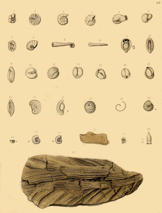
PLATE LXII.
Fossil Foraminifera.
With the exception of figs. 23, 24, 29, 31 and 32, all the specimens delineated in this Plate belong to the Foraminifera. The figures represent magnified views; the natural size is indicated in some instances by a minute outline. Under the article "Foraminifera," in the "Supplementary Notes," a general account is given of the structure and economy of the living animalcules. A list of names is subjoined.
Figs. 1, & 2. Rotalia trochiliformis, of Lamarck. Tertiary.
Fig. 3. Rotalia Beccarii, of Linnæus. Tertiary.
Fig. 4. Cristellaria rotulata, Lamarck. Chalk.
Figs. 5, 6, 7. Lituola nautiloidea, Lamarck. Chalk.
Fig. 8. Spirolina depressa, Lamarck. This and the specimens to fig. 21 inclusive, are tertiary fossils.
Fig. 9. Spirolina cylindracea, Lamarck.
Fig. 10. Orthocerina clavulus.
Fig. 11. Biloculina ringens, Lamarck.
Figs. 12, & 13. Quinqueloculina cor anguinum, Lamarck.
Figs. 14, 15, & 16. Quinqueloculina.
Figs. 17, 18, 19. Triloculina trigonula, Lamarck.
Fig. 20. Quinqueloculina opposita, Lamarck.
Fig. 21. Peneroloplis opercularis.
Fig. 22. Adelosina, of D'Orbigny; a recent species.
Figs. 23, & 24. Gyrogonites. The fossils here figured on a magnified scale as microscopic shells of the same family as those above described, received the name of Gyrogonites, or twisted stones. They prove to be the seed-vessels of a species of the common fresh-water plant, the Chara. The fruit of this genus consists of minute nuclei, with an external calcareous covering, composed of five spirally twisted plates, which unite at the summit. These fossils occur by myriads in many of the fresh-water secondary and tertiary limestones, as well as in the calcareous deposits now in progress of formation in our lakes. In the lacustrine limestones of the Isle of Wight (at Binstead, White Cliff, &c.), beautiful specimens may be obtained.[60] Professor E. Forbes has discovered Gyrogonites in the Wealden strata of the Isle of Purbeck, associated with shells of the genera Planorbis, Physa, Paluolina, &c.
Fig. 25. Polystomella crispa, of Linnæus. From the tertiary strata of the Apennines.
Fig. 26. Cristellaria ?
Figs. 27, & 28. Rotalia Beccarii. Apennines.
Fig. 30. Cristellaria galea, of Lamarck. Apennines.
Fig. 29. Cast of a species of Area; a bivalve shell, from tertiary strata, Bordeaux.
Fig. 31. A curious pteropodous shell (Vaginella depressa), from tertiary strata, Basterot.
Fig. 32. This appears to be an imperfect specimen of a bivalve having a fibrous structure, like Pinna. It is probably a fragment of an Inoceramus.
Plate LXIII.
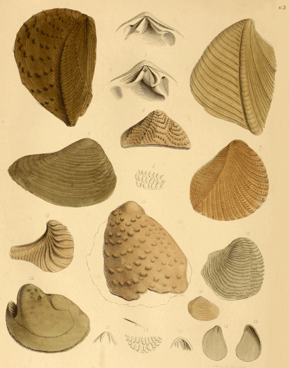
PLATE LXIII.
Trigoniæ.
Figs. 1, & 2, represent the structure of the hinge in both valves of a genus of bivalves of which numerous fossil species are met with in the secondary strata, and two or three species still exist in the Pacific Ocean. The genus is named Trigonia, from the form of the hinge, and the specific names below are those given by Mr. Parkinson.
Fig. 3. Trigonia clavellata, of Parkinson, from the Kimmeridge clay, Hartwell, Bucks.
Fig. 4. Trigonia costata, Oxford clay, Wilts.
Fig. 5. Trigonia excentrica; upper greensand, Blackdown. Like most of the shells from this locality, the Trigoniæ are transmuted into silex.
Fig. 6. Trigonia dædalea, Blackdown.
Fig. 7. —— spinosa, Blackdown.
Fig. 8. Enlarged view of the spines of the above.
Fig. 9. Trigonia alæformis, Blackdown.
Fig. 10. —— rudis, Blackdown.
Fig. 11. a bivalve shell of the genus Productus (P. antiquatus, of Sowerby?), from the Mountain limestone. See description of fig. 9, Plate LXVII.
Fig. 12. Cast of a species of Trigonia (T. clavellata), from the Portland rock. Many beds of this oolitic limestone are almost entirely made up of casts of Trigoniæ, and chiefly of this species.
Fig. 13. Trigonia sinuata, from Blackdown.
Figs. 14 to 18. "Different views of a species of Harpax."—Mr. Parkinson. (Plicatula spinosa). From the Lias, Gloucestershire.
Fig. 14. The inner surface of the flat valve.
Fig. 15. Inner surface of the convex valve.
Fig. 16. Magnified hinge teeth of the flat, and fig. 17, of the convex valve.
Fig. 18. Magnified view of the adpressed spines on the external surface of the shell.
Plate LXIV.
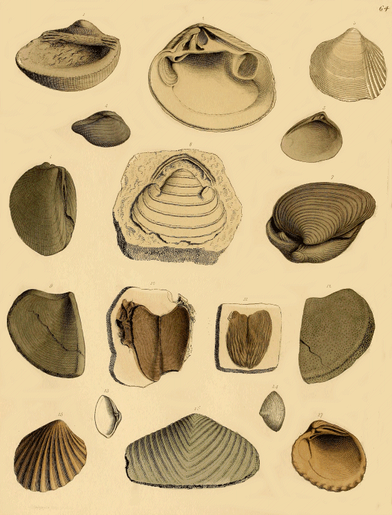
PLATE LXIV.
Fossil Shells.
Fig. 1. A perfect specimen of one valve, showing the character of the hinge of Cucullæa decussata, of Parkinson. London clay. Herne Bay.
Fig. 2. Interior view of Crassatella tumida, of Lamarck. Eocene strata, Paris.
Fig. 3. Cardium Hillanum, of Sowerby. A beautiful silicified bivalve from Blackdown.
Fig. 4. Nucula ovum, of Sowerby. A common bivalve, in the Lias, Yorkshire.
Fig. 5. Inner view of Cyrena deperdita, of Parkinson. Plastic clay, Woolwich.
Fig. 6. Lima gigantea, of Sowerby, from Lyme Regis. This is a young and small specimen of a large bivalve that occurs in great perfection in the Lias.
Fig. 7. Cardinia Listeri, of Sowerby. From the Lias, Gloucestershire.
Fig. 8. Cast of a bivalve; genus uncertain.
Figs. 9 to 12. These fossils are the Trigonellites of Mr. Parkinson; and have since been referred to a genus named Aptychus. Their true relations are very problematical. Though found in pairs, there is no hinge or natural connexion. Some naturalists suppose they may belong to the internal organization of Ammonites, because certain kinds have been found collocated with particular species of that genus of Cepholopoda. At present I do not think there is any satisfactory evidence as to their real nature. Species occur in the Kimmeridge clay, and other subdivisions of the Oolite formation.
Figs. 9, & 12. Trigonellites lata, of Mr. Parkinson.
Figs. 10, & 11. —— lamellosa.
Figs. 13, & 14. Corbida revoluta, of Sowerby. London clay, Highgate.
Fig. 16. An imperfect specimen of Lysianassa (Mya) literata, from the fullers' earth of the Oolite, Wiltshire.
Figs. 15, & 17. Cardita senilis, of Sowerby. From the Red crag of Suffolk.
Plate LXV.
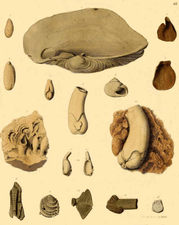
PLATE LXV.
Fossil Shells.
Fig. 1. A single valve, viewed interiorly, of a fine shell (Panopæa Aldrovandi, of Faujas St. Fond) from the Pleistocene or Newer Tertiary strata, that form a chain of low hills near Palermo, in Sicily. The shells in these deposits comprise almost all the genera and species that now inhabit the Mediterranean. They occur in the most beautiful state, deprived only of their colour; and groups are often met with of extreme elegance. The cabinet of the Marquess of Northampton contains an extensive and unrivalled series of these fossils, collected during his Lordship's residence at Palermo.
Figs. 2, & 4. A boring bivalve (Fistulana or Lithodomus) from the Oolite, Bath.
Figs. 3, & 5. Valves of a small Oyster from the Crag of Essex.
Fig. 6. A group of Lithodomi in limestone from the Oolite, Bradford, Wilts.
Fig. 7. A detached specimen from the same, showing the enclosed bivalve.
Figs. 8, & 10. Fine but imperfect specimens of a species of Teredo (Teredina personata, of Lamarck), from the Plastic clay of Epernay, France.
Fig. 9. A snail-shell (Helix arbustorum) found associated, and evidently contemporaneous, with bones of Mammoth, and extinct species of Deer, and other mammalia. From Brentford, in a bed of light calcareous earth, twenty feet below the surface.
Fig. 11. "A concamerated Teredo."—Mr. Parkinson. I am unable to ascertain the nature of this fossil.
Fig. 12. A species of Fistulana, from France.
Fig. 13. External surface of Chama squamosa of Brander. London clay, Hordwell.
Figs. 14, & 15, are the anchylosed caudal vertebræ of the tails of fishes. From the London clay, Isle of Sheppey.
Fig. 16. "A small oyster with a spathose structure."—Mr. Parkinson. This shell is probably the flat valve of a species of Dianchora, of Sowerby; from the Chalk.
Plate LXVI.
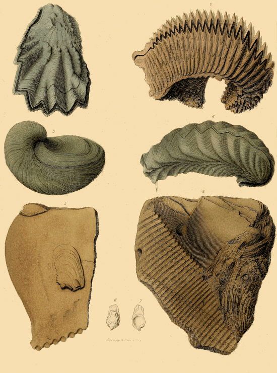
PLATE LXVI.
Fossil Bivalve Shells.
Fig. 1. A fossil Oyster (Ostrea Marshii, of Sowerby), from the Cornbrash of the Oolite, Wiltshire.
Fig. 2. The fossil Cockscomb Oyster, (Ostrea carinata, of Lamarck,) from the Lower chalk, Havre, France.
Fig. 3. The elegant fossil shell here figured is a peculiar and most abundant species in the Lias formation; specimens are not uncommon, in which every part of the shell is as perfect as if just thrown up on the sea-shore. It belongs to the genus Gryphites (Gryphea incurva, of Sowerby,) the shells of which are nearly related to the oysters, but are distinguished by the deep concave under-valve, and its curved beak, and the almost flat upper shell. The testaceous substance is of a finer laminated structure than in the Ostrea, and the hinge-ligament is inserted in an elongated curved groove.[61]
Fig. 4. "Ostrea vel frons folium."—Mr. Parkinson. This species appears to be the Ostrea gregarea (?) of Sowerby, which occurs in the chlorite marl or firestone of the Lower chalk in Sussex and Kent.
Fig. 5. The fossil is the cast of an oyster-like bivalve, called Perna, (Perna quadrata, of Sowerby,) which is easily recognisable, even in casts, by the line of distinct teeth which compose the hinge. This species is abundant in the Portland limestone, particularly in the quarries around Swindon, in Wiltshire; but from the close adhesion of the outer surface of the shell to the surrounding stone, they can seldom be extracted, the casts only being readily obtainable. In the Kimmeridge clay, which lies above the Portland rock, the shells may be met with in great perfection. The best locality is near Hartwell, in Buckinghamshire, where the clay is extensively dug for the brick manufactures.
Figs. 6, & 7. Two views of a small shell of the genus Crenatula, from Bedfordshire.
Fig. 8. Portion of a very large species of Perna (Perna maxillata, of Sowerby), from tertiary strata. Piedmont. The figure shows the inner surface of the shell with part of the broad crenulated hinge.
Plate LXVII.
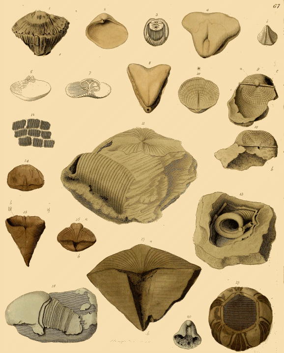
PLATE LXVII.
Fossil Shells of Brachiopoda, &c.
Fig. 1. A species of Radiolites (R. agariciformis, of M. D'Orbigny), from the Cretaceous strata of France. This genus is only known in a fossil state; it belongs to the same group of shells (order, Rudistes) as the Spherulites and Hippurites: the lower valve is conical, and much larger than the upper, which is slightly convex; it is deeply channelled longitudinally.
Fig. 2. Smooth valve of a species of Corbula (Corbula gallica, of Lamarck); abundant in some of the Eocene deposits of the Paris basin.
Fig. 3. A single valve; the inner surface is shown in the figure, of a remarkable genus of shells (Crania personata, of Lamarck), frequently occurring attached to Echinites and other bodies of the white chalk.
Fig. 4. A species of Terebratula (T. diphya, of Lamarck). The shells of this genus belong to that division of mollusks termed Brachiopoda (arm-feet), from their having internally two spiral fleshy arms developed from the sides of the alimentary orifice. These organs are supported by shelly processes, curiously modified in different genera, which often occur in a fossil state. Although the fossil Terebratulæ are very numerous, the recent species are but few, and are inhabitants of the seas off Australia. They form two natural groups; in the one the shells are smooth, but perforated all over with minute openings or foramina; and these are often filled with a dark substance, which is the carbonized soft parts: in the other division the shells are plicated or furrowed, and are not foraminiferous.[62] The Spirifers, another group of Brachiopoda, have a pair of internal spiral appendages.
Fig. 5. Terebratula coarctata, of Parkinson. Bradford clay, Wilts.
Figs. 6, & 7, show the internal structure of recent Terebratulæ from New Holland. The complicated shelly apophyses which supported the arms are quite perfect.
Fig. 8. Terebratula triquetra, of Parkinson (T. diphya, of Lamarck); another example of the species, fig. 4.
Figs. 9, & 10. Different parts of the same specimen of a brachiopodous bivalve belonging to the genus Productus, so named from the lengthened or produced form of the convex valve. "This is generally filled with limestone, which conceals the internal structure; but, with a slight blow, the shell divides, when the edge of the small valve rests against the inside of the produced cylindrical part of the larger one; generally about half an inch from the top of the shell: one side of the valve, before hidden, fig. 9 a, is then exposed, as shown in fig. 10."—Mr. Parkinson.
Fig. 9. a, the beak of the upper valve; c, a cavity in the superior part of the shell.
Fig. 10. The under part of the shell; b, a depression receiving the beak of the upper valve, a.
Fig. 10*. The inner surface of another upper valve, having a longitudinal fissure. The species figured is the Productus Martini of Mr. Sowerby. From the mountain limestone of Derbyshire; in which deposit numerous examples occur.
Fig. 11. a large species of Spirifer (Spirifer striatus, of Sowerby), from the mountain limestone of Derbyshire. In this species the upper valve is broken away, and one of the large spiral apophyses is seen lying imbedded in the limestone with which the cavity of the shell is filled.
Fig. 13, is a beautiful example of part of one of the spiral appendages of the same species.
Fig. 12. "A patch of square scales of a fish from Dorsetshire."—Mr. Parkinson. These evidently belong to a Lepidoid fish (Dapedius), whose remains are common in the Lias;[63] perfect specimens are often obtained. The British Museum contains some beautiful examples of this fossil fish.
Figs. 14, & 15. A curious fossil bivalve, from the Devonian strata of the Eifel. The flat valve is shown in fig. 14; and the deep conical valve in fig. 15; a, tooth in the posterior margin; b, a part of the surface magnified, to show its cellular structure. The species is Calceola sandalina, of Lamarck.
Fig. 16. A species of Spirifer; a, medial convexity of the upper valve; b, the triangular foramen at the beak.
Fig. 17. Spirifer (S. cuspidatus, of Mr. Martin), from the Mountain limestone of Derbyshire.
Fig. 18, represents a common appearance in certain chalk flints. Although I have examined hundreds, and some in which the form was more definite than in the specimen figured, I am not able to offer any probable suggestion as to their origin, should they be organic bodies, of which there is much doubt.
Fig. 19. "Coronulites diadema."—Mr. Parkinson. Probably a species of Balanus, from a tertiary deposit.
Fig. 20. Cast of one of the shells of a bivalve (Pentamerus), from the Wenlock limestone of Dudley.
Plate LXVIII.
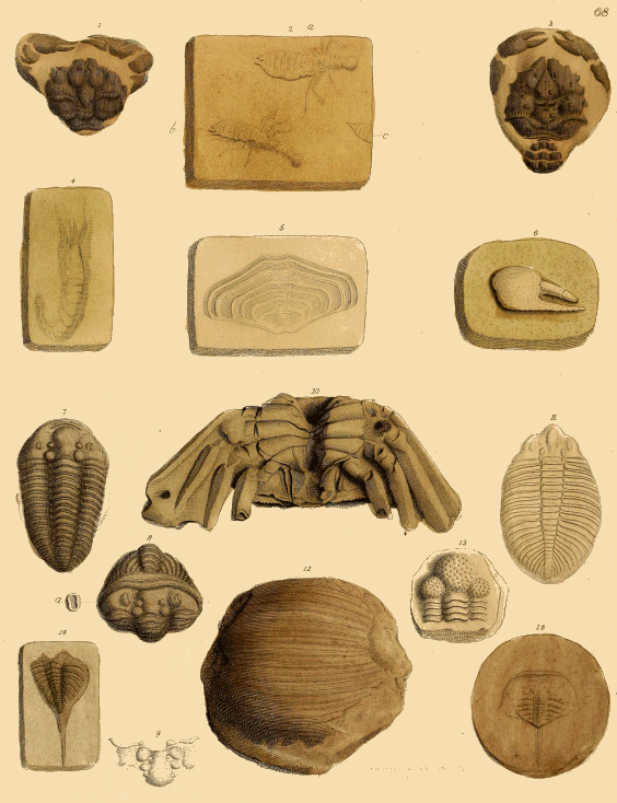
PLATE LXVIII.
Fossil Crustacea.
Figs. 1, & 3. "Fossil Crabs, from Sheppey."—Mr. Parkinson. The London clay of this celebrated locality contains an abundance of the fossil remains of Crustacea; and the visitor may purchase of the local collectors fossil crabs and lobsters, as readily as the recent species from the neighbouring sea. Good specimens are however rare, and command high prices. The specimens figured are two common species.
Fig. 1. Cancer Leachii, of MM. Desmarest and Brongniart.
Fig. 3. Inachus Lamarckii.
These fossils show the usual mode in which the crustaceæ occur in the hardened clay of Sheppey. The thorax is bent over the abdomen, and the pair of large chelate claws drawn towards each other.
Fig. 2. Fossil Insects from the lithographic stone of Pappenheim. "a, an insect with a bifurcated caudal extremity; b, the sting which has passed out of its sheath; c, the termination in a single point."—Mr. Parkinson.
Fig. 4. "A fossil Shrimp, from Anspach."—Mr. Parkinson.
Fig. 5. "Impression of an unknown fossil."—Mr. Parkinson.
Fig. 6. "The claw of a Crab, from Maestricht, &c."—Mr. Parkinson. Claws of this kind are frequent in the soft sandy limestone of St. Peter's Mountain, but no other vestiges of the Crabs to which they belonged have been met with. The cause of this has been ascertained: the claws belong to a species of Hermit Crab (Pagurus Faujasii, of Desmarest), which like the living species had the body covered by a delicate membrane, the claws only possessing a durable crustaceous shell.[64]
Fig. 7. "An extended trilobite, from Dudley."—Mr. Parkinson. Among the organic remains of the inhabitants of the seas, in whose abysses were formed the Silurian, Devonian, and other ancient sedimentary strata, an extinct family of crustaceans, comprising numerous genera, are among the most characteristic and remarkable. The name "Trilobite," first given by Mr. Parkinson, expresses the most obvious character of the longitudinally trilobed, convex, segmented, carapace of the body, of the most common forms; but so great is the number of species, and so dissimilar the groups, now known, that the nomenclature of this class of fossils is greatly extended. In Sir R. I. Murchison's splendid work on the Silurian System, the genera and species of the formations therein comprised are beautifully illustrated. The specimen figured is an expanded specimen of the species commonly known as the Dudley Locust or Insect, (Calymene Blumenbachii), from the Wenlock limestone, Dudley.
Fig. 8. A coiled-up specimen; in this view are seen both ends of the crustaceous covering of the animal: a, "the eye enlarged."
Fig. 9, is part of the head of the same species.
Fig. 10. "A fossil Crab from the East Indies."—Mr. Parkinson. Beautiful specimens of this species of Crab (Gonoplax Latreilli, of Mr. Edwards) have been obtained from the tertiary strata of India.
Fig. 11. Another form of Trilobite (Ogygia Buchii, (Asaphus,) of the Silurian System), from the Llandeilo flagstones.
Fig. 12. "Remains of some large unknown insect."—Mr. Parkinson. This figure is not sufficiently defined to admit of interpretation.
Fig. 13, "Part of a trilobite with tuberculated head," (Calymene variolare,) from the Wenlock limestone, of Dudley.
Fig. 14. Posterior part of a trilobite with a caudal style or process, (Asaphus caudatus,) from the Wenlock shale, Dudley.
Fig. 15. A nodule of ironstone from Coalbrook Dale, in which is imbedded a small crustacean allied to the recent King Crab or Limulus; a genus abundant in the seas of India and America.[65] (Limulus trilobitoides, of Dr. Buckland. Bellinurus bellulus, of Mr. König.)
Plate LXIX.
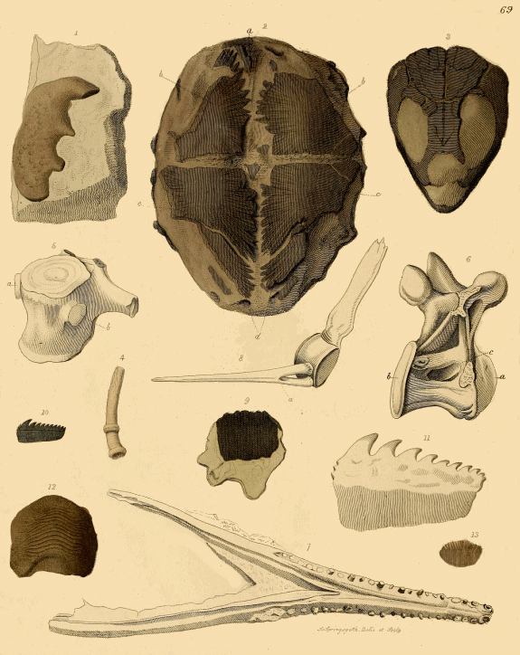
PLATE LXIX.
Fossil Fishes and Reptiles.
Fig. 1. "A fossil body resembling part of a Tortoise, from Gloucestershire."—Mr. Parkinson. This specimen is probably one of the mandibles of a remarkable extinct genus (Ceratodus) of fishes, whose dental organs, like those of the recent Chimæra, consisted of consolidated plates instead of separate teeth; each side of the jaw was formed by one of these mandibular processes; the upper margin is deeply undulated. The bone-bed of the Lias at Aust Cliff near Westbury, Somersetshire, is rich in these remains.
Fig. 2. The plastron, or inferior aspect of the carapace of a fossil Turtle (Chelonia breviceps), from the London Clay of the Isle of Sheppey. a, fragment of the entosternal plate; b, b, hyosternal plates; c, c, hyposternals; d, xiphisternals.[66]
Fig. 3. The cranium of the same species of Turtle, from the Isle of Sheppey. Equally rich in the remains of Chelonian reptiles, as in those of Fishes, Crustaceans, Serpents, and Mollusks, the little Island at the mouth of the Medway has yielded to the indefatigable researches of Mr. Bowerbank the most extensive series of fossil Turtles hitherto discovered in England. The various genera and species will be figured and described in a work now in progress by Professors Bell and Owen, under the auspices of the Palæontographical Society.
Fig. 4. A Serpula (S. antiquata ?), from the chalk, Sussex.
Fig. 5. A dorsal vertebra of a fossil crocodilian reptile (Steneosaurus), from the Oxford Clay of Honfleur. a, b, costal depressions.
Fig. 6. A dorsal convexo-concave vertebra of a crocodilian or gavial-like reptile (Streptospondylus), from the same locality. This figure shows the remarkable character whence the name of this genus: the convexity of the body of the vertebra (a) being situated anteriorly as in mammalia, the reverse of the position of the bones forming the vertebral column in the existing Crocodilians and Lacertians. b, the posterior concavity; c, a deep depression beneath the neural arch.
Fig. 7. Sketch of the lower jaw of an extinct gavial-like reptile (Steneosaurus): the vertebra, fig. 5, probably belongs to the same species. From Honfleur. This figure, and figs, 5, 6, and 8, are copied from Cuvier, "Annales du Muséum"
Fig. 8. A caudal vertebra of the Fossil Animal of Maestricht (Mosasaurus); a, the chevron bone or inferior spinous process (hœmapophysis), anchylosed to the middle of the body of the vertebra.
Fig. 9. Fossil scale of a ganoid fish (probably Lepidotus), from Kent.
Fig. 10. Fossil tooth of a fish of the Shark family (Notidanus microdon, of Agassiz,) from the chalk of Kent.
Fig. 11. Recent "tooth of one of the Dog-fish," (Mr. Parkinson,) for comparison with fig. 10.
Fig. 12. Tooth of an extinct group of squaloid fishes (Ptychodus decurrens, of Agassiz,) from the chalk of Kent.[67]
Fig. 13. a ctenoid (or comb-like) scale of a fish, (probably of a species of Beryx,) from the chalk of Kent.
Plate LXX.
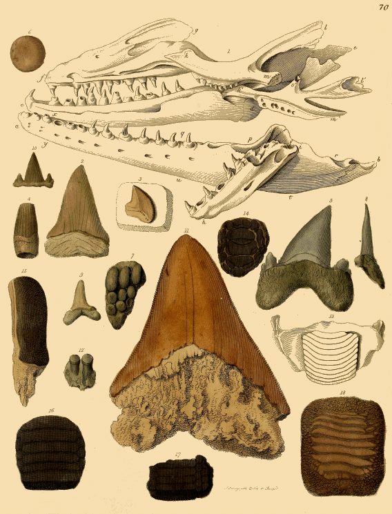
PLATE LXX.
Fossil Reptiles and Fishes.
Fig. 1. A reduced figure of the celebrated specimen of the jaws, &c. of the "Fossil Animal of Maestricht," (Mosasaurus Hoffmani,) from the cretaceous strata of St. Peter's Mountain. See "Supplementary Notes," art. Mosasaurus.
"a, b. The left side of the lower jaw, nearly whole, and seen on its outer side.
c, d. Right side of the lower jaw, viewed on the inner side, the posterior part of which, a little concealed by the palate bones, is continued to e.
f, g. The right side of the upper jaw, seen on its inner side, and with the palate bone. This part is nearly in its natural position in relation to the corresponding ramus of the lower jaw.
h, i. A fragment of the left side of the upper jaw, displaced and fallen across the lower jaw.
k, l, m; k', l', m', o'. The two palate bones displaced and thrown one over the other, and also over the right side of the lower jaw. In the original specimen a portion of bone is placed from m to p, and another at q, which are omitted to render the figure more intelligible."—Mr. Parkinson.
Figs. 2 to 18, are fossil teeth of various kinds of fishes, principally of the Shark and Ray families.
Fig. 2. Tooth of a shark (Lamna), from Malta.
Fig. 3. Tooth of a shark (Galeus pristodontus), chalk marl, Kent.
Fig. 4. Tooth of a Saurian, the upper and lower end imperfect: probably of a species of Steneosaurus, from Bath.
Figs. 5, & 8. Teeth of a shark (Otodus,) London Clay, Isle of Sheppey.
Fig. 6. Tooth of a fish, (Spherodus,) from the Oolite, Gloucestershire.
Fig. 7. Part of the fossil jaw with three rows of teeth of a fish, (of the Pycnoid[68] family,) from the Oolite, Gloucestershire.
Fig. 9. Tooth of a species of Lamna, from Sheppey.
Fig. 10. Tooth of a species of Hybodus,[69] Stonesfield.
Fig. 11. A very large tooth of a Shark, (Carcharias megalodon,) from the tertiary deposits of Malta.
Fig. 12. Fragment of a bone, with two teeth, probably of a species of Pycnodus.
Fig. 13. "The mandible and tooth of a recent fish (Diodon), to compare with the fossils figs. 16, and 17."—Mr. Parkinson.
Fig. 14. "Fossil palate of a fish, from Sheppey."—Mr. Parkinson. This evidently belonged to a species of Ray; possibly to the Eagle rays (Miliobatis).
Fig. 15. Tooth of a fish allied to the Cestracionts, or Port Jackson Shark, (probably of the genus Acrodus,[70]) from Bath; commonly called "Leech palates" by the quarry-men.
Figs. 16, & 17. "Fossil palates of fishes of the Ray kind, from Sheppey."—Mr. Parkinson. These appear to belong to the Miliobates (M. micropleuris, of Agassiz). Beautiful examples of these fossils have been obtained from the Bracklesham clay, on the coast of the West of Sussex. The late Frederic Dixon, Esq. of Worthing, whose untimely death is so much to be deplored, had a matchless suite of specimens from that locality.