Chapter XXIII · GENERAL ACCOUNT OF THE SKELETON IN MAMMALIA (continued). (continued 2)
Reading edition
In many Rodentia the ilia have their gluteal, iliac, and sacral surfaces of nearly equal extent; in the Hares, however, the gluteal and iliac surfaces are confluent. The pubes and ischia are always well developed and sometimes, as in the Hares, the acetabular bone also. In these animals the pubis does not take part in the formation of the acetabulum, and the ischium bears on its outer side a well-marked ischial tuberosity.
In the Carnivora the pelvis is long and narrow. The iliac surfaces (fig. 78, A, 5) are very small and the sacral large; the crest or supra-iliac border is formed by the union of the sacral and gluteal surfaces. The symphysis is long and includes part of both pubis and ischium. The ischial tuberosity (fig. 78, A, 10) is often well marked, and sometimes as in Viverra the acetabular bone is distinct. In the Pinnipedia the pelvic symphysis is little developed, or sometimes not developed at all, and the obturator foramina are remarkably large.
In some Insectivora such as Galeopithecus, there is a long pelvic symphysis, in others such as Erinaceus and Centetes, it is very short, in others again such as Talpa and Sorex, there is no pelvic symphysis. The acetabular bone is exceptionally large in Talpa and Sorex.
In Chiroptera the pelvis is small and narrow, and in the great majority of cases the two halves do not meet in a ventral symphysis. The pubis has a strongly developed pectineal process, which occasionally unites with a process from the ilium enclosing a large pre-acetabular foramen.
Primates. In Man and the Anthropoid Apes the pelvis is very large and wide, and the ilium has much expanded iliac and gluteal surfaces. The symphysis is rather short and formed by the pubis alone. The acetabulum is deep and the obturator foramen large, and there is frequently a well-marked ischial tuberosity. In the lower Anthropoidea the ilium is long and narrow and has a small iliac surface. The ischial tuberosities are large in the old world monkeys.
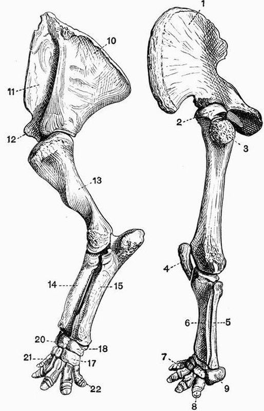
Fig. 108. Left anterior and posterior limb and limb girdle of
Uintatherium mirabile. The anterior limb is to the left, the posterior
to the right × 1/10. (From casts, Brit. Mus.)
1. ilium.11. prescapular fossa.
2. head of femur.12. coracoid process.
3. great trochanter.13. humerus.
4. patella.14. radius.
5. fibula.15. ulna.
6. tibia.17. unciform.
7. second digit of pes.18. cuneiform.
8. ungual phalanx of fifth20. lunar.
digit of pes.21. first metacarpal.
9. calcaneum.22. fifth metacarpal.
10. postscapular fossa.
The Thigh and Shin.
In the Monotremata the femur is short, rather narrow in the middle, and expanded at each end. The great and lesser trochanters are large and about equally developed, but there is no third trochanter. The fibula is very large and is expanded at its proximal end, forming a flattened plate much resembling an olecranon. The patella is well developed.
In the Marsupialia there is no third trochanter to the femur, the fibula is well developed but not the patella as a general rule. Notoryctes has a femur with a prominent ridge extending some little way down the shaft from the great trochanter; the tibia has a remarkably developed crest, and the fibula has its proximal end much expanded and perforated; there is an irregularly shaped patella closely connected with the proximal end of the tibia.
Edentata. In the Sloths the leg bones are all long and slender. The femur has no third trochanter, and the fibula is complete and nearly equal in size to the tibia. In the Megatheriidae the leg bones are extraordinarily massive, the circumference of the shaft of the femur in Megatherium equalling or exceeding the length of the bone. There is no third trochanter in Megatherium. In most of the remaining Edentata the leg bones are strongly developed. The femur in the Armadillos and Aard Varks has a strong third trochanter, and the tibia and fibula are both large and are commonly ankylosed together at either end. The limb bones are very massive also in the Glyptodonts.
Sirenia. In no living Sirenian is there any trace of a hind limb, but in Halitherium a vestigial femur is found, which articulates with the pelvis by a definite acetabulum.
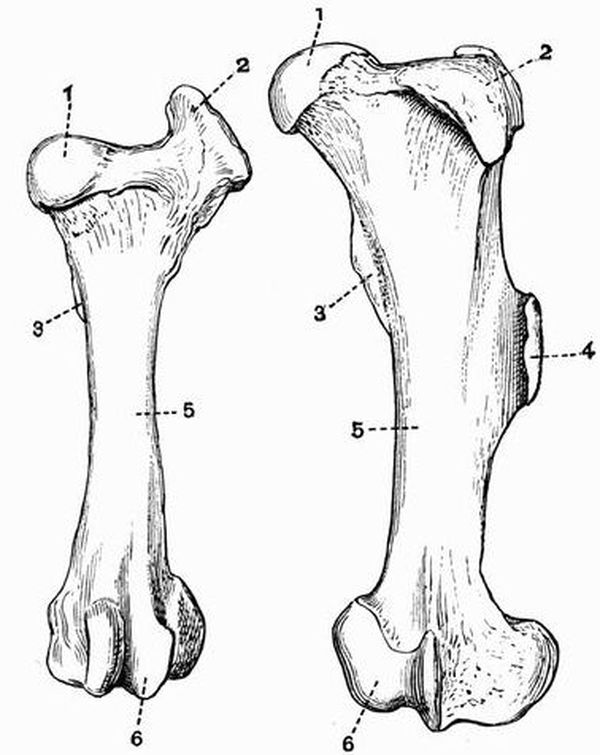
Fig. 109. Left femur of an Ox (Bos taurus) (to the left) and of
a Sumatran Rhinoceros (R. sumatrensis) (to the right). × 1/6.
(Camb. Mus.)
1. head.4. third trochanter.
2. great trochanter.5. shaft.
3. lesser trochanter.6. condyles.
In the Mystacoceti among the Cetacea small nodules of bone or cartilage occur connected with the vestigial pelvis, and may represent the femur and tibia. No trace of the skeleton of the hind limb is known in the Odontoceti.
In the Ungulata vera the femur is noticeable for the size of the great trochanter (fig. 109, 2); there is no definitely constricted neck separating the head from the rest of the bone, and the lesser trochanter (fig. 109, 3) is not very prominent. All Perissodactyles except the Chalicotheriidae show a strongly marked third trochanter, but this is absent in all known Artiodactyles. The development of the fibula in general corresponds to that of the ulna. In Rhinoceros, Macrauchenia, Tapirus and the Suina it is distinct and fairly well developed; in the Tragulina on the other hand it is vestigial, being reduced to the proximal end only. In the Ruminantia and Tylopoda also, it is much reduced forming merely a small bone attached to the distal end of the tibia, sometimes, as in the Red deer a slender vestige of the proximal end also is preserved quite detached from the distal portion; in the Horse this proximal portion is all that there is found of the fibula. The progressive diminution of the fibula can be well seen in the series of forms that are regarded as the ancestors of the Horse. The patella of the Ungulata vera is well ossified, but fabellae[177] are not usually found.
Subungulata. Of the Toxodontia, Toxodon has no third trochanter while Typotherium and Astrapotherium have one. In the Condylarthra the femur has well-marked lesser and third trochanters, and the fibula and patella are well developed. In the Hyracoidea there is a slight ridge on the femur in the place of the third trochanter, the fibula is complete, but is generally fused to the tibia at its proximal end.
Of the Amblypoda, Coryphodon has a third trochanter, but Uintatherium has none; in this respect, in the vertical position and general appearance (fig. 108) of the limb, and in the articulation of the fibula with the calcaneum, the leg of Uintatherium closely approaches that of the Proboscidea.
In the Proboscidea the femur is very long and straight, the development of trochanters is slight, and the fibula though slender is complete and articulates with the calcaneum.
A third trochanter is found in the Tillodontia.
In Rodentia the femur is variable, the great trochanter is generally large and so sometimes is the third as in the Hares. In most Rodents as in the Beaver the fibula is distinct, sometimes as in the Hares it is united distally with the tibia. The patella is well developed, and so too are the fabellae as a general rule.
Carnivora. In the Carnivora vera the femur (fig. 79, A) is generally rather straight and slender, and has a very distinct head. The fibula (fig. 79, C) is always distinct and there is generally a considerable interval between it and the tibia. Fabellae (fig. 79, 7) are commonly present.
In the Pinnipedia the femur is short, broad and flattened, having a prominent great trochanter. The fibula is nearly as large as the tibia, and the two bones are generally ankylosed together at their proximal ends.
The Creodonta differ from all living Carnivores in having a femur with a third trochanter.
In the Insectivora a third trochanter is sometimes developed. The fibula is sometimes distinct, sometimes fused distally with the tibia, thus differing from that of a Carnivore.
In Chiroptera the femur is straight, slender and rather short, with a small but well-developed head. The fibula may be well developed or quite vestigial or absent. Owing to the connection of the hind limb with the wing membrane the knee joint is directed backwards.
In Primates the femur is rather long and slender, having a nearly spherical head and large great trochanter. The tibia and fibula are always distinct and well developed. Fabellae are not found in the highest forms but are generally present in the others.
The Pes.
The skeleton of the pes is in most respects a counterpart of that of the manus. Just as in the manus if one digit is absent it is the pollex, so in the pes it is the hallux. But while in the manus the third digit is always well developed, however much the limb may be modified, in the pes any of the digits may be lost. In all mammals the tibiale and intermedium fuse to form the astragalus, and the fourth and fifth tarsalia to form the cuboid. Sesamoid bones are considerably developed. In almost every case the phalanges and first metatarsal have epiphyses only on their proximal ends, while the remaining four metatarsals have epiphyses only on their distal ends.
In the Monotremata all the usual tarsal bones are distinct, and the five digits have the normal number of phalanges. Several sesamoid bones are developed, the most important one, found only in the male, being articulated to the tibia and bearing the curious horny spur. The ungual phalanges of the pes like those of the manus, are deeply cleft at their extremities. In the Echidnidae the pes is turned outwards and backwards in walking.
In the Marsupialia the pes is subject to great modifications, but in every case the seven usual tarsal bones are distinct. In the Didelphyidae the foot is broad, all five digits are well developed, and the hallux is opposable to the others. In the Dasyuridae the foot is narrow, and the hallux may be very small, or as in Thylacinus completely absent. In Notoryctes the pes is much less abnormal than the manus, and all five digits have the usual number of phalanges. The fifth metatarsal has a curious projecting process, and there is a large sesamoid above the hallux. In the Wombats (Phascolomyidae) the foot is short and broad, the digits are all distinct, and the hallux is divaricated from the others.
In the remaining marsupials the second and third metacarpals and digits are very slender, and are enclosed within a common integument. This condition is known as syndactylism, and its effect is to produce the appearance of one toe with two claws. In the Kangaroos (Macropodidae) the pes is very long and narrow, owing to the elongation of the metacarpals. The fourth digit is greatly developed, the fifth moderately so, while the hallux is absent, and the second and third digits are very small. The Peramelidae have the foot constructed on the same plan as in the Kangaroos, and in one genus Choeropus the same type of foot is carried to a greater extreme than even in the Kangaroos. Thus the fourth digit is enormously developed, the second and third are small, and the fifth smaller still, while the hallux is absent. In the Phalangers and Koalas though the second and third toes are very slender, the hallux is well developed and opposable.
Edentata. In the Sloths the pes much resembles the manus, being long and narrow, but in both genera the second, third and fourth digits are well developed. Most of the other Edentates have a but little modified pes with the normal number of tarsal bones and the complete series of digits. In Cycloturus however the hallux is vestigial and it is absent in Glyptodonts. Megatherium has a greatly modified pes, the hallux is absent, and the second digit vestigial, while the third is very large, having an enormous ungual phalanx. The calcaneum too is abnormally large.
No trace of the pes occurs in either Sirenia or Cetacea.
In the Ungulata the pes like the manus is subject to much variation and is of great morphological importance.
In the Ungulata vera the pes is never plantigrade and never has more than four digits, the hallux being absent. The cuboid always articulates with the astragalus, and the tarsal bones strongly interlock. As was the case also with the manus, the pes is formed on two well-marked types characteristic respectively of the Artiodactyla and Perissodactyla.
Artiodactyla. Just as in the manus, the third and fourth digits are well and subequally developed; their ungual phalanges have the contiguous sides flat, and the axis of the limb passes between them, and between the cuboid and navicular. The astragalus has both the proximal and distal surfaces pulley-like, and articulates with the navicular and cuboid by two facets of nearly equal size. The calcaneum articulates with the lower end of the fibula if that bone is fully developed.
In the Suina four toes are developed, and though in the Peccaries the third and fourth metatarsals are united, they are all distinct in most members of the group, as are all the tarsal bones. In the Hippopotami the four digits are of approximately equal size, and the middle ones do not have the contiguous faces of their ungual phalanges flattened.
In the Tragulina the cuboid, navicular, and two outer cuneiforms are united forming a single bone; all four metatarsals are complete and the two middle ones are united. In the Tylopoda and Anoplotherium commune only the third and fourth digits are developed, their metatarsals are free distally, but are elsewhere united. In the Ruminantia the cuboid and navicular are always united and so are the second and third cuneiforms, while in Cervulus all four bones are united together. The third and fourth metatarsals in Ruminants are always united in the same way as are the third and fourth metacarpals, while the second and fifth are always wanting. In Deer the second and fifth digits are usually each represented by three small phalanges, but in the Giraffe and most Bovidae the bones of these digits are wanting.
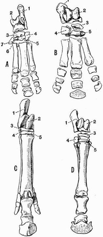
Fig. 110. A. Left pes of a Tapir (Tapirus americanus). × 1/6.
B. Right pes of a Rhinoceros (R. sumatrensis). × 1/8.
C. (Cast of) right pes of Hipparion gracile. × 1/7.
D. Right pes of a Horse (Equus caballus). × 1/10. (All Camb. Mus.)
1. calcaneum.5. external cuneiform.
2. astragalus.6. middle cuneiform.
3. navicular.7. internal cuneiform.
4. cuboid.
In the Perissodactyla the pes like the manus is symmetrical about a line drawn through the third digit; this line when continued passes through the external cuneiform, navicular and astragalus. The astragalus has its distal portion abruptly truncated, and the facet by which it articulates with the cuboid is much smaller than that by which it articulates with the navicular. The calcaneum does not articulate with the fibula. The tarsus in Macrauchenia like the carpus differs from that of other Perissodactyles and resembles that of Subungulates in having the bones arranged in lines with little or no interlocking. The calcaneum resembles that of Artiodactyles in having a small facet for articulation with the fibula. Tapirus (fig. 110, A), Rhinoceros (fig. 110, B) and Titanotherium have a short and broad foot with the usual tarsal bones and three well-developed digits,—a number never exceeded by any Perissodactyle. From this tridactylate limb a series of stages is exhibited by various extinct forms leading gradually to the condition met with in the Horse (fig. 110, D) in which the third toe is greatly developed, while the second and fourth are reduced to slender metatarsals attached to the proximal half of the third metatarsal.
In Chalicotherium and Agriochoerus the pes has the same abnormal characters as the manus, the digits being clawed and the ungual phalanges in Chalicotherium deeply cleft.
In the Subungulata the pes is sometimes plantigrade and pentedactylate, the cuboid sometimes does not articulate with the astragalus, and the tarsal bones sometimes do not interlock.
In Typotherium (Toxodontia) the hallux is absent and the other four digits are well developed; in Toxodon and Nesodon the pes is tridactylate. The tarsal bones have the regular Subungulate arrangement, the cuboid not articulating with the astragalus. The calcaneum articulates with the fibula as in Artiodactyles. The astragalus in most forms, but not in Astrapotherium, resembles that of the Ungulata vera in having a grooved proximal surface.
In Phenacodus (Condylarthra) the tarsus is very little modified, five digits are present, the first and fifth being small and not reaching the ground.
In Procavia only the three middle digits are present with a vestige of the fifth metacarpal.
In the Amblypoda the pes (fig. 108) is very short and broad, all five digits are functional, and at any rate in Coryphodon plantigrade, the hallux being the smallest. The astragalus is very flat, and the tarsals interlock to a slight extent, the cuboid articulating with both calcaneum and astragalus.
The pes in the Proboscidea much resembles that in the Amblypoda, but differs in that the astragalus does not articulate with the cuboid, the tarsals not interlocking at all.
In the Rodentia the structure of the foot is very variable. In Beavers the foot is very large, all five digits being well developed; the fifth metatarsal articulates with the outer side of the fourth metatarsal, and not with the cuboid, and there is a large sesamoid bone on the tibial side of the tarsus. In the Rats, Porcupines and Squirrels, there are five digits, in the Hares only four, and in the Capybara and some of its allies only three. In the Jerboa (Dipus) a curious condition of the pes is met with, as it consists of three very long metatarsals fused together and bearing three short toes, each formed of three phalanges. Lophiomys differs from all other Rodents in having the hallux opposable.
Carnivora. In the Carnivora vera the pes is regular and shows little deviation from the normal condition. All the usual tarsal bones are present, but sometimes as in the Dogs, Cats, and Hyaenas, the hallux is vestigial. Sometimes as in the Bears the pes is plantigrade, sometimes as in the Cats and Dogs it is digitigrade. In this respect and in the character of the ungual phalanges, the pes closely corresponds with the manus. In the Sea Otter (Latax) the foot is large and flattened and approaches in character that of the Pinnipedia.
In the Pinnipedia the pes differs much from that in the Carnivora vera. In the Seals in which the foot cannot be used for walking, and is habitually directed backwards, the first and fifth digits are much longer and stouter than any of the others. In the Sea Lions which can use the pes for walking, the digits are all of nearly the same length, and in the Walrus the fifth is somewhat the longest.
In the Insectivora the pes is almost always normal, and provided with five digits.
In the Chiroptera the pes is pentedactylate, and the digits are terminated by long curved ungual phalanges. In some genera the toes have only two phalanges. The calcaneum is sometimes produced into a long slender process which helps to support the membrane between the leg and the tail.
Among the Primates Man has the simplest form of pes. In Man all five digits are well developed, the hallux being considerably the largest. Sesamoid bones occur only under the metatarso-phalangeal joint of the hallux.
In the other Primates the internal cuneiform has a saddle-shaped articulating surface for the hallux, which is obliquely directed to the side of the foot and opposable to the other digits. Two sesamoid bones are usually developed below each metatarso-phalangeal joint, and one below the cuboid. The second digit in Lemurs, and all except the hallux in Chiromys have pointed ungual phalanges; in all other cases the ungual phalanges are flat. In some of the Lemuroidea, especially Tarsius, the tarsus is curiously modified by the elongation of the calcaneum and navicular.