Chapter XI · TUMOURS OF THE SKULL-BONES
Reading edition
In this the last chapter of this work, I originally intended to deal with tumours of the scalp and skull-bones. With respect to tumours of the scalp there is, however, but little to say. The various conditions are well recognized, their pathology is known, and there is in general but little to relate. Tumours of the skull-bones come under a different category and require some consideration.
Tumours of the skull-bones.
The more important of these tumours are as follows:—
Osteomata.
Sarcomata, primary and traumatic.
Secondary sarcomata and carcinomata.
OSTEOMATA
Exostoses of the skull, though by no means of frequent occurrence, occupy such definite positions that it would appear as if they were dependent on some local governing cause. They develop most frequently in the following positions:—
At the external angular frontal process.
At the frontal and parietal eminences.
In the region of the frontal sinus.
In the region of the external auditory meatus and mastoid process.
All pathological museums possess specimens illustrating the formation of such exostoses, tumours of a like nature in other parts of the skull being excessively rare.
In endeavouring to arrive at an explanation it would appear necessary to direct one’s attention to tumours of a similar nature occurring in other parts of the body, more especially in the long bones. There, it is well recognized that their development is dependent on irregularities of growth in the region of the epiphyseal lines, regions where activity of growth is long maintained.
On referring again to the skull, similar features appear. For instance, the frontal bone, besides possessing one primary centre for the frontal eminence, has secondary centres for the external angular frontal process, for the trochlear fossa, and for the nasal spine, and it is highly probable that those exostoses which develop in the region of the frontal sinus in reality arise from one or other of the secondary centres situated in that region.
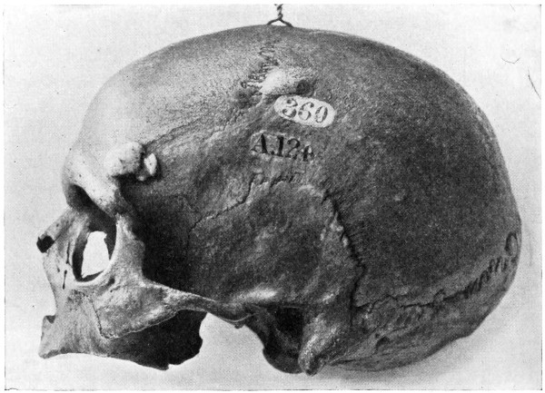
Fig. 92. Ivory Exostoses of the Skull.
Both frontal and parietal eminences are also sites of active and prolonged ossification, and the tumours there arising are to be explained on a like hypothesis.
Similar features are to be observed with respect to those bony tumours which develop in the aural region, the numerous centres of ossification for the periotic capsule accounting satisfactorily for their origin.
Whether originating in the region of the frontal sinus or in the aural area, the tumour naturally develops along the line of least resistance, filling up the frontal sinus and growing into the external auditory meatus and mastoid antrum.
More rarely, small exostoses develop on the inner aspect of the skull, chiefly from the frontal bone in the region of the crista galli. In some cases the inner aspect of the skull is studded with small bony tumours, more especially along the line of the superior longitudinal venous sinus.
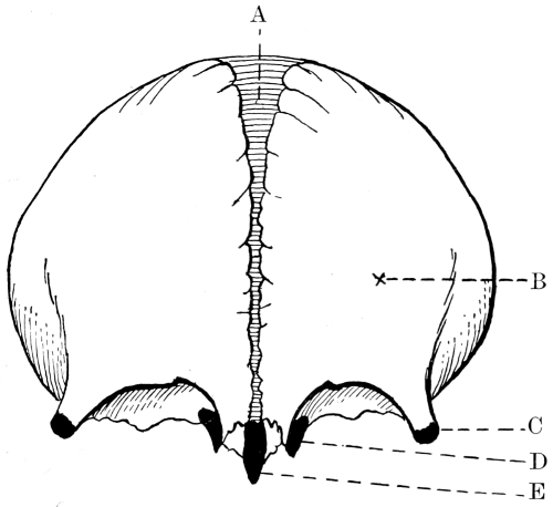
Fig. 93. The Development of the Frontal Bone. A, Metopic suture; B, Primary centre for frontal eminence; C, Secondary centre for external angular frontal process; D, Secondary centre for trochlear fossa; E, Secondary centre for nasal spine.
These internal exostoses seldom give rise to pressure symptoms, although, according to Wilks and Moxon,[77] they may push inwards the dura mater and even lead to idiocy and epilepsy. I have seen several cases of internal exostosis development, but in all cases their discovery was accidental.
Clinical characteristics.
Exostoses vary greatly both in size and consistency. Some are densely hard—ivory exostoses—others possess a covering of compact bone, whilst their interior is made up of cancellous tissue continuous with that of the bone from which they arise. The denser variety seldom attain any considerable size, but the less compact, growing in the direction of least resistance, often attain such dimensions as to be both unsightly and dangerous. Thus, a frontal exostosis may invade the frontal air sinus and grow into the orbital cavity, obliterating the sinus, interfering greatly with ocular movements, causing protrusion of the globe and even destruction of the eye.
An aural exostosis may block up the external auditory meatus, compress the facial nerve, and lead to the development of a mastoid empyema.
It might also be added that there are a few cases on record in which a frontal exostosis, by reason of extensive inward growth, has produced cerebral symptoms—general compression and intellectual deterioration.
Treatment.
In considering the question of treatment, it must be accepted that, although of slow growth, some of these exostoses are definitely progressive, tending to interfere with the character and functions of the region with which they are anatomically situated. There is also reason to believe that those secondary changes—sarcomatous, myxomatous, &c.—which are occasionally observed in the exostoses of long bones are also liable to develop in those cranially situated. The question of treatment hinges, therefore, to a large extent on the nature and position of the tumour.
When of the ivory type and growing from the flat bones of the skull, but so situated that no marked deformity or pressure symptoms are likely to ensue, they may be left alone, but when definitely progressive and situated in accessible regions, they should be removed. Their exposure is carried out by the formation of a suitable scalp-flap—designed as far as possible so as to be subsequently hidden by the hairy scalp—and the exostosis removed by the application to its base of a Gigli saw. This method is greatly superior to the older procedures whereby the tumour was chiselled away with hammer and gouge.
Occasionally the tumour is so dense and presents so wide a basal attachment that it becomes necessary to attack from a more distant line, cutting out a trench, deepened to the diploic tissue, circumferentially around the tumour and levering away the central mass. When the tumour extends more deeply, involving nearly the whole thickness of the skull, it may be removed by the application of a small trephine immediately to one side of the tumour, followed by the use of de Vilbiss forceps circumferentially around the main mass, thus freeing it from its surroundings. The resultant gap in the skull may be protected by one or other of those measures enumerated in Chapter VI.
Frontal and mastoid exostoses often necessitate formidable operations insomuch as their size and anatomical relations present considerable difficulties (see Fig. 94).
The indications for operation in the case of aural exostoses are as follows[78]:—
1. If there is middle-ear suppuration and signs of retention of pus.
2. When the pressure of the exostosis produces pain which cannot otherwise be relieved.
3. If the exostosis nearly blocks up the meatus of both ears, and there is prospect of each side becoming completely blocked in the near future. Here operation is carried out on the worst side.
4. If the meatus is nearly blocked by the exostosis and the patient going to a country where he cannot be within easy reach of a competent medical man.
The indications for operation in the case of a frontal exostosis are as follows:—
1. When the exostosis interferes with the actions of the ocular muscles, causes proptosis and threatens the integrity of the globe.
2. When associated with pain which cannot otherwise be relieved.
3. When the exostosis leads to blockage of the accessory sinuses of the nose, more especially when such blockage is associated with pus pent up within.
4. When very unsightly.
The accompanying figure illustrates the deformity and dangers associated with large frontal exostoses. The tumour developed from the inner angle of the orbital cavity, pushing the globe forwards and outwards, with diplopia and severe neuralgia.
It is barely possible to enter into the operative details suited to frontal and aural exostoses—the operations are so atypical. It is sufficient to say that the operation may be a very formidable one, that the details must be carefully thought out, and that every precaution must be adopted to avoid injuring neighbouring structures.
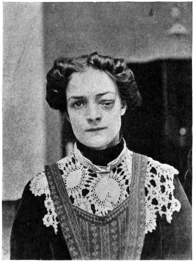
Fig. 94. An Exostosis of the Orbit.
SARCOMATA, PRIMARY AND TRAUMATIC
Primary sarcomata of the skull, when compared to sarcomata developing in other situations, is undoubtedly a rare disease. Still, many cases have been recorded, and four have come under my own personal care, one of which is depicted in the figure.
The disease is equally prevalent in the two sexes, and, excluding chloromata (see p. 334), usually develops at or after middle life. The growth may originate in the diploic tissue (as a myelogenous tumour), or may spring from the pericranium. The cellular structure varies accordingly. More commonly the cells are of the large round or spindle type, and are proportionately malignant.
As regards site of development, the temporal bone (squamous portion) is most commonly involved, next to which comes the frontal bone.
In considering the ætiology of sarcoma in general, trauma must always be taken into account, for it is an undoubted fact that it plays an important part in the development of this dire disease. With respect to the skull similar factors come into play. For instance, Fröhlking,[79] after collecting 48 cases of sarcoma of the skull, found a definite history of trauma in 9-21 per cent.
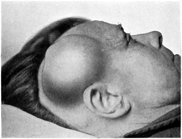
Fig. 95. An Extrinsic Sarcoma of the Skull.
Ziegler[80] lays down the following essentials in establishing the traumatic origin of the tumour: It must develop directly after the trauma on the basis of the swelling or directly in the scar of the wound.
The tumour must be palpable immediately after the acute swelling has diminished.
At the site of the trauma constant or intermittent pain must be present.
A considerable number of cases of sarcoma of the skull will fulfil even these arbitrary conditions. Such sarcomata may definitely be labelled ‘traumatic’ sarcomata.
Whether dependent on injury or not, the symptoms associated with sarcoma of the skull vary according to whether the tumour is extrinsic or intrinsic—whether, for instance, the growth develops in the inward direction and presses on the brain, or grows from the pericranium and is directed externally. Intrinsic tumours, with the exception of some local pain, œdema of tissues, and dilatation of superficial veins, give rise to symptoms closely resembling those observed in intracranial tumour formation. When extrinsic, the tumour varies in size, but is necessarily attached to the bone, the base being the widest part of the tumour. In the earlier stages the overlying skin, with the exception of a few dilated vessels, is more or less normal. Later on, the integument becomes adherent to the tumour, then red and inflamed, and finally ulcerated, the growth now fungating to the surface. The tumour itself is of variable consistency, first hard, then softer, and lastly semi-fluctuating.
Pain, though not very severe, is more or less constant—of a dull, aching character. The extrinsic tumours may, however, give rise to acute neuralgic pain in the event of implication of cutaneous nerves; whilst the intrinsic, in the later stages of the disease, lead to the more severe types of headache observed in intracranial tumour formation.
Secondary nodules appear in other parts of the scalp—all appertaining, in their clinical characteristics, to the primary growth; the cervical glands become infected, and death results from repeated hæmorrhages, pulmonary complications, &c., usually within one to two years from the date of primary development.
Treatment.
The removal of an extrinsic tumour should only be carried out when the tumour is small and non-adherent to the tissues of the scalp. With respect to the intrinsic variety greater circumspection is required. The presence of cerebral symptoms and the inward extension of the growth—verified by symptoms and by X-ray investigation—may be regarded as implying that the conditions are beyond the reach of surgery. In both varieties of tumour, extensive glandular implication acts as a contra-indication to operation.
Under the more favourable conditions an attempt may be made at the extirpation of the growth. The operation should be rendered as bloodless as possible, for which purpose it is essential that the scalp-tourniquet should be applied as a preliminary measure. A scalp-flap is then framed, suited to requirements, and allowing of free exposure of the tumour and surrounding healthy tissues. The skull is then trephined to one side of the growth, and the disk removed. The dura is separated from the overlying bone, and by the circumferential application of de Vilbiss or other craniectomy forceps the central mass is isolated and removed. During these manipulations free hæmorrhage may be experienced from the numerous dilated diploic veins. For the arrest of this the surgeon should have ready to hand, ivory pegs, bone-wax, and other aids for the control of hæmorrhage (see Chapter II). The scalp-flap is then replaced.
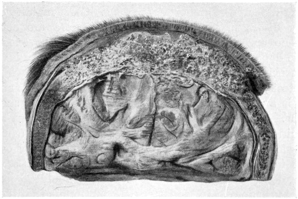
Fig. 96. An Intrinsic Sarcoma of the Skull.
The gap in the bone may be covered in, at a later date, if the patient’s condition is favourable.
The operation may be a formidable one, but records are to hand of 35 cases in which radical measures were adopted. Ten cases died from the operation, 13 were well for periods varying from six months to six years; and recurrence took place soon after the operation in 21 cases.
Chloromata.
Chloroma, a peculiar type of sarcoma characterized by the pale green hue of the tissues, usually develops in the young. The cells are small and round, the pigment distributed in and around the cell elements. The colour, said to be dependent on the presence of a pigmented fat, is most intense immediately after the removal of the tumour, fading rapidly on exposure to light.
The tumour develops from the periosteum of bones, more especially from those which enter into the formation of the orbit and base of skull. In fact, nearly all the cases reported have originated from the temporal and orbital regions. Great rapidity of growth and early dissemination throughout the viscera are conspicuous features—no organ of the body can be said to be exempt from metastatic deposits. The meninges and brain are early involved by direct extension.
Proptosis, as the result of cavernous sinus thrombosis and orbital invasion, is a prominent and early symptom. Death results within six months. No treatment, surgical or otherwise, is of any avail.
Carcinoma
can only involve the skull-bones as a metastatic deposit—more commonly in association with mammary cancer—or by direct invasion from an overlying epitheliomatous scalp ulcer. In the former instance, any radical treatment would be contrary to all surgical principles. In the latter case, presuming that the cervical glandular region is unaffected or capable of removal, the scalp ulcer should be freely excised together with the whole thickness of underlying bone. The resultant osseous gap may be covered in by means of a plastic flap derived from neighbouring healthy tissue.