IV. COMPARISON BETWEEN THE RESPIRATORY LESIONS OF INFLUENZA AND THOSE INITIATED BY THE INHALATION OF POISONOUS GASES
Reading edition
Immediately preceding the advent of the influenza epidemic in New Haven, there had been completed in this laboratory the experimental studies of the effects of a large number of different toxic gases upon the respiratory tract. This was begun with the support of the Bureau of Mines, and subsequently, after the formation of the Chemical Warfare Service, the work received the support, not only of this branch of the Army, but also of the Surgeon General’s Office. Exceptional opportunities for animal experimentation were offered. Every stage of the inflammatory process in the respiratory tract was studied, from the most acute, a few hours after the exposure of the animal to a high concentration of gas, to the very chronic types that resulted from sublethal concentrations and led to lesions encountered at the sacrifice of the animals many months later.
The criticism will unquestionably be introduced, that with the anatomical picture of the respiratory lesions initiated by irritating gases freshly in mind, an analogy between them and those of the pulmonary lesions of influenza may not be impartially drawn. However, such fundamental and striking similarities exist between the two processes (128) that the argument is simplified.
A. THE INFLAMMATORY RESPONSE VERSUS THE SYSTEMIC CAPACITY TO COMPENSATE
Symmers (141) and Oberndorfer (108) have already likened the influenzal pulmonary complications to plague pneumonia, and it is quite possible that others will find fundamental similarities with other fulminating, inflammatory reactions. Inflammation, a series of processes manifested after injury by a tissue which still retains its viability, is modified by another important factor usually neglected; namely, the state of the host. As a consequence, the inflammatory reaction might readily have similar manifestations even though the primary exciting agents are diverse. This has been amply demonstrated for many different reactive processes, and still it may be considered as a challenge for those who are of the opinion that specific bacterial agents necessarily produce characteristic anatomical manifestations. Consider the state of knowledge of the various reactions to the tubercle bacillus before this microorganism was isolated; and then bear in mind that we are dealing here with a disease whose causative agent, despite the present uncertainty regarding its specific nature, produces a lesion that paves the way for the invasion of the respiratory tract by many different organisms. The extent of the reaction may depend upon the systemic capacity to compensate, as well as upon the degree of primary damage in a specific system of organs. Unquestionably, these two factors must take their places in the balance, the opposite pan of which contains the one or the other secondary bacillary invader.
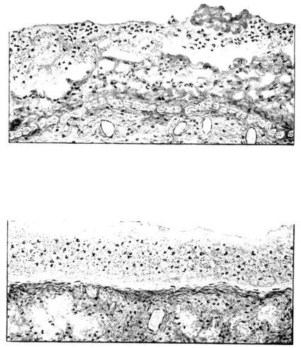
FIG. XXXVIII. THE UPPER ILLUSTRATION, AUTOPSY NO. 116, SHOWS A FRESH, FIBRINOPURULENT PLEURAL EXUDATE. THE PLEURAL LINING CELLS ARE SWOLLEN AND CONSPICUOUS. THE LOWER ILLUSTRATION, AUTOPSY NO. 100, SHOWS ANOTHER AND LATER TYPE. THE ELEMENTS OF THE EXUDATE HAVE DISINTEGRATED TO FORM A MUCILAGINOUS MASS. COMPARE FIGURE XXXVII.
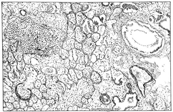
FIG. XLI. AUTOPSY NO. 140. IS A LOW POWER DRAWING OF THE CHARACTERISTIC LUNG IN THE MORE CHRONIC STAGE OF THIS DISEASE. NECROTIZING AND ORGANIZING BRONCHIOLITIS AND PERIBRONCHIOLITIS ARE ASSOCIATED WITH AN ORGANIZATION OF THE EXUDATE IN THE ALVEOLI. THE ORGANIZATION PROCESS IS SOMEWHAT OBSCURED BY THE EDEMA. COMPARE FIGURES XI, XXXIX, XI, AND XLIV.
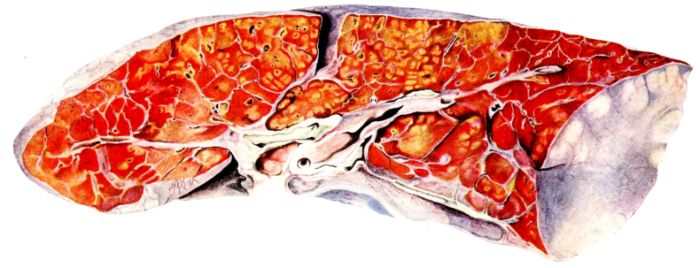
FIG. XXXIX. AUTOPSY NO. 140. RIGHT LUNG. THE HISTOLOGY OF THE PULMONARY CHANGES IN THIS CASE IS ILLUSTRATED IN FIGURES XI, XLI, AND XLIV. THE NECROTIZING PERIBRONCHIAL FOCI STAND OUT PROMINENTLY AND THE ORGANIZATION OF THE EXUDATE IN THE SURROUNDING ALVEOLI IS ALSO DISTINCTLY SEEN IN THE ABOVE FIGURE.
41Scientific medicine has reached that evolutionary period where, in addition to the specific infecting microorganism, associated etiological factors find an important place in the interpretation of a disease. More and more attention is being directed toward the latter factors and the rôle they play in respiratory infections. Many different factors open the pulmonary portal for organisms and, in this way, terminate by a serious respiratory inflammatory complication what otherwise might have been a relatively mild disease. In the past two years this has been demonstrated for measles.
For the above reasons, not only the immediate action but the complications and sequelæ, too, of the inhalation of irritating gases by normal healthy animals, emphasize themselves as a framework for a more comprehensive interpretation of respiratory disease in general.
B. THE PRIMARY INJURY
With few exceptions, the gases studied have their most marked effect upon the respiratory tract. They differ, however, in the localization of the lesions and in the extent of the damage. For instance, mustard gas, in high concentrations, has a necrotizing effect upon the entire respiratory tract even to the pulmonary parenchyma itself. Inhaled in a more dilute form it involves the larynx and the trachea and has spent itself before the lung is reached. With phosgene, the opposite is true. The upper respiratory tract is only slightly involved. The outspoken lesions involve the bronchioles, the ducti alveolares, and extend to the alveolar walls of the lung. Chlorine, while it produces a less severe lesion of the upper respiratory tract, extends more often through all of the ramifications of the tracheal tree to the lung.
With vital stains (trypan blue), it may be readily demonstrated that these gases kill the epithelium of the respiratory tract and extend through this superficial cellular coat to the deeper tissues of the bronchiolar wall and to the lung tissue, killing it entirely, just as a corrosive chemical destroys the wall of the stomach.
When chlorine and phosgene reach the lung, an intense reactive process follows immediately. The congestive changes, the hemorrhage into the pulmonary parenchyma both in the alveoli and in the subpleural and interstitial tissues, and the albuminous rich, serous exudates occupy the foreground of the acute picture and are associated clinically with intense cyanosis, great dyspnœa, hemorrhage, bronchorrhea, and also with pulmonary and subcutaneous interstitial emphysema. The microscope reveals a hyalinization of the walls of the bronchioles or ducti alveolares which are distended during this acute period and form a picture that is very unusual in the more frequent types of respiratory inflammation. It shows also a hyalinization of the alveolar walls, usually those in direct continuity with the larger air passages. The interstitial edema and hemorrhage, often perivascular in distribution, is only overshadowed by the thick, almost colloid-like material within the alveolus itself. Fibrin stains at this stage show this exudative element in surprisingly large amounts. Fibrin not only covers the alveolar wall, but crosses this structure and often forms intracapillary plugs. In fact, besides the red blood cells, the serum and the fibrin, one often sees, not only an excess of polymorphonuclear leucocytes in the vessels of the lung, but these appear in process of migration within a few hours after the animal has been exposed.
Animals surviving this stage often succumb later with a typical pneumonic process. Although frequently pseudolobar, the process may be lobar in extent, and as time goes on more definite lobular involvement is frequently encountered. With the localization of the 42inflammatory process, the general edema, congestion and milder reactions in other portions of the lung tend to subside, and may be entirely absent in a few days. The clinical picture, in the experimental animal at least, also assumes a more typical expression of respiratory infection,—leucocytosis, chlorine retention, etc., appear. Often the pneumonias, both lobar and lobular, are complicated, grossly, by softening, and histologically, by necrotization of the bronchiolar and alveolar walls. Recrudescence of the active pulmonary infection is not uncommon, and perhaps is associated with these focal necrotizing areas.
C. THE TENDENCY TO ORGANIZATION OF BRONCHIOLAR AND ALVEOLAR EXUDATES
The sequelæ after exposure to gas may be markedly delayed. They occur, not only in animals which have exhibited characteristic acute symptoms, but also, and especially with phosgene, where no serious immediate clinical effects followed exposure to the gases. They are dependent upon the fact that the pneumonic process has a very striking tendency to undergo organization, a process which involves not only the exudate in the alveoli, but also that within the bronchioles. The organizing process may be present in both these portions of the lung or may be confined to the bronchiole alone, and lead to a progressive interference with the ingress and egress of alveolar air, so that atelectasis and emphysema become permanent, and in turn lead to narrowing of the vascular bed, ultimately producing right-sided cardiac decompensation. Bronchiectases also occur; they may be tubular, associated with organization of the pneumonic process in their vicinity, or saccular if death has not followed the destruction of the bronchiolar wall.
Compare this picture with that of influenzal pneumonia and its sequelæ; in both there is, first the acute diffuse involvement of the lung, initiated by, or occurring simultaneously with, an acute tracheobronchitis and presenting clinically dyspnœa, cyanosis, blood-stained, abundant sputum, and even interstitial emphysema; next, the tendency for the pneumonic process to localize and to necrotize; and finally, if the acute period is survived, organization of the pneumonic and bronchiolar exudates with resulting bronchiolitis and bronchiectasis. With even more minute comparison, the resemblance is sustained. The initial or early dilatation of the ducti alveolares, so characteristic in gas poisoning, also occupies the foreground of the histological picture in influenza. These dilated structures are the more prominent on account of the red, ribbon-like strands that cover the surface and often involve the walls. Similar necrotization, varying in its depth even to involve the whole of the alveolar walls, occurs in both of these conditions. Similar hemorrhages in the early stage, rupture of the alveolar wall with interstitial and subcutaneous emphysema, necrotization, gangrene, and organization in the same localities, characterize the two lesions, and are not found with the same uniformity in any other type of respiratory disease.
D. THE IMPORTANCE OF THE TRACHEA AND ITS RAMIFICATIONS AS A PROTECTIVE MECHANISM AGAINST INFECTION OF THE PULMONARY PARENCHYMA
In gas poisoning it has been demonstrated that the initial damage to the epithelium of the larger air passages is followed by an invasion of the pulmonary parenchyma by the bacteria of the mouth. Repeated cultures from the mouth before the exposure of the animal to gas have been followed by the recovery of the same organisms, including the pneumococci, the streptococci, the staphylococci, and a gram-negative, hemoglobinophilic, small bacillus from the pneumonic lung. They find their way into the lung after the destruction or incapacitation of the protective mechanism (70) of the upper respiratory tract,—into a lung which has been so damaged by the irritative gas that bacteria innocuous in the normal pharynx now find a favorable medium for their development. The inflammatory reaction which develops into pneumonia, perhaps necrotizing, perhaps later organizing, can only be explained by the combined action of the corrosive gas and the organisms saprophytic in the normal mouth, but now pathogenic in varying degrees in the lung whose vital reactions have either been inhibited or impaired by the gas.
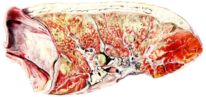
FIG. XL. AUTOPSY NO. 140. LEFT LUNG. THE HISTOLOGY OF THE PULMONARY CHANGES IN THIS CASE IS ILLUSTRATED IN FIGURES XI, XLI, AND XLIV. THESE PROCESSES, ILLUSTRATED IN FIGURE XXXIX, ARE ACCENTUATED AND THERE IS ALSO AN ORGANIZATION OF THE EXTENSIVE PURULENT PLEURISY TO BE MADE OUT IN THE ABOVE FIGURE.
43There is no reason why this analogy should not be drawn, no reason why we should not consider that the unknown etiological agent in influenza produces a similar injury to, or even destruction of, the protective mechanism of the respiratory tract. Similarly, gas and influenza damage the pulmonary parenchyma itself, so that the bacteria of the air and of the mouth which find their way into the damaged lung[16] initiate processes and produce complications which may not be distinguished.
44