Part 1 — CHAPTER V. CLASSIFICATION.
Reading edition
The arrangement of living organisms in groups according to their resemblances and the adoption of fixed names is of the greatest advantage in their scientific study. For animal forms and for the higher plants this classification is gradually becoming standardized through the International Congress of Zoölogists and of Botanists respectively. Unfortunately, the naming of the bacteria has not as yet been taken up by the latter body, though announced as one of the subjects for the Congress of 1916 (postponed on account of the war). Hence there is at present no system which can be regarded as either fixed or official.
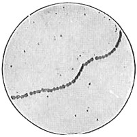
Fig. 49.—Illustrates the genus Streptococcus. Typical chains, no staphylococcus grouping, no sarcina grouping, no flagella.
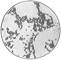
Fig. 50.—Illustrates the genus Micrococcus. Diplococcus, tetrads short chains and staphylococcus; no sarcina, no flagella.
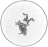
Fig. 51.—Illustrates the genus Sarcina. Sarcina grouping, no flagella.
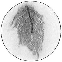
Fig. 52.—Illustrates the genus Bacillus. A bacillus with peritrichic flagella. (Student preparation.)
Since Müller’s first classification of “animalcules” in 1786 numerous attempts have been made to solve the problem. Only those beginning with Ferdinand Cohn (1872–75) are of any real value. As long as bacteria are regarded as plants it appears that the logical method is to follow the well-established botanical principles in any system for naming them. Botanists depend on morphological features almost entirely in making their distinctions. The preceding chapters have shown that the minute plants which are discussed have very few such features. They are, to recapitulate, cell wall, protoplasm, vacuoles, metachromatic granules, capsules, flagella, spores, cell forms and cell groupings. Most bacteria show not more than three or four of these features, so that itphysiological activities. One of these systems was adopted by the laboratory section of the American Public Health Association and by the Society of American Bacteriologists and was practically the standard in this country until superseded by the Society’s own classification. It is that of the German Bacteriologist Migula and is given below for comparison. Since practically the entire discussion in this book is concerned with the first three families the generic characteristics in these only will be given. The full classification as well as a thorough discussion of this subject is given in Lafar’s Handbuch, whence the following is adopted:
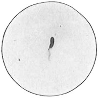
Fig. 53.—Illustrates the genus Pseudomonas. A bacillus with flagella at the end only.
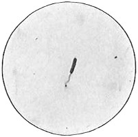
Fig. 54.—Illustrates the genus Microspira. It is (though the photograph does not prove it) a short spiral with one flagellum at the end.
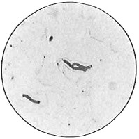
Fig. 55.—Illustrates the genus Spirillum. Spiral bacteria with more than three, in this case four, flagella at the end.
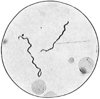
Fig. 56.—Illustrates the genus Spirochæta
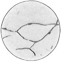
Fig. 57.—Illustrates the genus Chlamydothrix. Fine threads with a delicate sheath.
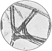
Fig. 58.—Illustrates the genus Crenothrix. The thickness of the cell walls is due to deposits of iron hydroxide. (After Lafar.)
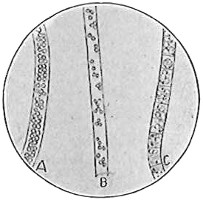
Fig. 59.—Illustrates the genus Beggiatoa. The filament A is so full of sulphur granules that the individual cells are not visible. B has fewer sulphur granules. In C the granules are nearly absent and the separate cells of the filament are seen. (After Winogradsky, from Lafar.)
ORDER I. Eubacteria.
Cells without nuclei, free from sulphur granules and from bacteriopurpurin (p. 112); colorless, or slightly colored.
1. Family: Coccaceæ (Zopf) Migula, all cocci.
Non-flagellated, Non-motile Genus 1. Streptococcus Billroth: division in one plane only (Fig. 49). Genus 2. Micrococcus (Hallier) Cohn: division in two planes only (Fig. 50). Genus 3. Sarcina Goodsir: division in three planes only (Fig. 51).
Flagellated, motile Genus 4. Planococcus Migula: division in two planes only. Genus 5. Planosarcina Migula: division in three planes only.
2. Family: Bacteriaceæ Migula, all bacilli.
Genus 1. Bacterium (Ehrenberg) Migula: no flagella; non-motile. Genus 2. Bacillus (Cohn) Migula: flagella peritrichic (Fig. 52). Genus 3. Pseudomonas Migula: flagella at the end: monotrichic, lophotrichic, amphitrichic (Fig. 53).
3. Family: Spirillaceæ Migula, all spirilla.
Cells stiff Genus 1. Spirosoma Migula: non flagellated; non-motile. Genus 2. Microspira (Schrœter) Migula: flagella one to three at the end (Fig. 54). Genus 3. Spirillum (Ehrenberg) Migula: flagella more than three at the end (Fig. 55).
Cell flexible Genus 4. Spirochæta Ehrenberg: motile; no flagella (Fig. 56).
4. Family: Chlamydobacteriaceæ.
Cells cylindrical in long threads and surrounded by a sheath. Reproduction also by gonidia formed from an entire cell.
Genus 1. Chlamydothrix Migula (Fig. 57). Genus 2. Crenothrix Colin (Fig. 58). Genus 3. Pragmidiothrix Engler. Genus 4. Spherotilus (including Cladothrix).
ORDER II. THIOBACTERIA: SULPHUR BACTERIA.
Cells without a nucleus, but containing sulphur granules, may be colorless or contain bacteriopurpurin and be colored reddish or violet.
1. Family Beggiatoaceæ.
Genus 1. Thiothrix Winogradsky.
Genus 2. Beggiatoa Trevisan. Of interest since it is without aFig. 59).
2. Family Rhodobacteriaceæ.
This has five subfamilies and twelve genera, most of which are due to the Russian bacteriologist Winogradsky who did more work than anyone else with the sulphur bacteria.
THE CLASSIFICATION OF THE SOCIETY OF AMERICAN BACTERIOLOGISTS.
The Committee on Classification of the Society of American Bacteriologists at the meeting held in December, 1919, submitted its final report. This report has not been formally adopted as a whole, but in all probability will be substantially as outlined below. This outline does not attempt to give the detailed characterizations of the different groups as defined by the committee, but does show the names to be applied to the commoner organisms. These organisms are included in the 4th and 5th orders. Details of the first three orders have not been worked out. They are listed merely for completeness.
CLASS SCHIZOMYCETES.
Unicellular, chlorophyl-free plants, reproducing by transverse division (some forms by gonidia also).
Orders:
A. Myxobacteriales—Cells united during vegetative stage into a pseudo-plasmodium which passes over into a highly developed cyst-producing resting stage.
B. Thiobacteriales—Sulphur bacteria.
C. Chlamydobacteriales—Iron bacteria and other sheathed bacteria.
D. Actinomycetales—Actinomyces, tubercle and diphtheria bacilli.
E. Eubacteriales—All the other common bacteria.
Genera of Orders D and E.
D. ACTINOMYCETALES—
Family I. Actinomycetaceæ Buchanan, 1918.
Genus 1. Actinobacillus, Brampt, 1900.
Type species, Actinobacillus lignieresi Brampt, 1900.
Genus 2. Leptotrichia Trevisan, 1879.
Type species, Leptotrichia buccalis (Robin, 1847) Trevisan.
Genus 3. Actinomyces Harz, 1877.
Type species, Actinomyces bovis Harz.
Genus 4. Erysipelothrix Rosenbach, 1909.
Type species, Erysipelothrix rhusiopathiæ (Kitt, 1893) Rosenbach, swine erysipelas.
Family II. Mycobacteriaceæ Chester, 1897.
Genus 1. Mycobacterium Lehmann and Neumann, 1896.
Type species, Mycobacterium tuberculosis (Koch, 1882) L. and N.
Genus 2. Corynebacterium Lehmann and Neumann, 1896.
Type species, Corynebacterium diphtheriæ
Genus 3. Fusiformis Hoelling, 1910.
Type species, Fusiformis termitidis Hoelling. Vincent’s angina.
Genus 4. Pfeifferella Buchanan, 1918.
Type species, Pfeifferella mallei (Loeffler, 1896) Buchanan. Glanders bacillus.
E. EUBACTERIALES
Family I—Nitrobacteriaceæ—Proto- or autotrophic for N or C and sometimes for both (except Acetobacter).
Tribe I—Nitrobactereæ—autotrophic for C.
Genus 1. Hydrogenomonas Jensen, 1909.
Type species, Hydrogenomonas pantotropha (Kaserer, 1906) Jensen; oxidizes free H.
Genus 2. Methanomonas Jensen, 1909.
Type species, Methanomonas methanica (Söhngen) Jensen; oxidizes CH4.
Genus 3. Carboxydomonas Jensen, 1909.
Type species, Carboxydomonas oligocarbophila (Beijerinck and Van Delden, 1903) Jensen; oxidizes CO.
Genus 4. Acetobacter Fuhrman, 1905.
Type species, Acetobacter aceti (Thompson, 1852) Fuhrman; oxidizes alcohol to acetic acid.
Genus 5. Nitrosomonas Winogradsky, 1892.
Type species, Nitrosomonas europoea Winogradsky; oxidizes ammonia or ammonium salts to nitrous acid, hence nitrites.
Genus 6. Nitrobacter Winogradsky, 1892.
Type species, Nitrobacter Winogradskyi Committee of 1917; oxidizes nitrous acid (nitrites) to nitric acid (nitrates).
Tribe II—Azotobactereæ—prototrophic for N.
Genus 7. Azotobacter Beijerinck, 1901; large, free-living, aerobic N absorbers.
Type species, Azotobacter chroococcum Beijerinck.
Genus 8. Rhizobium Frank, 1889.
Type species, Rhizobium leguminosarum Frank; root tubercle bacteria of legumes.
Family II—Pseudomonadaceæ, Committee of 1917.
Genus 1. Pseudomonas Migula, 1894.
Type species, Pseudomonas violacea (Schroeter, 1872) Migula.
Family III—Spirillaceæ Migula, 1894—all spiral bacteria.
Genus 1. Vibrio Müller, 1786, emended by E. F. Smith, 1905.
Type species, Vibrio choleræ (Koch, 1884) Schroeter, 1886.
Genus 2. Spirillum Ehrenberg, 1830, emended Migula, 1894.
Type species, Spirillum undula (Müller, 1786) Ehrenberg.
Family IV—Coccaceæ Zopf, 1884, emended Migula, 1894—all cocci.
Tribe I—Neissereæ.
Genus 1. Neisseria Trevisan, 1885.
Type species, Neisseria gonorrhoeae Trevisan.
Tribe II—Streptococceæ Trevisan, 1889.
Genus 2. Diplococcus Weichselbaum, 1886.
Type species, Diplococcus pneumoniae Weichselbaum.
Genus 3. Leuconostoc Van Tieghem, 1878.
Type species, Leuconostoc mesenterioides (Cienkowski) Van Tieghem.
Genus 4. Streptococcus Rosenbach, 1884; emended Winslow and Rogers, 1905.
Type species, Streptococcus pyogenes Rosenbach.
Tribe III—Micrococceæ Trevisan, 1889.
Genus 5. Staphylococcus Rosenbach, 1884; animal parasites.
Type species, Staphylococcus aureus Rosenbach.
Genus 6. Micrococcus Cohn, 1872, emended Winslow and Rogers, 1905. Facultative parasites or saprophytes.
Type species,Micrococcus luteus (Schroeter, 1872) Cohn.
Genus 7. Sarcina Goodsir, 1842, emended Winslow and Rogers, 1905.
Type species, Sarcina ventriculi Goodsir.
Genus 8. Rhodococcus Zopf, 1891, emended Winslow and Rogers, 1905; cocci with red pigment.
Type species, Rhodococcus rhodochrous Zopf.
Family V—Bacteriaceæ Cohn, 1872, emended by Committee of 1917; bacilli without spores not above included.
Tribe I—Chromobactereæ Committee of 1919; producing red or violet pigment, mainly water forms.
Genus 1. Erythrobacillus Fortineau, 1905.
Type species, Erythrobacillus prodigiosus (Ehrenberg, 1848) Fortineau.
Genus 2. Chromobacterium Bergonzini, 1881.
Type species, Chromobacterium violaceum Bergonzini.
Tribe II—Erwineæ Committee 1919; plant pathogens.
Genus 3. Erwinia Committee 1917.
Type species, Erwinia amylovora (Burrill, 1883) Committee 1917.
Tribe III—Zopfeæ Committee of 1919; Gram +, no pigment, non-carbohydrate-fermenting.
Genus 4. Zopfius Wenner and Rettger, 1919.
Type species, Zopfius zopfii (Kurth) Wenner and Rettger.
Tribe IV—Bactereæ Committee of 1919; Gram -, carbohydrate fermenters.
Genus 5. Proteus Hauser, 1885; liquefy gelatin.
Type species, Proteus vulgaris Hauser.
Genus 6. Bacterium Ehrenberg, 1828, emended Jensen, 1909; liquefy gelatin rarely.
Type species, Bacterium coli.
Tribe VI—Lactobacilleæ Committee of 1919; Gram +, high acid, thermophils.
Genus 7. Lactobacillus Beijerinck, 1901.
Type species, Lactobacillus caucasicus (Kern?) Beijerinck; Bulgarian bacillus.
Tribe VI—Pasteurelleæ Committee of 1919; organisms of hemorrhagic septicemia.
Genus 8. Pasteurella Trevisan, 1888.
Type species, Pasteurella cholerae-gallinarum (Flügge, 1886); Trevisan.
Tribe VII—Hemophileæ Committee of 1917; require hemoglobin for growth.
Genus 9. Hemophilus Committee of 1917.
Type species, Hemophilus influenzae (Pfeiffer, 1893) Committee of 1917.
Family VI—Bacillaceæ Fischer, 1895. Spore forming rods.
Genus 1. Bacillus Cohn, 1872; aerobic, no change of form around the spore.
Type species, Bacillus subtilis Cohn.
Genus 2. Clostridium Prazmowski, 1880; anaërobic
Type species, Clostridium butyricum Prazmowski.
As compared with Migula’s classification it is to be noted that there are 38 genera listed by the Committee instead of 13 in the same general groups.
The following list of Genera conservanda submitted by the Committee was formally adopted by the Society and these are therefore its official names for the organisms included in these genera.
Acetobacter Fuhrman
Actinomyces Harz
Bacillus Cohn
Bacterium Ehrenberg
Chromobacterium Bergonzini
Clostridium Prazmowski
Erythrobacillus Fortineau
Leptotrichia Trevisan
Leuconostoc Van Tieghem
Micrococcus Cohn
Rhizobium Frank
Sarcina Goodsir
Spirillum Ehrenberg
Staphylococcus Rosenbach
Streptococcus Rosenbach
Vibrio Müller
It is greatly to be desired that the Society’s Classification when finally completed shall become the standard in the United States at least.
Such names as have been adopted by the Society are used throughout this work.
The Committee also submitted the following artificial key for determining the genera in the two orders ACTINOMYCETALES AND EUBACTERIALES:
A—Typically filamentous forms ..........Actinomycetacae
B—Mycelium and conidia formed ..........Actinomyces
BB—No true mycelium
C—Cells show branching
D—Gram negative ..........Actinobacillus
DD—Gram positive ..........Erysipelothrix
CC—Cells never branch. Gram positive threads later fragmenting into rods ..........Leptotrichia
AA—Typically unicellular forms (though chains of cells may occur)
B—Cells spherical—COCCACEÆ
C—Parasitic forms (except Leuconostoc), cells generally grouped in pairs or chains, never in packets, generally active fermenters.
D—Cells in flattened coffee-bean-like pairs, gram -. ..........Neisseria
DD—Not as D
E—Saprophytes in zoögloea masses in sugar solutions. ..........Leuconostoc
EE—Not as E. Gram +.
F—Cells in lanceolate pairs or in chains. Growth on media not abundant.
G—Cells in lanceolate pairs. Inulin generally fermented. ..........Diplococcus
GG—Cells in chains. Inulin not generally fermented. ..........Streptococcus
FF—Cells in irregular groups. Growth in media fairly vigorous. White or orange pigment. ..........Staphylococcus
CC—Saprophytic forms. Cells in irregular groups or packets, not in chains. Fermentative powers low.
D—Packets ..........Sarcina
DD—No packets.
E—Yellow pigment ..........Micrococcus
EE—Red pigment ..........Rhodococcus
BB—Rods:
C—Spiral rods
D—Short, comma-like rods. One to three flagella. ..........Vibrio
DD—Long spirals. Five to twenty flagella. ..........Spirillum
CC—Straight rods.
D—No endospores.
E—Rods of irregular shape or showing branched or filamentous involution forms.
F—Cells irregular in shape. Staining unevenly. Animal parasites.
G—Acid fast ..........Mycobacterium
GG—Not acid fast.
H—Cells elongated, fusiform ..........Fusiformis
HH—Cells not elongated, sometimes branching.
I—Gram positive. Slender, sometimes club-shaped. ..........Corynebacterium
II—Gram negative. Rods sometimes form threads. Characteristic honey-like growth on potato. ..........Pfeifferella
FF—Cells staining unevenly but with branched or filamentous forms at certain stages. Never acid fast. Not animal parasites.
G—Metabolism simple, growth processes involving oxidation of alcohol or fixation of free N (latter in symbiosis with green plants).
H—Cells minute. Symbiotic in roots of legumes. ..........Rhizobium
HH—Oxidizing alcohol. Branching forms common. ..........Acetobacter
GG—Not as G. Proteus-like colonies.
H—Not attacking carbohydrates ..........Zopfius
HH—Proteus
EE—Regularly formed rods.
F—Metabolism simple, growth processes involving oxidationNITROBACTERIACEÆ.
G—Fixing N or oxidizing its simple compounds.
H—Fixing N, cells large, free in soil ..........Azotobacter
HH—Oxidizing N compounds.
I—Oxidizing NH4 compounds ..........Nitrosomonas
II—Oxidizing nitrites ..........Nitrobacter
GG—Not as G.
H—Oxidizing free H ..........Hydrogenomonas
HH—Oxidizing simple C compounds, not free H.
I—Oxidizing CO ..........Carboxydomonas
II—Oxidizing CH4 ..........Methanomonas
FF—Not as F.
G—Flagella usually present, polar—PSEUDOMONADACEÆ ..........Pseudomonas
GG—Flagella when present peritrichic—BACTERIACEÆ
H—Parasitic forms showing bi-polar staining. ..........Pasteurella
HH—Not as H.
I—Strict parasites growing only in presence of hemoglobin ..........Hemophilus
II—Not as I.
J—Water forms producing red or violet pigment.
K—Pigment red ..........Erythrobacillus
KK—Pigment violet ..........Chromobacterium
JJ—Not as J.
K—Plant pathogens ..........Erwinia
KK—Not plant pathogens.
L—Gram +, forming large amount of acid from carbohydrates, sometimes CO2, never H ..........Lactobacillus
LL—Gram -, forming H as well as CO2 if gas is produced ..........Bacterium
DD—Endospores present—BACILLACEÆ
E—Aerobes, rods not swollen at sporulation. ..........Bacillus
EE—AnaërobesClostridium