PART I TROPICAL DISEASES AND THEIR TREATMENT
Reading edition
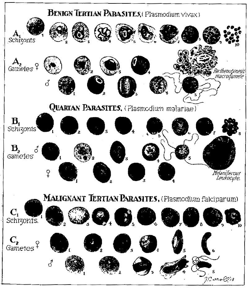
DESCRIPTION OF PLATE OF MALARIAL PARASITES
Benign Tertian Parasites
A1. Schizonts. 1. Normal red cell. 2. Young ring form. 3. Amoeboid or figure-of-eight form showing Schüffner’s dots. 4. Amoeboid form showing increased chromatin (twenty-four to thirty hours). 5. Segmentation of nucleus. 6. Nuclear halves further apart, red cells enlarged and pale. 7. Further division of nucleus. 8. Unusual division form. 9. Typical merocyte. 10. Rupture of merocyte liberating merozoites.
A2. Female gametes. 1. Young form showing solid instead of ring-form staining. 2. Half grown form. 3. Rapidly growing form with compact nucleus and clear vacuolated zone. 4. Full grown macrogamete showing eccentrically placed chromatin and much pigment in deep blue stained protoplasm. Male gametes. 1. Young form similar to female one. 2. Half grown form showing central chromatin. 3. Full grown microgametocyte showing large amount of centrally placed chromatin with light blue protoplasm surrounding. 4. Division of chromatin occurring in microgametocyte and developing in wet preparation. Note.—Chromatin division in gametes does not take place until blood is withdrawn. 5. Spermatozoon like microgametes developing from the microgametocyte. This only occurs in wet preparations or in the stomach of the mosquito.
Quartan Parasites
B1. Schizonts. 1. Normal red cell. 2. Young ring form. 3. Older ring form. 4. Narrow equatorial band. 5. Typical band-form. 6. Oval form showing division of chromatin. 7. Early stage merocyte. 8. Daisy form merocyte.
B2. Male gametes. 1. Young solid form. 2, 3, 4. Developmental stages microgametocytes. 5. Flagellated body in wet preparation showing microgametes developing from microgametocyte. Female gametes. 1. Young oval form. 2. Somewhat older stage. 3 and 4. Mature macrogametocytes (same as benign tertian).
Malignant Tertian Parasites
C1. Schizonts. 1. Normal red cell. 2, 3, 4, 5, 6. Young ring forms. These are hair-like rings and are the only forms besides crescents to be found in the peripheral blood. In very heavy infections or in smears from spleen the following forms are found. 7. Beginning division of chromatin. 8 and 9. Further division. 10. Merocyte.
C2. Female gametes. 1 and 2. Young macrogametes. 3. Older stage. 4. Development in red cell. 5 and 6. Fully developed female crescents showing clumping of pigment and rich blue colour. Male gametes. 1 and 2. Developing forms. 3 and 4. Fully developed microgametocytes. 5. Flagellated body developed in wet preparation.
PLATE OF MALARIAL PARASITES