Part 1 — CHAPTER I MALARIA
Reading edition
Definition and Synonyms
Definition.—Malaria is a protozoal disease caused by three species of Plasmodium. In the clinically benign types of malaria we have that of benign tertian, due to P. vivax, with a tertian periodicity and that of quartan, due to P. malariae and showing a quartan or seventy-two hour periodicity. The clinically malignant type of malaria is due to P. falciparum, the parasite of malignant tertian or aestivo-autumnal malaria.
The benign malarial fevers are characterized by a frank chill and well marked distinctions of cold, hot and sweating stages. In malignant tertian there is an indefinite or dumb chill with prolonged hot stage. Diagnostic of malaria are periodicity, parasites and splenic enlargement. The malignant tertian parasite is the one responsible for the so-called cerebral and algid manifestations of perniciousness. Man is the intermediate host of the parasite while the sexual cycle or sporogony goes on in some species of mosquito of the anopheline subfamily, the definitive host.
Synonyms.—Remittent Fever, Intermittent Fever, Ague, Marsh Fever, Paludism, Jungle Fever.
French: Paludisme. German: Wechselfieber.
History and Geographical Distribution
History.—Hippocrates, who considered malaria as intimately connected with bile, divided the disease into quotidian, tertian and quartan, differentiating such types of fever from continuous fevers. It is interesting to note that Celsus recognized two types of tertian fever, the one benign and similar to quartan fever, the other far more dangerous, with a fever occupying thirty-six of the forty-eight hours, not entirely subsiding in the remission, but being only mitigated.
In the time of Caesar views were expressed by Varro that swamp air might be the cause of malaria and furthermore that animals, so small that the eye could not follow them, might transmit diseases by way of the mouth or nose.
In the view of our present knowledge it is remarkable that Lancisi, in 1718, should have associated marshes with the development of gnats, which insects he thought could not only introduce with their proboscides the putrefying organic matter of such swamps but animalcules as well.
In 1638 Countess del Chinchon, the wife of the Viceroy of Peru, was cured of an intermittent fever by the employment of the bark of certain trees which bark was introduced into Europe in 1640. The origin of the name cinchona is thus explained.
While Morton and Sydenham in 1666 noted the specific action of cinchona in certain fevers it remained for Torti, in 1753, by the use of cinchona, clinically to differentiate those fevers which were cured by cinchona from those which failed to yield to this specific. Quinine was not introduced until after 1820. Audouard, in 1803, was the first to draw attention to the splenic enlargement of malaria.
The views of Nott and Beauperthuis as to transmission of malaria and yellow fever by insects are considered under the latter disease.
In 1847 Meckel announced that the dark color of malarial organs was due to a pigment and in 1848 Virchow noted that this pigment was contained in cells. In 1875, Kelsch observed pigmented bodies in malarial blood and in 1880 came to the conclusion that these pigmented cells were diagnostic of malaria.
The year 1880 is the most important one in the history of malaria for on November 6, 1880, Laveran, at Constantine, first saw the parasites of malaria while carrying on investigations as to the origin of the pigmented bodies and melaniferous leucocytes. He not only noted the findings of spherical pigmented bodies but also of crescents and in particular the flagellation of the male gamete which demonstrated to him that these were living bodies.
The name Oscillaria malariae was proposed on account of the movements of the flagellate body, but had to be dropped as not valid, the generic name Oscillaria having been previously applied.
When these bodies were demonstrated to various Italian authorities, in 1882, they were thought by them to be degenerated red cells.
It may be stated that at this time the Italians, influenced by the work of Pasteur, were convinced that an organism, Bacillus malariae, reported by Klebs and Crudeli (1879) to have been isolated from water and soil of malarious districts, was the cause of malaria. This bacillus was said to be cultivable on ordinary media and to be capable, when injected into man, of producing malaria.
By 1885 the Italians were convinced that the bodies discovered by Laveran were the cause of malaria and Marchiafava, by staining with methylene blue, noted the ring forms and the increase in size up to that of the sporulating parasites. To Golgi we not only owe the discovery that the malarial paroxysm coincides with the period when the sporulating forms (merocytes) simultaneously reach maturity but also the exact working out of the cycle of quartan malaria. He even showed three stages of development of the parasites in a triple quartan. It may be stated that Golgi, Marchiafava and Celli are the ones to whom we owe our first knowledge of the existence of different species of parasites for different kinds of malaria. In these investigations they showed that as a rule they could reproduce a certain type of malaria by injecting the blood of such a case of malaria into a well man. Gerhardt, in 1884, was the first to produce malaria by the injection of malarial blood. Laveran insisted all this time that there was but a single species of malaria. About this period a great deal of research was carried on as to the origin of malarial parasites and it was found that many animals harbored parasites similar to the malarial parasites of man. In 1891 the chromatin staining method of Romanowsky was introduced which by bringing out the variations in chromatin distribution led to more accurate study of species and cycles.
Our present exact knowledge as to the existence of 3 species of malaria is largely due to the careful examinations made by Koch of fresh and stained malarial blood preparations.
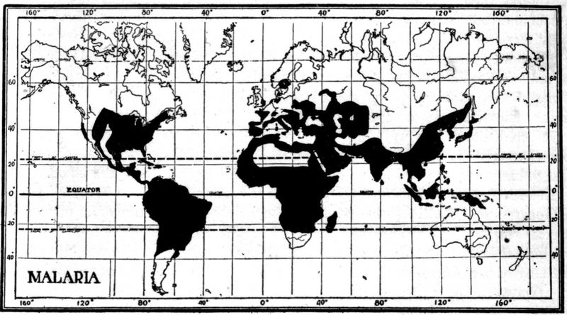
Fig. 1.
—Geographical distribution of malaria.
In 1894 Manson formulated the hypothesis of the mosquito transmission of malaria. He based this upon the fact that the flagellation of the male gamete does not take place for several minutes after the removal of the blood from the peripheral circulation. He also suggested that larvae might feed upon infected mosquitoes dying upon the water and thus acquire the disease.
Ross for two years had mosquitoes feed upon the blood of malarial patients which contained crescents but as he used insects of the genera Culex and Stegomyia he failed to observe development in the tissues of the mosquitoes. In 1897 he used 8 dappled-wing mosquitoes (Anopheline) and in two of these, upon dissection, he noted pigmentary bodies different from anything he had observed in hundreds of dissections of other mosquitoes. At this time he was forced to discontinue this work for about six months.
In 1886 Metschnikoff from observation of sporulating parasites in the brain capillaries at the autopsy of a malarial case considered them to be coccidial in nature. In 1892 Pfeiffer, studying the Coccidia showed that there was an endogenous cycle going on in the epithelial cells as well as the long known exogenous cycle connected with the ingestion of oocysts passing out in the feces of an animal infected with coccidiosis. He suggested that malaria might similarly have an exogenous cycle as well as the well-known endogenous one. Opie noted hyaline and granular forms of parasites in the blood of crows and MacCallum, working with this malaria-like disease of birds (Halteridium), observed the fecundation of a granular female parasite by the flagellum-like process of the hyaline male cell.
In 1898, in India, working with a malarial disease of sparrows (Proteosoma), Ross infected 22 out of 28 healthy sparrows by mosquitoes which had previously fed on sick sparrows. He noted in the culicine mosquito employed for transmission the same cycle of development as that subsequently worked out for human malaria, in anopheline mosquitoes, by Grassi and Bignami, in Italy.
Koch’s great work in connection with malaria was to demonstrate that the malaria-like infections of other animals had no part in the causation of human malaria and that the malarial parasite could only circulate between man and certain mosquitoes.
In order to demonstrate conclusively the connection between infected mosquitoes and malaria Sambon and Low lived for three of the most malarious months of 1900, in one of the most malarious sections of the Roman Campagna, in a mosquito screened hut and did not contract malaria.
Infected mosquitoes were also sent to London from Italy and allowed to feed upon Doctor P. T. Manson and Mr. George Warren. After a period of incubation these volunteers came down with typical malaria with parasites in the blood.
In 1911 Bass first cultivated the parasites of malaria.
Geographical Distribution.—Malaria is so widely distributed over all parts of the tropical and subtropical world that it would require too much space to give its geographical distribution other than as given in the accompanying chart. The malaria belt may be said to extend from 60° N. to 40° S. Many of the islands of the Pacific are exempt.
Etiology and Epidemiology
Etiology.—There are at least three species of animal parasites which produce human malaria, Plasmodium vivax, the cause of benign tertian, P. malariae of quartan and P. falciparum of aestivo-autumnal. These parasites belong to the haemamoeba type of the order Haemosporidia, of the class Sporozoa and of the phylum Protozoa.
This type of Haemosporidia is characterized by invasion of red cells, amoeboid movement, pigment production and the extrusion of flagellum-like processes from the male sporont after the blood is taken from the animal and allowed to cool.
Other Haemosporidia which are very important in diseases of domesticated animals, but not for man, are those of the piroplasm type.
These parasites of the red cells do not produce pigment and do not “exflagellate.” It is to parasites of this type that some authorities have ascribed the cause of blackwater fever, a condition undoubtedly connected with malaria.
It has been thought proper by some to consider the malarial parasites as belonging to two genera, the genus Plasmodium, characterized by round sexual forms and including P. vivax and P. malariae and the genus Laverania, characterized by crescent-shaped sexual forms and including but one species L. malariae, that of aestivo-autumnal malaria.
Craig recognizes a quotidian form and a tertian form for the aestivo-autumnal parasite. Manson formerly held the view that three different species of crescent-bearing parasites were concerned in malignant infections; one, of tertian periodicity, Laverania malariae, and two, of quotidian periodicity, L. praecox, a pigmented form, and L. immaculata, a form in which pigment is only observed in the crescent formation and does not exist in the ring form schizonts. He has abandoned this view. Stephens has noted a parasite which has more nuclear material than P. falciparum (P. tenue).
Malaria of Animals.—Other Haemosporidia of the haemamoeba type are found in birds, monkeys, bats, squirrels and possibly in reptiles (the parasites of reptiles, while intracorpuscular and pigment producing, do not exflagellate). Of particular interest is the so-called bird malaria or Proteosoma, a parasite very similar to the human malarial ones.
The life cycle of this parasite was demonstrated before that of the malarial parasites of man.
Although Koch in his work showed that these malaria-like parasites of other animals were not infectious for man, Fermi has recently carried out well-controlled experiments, by feeding laboratory bred anophelines on the blood of various animals showing such infections, and subsequently on men, with invariably negative results.
Accumulated experience shows that man is not susceptible to any of the animal malarias and that the three human species can only exist in man as an intermediate host and in certain species of anopheline mosquitoes as definitive hosts. Culicine mosquitoes never transmit malaria.
Malaria-Transmitting Mosquitoes.—In the United States, Cellia albimana, C. argyrotarsis, Anopheles crucians, A. quadrimaculatus and A. pseudopunctipennis are efficient transmitters of malaria. Rather remarkable is the experience of Beyer in New Orleans that A. crucians will only transmit P. falciparum while A. quadrimaculatus will transmit P. vivax and P. malariae, but not P. falciparum. Further experiments have shown that A. crucians will transmit P. vivax as well as P. falciparum.
As showing the uncertainty attaching to the question of a certain anopheline species being efficient hosts for malaria may be cited the case of A. punctipennis. This species has been frequently reported as incapable of transmitting malaria and quite recently Mitzmain reported experiments on 219 females of the species which had fed on crescent containing blood and which were dissected from three to thirty-eight days after such feedings with negative findings in stomach and salivary glands. Furthermore, these mosquitoes failed to transmit malaria to healthy persons. Control experiments with A. quadrimaculatus and A. crucians were successful. In June, 1916, Dr. King reported 33% of positive findings after dissection of A. punctipennis which had fed on malignant tertian cases and 85% of success where the man bitten had benign tertian malaria. These results showed as high a degree of success as that obtained with the control A. crucians and A. quadrimaculatus.
From the above it must be evident that there are other factors involved besides that of the host species as both Mitzmain and King are expert epidemiologists.
A species which may be the chief transmitter in one country may be unimportant, though present, in another country. Thus Cellia albimana is the chief malarial transmitter of Panama although C. argyrotarsis is present. In Brazil the conditions are reversed, probably due to C. albimana thriving best where slightly brackish pools of standing water abound, as in Panama.
In the Philipines A. febrifer seems the important transmitter. It freely enters houses and is a vicious biter.
In India the species which seem most active in transmitting malaria are Myzomyia culicifacies and M. listoni; while in Africa, M. funesta is very efficient.
In Europe A. maculipennis and A. bifurcatus are important.
The following species of anophelines selected from the different genera are important transmitters of malaria.
Anopheles maculipennis.—Wings with four spots located at bases of both forked cells and of second and third longitudinal veins. No costal spots. Palpi yellowish brown and unbanded. Legs unbanded.
Anopheles punctipennis.—Wings with black costa showing yellow spots at apical third and at apex. The apical spot involves the first long vein and upper branch of first fork cell. The larger spot at the apical third passes through the first long vein and to the second vein just before it branches. In A. pseudopunctipennis the markings are as above but the fringe has yellow spots.
Myzomyia funesta.—Wings with four yellow spots on a black costa and two black line spots on third longitudinal vein. Palps with three white rings. Proboscis unbanded. Legs with faint apical bands.
Pyretophorus costalis.—Costa black with five or six small yellow spots. Palps with two narrow white bands and white tip. Femora and tibiae with yellow spots. Apical tarsal bands.
Myzorhynchus pseudopictus.—Black costa with two pale yellow spots. Wing fringe unspotted. Black palps with four pale bands. Apex of palps white.
Nyssorhynchus fuliginosus.—Black costa with three large yellow spots. Numerous black dots on the longitudinal veins. Palpi black with white tip and two narrow white bands. Last three hind tarsal segments white.
Cellia argyrotarsis.—Black costa with two distinct and several smaller white spots.
While anophelines are usually rural or at any rate preferring the suburbs of cities yet we can differentiate between domesticated and wild anophelines, these latter keeping away from man and consequently not playing a transmitting rôle.
Another factor in their becoming an efficient host appears to rest in the feeding habits of such anophelines, one which is voracious and fills and then ejects by rectum the blood taken from the malarial patient is more apt to be a transmitter than a species less greedy.
By an efficient host is meant a species in which full development of the parasite takes place.
Life History of the Malarial Parasite
Malaria can be transmitted by subcutaneous or intravenous injection of the blood of a patient with the disease into a well person, the same type being reproduced.
Transmission of Malaria.—Such a method of transmission is only of scientific interest and the regular method is as follows: An infected anopheline at the time of feeding on the human blood introduces through a minute channel in the hypopharynx the infecting sporozoite of the sexual cycle.
When man is first infected by sporozoites we have starting up a nonsexual cycle (schizogony) which is completed in from forty-eight to seventy-two hours, according to the species of the parasite. The falciform sporozite bores into a red cell, assumes a round shape and continues to enlarge (schizont). Approaching maturity, it shows division into a varying number of spore-like bodies. At this stage the parasite is termed a merocyte. When the merocyte ruptures, these spore-like bodies or merozoites enter a fresh cell and develop as before.
Malarial Toxin.—At the time that the merocyte ruptures it is supposed that a toxin is given off which causes the malarial paroxysm.
Rosenau, by injecting, intravenously, filtered blood, taken from a patient at the time of sporulation of the parasites caused a malarial paroxysm. No parasites developed later. Another man who received a small amount of unfiltered blood allowed a slight paroxysm and four days later showed parasites in his blood. Hence the parasite will not pass through the pores of a Berkefeld filter.
Schizogony.—The nonsexual cycle goes on by geometric progression from the first introduction of the sporozoite, but it is usually about two weeks before a sufficient number of merocytes rupture simultaneously to produce sufficient toxin for symptoms (period of incubation). This cycle is termed schizogony. It is considered that there must be several hundred parasites per cubic millimeter sporulating to be capable of producing symptoms.
Gametes.—After a varying time, whether by reason of necessity for renewal of vigor of the parasite by a respite from sporulation, or whether from a standpoint of survival of the species, sexual forms (gametes) develop. Some think that sporozoites of sexual and nonsexual character are injected at the same time. It is usually considered, however, that sexual forms develop from preexisting nonsexual parasites. The developing gametes are often termed sporonts. Strictly, the sexual parasites in the blood should be called gametocytes. The gametes take about twice as long to reach maturity as schizonts. The life of a crescent has been estimated as about ten days and that of the gametes of benign tertian and quartan about one-half this period.
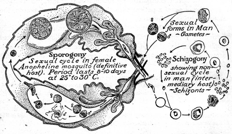
Fig. 2.—Sexual (sporogony in mosquito) and nonsexual (schizogony in man) cycle of the malarial parasite. The sporogony diagram to the left shows in lower portion the fertilization of the female gamete by the microgamete. The vermiculus stage of the zygote is shown boring into the walls of the mosquito’s stomach to later become the more mature zygote packed with sporozoites as shown in the upper diagram of the developmental processes in the mosquito’s stomach.
Sporogony.—The gametes show two types the one which contains more pigment, has less chromatin, and stains more deeply blue is the female—a macrogametocyte; the other with more chromatin, less pigment, and staining grayish green or light blue is the male—a microgametocyte. When the gametes are taken into the stomach of the Anophelinae, the male cell throws off spermatozoa-like projections, which have an active lashing movement and break off from the now useless cell carrier and are thereafter termed microgametes. These fertilize the macrogametes and this body now becomes a zygote. (Following nuclear reduction with formation of polar bodies the macrogametocyte becomes a macrogamete). This process of exflagellation can be observed in a wet preparation under the microscope. There is first seen a very active movement of the pigment of the male gamete and finally long delicate bulbous-tipped flagellum-like processes are thrown off (exflagellated) and push aside the red cells by their progressive motion. MacCallum saw a female Halteridium fertilized by the microgamete, after which it was capable of a worm-like motion (vermiculus or ookinete).
By a boring-like movement the vermiculus stage of the zygote goes through the walls of the mosquito’s stomach, stopping just under the delicate outer layer of the stomach or mid-gut. In three or four days after fertilization the zygote becomes encapsulated and is then often called an oocyst. It continues to enlarge until about the end of one week it has grown to be about 50µ in diameter and has become packed with hundreds of delicate falciform bodies. Some only contain a few hundred, others several thousand.
Zygotes.—In some of his observations Darling has noted that the zygote of benign tertian malaria grows larger and more rapidly than that of aestivo-autumnal and that the pigment is clumped rather than in belts or lines as with aestivo-autumnal. Darling has also noted that mosquitoes do not tend to become infected unless the gamete carrying man has more than 12 gametes to the cubic millimeter of blood. Rouband notes that the oocysts of P. vivax are feebly refractile with fine granules of gray pigment in loose chains while P. falciparum ones are highly refractile with large grains of black pigment. At a temperature of 25°C. vivax completes its cycle in 11 days while the zygote of the crescent requires 14 days. Apparently it is possible for a mosquito to carry both types of parasites.
The capsule of the mature zygote ruptures about the tenth day and the sporozoites are thrown off into the body cavity. They make their way to the salivary glands and thence, by way of the veneno-salivary duct, in the hypopharynx, they are introduced into the circulation of the person bitten by the mosquito, and start a nonsexual cycle. As the sexual life takes place in the mosquito, this insect is the definitive host and man only the intermediate host. The sexual cycle or sporogony in the mosquito takes about ten to twelve days.
Efficient Mosquito Hosts.—It must be remembered that only certain genera and species of Anophelinae are known malaria transmitters; thus Stephens and Christophers, in dissecting 496 mosquitoes of the species M. rossi, did not find a single gland infected with sporozoites.
With M. culicifacies, however, 12 in 259 showed infection. A mosquito which is capable of carrying out the complete sporogonous cycle is an efficient host and in the case of malaria the mosquito is the definitive host (sexual life of parasite).
Malarial Index.—Mosquito dissection is one method of determining the endemicity of malaria or the malarial index. There are two other methods: 1. by noting the prevalence of enlarged spleens, and 2. by determining the number of inhabitants showing malarial parasites in the blood. This index is best determined from children between two and ten years of age, as children under two show for a general average too high a proportion of parasites in the peripheral blood while those over ten years of age show too great an incidence of enlarged spleens.
Barber working in the Philippines with children from five to ten years of age obtained a spleen index of 13.3 and a parasitic index of 11.
As Before Stated there are Three Species of Malarial Parasites: 1. Plasmodium vivax, that of benign tertian—cycle, forty-eight hours; 2. Plasmodium malariae, that of quartan—cycle, seventy-two hours; and 3. Plasmodium falciparum, that of aestivo-autumnal or malignant tertian—cycle of forty-eight hours.
Multiple Infections.—Variations in cycles may be produced by infected mosquitoes biting on successive nights, so that one crop will mature and sporulate twenty-four hours before the second. This would give a quotidian type of fever. In aestivo-autumnal infections anticipation and retardation in the sporulation cause a very protracted paroxysm, lasting eighteen to thirty-six hours; this tends to give a continued or remittent fever instead of the characteristic intermittent type.
Plasmodium Vivax.—In fresh, unstained preparations, taken at the time of the paroxysm or shortly afterward, the benign tertian schizont, or nonsexual parasite, is seen as a grayish white, round or oval body, whose outlines cannot be distinctly differentiated from the infected red cell. They are about one-fifth of the diameter of the red cell and are best picked up by noting their amoeboid activity. In about eighteen hours fine pigment particles appear and make them more distinct. After twenty-four hours the lively motion of the pigment and the projection of pseudopod-like processes, in a pale and swollen red cell, make their recognition very easy. When about thirty to thirty-six hours old the amoeboid movement ceases. Approaching the merocyte stage the pigment tends to clump into one or two pigment masses and one can recognize small, oval, highly refractile bodies within the sporulating parasite.
The gametes or sexual forms do not show amoeboid movement, but the fully developed gamete, which is generally larger than the red cells, has abundant pigment, which is actively motile in the male gamete and nonmotile in the female. The male gamete is more refractile, is rarely larger than a red cell and shows yellow-brown, short rod-like particles of pigment. About fifteen minutes after the making of a fresh preparation these male gametes throw out four to eight long, slender, lashing processes, which are about 15 to 20 microns long. These spermatozoon-like bodies now break off from the useless parent cell and with a serpent-like motion glide away in search of a female gamete, knocking the red cells about in their passage through the blood plasma.
The female gamete is larger than a red cell, is rather granular and has more abundant dark-brown pigment than the male.
Stained Smears.—In dried smears, stained by some Romanowsky method, as that of Wright, Leishman or Giemsa, we note small oval blue rings, about one-fifth of the diameter of the infected yellowish-pink erythrocyte. One side of the ring is distinctly broader than the rather fine opposite end, which seems to hold a round, yellowish-brown dot, the chromatin dot, and has a resemblance to a signet ring. These small tertian rings of the nonsexual parasites (schizont) are seen about the time of the commencement of the sweating stage of the paroxysm. Two chromatin dots in the line of the ring are rare as is also true of more than one ring in a red cell.
When the parasite is about twenty-four hours old we note that it contains much pigment and has an amoeboid or multiple figure-of-eight contour, is about three-fourths the size of a red cell and that the infected red cell is about one and one-half times as large as in the beginning and presents a washed-out appearance. It is an anaemic-looking cell. We also note, as characteristic of a benign tertian infection, reddish-yellow dots in the pale red cell, which are known as Schüffner’s dots. These, practically, are characteristic for benign tertian.
A few hours before the completion of its forty-eight-hour cycle the contained pigment begins to clump, the chromatin to divide and, finally, we have a sporulating parasite, in which the 16 to 20 small, round, bluish bodies, with chromatin dots, are irregularly distributed over the area of the merocyte.
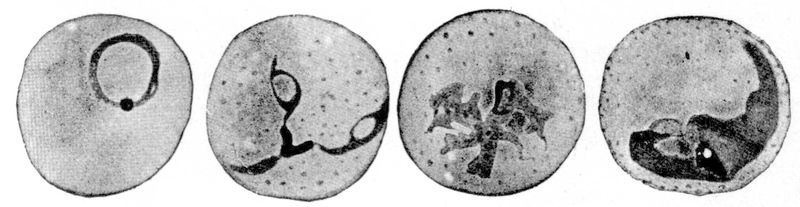
Fig. 3.—Plasmodium vivax.(Benign tertian) Development of schizonts of nonsexual cycle in peripheral blood of man. Red cell swollen and stains feebly. Note Schüffner’s dots. X 2200. (MacNeal after Doflein.)
The gametes, or sexual parasites, show a thicker blue ring and have the chromatin dot in the center of the ring. The pigmentation of the half-grown gametes is more marked than that of schizonts of equal size. The shape of the gametes is not amoeboid, as is that of the twenty-four to thirty-six-hour-old schizont, but round or oval. The full-grown gametes have the pigment distributed and the chromatin in a single aggregation—just the opposite of nonsexual parasites. The male gamete stains a light grayish blue and has a very large amount of chromatin, usually centrally placed. The female gamete stains a pure blue, has only about one-tenth as much chromatin as plasma, with the chromatin often placed at one side. The pigment of the female gamete is dark brown while that of the male is yellowish brown.
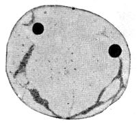
Fig. 4.—Plasmodium vivax. (Benign tertian.) Double infection of a red blood cell which is enlarged and shows Schüffner’s dots. X 2200. (MacNeal after Doflein.)
Plasmodium Malariae.—In fresh preparations the young quartan schizont has only slight amoeboid movement and, as development proceeds, the rather dark brown, coarse pigment tends to arrange itself peripherally about the band-shaped or oval parasite.
The infected red cell shows but little change. At the end of seventy-two hours the rather regular daisy form of the merocyte is more distinct than that of the benign tertian merocyte.
The distinctions between the male and female gametes are similar to those of the benign tertian gametes. In Romanowsky-stained smears it is difficult to distinguish the young quartan schizont from the benign tertian one but, after twenty-four hours, the tendency of the quartan schizont to assume equatorial band forms across a red cell of normal size and staining characteristics and without Schüffner’s dots makes the differentiation easy. In the fully developed sporulating parasite or merocyte the eight merozoites assume a regular distribution, giving it a daisy appearance.
The gametes show practically the characteristics of the benign tertian ones but are smaller.
Plasmodium Falciparum.—The young schizont of malignant tertian is extremely difficult to detect in fresh preparations, there being noted early in the rather long continued, hot stage, as small crater-like dots, about one-sixth of the diameter of a red cell which, however, show an active amoeboid movement.
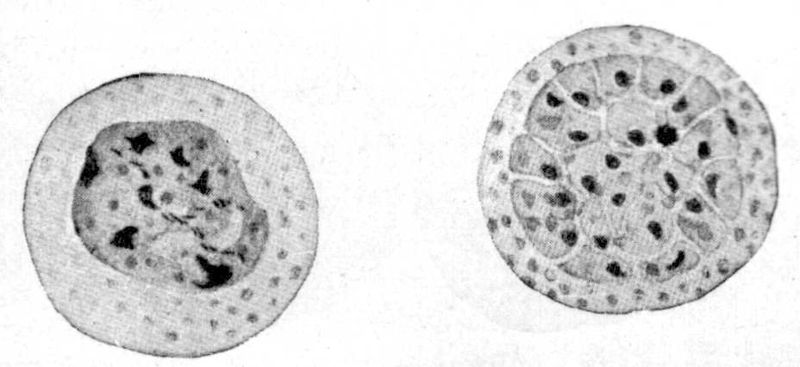
Fig. 5.—Plasmodium vivax. Mature schizont and merocyte. Found in the blood just before and at onset of chill. X 2200. (MacNeal after Doflein.)
Malignant tertian blood tends to show rather marked vacuolation of the red cells and these central vacuoles have a resemblance to young ring forms. The malarial parasites are most often peripherally placed and they do not enlarge and diminish in size on focusing up and down as do the vacuoles.
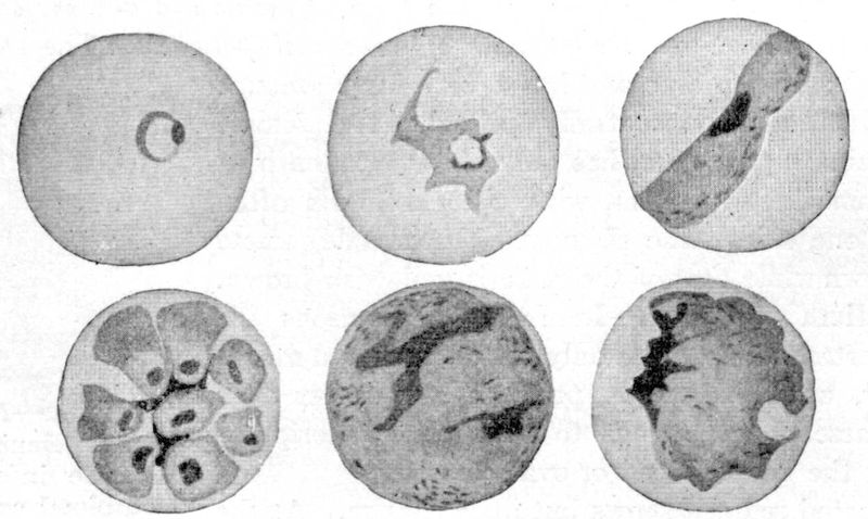
Fig. 6.
—
Plasmodium malariae.
(Quartan.) Development of nonsexual parasite in blood of man. X 2200. (From MacNeal after Doflein.)
Later on in the hot stage these ring-like dots enlarge to become about one-third of the diameter of a red cell, most often occupying the periphery of the infected red cell. About this time, or at the very commencement of the pigmentation, the schizont-containing red cells disappear from the peripheral circulation so that the further development is rarely observed in blood specimens.
The infected cell is brassy in color and shrunken in shape—it shows evidences of degeneration. The gametes appear as crescent-shaped bodies, which are absolutely characteristic of malignant tertian, the male gamete being more hyaline and delicate while the female one is more granular and larger.
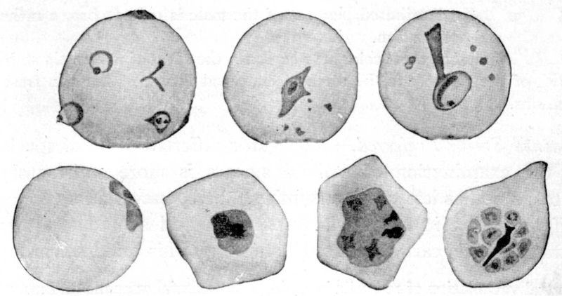
Fig. 7.—Plasmodium falciparum. (Malignant tertian) Nonsexual cycle in blood and internal organs of man. Note multiple infections of single red cell. (From MacNeal after Doflein.)
In Romanowsky-stained preparations we see, while the fever is sustained, small hair-like rings, with geometrical outline, with frequently two chromatin dots in one end of the ring and a single red cell often showing two or more of these young rings. The rings are often seen as if plastered on the periphery of the red cells or as if having destroyed a rounded section of the rim of the red cell. As the fever declines the rings tend to disappear from the peripheral circulation. The infected red cells often show polychromatophilia and distortion.
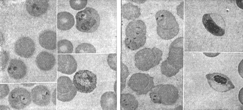
Fig. 8.—Tertian malarial parasite, one red cell showing malarial stippling. (Todd.)
Fig. 9.—Estivo-autumnal malarial parasites, and small ring forms and crescents. (Todd.)
In old aestivo-autumnal cases, or those with severe infection, we may see adult rings and merocytes, which latter are smaller than those of benign tertian, show from 10 to 12 irregularly placed merozoites and a sharply clumped mass of pigment.
The gametes are the striking crescent-shaped bodies and these show the distinctions of blue-staining for the female, with lighter gray-blue to purplish staining and abundance of chromatin for the male. The chromatin staining of crescents does not stand out so well as that of the round form gametes of benign tertian and quartan.
The black pigment of the female tends to be clumped toward the center while the rather generally distributed pigment of the male is reddish brown rather than black in a stained preparation.
This variation of pigment color may be due to the effect of chromatin staining, as the black of the pigment is the same in male and female gametes in fresh blood preparations.
Stained Smear Preferred.—As regards differentiation of species and cycle the examination of stained smears is more satisfactory and definite, as well as less time consuming. Still, one obtains many points of differentiation in the fresh preparation and should study such a preparation while carrying out the staining of his dried smear.
Unstained Specimen (fresh Blood)
P. vivax (benign tertian) P. malariae (quartan) P. falciparum (malignant tertian) (aestivo-autumnal)
Character of the infected red cell. Swollen and light in color after eighteen hours. About the size and color of a normal red cell. Tendency to distortion of red cell rather than crenation. Shriveled appearance. (Brassy color.)
Character of young schizont. Indistinct amoeboid outline. Hyaline. Rarely more than one in r.c. Active amoeboid movement. One-third diam. of r.c. Distinct frosted glass disc. Very slight amoeboid motion. Small, distinctly round, crater-like dots not more than one-sixth diameter of red cell. Two to four parasites in one red cell common. Shows amoeboid movement until appearance of pigment.
Character of mature schizont. Amoeboid outline. No amoeboid movement. Rather oval in shape. Sluggish movement of peripherally placed coarse black pigment. Only seen in overwhelming infections. Have scanty fine black pigment clumped together.
Pigment. Fine yellow-brown, rod-like granules which show active motion in one-half-grown schizont. Motion ceases in full-grown schizont. Coarse almost black granules. Shows movement only in young to half-grown schizont. Pigmented schizonts very rare in peripheral circulation except in overwhelming infections. Tends to clump as eccentric pigment masses almost black in color.
Stained Specimen
P. vivax (benign tertian) P. malariae (quartan) P. falciparum (malignant tertian) (aestivo-autumnal)
Character of infected red cell. Larger and lighter pink than normal red cell. Shows “Schüffner’s dots.” About normal size and staining. Shows distortion and some polychromatophilia and stippling. Rarely we have coarse cleft-like reddish dots—Maurer’s spots.
Character of young schizont. Chromatin mass usually single and situated in line with the ring of the irregularly outlined blue parasite. Rather thick round rings which soon tend to show as equatorial bands. Very small sharp hair-like rings, with a chromatin mass protruding from the ring. Often appears on periphery of red cell as a curved blue line with prominent chromatin dot. Frequently two chromatin dots.
Character of half-grown schizont. Vacuolated or Fig. 8 loop-like body with single chromatin aggregation. Schüffner’s dots. More marked band forms stretching across r.b.c. Not often found in peripheral circulation. Chromatin still compact.
Character of mature schizont. Fine pigment rather evenly distributed in irregularly outlined parasite. Coarse pigment rather peripheral- ly arranged in an oval parasite. Very rarely seen in peripheral circulation in ordinary infection. Pigment clumps early.
Character of merocyte. Irregular division into 15 or more spore-like chromatin dot segments. Rather regular division into eight or ten merozoites—Daisy. Sporulation occurs in spleen, brain, etc. Rarely in peripheral circulation. 6 to 10 irregularly placed merozoites. (In culture 32.)
Character of macrogamete. Round deep blue. Abundant, rather coarse pigment, chromatin at periphery. Round, similar to P. vivax but smaller. Crescentic, pure blue pigment clumped at center, chromatin scanty and in center.
Character of microgametocyte. Round, light green-blue, pigment less abundant, chromatin abundant and located centrally or in a band. Round like P. vivax. More sausage-shaped than crescent. Light grayish blue to purplish. Pigment scattered throughout. Chromatin scattered and in greater quantity but difficult to stain.
Central vacuolation of red cells is common in malarial anaemia and may be mistaken for nonpigmented parasites.
Malarial rings are usually peripheral and do not vary in size as one focuses up and down as do the central vacuoles.
Quinine-affected Parasite.—A very puzzling but well-recognized finding in cases treated with quinine or salvarsan is the so-called quinine-affected parasite. Such parasites lack definiteness of outline and show poor chromatin staining. The gametes do not seem to show these effects from the drug.
Certain questions connected with the life history of the malarial parasite in man which are of interest.
1. Extracellular location.—It is usual to consider the parasite as developing within a red cell and in this position to destroy the red cell. Rowley-Lawson, however, thinks that the parasites are exclusively extracellular and that they adhere to the red cells by loop-like pseudopodia which encircle a portion of the red cell and digest the haemoglobin of such an area.
2. Relapses.—There are several views as to the etiology of relapses in malaria. These views are taken up under relapses (see page 35).
3. Malarial toxin.—Nature of the toxic material thrown off by the parasite at the time of simultaneous sporulation. Rosenau’s experiments tend to show that there is a fever-producing toxin thrown off at this time. Other authors have thought that a haemolysin and an endotheliolysin were thrown off at the same time. Brown considers that the pigment produced by the parasite, in its metabolism of the haemoglobin of the red cell, may act as a haemolysin, he having found that intravenous injections of haematin were capable of producing marked anaemia. It is well known that a far greater number of red cells are destroyed in a paroxysm than would be accounted for by the actual percentage of red cells destroyed by parasites. The endothelial cells take up actively this malarial pigment or haemozoin and are damaged or destroyed thereby. Haematin injections also tend to destroy leucocytes and platelets.
Rowley-Lawson is of the opinion that the greater red cell destruction than would be represented by percentage of cells showing parasites is explained by parasites migrating from cell to cell so that many red cells may be destroyed by a single parasite.
4. Transmission to larvae.—There has been an idea that sporozoites might enter the ovaries and ova as well as the salivary glands so that a second generation of mosquitoes might transmit malaria. There is no proof that such a method is ever operative.
5. Congenital malaria.—There has been some question as to the possibility of congenital malaria. Heiser has recently reported the case of an infant which showed crescents in its blood by the end of one week from birth. The mother showed the same infection and the child must have been infected through the placental circulation.
Clark in numerous examinations of the blood of the new-born failed to find infection even when the mother’s blood teemed with parasites. In one case where the child showed infection shortly after birth there had been an accident to the placenta and he believes that instances of so-called congenital malaria are to be explained in this way.
6. Cultivation of parasite.—As to cultivation of malarial parasites. Bass takes from 10 to 20 cc. of blood from the malarial patient’s vein in a centrifuge tube which contains 1/10 cc. of 50% glucose solution. A glass rod, or a piece of tubing extending to the bottom of the centrifuge tube is used to defibrinate the blood. After centrifugalizing there should be at least one inch of serum above the cell sediment. The parasites develop in the upper cell layer, about 1/50 to 1/20 inch from the top. All of the parasites contained in the deeper-lying red cells die. To observe the development, red cells from this upper 1/20 inch portion are drawn up with a capillary bulb pipette.
Should the cultivation of more than one generation be desired, the leucocyte upper layer must be carefully pipetted off, as the leucocytes immediately destroy the merozoites. Only the parasites within red cells escape phagocytosis. Sexual parasites are much more resistant. Bass thinks he observed parthenogenesis. The temperature should be from 40° to 41°C. and strict anaerobic conditions observed. Aestivo-autumnal organisms are more resistant than benign tertian ones. Dextrose seems to be an essential for the development of the parasites.
Bass considers that P. vivax has a disc-like structure which enables it to squeeze through the brain capillaries while adult schizonts of P. falciparum have a solid oval form which causes them to be caught in the capillaries.
The Thompsons have rather simplified the method of Bass. They draw 10 cc. of blood into a test tube containing the usual amount of glucose solution. They then defibrinate the blood by stirring with a thick wire for about five minutes and remove the wire with the adhering clot. They then pour this defibrinated blood into several small sterile test tubes, which should contain at least a one-inch column. Rubber caps are adjusted over the cotton plugs and the tubes placed in the incubator. They note the tendency of cultures of P. falciparum to agglutinate which is not true of P. vivax.
They think this agglutination the great cause of the plugging of capillaries in pernicious malaria. They note 32 merozoites as maximum number in sporulation of P. falciparum while P. vivax has usually 16 or more, but never as many as 32.
This would explain the shorter incubation period of malignant tertian. The pigment of P. falciparum clumps much earlier in the developing schizont than that of P. vivax and is much coarser and more discrete.
While Bass thought he noted parthenogenesis in cultures others have failed to observe any evidence of it.
7. Immunity.—As to immunity. There is no real immunity to malaria, it is a continuance of the infection, but the parasites are not in sufficient numbers to give rise to fever. If, however, the patient becomes chilled or fatigued or otherwise depressed, fever results.
This apparent immunity is also kept up by reinfection, because if natives leave the locality for a length of time they lose it. Patients who show this apparent immunity to one form of malaria have no such resistance to the other types. Bass states that immune bodies are produced in malaria and that immune processes contribute to control of the infection, but that it is not lasting and is not effective against new infection.
8. Perniciousness.—Causes of perniciousness. This is taken up under perniciousness in malaria. (See page 31.)
9. Quinine-affected parasite.—Effect of quinine on malarial parasites. It is usually thought that the merozoites at the time of being thrown off from the merocyte are most vulnerable, while the gametes are only slightly affected, if at all. Still, the young forms from which gametes develop are destroyed. Quinine causes parasites to disappear from the peripheral circulation and produces degenerative changes in such parasites as may remain. Bass thinks that quinine makes the red cell permeable to the lytic action of serum. Anaemia may cause degenerative changes in parasites similar to that from quinine.
10. Anaphylaxis and the paroxysm.—Abrami has brought forward evidence in favor of the malarial paroxysm being due to the outpouring of merozoites into the blood plasma which act as foreign antigen. It is noted that the dissemination of merozoites takes place some hours before the cold stage which is one of the manifestations of anaphylactic shock. They note a leucopenia and lowering of the blood pressure preceding the paroxysm as evidence of a haemoclastic crisis.
The Anopheline Mosquito
The ova of culicine mosquitoes are usually deposited in a scooped-out raft-like mass of about 250 eggs set vertically. The raft is easily seen with the eye, being about ⅕ inch long. The anopheline eggs are oval in shape with pleated air cell projections laterally. They are laid upon the surface of the water, to the number of about 100, in star, triangle or ribbon patterns. The egg stage is two to four days but shorter, however, in the tropics.
The larval stage is the most important one to be acquainted with because in this stage one can most readily distinguish the anopheline or possible malaria transmitter from a culicine species. One can more readily and quickly make a survey for anophelines by examining the collections of water for larvae than in any other way. The anopheline larva seems to prefer the surface, on which it lies flat and out of the water. To keep it from turning over on its long axis, it has little rosette-like hair tufts on the dorsal surface of the 5 or 6 middle abdominal segments (palmate hairs). There are feathered lateral hairs projecting from thorax and abdominal segments. The head is very small in comparison with the thorax and can be rotated with lightning-like rapidity. There is no projecting breathing tube or syphon from the next to the last abdominal segment, as is characteristic of Culex, Stegomyia or any other culicine genus.
In addition, culicine larvae do not float parallel to the surface of the water, but hang suspended at an angle, with only the tip of the syphon pushed upward to the surface. The lateral hairs or bristles are not feathered and the head is much larger than that of the anopheline larvae. It is the fact of the surface position of these anopheline larvae which enables them to worm their way over film layers of water or between blades of grass, in grass or rush studded pools or swamps.
In the pupal stage it is rather difficult to differentiate species of mosquitoes from each other, so that, other than to recognize that the bloated shrimp-like body is a mosquito pupa, is unnecessary.
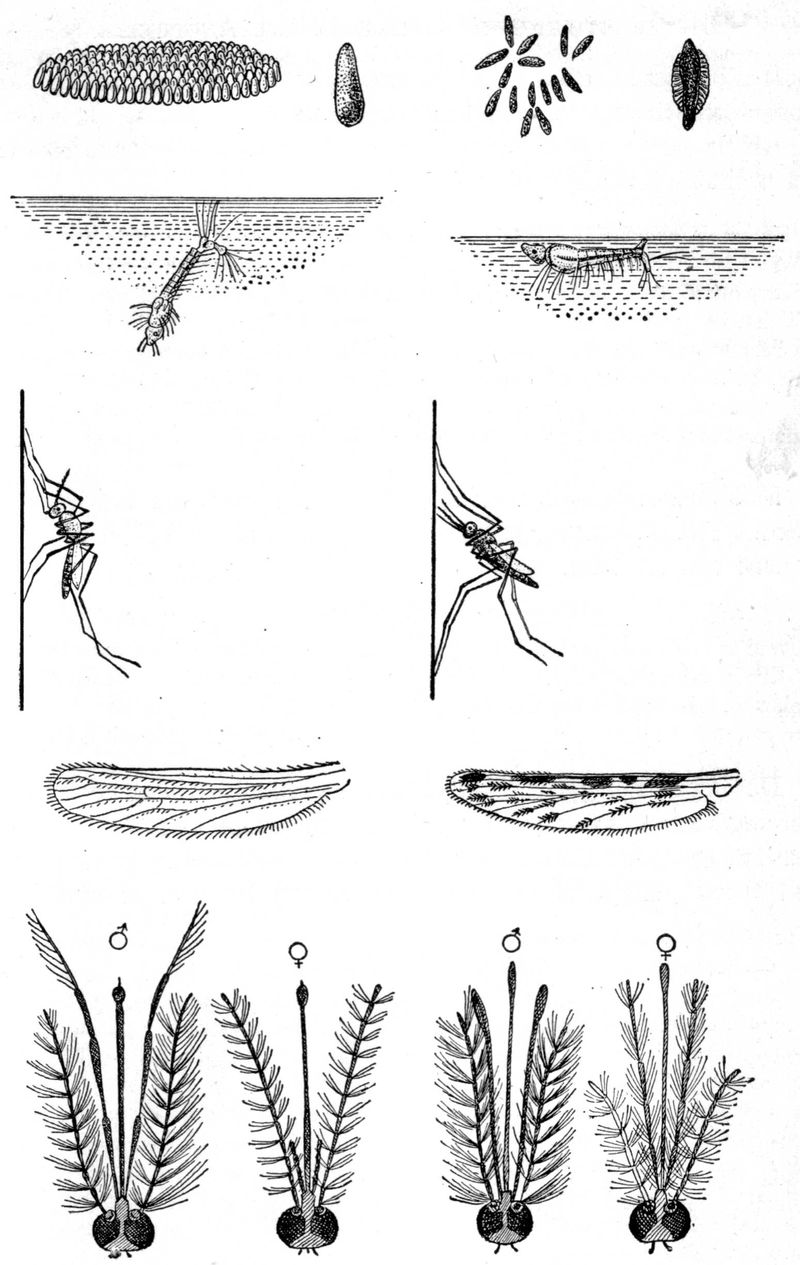
Fig. 10.—In the above figure note the culicine egg raft, 45° angle position of syphonate larva, parallel attitude of resting mosquito, nonbulbous palpi of male and short palpi of female as contrasted with the anopheline star or ribbon arrangement of eggs, horizontal attitude of asiphonate larva, bradawl attitude of resting mosquito, spotted wings, bulbous palpi of male and long palpi of female mosquito. (From Jordan after Kolle and Hetsch.) MacNeal.
Differentiation of Culicinae and Anophelinae
It is impossible even for an entomologist to determine the species of mosquitoes without recourse to elaborate keys and tables. It is a comparatively easy matter, however, to decide as to whether the mosquito is a probable malaria transmitter or not.
The male anopheline.—While certain characteristics of the male are used to separate the Aedinae from other subfamilies, yet it is only with the female that we concern ourselves in differentiating the Culicinae from the Anophelinae. Therefore, it is first necessary to distinguish the male from the female. If the antennae have not been torn off, this can be decided by the highly adorned plumose antennae of the male, those of the female being sparsely decorated with short hairs. The palpi of the male Anopheles tend to be clubbed, while those of the Culex are straight. If the antennae have been broken off, look for the claspers at the end of the abdomen.
Male mosquitoes do not feed on blood but on fruits and flowers instead. The puncturing parts of the male are not sufficiently resistant to penetrate the skin.
The female anopheline.—Having determined that the insect is a female, we then proceed to place it either in the subfamily Culicinae or Anophelinae by a study of the relative length of the palpi to the proboscis. If the palpi are much shorter than the proboscis, it belongs to the Culicinae; if about as long or longer, to the Anophelinae. The palpi of the female Megarhininae are also long, but the proboscis is curved.
Having settled on the subfamily, we separate the genera by considering such points as character and distribution of scales on back of head, wings, thorax, and abdomen; banding of proboscis, legs, abdomen, and thorax, shape of scales on wings, and location of cross veins.
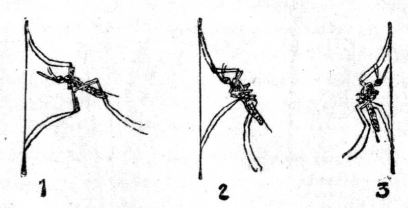
Fig. 11.
—Resting posture of mosquitoes; 1 and 2,
Anopheles
; 3,
Culex pipienes
. (
After Sambon.
) From P. H. Reports.
Anophelinae show abundant upright forked scales on occiput. The mesothorax shows sparse hairs or scales with a smooth scutellum. As a rule, the wings are spotted (dappled) and the location of these spots gives the best clue to the different species of the genera. With the exception of Bironella the first submarginal cell is large. This cell is longer than the second posterior one.
In the resting position Culex allows the abdomen to droop, so that it is parallel to the wall. The angle formed by the abdomen with head and proboscis gives a hunchback appearance.
Anopheles when resting on a wall goes out in a straight line at an angle of about 45°. It resembles a bradawl.
The scutellum of Anopheles is simple, that of Culex trilobed. Anopheles has but one spermatheca; Culex has three.
Anophelinae
┏ 1. Scales on wings, large and lanceolate. Anopheles. Palpi only slightly scaled.
1. Scales on head only; hairs on thorax and abdomen. ┃ 2. Wing scales small and narrow and lanceolate. Myzomyia. Only a few scales on palpi.
┗ 3. Large inflated wing scales. Cycloleppteron.
2. Scales on head and thorax (narrow curved scales). Abdomen with hairs. [ 1. Wing scales small and lanceolate. Pyretophorus.
┏ 1. Abdominal scales only on ventral surface. Thoracic scales like hairs. Myzorhynchus. Palpi rather heavily scaled.
3. Scales on head and thorax and abdomen. Palpi covered with thick scales. ┃ 2. Abdominal scales narrow, curved or spindle-shaped. Abdominal scales as tufts and dorsal patches. Nyssorhynchus.
┗ 3. Abdomen almost completely covered with scales and also having lateral tufts. Cellia. 4. Abdomen completely scaled. Aldrichia.
Note.—Of the above genera only Cycloleppteron and Aldrichia are unproven malarial transmitters.
The female anopheline mosquito alone bites man, the male feeding on fruits and flower juices. The female absolutely requires blood for the development of her eggs after fertilization by the male mosquito.
The anopheline mosquito bites at night or toward evening and selects some dark place or dark colored wall to sleep against during the day. Hence the advantage of a buff colored wall interior. It is well to remember that the malarial incidence may be kept down by killing the mosquitoes inside of a house by striking them with a folded paper or piece of wire gauze on a handle (fly swatter).
It is not a bad plan to have a dark colored surface in a room to attract them and make their destruction easy.
Anophelines do not like wind and seek protection of underbrush. As regards distance of flight from breeding places Metz has noted that A. crucians were not distributed generally over 7000 feet and rarely were found between 7000 and 9000 feet beyond which distance they were not found. Some anophelines get accustomed to feeding exclusively on animals. Mosquitoes may hibernate through the winter and possibly cause new infections the following spring. Cases of malaria in the spring are however usually due to relapses. Mitzmain’s negative experiments with hibernating mosquitoes prove man to be the winter carrier.
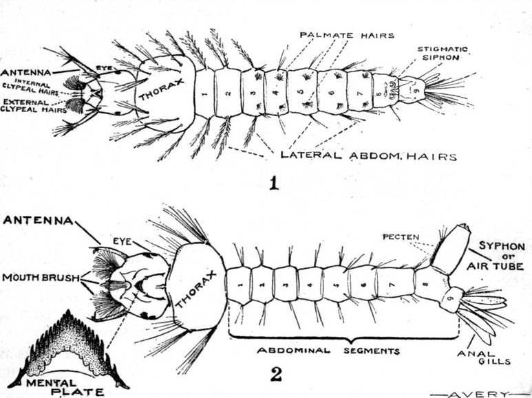
Fig. 12.
—Asiphonate (Anopheline) larva
Anopheles
. 2 Siphonate (Culicine) larva
Stegomyia
The malarial zygote will not develop in the stomach of the mosquito if the temperature is below 16°C. (60°F.). It would seem that the zygote of P. malariae will develop at a lower temperature than that of the other two species, P. falciparum requiring the highest temperature.
Our views as to temperature requirements for the development of zygotes in the mosquito must be changed as King has recently shown that P. vivax sporonts will survive exposure to temperatures of 30°F. for two days and P. falciparum temperatures of 35°F. for one day. This proves that temperatures approximating freezing ones will fail to destroy the parasites of hibernating mosquitoes.
Wenyon has found experimentally that mosquitoes which had fed on malarial blood and kept at incubator temperatures for a week to allow development of zygotes showed inhibition of development of zygotes when kept at temperatures corresponding to hibernating ones. This treatment did not kill the zygotes but complete development took place when subsequently the mosquitoes were again subjected to incubator temperatures. From this it would seem that the zygotes remain viable during the winter hibernation. This is at variance with Mitzmain’s views who regarded hibernation as destructive to zygotes.
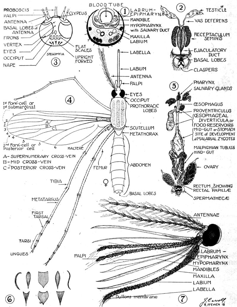
Fig. 13.
—Anatomy of the mosquito. No. 6 shows various types of scales.
The mosquito does not seem to suffer from her malarial infection—quite different from the serious affection that filariasis causes in the mosquito.
Epidemiology.—This matter has been considered rather extensively under the historical and etiological discussions.
It may be stated however that the requirements for the spread of malaria are: (1) Men who have sexual forms of the malarial parasite in their peripheral circulation; (2) efficient anopheline hosts, and (3) an atmospheric temperature above 60°F. (16°C.).
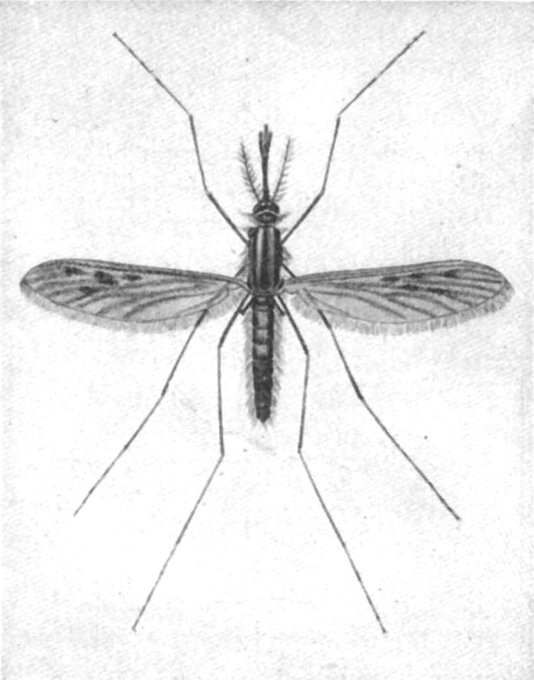
Fig. 14.
—
Anopheles maculipennis (quadrimaculatus)
, female. (
Castellani and Chalmers, after Austen.
) From P. H. Reports.
It is a well recognized fact in the tropics that the natives seem to have an immunity to malaria yet may carry parasites in their circulation and serve as carriers. The native children to a striking degree harbour parasites and to them malaria is a prime cause of death. After repeated infections, if not fatal, a temporary immunity is acquired. Many localities in the tropics owe freedom from malaria to an absence of anophelines, as for instance Barbadoes. Again malaria-bearing mosquitoes may acquire the habit of feeding on animal blood other than that of man. It is well recognized that rural populations are more liable to malaria than those of towns and as the population of a country moves to the industrial centres human blood may become difficult to obtain and the anophelines turn to other sources of blood supply. It has been suggested that mosquitoes may suffer from other infections which may be inimical to the development of malarial zygotes (black spores of Ross). Anophelines bite chiefly at sunset and at night from which fact there would seem to be some value in shutting the windows towards nightfall as is the custom in many malarious parts of the world.
Pools containing a border growth of grass or rushes are often selected by anophelines for depositing eggs. The small fish or tadpoles, which prey on the larvae, cannot work their way through the obstacles and, again, petroleum oil cannot be easily distributed in a network of grass. Anophelines of different species and of different countries seem to vary in their selection of water for depositing their eggs. We should not generalize but go out and search for breeding places.
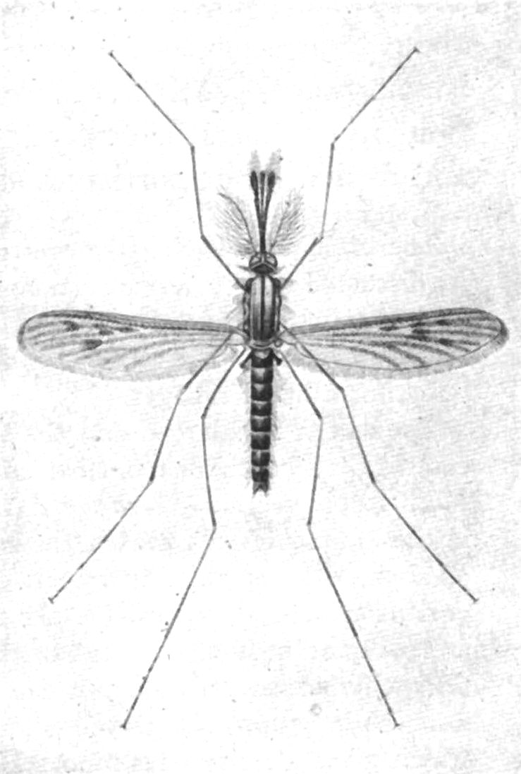
Fig. 15.
—
Anopheles maculipennis (quadrimaculatus), male.
(
After Castellani and Chalmers
.) From P. H. Reports.
Anophelines seem to prefer small collections of water or sluggish clear streams. The pools made by excavations following railway or other similar construction are favorite breeding places. Proper cultivation of rural districts makes the country more healthful and Carter has stated that tile drainage is the key to rural malaria control.
The most practical method for the identification of anopheline species is to collect the larvae and later to study the adults which develop from the pupae. On the whole culicines do not seem to object to foul collections of water while anophelines avoid such breeding places.
Pathology and Morbid Anatomy
The pathological lesions are those connected with the destruction of enormous numbers of red cells, not only each infected red cell being destroyed but others not so parasitised. There has been an idea that at the time of sporulation and rupture of the merocyte a pyrogenetic toxin was given off and along and with this there were haemolysins and endotheliolysins. Following Brown we are justified in thinking that the malarial pigment (melanin or haemozoin) can act as a haemolysin and by being taken up by endothelial cells bring about their degeneration with associated capillary haemorrhages. All three factors—red-cell destruction by parasites, haemolytic action on red cells and capillary haemorrhages lead to anaemia.
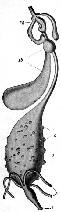
Fig. 16.—Digestive tract of Anopheles the stomach of which is covered with numerous zygotes or oocysts of Plasmodium falciparum. c, cloaca; mt, malpighian tubules; o, oocyst; s, stomach; sb, sucking bladders or pumping organ; sg, salivary gland. (MacNeal from Doflein, modified after Ross and Grassi.)
The brain has a leaden hue caused by the black pigment. As discussed under pernicious manifestations the blocking of the capillaries may be explained in several ways. When examining sections of a malarial brain one often encounters punctiform haemorrhages.
The spleen is enlarged and the surface dark. In acute cases it may be diffluent instead of hard, as in ague cake. Microscopic sections show a striking absence of pigment in the Malpighian corpuscles, the haemozoin being pushed off into the surrounding spleen pulp. Bone marrow is dark from deposit of pigment. In the liver the endothelial and Kupfer cells are packed with black pigment. The parenchymatous cells do not contain this pigment but may show grains of a yellow pigment, haemosiderin, which gives the iron reaction. Haemozoin, although it contains iron, does not give this reaction. Haemozoin is soluble in alkalis, but not in alcohol while haemosiderin is soluble in alcohol but not in alkalis.
The splenic blood is more rich in haemozoin than that of the other vessels, this indicating the spleen as the place of destruction of infected red cells or as the nursery for the development of malarial parasites. As a matter of fact splenectomy may cure an old malarial cachectic.
The finding of pigmented mononuclears or pigmented parasites in a cross section of a blood vessel makes for a diagnosis of a malarial infection.
Malarial manifestations are common in tropical autopsies and one must be very chary about reporting malaria as the real rather than contributing cause of death.
There is usually a marked increase in large mononuclears in malaria and if this is noted along with a leucopenia it is very suggestive. Melaniferous leucocytes occur in malaria only.
The kidneys may show degenerative changes and the presence of urobilin in the urine is an important indication of latent malaria.
Symptomatology
Clinically, we have two types of malarial paroxysms, (1) Those presenting a cold stage, followed by a hot stage, with a terminal sweating stage. Such attacks are brought about by the benign infections which include the benign tertian and the quartan. Owing to the fact that in such paroxysms the temperature makes a critical fall to normal or subnormal readings such fevers are frequently designated intermittent fevers.
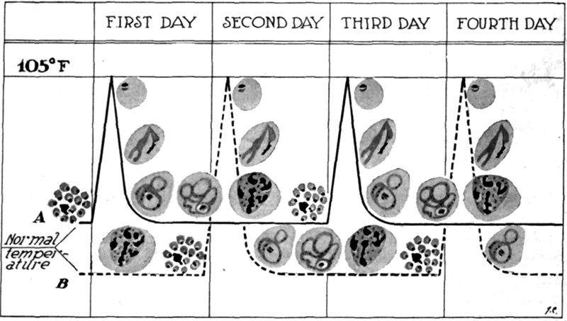
Fig. 17.—Diagram of the temperature chart of a double tertian malarial fever showing the succeeding development of two generations of parasites, causing thereby a quotidian fever. The solid line, A, shows the development of the generation of parasites first introduced and the dotted line, B, the cycle of the generation introduced later on.
While these benign infections rarely or never exhibit pernicious manifestations, they may, equally with the more dangerous aestivo-autumnal parasite, lead to the production of malarial cachexia, in which the clinical manifestations are similar whether produced by a benign or malignant species.
(2) Those in which the succession of cold, hot and sweating stages is lacking. There is not the frank well-defined chill of the former group, so that the term dumb chill is frequently applied. With the possible exception of the first paroxysm the temperature tends to remain well above normal giving a continuous, or at any rate a remittent type of fever, instead of the intermittent temperature curve of the benign infections. The designation remittent fever, is often applied to such fevers. Clinically there is a resemblance to typhoid fever.
Such malarial fevers are caused by the small hair-like ring parasite with its crescent sexual forms. There are many designations for this type of malarial fever of which the best recognized are: malignant tertian, subtertian, aestivo-autumnal and tropical. It is preëminently the malarial fever of the tropics and, from its appearing in temperate climates chiefly in the late summer and through the autumn months, received from the Italians the designation aestivo-autumnal.
Such fevers were called subintrant by Torti because the succeeding paroxysm set in before the completion of the long-continued preceding one. This type of fever was greatly dreaded. The designation malignant tertian is to be preferred as indicating the greater seriousness of this type of malaria.
Incubation Period
Depending in great part on the number of sporozoites introduced by one or more infecting anophelines at the time of biting we have with quartan fever (8-12 merozoites) a period of incubation of approximately three weeks, for benign tertian (16-24 merozoites) two weeks and for malignant tertian (32 merozoites in culture) eight to twelve days. These periods however may be much longer.
Prodromata
There may be prodromata of the nature of malaise, vague muscular pains, headache and anorexia, possibly showing a periodicity in their appearance or intensity.
It is only when a sufficient number of parasites sporulate simultaneously and pour out into the circulation sufficient toxic material to cause a well-marked paroxysm that such occurs—with less poison we may only have vague suggestions of an attack of ague.
In a large proportion of cases there are no prodromata, they begin with a sudden onset.
Malarial paroxysms show a preference for the forenoon or at any rate tend to occur in the early afternoon, rather than in the evening.
Mixed and Multiple Infections
When there are two generations of tertian parasites, each maturing on successive days, we have a paroxysm every day—a quotidian fever. Such a tertian infection is called a double tertian. In quartan infections, with the seventy-two-hour cycle of development, if we have two generations of parasites sporulating on succeeding days, but with an apyretic day intervening, we have a double quartan. If three generations of quartan parasites sporulate on three successive days we have a triple quartan infection. When two species of parasites are present in the same case we have a mixed infection. Mixed infections of malignant tertian and benign tertian are the most common, next, those of quartan and malignant tertian and very rarely those showing quartan and benign tertian. All three species have been found in a single individual.
Clinical Types
A Typical Benign Tertian or Quartan Paroxysm.—(Other than for the difference in periodicity the paroxysms of these two malarial infections are alike.)
The ague attack generally commences with malaise and slight headache, frequently accompanied by yawning and stretching. Chilly sensations radiating from the spinal column to the extremities and the jaws give way to actual chill with shaking body and chattering teeth, face pinched and bluish and cutis anserina.
The pulse is frequent, small and of rather high tension, there is increased frequency of urination and nausea and vomiting may be present.
Notwithstanding the fact that the rectal temperature is steadily rising five or six degrees during this cold stage there is a desire on the part of the patient to cover himself with all the wraps obtainable.
The cold stage, which usually lasts from twenty to sixty minutes, is succeeded by the hot stage.
At first there is a feeling of slight relief from the misery of the chill but this is soon lost sight of in the increasing headache and feeling of intense heat.
The previously welcome blankets are cast aside. The face now becomes flushed, the eyes shining, and the pulse more full. Epigastric discomfort, nausea and vomiting are apt to become more prominent in this stage. The patient often complains of a throbbing headache. It is at this time that he may become slightly delirious. A sense of tension or even pain may be experienced in the region of the spleen, which organ will be found tender even if not already palpable. Herpes about the nose and lips is almost as common as in lobar pneumonia.
An attending bronchitis is not uncommon.
The fever remains high, from 105° to 106°F., and continues so elevated for from four to six hours to be succeeded by the sweating stage. In this the dry skin becomes moist and perspiration breaks out first on the forehead to be followed by a more or less marked profuse sweating of the entire body. The pulse becomes slower, the temperature falls rapidly and the patient falls asleep to awake slightly exhausted but feeling well.
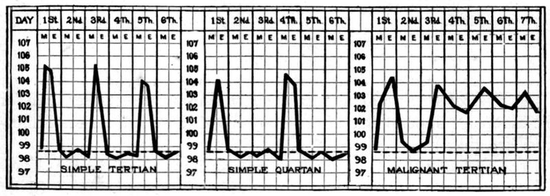
Fig. 18.
—Typical fever charts of the 3 types of malaria.
This feeling of well-being continues during the fever-free day which is often referred to by a patient as “my good day.”
The sweating stage lasts usually about four hours so that the entire paroxysm of cold, hot and sweating stages occupies approximately eight to twelve hours.
While most cases of the benign infections show the typical stages yet we meet cases where the cold and sweating ones are absent or but slightly marked.
Blood examination will show the parasites of the benign infections to be in the peripheral circulation during the entire apyrexial period. During the paroxysm we have a moderate leucocytosis and during the afebrile period a leucopenia with an increased percentage of large mononuclears.
Billet thinks that quartan paroxysms can be distinguished from benign tertian ones by their showing a less abrupt fever rise and a more rapid fall of temperature with a shorter duration of the paroxysm, four or five hours as against eight to twelve hours for benign tertian.
A Typical Malignant Tertian Paroxysm.—The characteristic features of the paroxysm are slight chilliness instead of a frank chill, prolonged and intensified hot stage, lack of marked terminal sweating and a tendency to exhibit a continuous or at least remittent fever curve instead of the distinct intermittence, with an apyrexial period, of the benign infections.
During the period of the remittence the patient fails to experience a sense of well-being. He is sick and does not have a “well day.”
The temperature of a malignant tertian paroxysm may fall to normal during the first attack but succeeding attacks only show the tertian periodicity by an exacerbation of the more or less continuous fever.
In these cases the temperature rise is gradual rather than abrupt and the fall rather by lysis than crisis.
The paroxysm lasts from twenty to thirty-six hours instead of ten hours.
To explain the continuous type of fever it is often stated that anticipation and retardation are characteristic of malignant tertian infections. This simply means that the new paroxysm tends to come on before the tertian periodicity of forty-eight hours has expired and, having appeared, tends to delay its termination. At any rate there is an extreme irregularity in the course of the paroxysm. These attacks are often termed “dumb chills” and are greatly dreaded.
The onset is insidious, occurring as a rule in the forenoon or early afternoon, with rarely a chill but only chilly sensations. The headache and backache are severe, the face is flushed, the pulse quickened and the thirst urgent.
The patient feels more prostrated and ill than does one in a benign paroxysm and there is a distinct tendency to mental confusion or delirium. Nausea and vomiting may be prominent features of an attack. At times an apathetic state may suggest typhoid fever. In these malignant malarial attacks the spleen is palpable and very tender. There is also a sense of weight in the region of the liver.
In a blood examination one is not apt to find any other parasites than the young hair-like ring forms which begin to appear a few hours after the onset of the paroxysm. The rings may be observed to broaden, but prior to that development in which pigment would appear in the ring, the parasite-containing red cell is caught in the capillaries of spleen or other organs. The finding of young ring forms while fever continues is suggestive of a malignant tertian infection.
In the absence of quinine administration the finding of parasites is to be expected in benign tertian and quartan infections, but with the tropical parasite a smear may fail to show any organisms where a few hours previously a blood examination would have shown a large percentage of infected red cells in every field of the microscope.
Pernicious Manifestations of Malaria.—These grave manifestations arise almost exclusively in the course of malignant tertian infections. In his study of malaria Stott had about 1% of his cases showing well-marked pernicious symptoms.
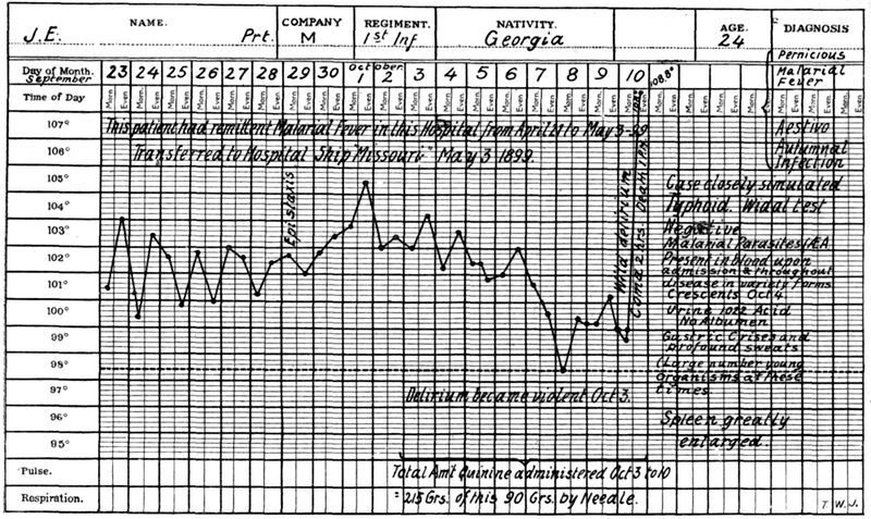
Fig. 19.
—Temperature chart of malignant tertian fever showing how readily one might confuse such a chart with that of typhoid fever. (From Jackson’s Tropical Medicine.)
As explanations of perniciousness are given: (1) the very large number of red cells infected and destroyed by the malarial parasites; (2) the throwing off at the time of sporulation of the merocyte of a large amount of toxic material owing to the presence of such a large number of disintegrating merocytes, and (3) from the plugging of the capillaries of important internal organs by adult parasites. This may arise as the result of (a) the sporulating parasites acting as emboli, being too large to pass the lumen of the capillary; (b) from degenerative changes or distension with pigment of the endothelial cells lining the capillaries, or (c) as the result of an ovoid shape on the part of the malignant tertian parasite there is an inability to pass through capillaries which the flattened benign parasites can do by infolding (Bass), or (d) resulting from the tendency of malignant tertian parasites to agglutinate.
Types of Pernicious Malaria.—It is customary to divide pernicious malaria into the following divisions—(1) Cerebral, (2) Algid, (3) Bilious Remittent and, possibly, also (4) Pneumonic and (5) Cardiac types.
Blackwater fever is often included in the grouping but would appear to be best considered as a separate disease although almost surely brought about by malaria.
We do not understand why in one case sporulating parasites should plug the capillaries of the central nervous system, with the production of conditions resembling well-recognized nervous diseases, while in another case the damage is done the intestinal mucosa, pancreas or lungs. At any rate these pernicious manifestations of malaria should always be kept in mind when a case of sudden cerebral involvement or acute abdominal disease shows itself in a patient in a malarious country and a blood examination should be promptly made.
Cerebral Manifestations of Pernicious Malaria.—Various authorities give different clinical pictures but the more commonly accepted types are:
(1) The hyperpyrexial, when the symptoms are those of heat stroke, with a temperature going up as high as 110°F. or even higher. Such patients rapidly become comatose and as a rule die.
(2) The delirious and comatose forms are apt to be associated, the comatose condition following a delirious state. Such manifestations may or may not set in with a chill. Cases belonging to this group may arise from a typical malignant tertian infection in which the headache and restlessness have been unusually marked. The pulse is full and fast with sighing respiration, hot dry skin and flushed face. There may be rigidity of the neck muscles.
(3) Such terms as epileptiform, tetanic, aphasic, cerebellar and bulbar have been applied to malarial manifestations and are self-explanatory.
Cerebral malaria may give rise to a delusional insanity. Various psychoses or amnesia at times follow cerebral types of pernicious malaria.
Algid Manifestation of Pernicious Malaria.—In such cases we have a small thread-like pulse and a cold clammy skin. There are signs of collapse. The respiration is slow and shallow and the voice weak. It is customary to consider some of these cases, when there is vomiting and diarrhoea, with painful cramps of the legs and scanty or suppressed urine, as of choleraic type, while other cases, with blood and mucus in the stools and marked abdominal pain are termed dysenteric. Most dysenteric types only show a diarrhoea with the presence of blood.
The dysenteric type is more common but the question always arises whether the case may not have been really dysentery lighting up a latent malaria or the lowering of resistance from the malaria favoring a dysenteric infection. Stott had five algid cases of dysenteric type but not one of choleraic. The choleraic types have often been reported during outbreaks of cholera.
When epistaxis and haemorrhages from the intestines or stomach are marked features of an attack the cases are termed haemorrhagic and, if a prostrating, collapse-producing sweat be a characteristic feature, they are called diaphoretic.
Cases have been observed when the excessive sporulation was apparently taking place in the pancreas, giving the symptomatology of acute haemorrhagic pancreatitis.
Bilious Remittent Fever.—This is the most common and the least dangerous of the pernicious manifestations but tends rapidly to produce malarial cachexia. Slight jaundice and bilious vomiting may appear in the course of an ordinary malignant tertian paroxysm and only severe types, with fatal tendency, should be classed as pernicious. It sets in with marked nausea followed by bilious vomiting and bile-rich stools. Jaundice shows itself by the second day; earlier than in yellow fever, but much later than the rapidly appearing jaundice of blackwater fever. The urine shows bile pigment and a yellow foam. Epigastric distress and liver tenderness are marked features and there may even be gastric haemorrhage.
Pneumonic and Cardiac Types.—Other recognized types are when, with the symptoms of a broncho-pneumonia, we find an element of periodicity and a response to quinine—the so-called pneumonic type.
Again, usually in elevated regions, dilatation of the right heart and death have been noted as occurring in cardiac types of pernicious malaria.
Another type is one in which the sweating stage is excessive, the so-called diaphoretic type. These cases may result in collapse and such a termination may be syncopal in character.
Relapses.—Relapses are distinct features of malarial diseases, the tendency being most marked in quartan and least so in malignant tertian. A relapse after an interval of two years is very rare in malignant tertian but periods as long as nine years may intervene between attacks of quartan fever.
Relapses are intimately associated with conditions which tend to lower the body resistance, so that exposure to cold or wet or hot sun may bring on an attack. Alcoholic or venereal excesses, as well as errors of diet, may be provocative. Persons returning home from the tropics often experience relapses as they approach the cooler climate of the temperate zone. It has been well stated that the old resident of the tropics owes his condition of health rather to education than acclimatization—experience has taught him discretion.
There are three explanations of relapses of which the one supported by Ross and Bignami seems more reasonable and is that the disappearance of nonsexual parasites is only apparent and that they continue their cycle but in insufficient numbers to excite symptoms.
Parthenogenesis.—Schaudinn thought that, either spontaneously or as the result of treatment, there was a disappearance of the nonsexual forms and the male gametes, the female gametes however surviving and, eventually, through the process of parthenogenesis, producing a set of spores or merozoites which set up a nonsexual cycle. It would seem probable that Schaudinn saw red cells containing a merozoite along with a female gamete and interpreted his findings as a sporulating sexual form.
Craig thinks that as the result of the conjugation of two young schizonts a more resisting parasite is evolved, which under favorable circumstances for its development may start anew a nonsexual cycle.
Latent Malaria.—The persistence of a malarial infection, in the absence of clinical and to a great extent of laboratory manifestations, is shown by the occurrence of relapses, so that the section treating of malarial relapses applies equally to this paragraph. In addition to the factors influencing relapses, such as exposure to sun, rain and excesses of various kinds, we note a particular tendency for a latent malaria to develop activity following surgical operations and childbirth. In untreated latent cases we may have delayed healing of surgical operations.
In another paragraph there is noted the importance of examining placental smears for evidence of a latent malarial infection. Persons returning to a cool climate from the tropics, who may not have shown evidence of active malaria for months, may come down with a paroxysm upon encountering cool weather (refrigeration). Latency may be complete or there may be vague manifestations of ill health such as anorexia, malaise, irritability, headaches, anaemia and alimentary tract disturbances. Not infrequently tropical residents without symptoms may show crescents in their blood and such cases are of prime importance in connection with infection of mosquitoes. To a certain extent they are the typical carriers and should be actively treated from a standpoint of malarial prophylaxis.
Masked Malaria.—While as a rule one should not accept such a diagnosis, unless the possibility of some other explanation than malaria is excluded, yet there are manifestations, chiefly neuralgic, gastro-intestinal or in the form of varied skin eruptions which at times show periodicity and which respond to treatment with quinine.
Fig. 20.—Abnormal malaria parasites. 1, Normal red corpuscle; 2, gametocyte and schizont; 3, gametocyte and gametocyte; 4, gametocyte and schizont; 5, schizont and schizont, both undergoing schizogony. (After Dr. J. D. Thompson, “Jl. R. A. M. C.”) By permission from Manson’s Tropical Diseases.
Malarial Cachexia.—As the result of repeated attacks of any type of malaria a condition of anaemia and physical and mental incapacity may be produced. The skin has a dirty earthy hue, particularly of the face, and the sclerae show a yellowish tinging. The patient is sensitive to the slightest cold and is the victim of mental depression with deterioration of memory or at any rate lack of concentration.
There may be long periods in which the temperature is normal or subnormal but slight febrile accessions may occur from time to time and at such times the blood may show parasites.
The spleen is enlarged as may also be the liver. Twisting of the pedicle of the spleen or its rupture from even slight blows may necessitate surgical intervention.
There is anorexia and alimentary tract disturbances. A very important feature of malarial cachexia may be the occurrence of haemorrhages, particularly serious being those from the retinal vessels.
It is probable that hookworm infection has frequently been confused with the anaemia of malarial cachexia as in both of these conditions we may have a high-grade anaemia with swelling about the ankles, palpitation of the heart and shortness of breath. Some authorities have recently called attention to splenic enlargement in hookworm disease, but this is not generally accepted. There may be also ascites in malaria. Urobilinuria is an important sign in malaria where other causes for red cell destruction are excluded.
The Sequelae of Malaria.—The anaemia and other manifestations of malarial cachexia have been described above. The enlarged spleen not only is a source of danger from rupture but it may cause sensations of pain or tension. The skin of those with chronic malaria tends to ulcerate from slight wounds and phagedenic lesions may occur. There may be various disorders of the nervous system varying from mental confusion or lack of mental concentration to melancholia. Neuritis and possibly peripheral neuritis may have origin in repeated attacks of malignant tertian malaria. Ulceration of the cornea is the most frequent of the ocular sequelae although even this is rare. It only occurs after many relapses. It is painful, heals slowly and tends to recur with relapses. Iritis may accompany it. Abortions are frequent unless the malaria is adequately treated.
Symptoms in Detail
General Appearance.—In the cold stage of the benign infections the face is pinched and blue to become decidedly flushed when the hot stage sets in. In malarial cachexia there is an earthy color with the pigmentation more marked about the face and knuckles. In the algid forms of pernicious malaria the skin is pale, cold and clammy, in a measure simulating cholera. Herpes labialis is very common in the benign infections, but less so in the malignant tertian ones. Jaundice is a feature of bilious remittent fever.
The Temperature.—Even in the cold stage the temperature is steadily rising and may have reached 105°F. or higher by the time of onset of the hot stage. It remains elevated during the four to six hours of the hot stage and then falls rapidly to normal during the sweating stage. The paroxysm tends to occur in the forenoon or early afternoon. In 793 typical paroxysms Stott found only 37% to occur before noon. Intermittent fever curves are characteristic of benign infections. In malignant tertian a prolonged hot stage (fifteen to thirty-six hours) is a marked feature. The onset also is more gradual and the fever tends only to remit or may remain continuous over several days, but even with such a chart there are apt to be indications of slight rises every other day.
In the hyperpyrexial form of cerebral perniciousness the temperature may rise to 112°F. and the case resemble sun stroke. In the algid forms the axillary and rectal temperatures are usually elevated.
The Circulatory System.—The pulse is small, rapid and of high tension in the cold stage to become full and bounding in the hot stage. A cardiac type of perniciousness in which the right heart dilates has been referred to.
The Alimentary Tract.—Nausea and vomiting are common manifestations of malarial paroxysms and in bilious remittent fever the bilious vomiting is an especially distressing feature.
So-called choleriform and dysenteric manifestations of perniciousness of the algid type are rarely observed.
Cases with the clinical picture of acute haemorrhagic pancreatitis have been reported as incident to excessive sporulation of malarial parasites in the capillaries of the pancreas.
The Respiratory System.—There may be a slight bronchitis in ordinary types of malarial fever. In the cerebral types of perniciousness the breathing may be markedly altered—even of Cheyne-Stokes character.
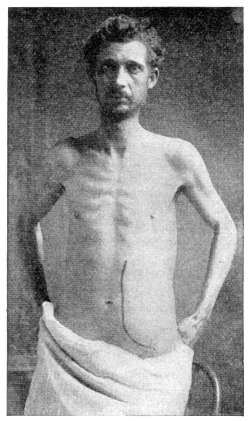
Fig. 21.
—Malarial cachexia. (Deaderick.)
A broncho-pneumonia which shows a periodicity and responds to quinine is often considered as a pernicious type of malaria.
The Skin.—Herpes labialis is a common manifestation of benign tertian and not rarely of malignant tertian infection. Urticaria may also be noted. The skin of malarial cachexia is earthy. Of course, one must always keep in mind the skin eruptions due to quinine administered in treatment, and of these urticaria is probably the most frequent.
The Nervous System.—In both the benign and malignant infections headache is a marked feature and is accentuated during the hot stage. There may be a “flighty” condition in the hot stage of benign tertian and quartan but in aestivo-autumnal infections there may be actual delirium.
Delirious and comatose states are prominent features of cerebral pernicious attacks. At times there may be an apathetic condition suggesting typhoid fever.
Almost any type of central nervous system disease may be simulated as the result of focal sporulation so that we have aphasic, epileptiform, hemiplegic, bulbar and other clinical types.
Some authors have recorded cases of multiple neuritis of malarial origin. Catto has recently examined the blood of a number of cases of multiple neuritis in Jamaica and has obtained negative malarial findings in every case. Neuralgic manifestations are features of latent malaria. Some loss of memory may be apparent after severe malaria.
The Special Senses.—Plugging of the retinal arteries may lead to blindness which may be either transient or lasting. The discs are grayish red instead of white as is the case with quinine amblyopia. The ringing in the ears is connected with the quinine treatment.
The Genito-urinary System.—In the cold stage there is apt to be frequent urination with increased secretion. Later on, there is a scanty febrile urine.
Albuminuria is rather common in aestivo-autumnal attacks and true nephritis occurs in about 2% of cases.
Plehn attaches great importance to the examination of the urine for urobilin as showing malarial infection when parasites cannot be found. The pigment particles in urinary sediment (Uriola) do not give reliable information. Bile in the urine is an important sign of bilious remittent fever.
Orchitis has been reported as a malarial complication.
The Liver and Spleen.—There is very little of importance to note in connection with the liver except tenderness and jaundice in bilious remittent fever. The spleen, however, is the organ in which centers the infection and its tenderness and enlargement are of special diagnostic value in malaria.
Even in comatose conditions pressure on the spleen may bring about indications of pain. The liability to rupture of the friable spleen of aestivo-autumnal infections is a real danger and the patient should not expose himself to injury.
The Blood Examination.—This is of prime value in the recognition of malaria, and one should examine both fresh blood preparations and stained films as well. More information is gotten from the stained films but we should also avail ourselves of the different characteristics of the 3 malarial species, which can be noted in a preparation made by taking up a small drop of exuding blood on a cover-glass and allowing it to drop on a slide and run out without any pressure on the cover-glass.
The crescents, when found, show a malignant tertian infection but there may also be present one of the benign parasites. A stained film should be used to identify malignant tertian young ring forms.
Pigmented rings are rarely observed in aestivo-autumnal fever, such parasites being caught in the capillaries as they enlarge to the stage where pigment begins to be present. Flagellated forms only develop in fresh blood preparations, 15 to 20 minutes after the taking of the blood. Of the greatest differential value is the swollen pale infected red cell of benign tertian, the normal red cell of quartan and the distorted shrunken red cell of malignant tertian.
Quinine administration may cause parasites to disappear from the peripheral circulation or it may so affect the parasite that the staining would indicate a degenerated parasite—the so-called quinine-affected parasite. It is difficult to diagnose the species of malaria from such a parasite.
Large mononuclears and transitionals containing phagocytized pigment (melaniferous leucocytes) are characteristic of malaria—the pigment however must be in the leucocyte and not free. There is a leucocytosis during the malarial paroxysm with a leucopenia and increase in the large mononuclears during the apyrexial period.
Among natives of India the large mononuclears and transitionals averaged 21% in the apyrexial stage of malaria while healthy natives rarely showed as much as a 10% count (Stott).
Some authorities have reported positive Wassermann reactions in serum of malarial patients taken during a paroxysm. All agree, however, that the serum of malarial patients at other times is negative.
Diagnosis
In the diagnosis of malaria the special points to consider are: (1) presence of malarial parasites, (2) periodicity, (3) splenic enlargement, (4) response to quinine therapy, (5) the presence of melaniferous leucocytes and (6) a high large mononuclear percentage when leucopenia is present. In the examination for parasites one should not only consider the species of parasite present but, as well, the stage of development and the presence of the sexual forms.
In an intensive investigation Bass has shown that 55.09% of those showing parasites in the blood give a clinical history of malaria while 44.91% of those with parasites in the blood fail to be associated with clinical manifestations.
Blood platelets are the findings most frequently mistaken for malarial parasites in stained blood, and the vacuoles in fresh blood. Quartan and tertian periodicity is only found in malaria, but quotidian periodicity is a feature of a host of diseases.
There are very few tropical diseases which have not been mistaken for malaria and many of these have been considered as of malarial etiology before the discovery of the real cause.
Of the cosmopolitan diseases, typhoid fever, septic conditions, including malignant endocarditis, tuberculosis, influenza, pyelitis and even syphilis are to be considered in a diagnosis of malaria.
As regards tropical diseases, kala-azar, Malta fever, liver abscess, filariasis, trypanosomiasis, leprosy, relapsing fever and yellow fever are to be thought of in differential diagnosis.
As was noted under the discussion of the pernicious manifestations of malaria, scores of diseases may be simulated by the sporulation of the malarial parasite in certain organs or areas of organs. One should always keep in mind the possibility of pain in the appendix region or in the gall bladder area as connected with malaria if in the tropics. A polynuclear increase negatives malaria and indicates appendicitis or cholecystitis. Malarial pancreatitis has been referred to before.
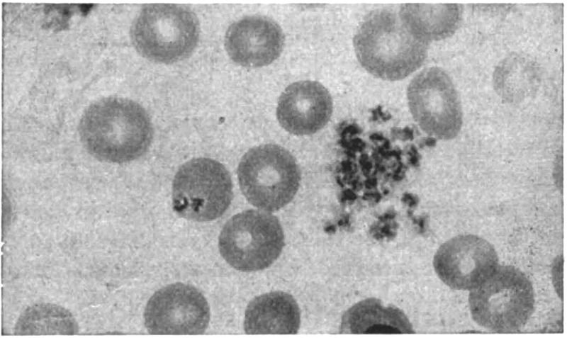
Fig. 22.
—A cluster of blood-plaques and two plaques lying upon a red cell and simulating malarial parasites (× 1000). (Todd.)
With malarial cachexia we must in particular keep from mistaking it for hookworm disease or other secondary anaemias due to intestinal parasites.
Provocative Measures.—Kohlbrugge’s recommendation to have patients suspected of malaria climb mountains and drink copiously of cold water, in order to bring on a relapse, is of value in the diagnosis. (Effects of fatigue and refrigeration.) It must always be borne in mind that quinine causes the parasites to disappear from the peripheral circulation. It is interesting to note that small doses of quinine given over ten days or two weeks may make a latent case active. Other provocative agents are subcutaneous injections of adrenalin (the best), or anti-typhoid inoculations. Certain physical methods, as hot and cold douches or alternating the hot air chamber at 55°C. for 10 minutes, followed by a cold bath for 3 minutes have been recommended. After injection of adrenalin the presence of parasites in the blood is at its height at the end of an hour. Sunlight is a factor in relapse.
The laboratory diagnosis of malaria has already been fully gone into in the section on etiology and that on blood examination under the heading of symptoms in detail.
The evenly spread stained film undoubtedly gives more accurate information as to species and stage of cycle than any other method. Still one should always examine a fresh specimen and if the parasites are very scarce, a thick film preparation. The thick film methods of Ross, Ruge and James are given under the chapter on the blood in tropical diseases. During winter parasites tend to disappear from the circulation regardless of treatment.
Prognosis
The prognosis in benign tertian and quartan is most favorable when proper treatment is instituted, as such infections are never fatal in first attacks. Not only may malignant tertian kill in a first attack but it leads rapidly to a cachexia while the cachexia following upon benign infections is more gradual.
It is the tendency to perniciousness which makes us dread malignant tertian as we can never be sure that a paroxysm may not develop cerebral or algid manifestations and these show a very high death rate, 25 to 50%, even when promptly treated.
As regards relapses quartan is the malarial fever which is most apt to show this feature and aestivo-autumnal the least. Deaderick gives the percentage of cases showing relapses in quartan, benign tertian and aestivo-autumnal as 65, 55 and 45.
The great importance of malaria is rather its invaliding tendency and by thus reducing the powers of resistance it makes the death rate from intercurrent diseases higher. Tropical malaria does not seem to affect the native as it does the European but the high death rate of infants among the natives is undoubtedly largely connected with this disease.
Statistics vary greatly as to the percentage of fatal cases in malaria. Certain figures from tropical countries give fatal results as occurring in from 2 to 10% of cases, while statistics from temperate climates show a death rate below 1%. The mortality from pernicious types of malaria is about 25%.
Prophylaxis and Treatment
Prophylaxis.—There are three methods in the prevention of malaria, all of which may be combined, as was the case in the Canal Zone region of Panama. These are: (1) Destruction of anopheline mosquitoes; (2) protection of the individual from the bites of mosquitoes, and (3) quinine prophylaxis.
It may be stated that it is frequently advisable to carry on the mosquito warfare without regard to the question of the kind of mosquitoes destroyed. In general terms the malarial mosquito breeds in the suburbs of towns or in districts more distinctly rural, while the transmitter of the more dreaded yellow fever, prefers breeding places in the immediate vicinity of city houses.
Bentley has recently noted that, with improvement in agricultural methods and utilization of marshy lands, malaria tends to disappear as much from the physical improvement and thereby greater resistance of the people as from the destruction of mosquitoes by the draining of the swamps. The resulting greater prosperity makes better food and shelter obtainable.
1. Destruction of Mosquitoes.
Such measures may be directed either toward the larva or fully developed insect.
(a) Measures against larvae. When practicable permanent measures should be preferred to temporary ones and when agricultural development goes along with drainage of swamps the cost is repaid.
The doing away with mosquito breeding places may be accomplished by filling in pools or by making ditches with smooth sloping sides to carry away the water. These ditches require a great deal of attention to prevent their filling up with tropical vegetation and thereby adding to breeding places. Subsoil drainage with tiled drains is better. Care should be exercised that public works operations do not raise the level of the subsoil water.
Anophelines tend to breed in sluggishly moving streams or in stagnant pools especially where there is a luxuriant growth of weeds or grass, and are not apt to be found in rapidly flowing streams, hence the necessity for constant care of ditches and the like to prevent their becoming obstructed by vegetation or silt. When filling in or drainage is not practicable the method of oiling the surface of the pool with crude petroleum is to be recommended. One uses about ½ pint for every 100 square feet of surface and the process should be repeated every two weeks.
In places where oil is not effective, Barber recommends Paris green mixed with dust and so used as to form a scant surface deposit. Anopheline larvae, being surface feeders, ingest it and are killed. It does not affect Culex larvae. On account of its ease of transportation, and adaptability to weedy places where oil does not penetrate, Paris green dust will doubtless prove a valuable selective larvicide. Mayne and Jackson recommend cresol as the best larvicide. In 1 to 1,000,000 parts it is an effective larvicide, and even in 1 to 1,000,000,000 it is destructive to young larvae.
Mixtures of soft soap and petroleum are better than petroleum alone.
Winds are apt to blow away the surface coating of oil and it is difficult to oil the surface of a pool filled with grass. Wise recommends crude carbolic acid, using 1 ounce to 16 cubic feet of water.
In using any larvicide it is well to introduce it along the banks of water collections with a long-spout can and mix it thoroughly with a stiff reed broom.
There are many enemies of mosquito larvae, such as tadpoles, water-beetle larvae and various small fish such as “millions.”
Terni suggests the using of such fish as carp and tench which have a food value as well as a larvicidal one.
(b) Measures against the mosquito. The clearing away of grass and brush from around houses exposes the mosquitoes to the sun in which they cannot live long.
When inside the house they may be destroyed by sulphur fumigation, 1 or 2 pounds of sulphur for each 1000 cubic feet and with an exposure of two hours.
It is usually stated that mosquitoes may hibernate during winter following infection in the autumn and that cases of malaria in early Spring may be explained by their bites. Examination of hibernating mosquitoes for zygotes does not give strong proof to this view but such mosquitoes, becoming active with a rise in temperature, may bite gamete carriers in the house and thus spread malaria.
Pyrethrum powder, which is set on fire with a little alcohol, may be burned, using 2 pounds per 1000 cubic feet, and an exposure of four hours. This does not certainly kill the insect and the stupified mosquitoes should be swept up and burned.
Giemsa’s spray is now considered an excellent measure for killing mosquitoes in rooms. The composition is as follows: Pyrethrum tincture (20 parts powdered pyrethrum blossoms to 100 parts alcohol), 480 grams; odorless potash soap, 180 grams; glycerine, 240 grams. Before using it dilute with 20 times its own weight of water, and spray the walls of the room with a spray pump.