Volume 2 — From The Popular Science Review. INSIDE THE EYE: THE OPHTHALMOSCOPE AND ITS' USES. BY ERNEST HART, OPHTHALMIC SURGEON.
Reading edition
There are few spectacles more affecting—and there were few more hopelessly distressing—than that which many have seen, of the blind man, with eyes unaltered in their human aspect of beauty, searching vainly to penetrate the unchangeable darkness of a noonday, bright to others, and replete with the splendor of light and color. There have always been many of these sufferers from a disease which claims the most profound sympathy, and which seemed bitterly to reproach our science that it could not timely penetrate the mystery of that obscure chamber which lies behind the iris, and had found no means for enabling us to see through the clear but darkened space of the pupil. That reproach, at least, exists in part no longer. Since some few years now we have learnt how to explain the obscurity of the interior of the eye, and by what optical contrivances we can overcome this darkness and look into the depths of the ocular globe; thus inspecting with ease, and quite painlessly to the individual, the lenses and humors of the eye, the nerve of sight and its transparent retinal expansion, and even the vascular tissue which lies behind and surrounds this. This is a great triumph of physical science, and it is no barren triumph. The insight which we gain into the host of affections of the refracting media and deep membranes of the eye has given to our diagnosis and therapeutical treatment of the most obscure forms of disease leading to blindness, a certainty and precision to which we were formerly strangers.
The optical instrument by which we are able to effect this inspection is known by the fitting title of the Ophthalmoscope ( the eye; I survey). With this instrument, the manner of using it, and its valuable applications, I am necessarily professionally much occupied in daily work; and as the editor of the "Popular Science Review" has requested me to give some plain account of the matter, I will endeavor to afford an untechnical statement of what the ophthalmoscope is, and what are some of the most useful results which have been obtained by its use. Let me first remind the general reader that in the human eye, behind the pupillary aperture of the colored iris, which presents to the unaided eye of the observer the mere aspect of black darkness, lies, first, a clear bi-convex lens; and behind this, filling the eye, and giving to it the character of a solid ball, a transparent globular mass, known as the vitreous body, or humor. It is into a depression in the front of this that the aforesaid lens is fitted, so that the whole space of the eye behind the iris is filled by the lens and vitreous body. The optic nerve, or nerve of sight, which pierces the tunics of the eye at the back and near the centre, spreads out and forms an expanded tunic of nerve-structure which enwraps the vitreous body as far as its most forward edge, where the colored iris descends in front of it. Enwrapping again this nerve-tunic or retina is a vestment, chiefly made of blood-vessels, connected by fine tissue and thickly coated with black pigment, having its own optical uses. This second outer pigmented vascular tunic is the choroid. This again is enclosed within the external strong fibrous membrane, which includes and protects all the sclerotic membrane {120} ( hard). These are the two humors and three tunics of the eye which can to a greater or less extent be examined during life by the aid of the ophthalmoscope. They can all be more or less investigated in the living eye by the aid of the ophthalmoscope, because by the aid of this instrument we are able to see through the pupillary space. If one considers what is the reason of the apparent darkness of the pupillary aperture and the chambers of the eye behind it, it is not difficult to gain an idea of the means by which this optical condition may be altered so as to enable us to see where all seem to the unaided vision obscure.

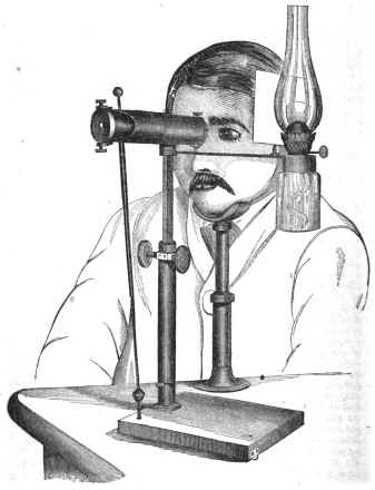
Doctor looking through ophthalmoscope.
{121}
This darkness of the pupillary aperture is attributable partly to obvious causes, such as the natural contraction of the pupil or iris which occurs under light—this contraction limiting the number of rays which can enter the eye. Then that black pigment which lines the iris absorbs a great deal of light; and thus, as in the case of albinos, whose eyes are deficient in pigment, or where the pupil is dilated, either through disease or by artificial agents, these obstacles for seeing into the living eye are removed. But still the main difficulties are not cleared away; and if you take for example an albino animal, such as one of those beautiful little white-furred rabbits, whose rosy eyes look like fiery opals edged with swan's down, and dilate the pupils with atropine, it is still not possible to see clearly the details of the structure within and at the back of the eye. This is by reason of the structure of the eye as an optical instrument, and because the rays of light in entering and in emerging from it undergo refraction, according to definite laws. The light which penetrates the eye traverses the transparent retina, producing the impression necessary for sight, and is partly absorbed by the black pigment of the choroid; but a great number of the rays are reflected; for here there is no exception to the general rule that some of the rays of light falling upon any substance are always reflected. These rays, in returning, are refracted through the vitreous body and lens, just as they were in entering the eye, with the object then of causing them so to converge as to produce upon the retina a clear and definite image of whatever external object they started from. Similarly, then, on their emergence they are refracted chiefly by the lens and cornea, so as to form an image in the outer air, the emergent rays coinciding in their path with that which they took when entering, and the image formed in the air being conjugated with the retinal image; being formed, therefore, on the same side, varying with the position of the lens and object, and the accommodation of the eye. Thus, then, to perceive this aerial image, derived from the retinal reflection, the eye of the observer needs to be placed in the axis of the converging rays; but since this is also the axis of the entering rays, he will of necessity in that position cut off those rays altogether of the light proceeding, say, from a lamp, or the source of light opposite to the eye to be illuminated.
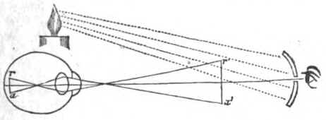
The problem to be solved consists, then, in the simple illumination of the eye to be observed by a source of light so arranged that the observer can be placed in the axis of the rays entering and emerging without intercepting those rays. This may be most conveniently effected by placing the source of light aside of the eye to be observed, and observing through a pierced concave mirror, which reflects that light into the eye. We can then, by looking through the central aperture of this mirror, place ourself in the path of the entering and emerging rays. The mirror becomes the source of light to the observed eye; the rays which it flashed into the eye emerge {122} in part, and return along the same path, forming the aerial image at a distance and under circumstances regulated by the optical conditions of the eye observed, and within view of the observer who is looking through the mirror. A very simple diagram will suffice to explain this: r a is the circle of diffusion of the retina, and the lines indicate how the reflected rays will pass through the media of the eye, and form at r' a' real enlarged but inverted image of the fundus of the eye. This will be placed at the distance of distinct vision of the subject, and has relation to the accommodation of the eye.
Diagram of preceding discussion.
As these are variable quantities, the practice of ophthalmoscopy demands a little address, which habit quickly gives. It is for want of understanding this, and from impatience of these preliminary difficulties, that many have been discouraged at the outset, and have abandoned unwisely the attempt to learn the use of the ophthalmoscope.
The image obtained in the way mentioned is not so distinct as to give that full perception of details which is necessary for scientific and medical purposes. A more defined image is obtained by interposing, for example, a bi-convex lens on the path of the luminous rays emerging from the eye observed. The effect of holding such a lens of short focus before the observed eye whilst examining it with a concave ophthalmoscopic mirror is to cause the rays emerging from the eye to undergo a further refraction, and to modify the actual image which they form, producing one which is smaller, more defined, but still inverted. This is the most simple and one of the most satisfactory methods of exploring the eye with the ophthalmoscope. It is that of the most general and easy application, and I will, therefore, add a few words to explain how it may most conveniently be practised.
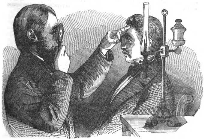
We will suppose that it is the human eye which is to be examined. The room is to be made dark; the person to be seated; a light—the white flame of an oil-lamp or an Argand gas-burner—to be placed near his head, on the side, and at the level of the eye to be observed. The observer takes then the concave mirror in the hand of the side toward the lamp, and placing it against the front of his eye, so that the upper edge rests against his eyebrow, brings his head to the level of that of the person seated, looks through the central perforation at the eye to be observed, and by a little careful change in the direction of the mirror casts, by its aid, upon the eye examined the light of the lamp.
He will now perceive that the pupillary aperture is illuminated, and, no longer black, shines with a silvery or reddened light. He takes now the bi-convex lens of short focus in the hand hitherto free, and places it in front of the examined eye, and at such a distance as to make the focus of the lens coincide with the pupil of that eye —distance varying from two to three inches. He himself will usually need to be at a distance from twelve to eighteen inches. This is for normal eyes. The slight movements backward and forward necessary to adjust these distances correctly, are effected very easily and precisely after practice; but at first it is a little difficult to avoid changing the direction of the mirror while thus slightly advancing or retiring the head; and this is a point on which it is well to give a warning, for it is a frequent source of discouragement to beginners, who find that at every movement they interfere with the illumination of the eye, and so suffer from a series of little failures at the outset. The first thing, in fact, that every one sees amounts to a little more than a red, luminous disc; those who begin by seeing nothing more, therefore, need not to be discouraged; a little patience and time will enable them to see what more practised persons describe. The eye to be examined may be more fully observed by dilating the pupil {123} with atropine—a drop of a solution, one grain to a pint of water, or one of the atropized gelatines prepared for me by Savory and Moore, each of which contains one hundred thousandth of a grain of atropine, and will maintain dilation during several hours. This acts also perfectly well with rabbits or cats.
Doctor examining patient.
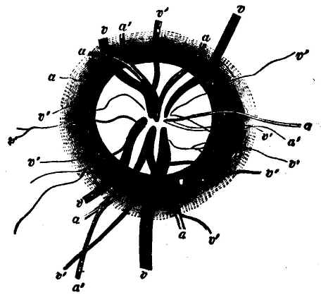
The first thing seen is the red reflection of the choroidal vessels showing through the transparent retina; and when the eye observed is directed upward and inward, we see the usually circular disc of the optic nerve, encircled by a double ring, cream-colored, or very faintly roseate or grey, and surrounded by the red choroid. The two rings are the apertures in the choroid and sclerotic, of which the former is the smaller. From out this disc we see springing the retinal artery and retinal veins, sometimes centric, at others excentric, in their passage. The artery is easily recognized as being somewhat smaller in calibre, and of a lighter red. The artery usually divides into a superior and inferior branch, each of which subdivides forthwith into two secondary branches, and these again continue to subdivide, dichotomously, running forward to the anterior limits of the retina. The veins, which are somewhat larger and deeper colored, usually pierce the disc of the optic nerve in two trunks. Pulsation may occasionally be detected in the veins by watching carefully their color, which seems to change at each impulse just where they pass over the edge of the optic disc and bend to pierce the nerve.
Fuller details of the ophthalmoscopic appearances of healthy eyes, both human and animal, will be found in Zander's treatise, excellently edited and translated by Mr. R. B. Carter, of Stroud. In the healthy eye the aqueous humor, lens, and vitreous humor are clear, and do not in any way obstruct the passage of the light. It is otherwise in disease; and this brings us to the discussion of some of the practical applications of the ophthalmoscope. Here, perhaps, I may be permitted to quote some of the {124} paragraphs of a paper which I read lately on the subject before the Hanveian Society:
Interior of eye.
"Taking up the diagnosis of the various forms of disease any of which would have been held to constitute the condition known as amaurosis, it may be noted, first of all, that even in the hands of the novice ophthalmoscopic examination supersedes those chapters in ophthalmology which were formerly devoted to the means of distinguishing between incipient cataract and amaurosis. In the past, and even at present, with those surgeons who are content to treat deep-seated diseases of the eye by guessing at their nature, and have not adopted the systematic use of the ophthalmoscope into their practice, the functional annoyances which commonly occur at the outset of the formation of lenticular cataract, have been, and are, fertile sources of deception. The patient complains of frontal pain, of confused vision, stars of light, and some other vague symptoms which characterize the outset alike of many forms of deep-seated disease of the eye, and of the fatty degeneration of the lens which commonly gives rise to lenticular cataract, probably from coincident swelling of the lens. An error arising from this source has many times condemned the unfortunate subject of a commencing cataract to the severe treatment thought appropriate to the unhappy class of amaurotics. The kind of alteration in the lens, imperceptible by any other means than the ophthalmoscope, is the slightly opaque striation of the substance of the lens sometimes seen in an early stage. These opaque striae may occupy either the anterior or the posterior segment of the lens, and spring from the centre of the crystalline or converge toward the centre from the circumference. In order to see the latter, the pupil must be fully dilated with atropine; as, indeed, for the purposes of complete ophthalmoscopic examination it always needs to be; and then, just as the greatest expert cannot discover them except by ophthalmoscopic illumination, so, neither with its aid, can they be passed over with ordinary care. In order to be quite sure in any delicate case, it is well to lower the light a little, and use only a feebly illuminating power, as a very strong light may overpower a {125} commencing opacity, and render us unable to detect the striae. This practical caution applies equally to all other conditions of opacity in the transparent media. In two cases, lately, I have been able to set at rest doubts of this kind, which happened to be in the persons of medical men, who were much disquieted by the symptoms—one a member of this society. In a third case I have recently detected incipient cataract (peripheric striae) in a gentleman supposed to be suffering from commencing glaucoma. "It is of frequent occurrence to find the capsule of the lens stained with black spots; these are stains left by the uveal pigment, and occur usually after an attack of iritis, when the iris has been in contact with the lens. When the iris has been adherent, a complete ring of pigment may often be seen on the surface of the lens. A day's experience at any ophthalmic clinique can mostly show examples of this condition; but it is only when these deposits are numerous, and in the central line of vision, that they become troublesome. They are then met with as the sequences of severe choroido-iritis, and usually coincide with further mischief in the vitreous and choroid. "The vitreous, under the influence most commonly of choroiditis, and usually syphilitic choroiditis, presents alterations of the most striking character for ophthalmoscopic observation. The patients who offer these changes complain usually of considerable dimness of sight, which on examination is found to include both diminution in the acuteness of visual perception, and restriction in the field of vision, or extent of any object seen at once. The great source of trouble to them is, that when they lift the eye or move the head, black corpuscles, or streaks, or webs float before their eyes, and obscure the object at which they are looking; and when the eyes are kept still, these fall again and disappear. Examine now the eyes of such an one, and you will see that the phenomena described are due to the existence of actual shreds, corpuscles, or webs of fibrous and albuminous exudation, which float in the vitreous, and at each motion of the eye rise in clouds and obscure the fundus, so that you can barely see it, or perhaps not at all. These conditions, I say, are mostly specific, but not invariably. They are sometimes the result of scrofula, and probably of other forms of choroiditis."
Here, then, are a large number of cases in which the ophthalmoscope transports us at once from the regions of the known to the unknown. There are other classes of cases equally striking. Let me take illustrative examples. Two persons apply for advice, complaining that the sight has been gradually growing more and more dim, perhaps in one eye,—it may be in both. The progress of the disease has been insidious and nearly painless. The eyes are to all external appearance healthy, except probably that in both patients the pupils are partially dilated and sluggish. The ophthalmoscope helps us to solve the problem.
The one is a case, it may be, of slow atrophy of the optic nerve, proceeding from central disease of the brain—from pressure on the optic tracts of nerve within the skull, or from defective nutrition following losses of blood. We find the nerve glistening white and slightly cupped, the arteries small, the fundus otherwise healthy. In the other we recognize at once, in the fulness of the veins, their pulsation, and the marked excavation of the optic disc, the indications of excessive tension of the eyeball and undue pressure of the nerve. The first requires careful constitutional treatment and a long course of studied hygiene and medication; the second calls for direct and immediate interference, with the view of relieving the intra-ocular pressure. In the diagnosis of this great class of glaucomatous disease of the eye—disease {126} characterized of loss of vision, sometimes slow and sometimes rapid, but always characterized by definite ophthalmoscopic signs: cupping of the disc, pulsation, fullness of the veins, and it may be more or less haziness of the transparent media—ophthalmoscopy has rendered a most brilliant and inestimable service. Prior to the introduction of the use of this instrument the disease was of an unknown pathology; its results were fatal to vision, but there were no means of diagnosing the conditions attending the earlier stages, and blindness followed almost certainly and inevitably. The investigation of the disease has brought us a remedy in the excision of a portion of the iris—a practice introduced by Von Gräfe, of Berlin, and of which the success is in suitable cases most gratifying.
Another series of examples may be chosen to illustrate the application of ophthalmoscopy. I avoid giving details here, but it is perhaps right to say that these are not fanciful sketches, but notices of cases in my experience and taken from my note-books of practice. Two persons are asking for advice as to the management of their eyes for short-sightedness. Are both to receive the same advice? The ophthalmoscope alone can furnish positive data. With this we may discover a staphylomatous condition of the back of the eye, a bright excentric margin around the optic disc and edge with black pigment. Examining it closely, we may find that this pigmented edge gives evidence of progressive inflammation at the back of the eye, and extending to continuous and increasing atrophy and retrocession of the coats of the eye. This person is in danger of becoming rapidly made short-sighted or of losing sight altogether. We must prohibit the use of concave glasses for a certain length of time, and must adopt active and effectual measures for subduing the atrophic inflammation. In the other patient the ophthalmoscope may show us but little stretching or waste, and that not progressive, and will enable us then to calm his fears, to prescribe appropriate glasses, and to dismiss him to his occupation with ease of mind and safety. So with sudden lose of sight from intra-ocular haemorrhage, the ophthalmoscope gives us information which could never have been guessed at without it, and guides us, not only to the local knowledge, but to the constitutional information essential for cure.
There are certain conditions of the eye which may warn any one that it is desirable that the condition of the vision ought to be investigated by the ophthalmoscope. Rapidly increasing short-sightedness is one of the most marked, and when this becomes associated with weakness of sight and loss of acuteness in the perception of small objects, the warning is very urgent. A diminution in the field of vision is another important indication of internal changes in the eye, of which only the ophthalmoscope can detect the true nature. It would be difficult, perhaps, to say whether more mischief is done and more suffering is caused by the total neglect of such symptoms or by their ignorant palliation by the aid of common spectacles, chosen empirically, because they facilitate vision for the time. The great use of the ophthalmoscope, then, is this: that it arms us with an instrument of precision, by which we can determine the precise local condition of the parts of the eye in which the function of sight is resident and through which it is regulated. If it cannot do all that we might ask, it is because the sense of sight is in truth a cerebral function, of which the eye is only an instrument; and in dealing with cerebral affections of the sight, it can indeed give us information which without it we should lack, but it leaves still to be desired more intimate acquaintance with first causes, which at present we can only discuss inferentially. To the amateur in science, and to the lover of nature, it discloses an exquisite spectacle, unknown till now, that carries {127} observation into the inner chambers of the living eye, and displays its wonders and its beauties. The observation is perfectly painless, and may easily be effected: rabbits, for example, submit to it with great calmness and composure, and at the College of Physicians' soirée last year, a little pet white rabbit of mine sat up calmly in a box which I had made for the purpose, and was examined, by the aid of a modification which I devised of Liebreich's demonstrating ophthalmoscope, by many score of observers. Mine has the advantage of being adapted for use even amid a blaze of light, and it cannot easily be disarranged; two qualities valuable in an instrument for demonstration.