Chapter IX · THE DEVELOPMENT OF THE ALLIGATOR (A. mississippiensis) (continued 2)
Reading edition
STAGE XI
Figure 14 (Plate XXI.)
Only the anterior region of this embryo is shown in the figure, which is a ventro-lateral view. While there is some change in the general shape and in parts of the head, the reason for figuring this stage is to show the first gill cleft (g′), which lies at an acute angle to the long axis of the neck behind the eye (e). The cleft is narrow but sharp and distinct in outline; it shows neither in this nor in the following stages the branched, Y-shaped outline mentioned by Clarke.
STAGE XII
Figures 15-15f (Plates XXI., XXII.)
In this stage, also, only the anterior region of the embryo is figured in surface view. The shape of the head is about the same as in the preceding stage, but it is drawn in exact profile. Three gill clefts (g¹⁻³) are now present, and are wide and distinct. The first cleft, as in the preceding stage, lies at an acute angle to the long axis of the pharynx and nearly at right angles to the second cleft. The third cleft sends a wide branch (g⁴) toward the posterior, as has been described by Clarke, from which, or in connection with which according to Clarke, the fourth cleft will develop. All three clefts may be distinctly seen to open entirely through the pharyngeal wall. The outlines of the visceral folds, especially of the mandibular, begin to be apparent. The nasal pit (n) now shows as a round depression in front of the more definitely outlined eye (e). The auditory vesicle (o) is so deep beneath the surface that it may be seen only by transmitted light.
Figures 15a-e represent transverse sections of an embryo of about this general state of development, except that the gill clefts are not so definitely open as in the surface view.
Figure 15a, the most anterior section of the series, passes through the forebrain (fb) in the region of the eyes, and through the hindbrain (hb) anterior to the auditory vesicles. The forebrain is here a large cavity with a dense wall of a comparatively even thickness. Owing probably to the section not being exactly in the transverse plane, the eyes are cut in different regions, that on the left (ov) being cut through its stalk, while that on the right (oc) is cut near its middle region and hence does not show any connection with the forebrain. The almost complete obliteration of the cavity of the optic vesicle to form the optic cup by the invagination of the outer wall of the vesicle is shown on the right side of the section (oc). The lens vesicle (lv) is completely cut off from the superficial ectoderm (ec), which is comparatively thin. The hindbrain (hb) has the usual shape for that structure. Its ventral wall is dense and thick, while its roof is reduced to the usual thin, wrinkled membrane. Close to the floor of the hindbrain lies the notochord (nt), which is large and is distinctly vacuolated. To the right of the hindbrain a large mass of darkly stained cells (cn) is one of the cranial nerves, which is connected with the hindbrain a few sections anterior to the one under consideration. The pharynx (ph), which is cut near its extreme anterior end, is represented by three irregular cavities near the base of the forebrain. Scattered throughout the mesoblast, which makes up the greater part of the section, are numerous blood-vessels (bv).
Figure 15b is twenty sections posterior to Figure 15a and passes through the tip of the bent-under forebrain (fb). On the left the section is anterior to the optic vesicle and barely touches the side of the optic stalk, which is seen as a small lump on the ventro-lateral wall of the brain. On the right the connection of the optic vesicle (ov) with the forebrain is shown. Dorsal to the optic vesicle just mentioned is a markedly thickened and slightly invaginated region of the ectoderm (n); this is the nasal pit; on the left side of the figure the thickening is shown, but the section did not pass through the invagination. The hindbrain (hb) is somewhat narrower than in the preceding figure, but is otherwise about the same; the origin of a cranial nerve is seen on its left side (cn). The notochord (nt) has the same appearance as in the preceding section. A number of blood-vessels may be seen, the pair lying nearest the notochord being the aortæ (ao), while the two other pairs, on either side of the fore- and hindbrains, are the anterior cardinals (ac). The first aortic arches are shown at ar. On the left the section passes through the exterior opening of the first gill cleft (g′), so that the mandibular fold (md) on that side is a distinct circular structure, made of a dense mass of mesoderm surrounded by a rather thick ectoderm. The mesoderm of this fold is especially dense near the center, probably the beginning of the visceral bar. Near the center is also seen the aortic arch that has already been mentioned. On the right the section does not pass through the external opening of the first gill cleft (g′) so that the tissue of the mandibular fold is continuous with the rest of the head. It is of course the slight obliquity of the section that causes the pharynx (ph) to be completely enclosed on the right, while on the left it is open to the exterior both through the gill cleft and between the mandibular fold and the tip of the head. The superficial ectoderm shown here as a heavy black line varies considerably in thickness, being thickest in the region of the nasal pit already mentioned and thinnest over the roof of the hindbrain. The amnion (a) in this, as in the other sections of the series, has the appearance of a thin, very irregular line.
Figure 15c is posterior to the region affected by cranial flexure and so shows only one region of the embryo, that of the hindbrain (hb), which is here of essentially the same structure as above described. On each side of the hindbrain is a large auditory vesicle (o); that on the left is cut through its center and shows the beginning of differentiation, its lower end being thick-walled and rounded, while its upper end is more pointed and has a thin, somewhat wrinkled wall. The notochord (nt) is slightly larger than in the more anterior sections. Numerous blood-vessels (bv, ar) are seen in the mesoblast. The pharynx (ph) is here open ventrally and also through the gill cleft of the left side; on the right side the plane of the section did not pass through the external opening of the cleft. The mesoblast of the visceral folds is much more dense than that of the dorsal region of the section.
Figure 15d, as is evident, is a section through the region of the heart, which appears as three irregular cavities (ht) with fairly thick mesoblastic walls (mes′) lined with endothelium (en′). The body wall, though consisting of but little besides the ectoderm (ec), completely surrounds the heart, and the pericardial or body cavity thus formed extends dorsally as a narrow space on either side of the foregut, giving the appearance of a rudimentary mesentery, though no especial development of such a structure would naturally be expected in this region of the embryo. The foregut (ent) is a moderately large cavity lined with a very distinct entoderm of even thickness. Dorsal to the foregut are three large blood-vessels, a median, and now single, dorsal aorta (ao), and a pair of cardinal veins (cv). The notochord (nt) is small and is flattened against the ventral side of the spinal cord (sc), which latter structure needs no special mention. The muscle plates (mp) are considerably elongated, so that they now extend ventrally to a point slightly below the upper angles of the body cavity.
Figure 15e is through the middle region of the embryo, and, owing to the curvature of the body, is not an exact dorso-ventral section; this accounts, in part at least, for the unusual diameter in a dorso-ventral direction of the aorta (ao), which is very large in proportion to the other structures. The posterior cardinal vein is shown on the left, but not on the right. The relative sizes of the spinal cord (sc) and notochord (nt) are very different from what was seen in the preceding figure. In this section the spinal cord is considerably smaller than in the preceding, while the notochord is very much larger; in fact the notochord here seems abnormally large when compared to corresponding sections of other series. It is true, however, that while the spinal cord has been diminishing in diameter the notochord has been increasing. The spinal cord, notochord, and dorsal aorta are all so large that they are flattened against each other, the pushing in of the ventral side of the spinal cord being even more marked than is shown in the figure. On either side of the spinal cord a large spinal ganglion (sg) is seen, closely wedged in between the spinal cord and the adjacent muscle plate (mp). As in the preceding stage, there is a marked space between the muscle plate and the adjacent ectoderm (ec). The somatic mesoblast at the upper angle of the unenclosed body cavity is thickened on each side and somewhat bulged out by the Wolffian body to form what might be termed a Wolffian ridge (wr). In the mid-ventral line is the considerably developed mesentery (ms), from which the intestine has been torn. The Wolffian bodies now consist, on each side, of a group of five or six tubules (wt) of various sizes, near which in a more ventro-lateral position, close to the upper angle of the body cavity, is the more distinct Wolffian duct (wd). The allantois is fairly large by this time, and may be seen in the most posterior sections as an irregular, thick-walled outgrowth from the hindgut.
A horizontal section through the anterior end of an embryo of this age is shown in Figure 15f. While enclosed of course in the same membranous amnion (a), the pharyngeal region of the section is separated by a considerable space from the more anterior region where the section passes through the forebrain (fb) and eyes. The spinal cord (sc), notochord (nt), muscle plates (mp), aortæ (ao), and anterior cardinal veins (ac) need no special description. The appearance of the pharynx (ph), with its gill clefts and folds, is quite similar to that of the corresponding structures in the chick. None of the four clefts (g¹⁻⁴) show, in the plane at which the section was cut, any connection with the exterior; in fact the fourth cleft (g⁴) would scarcely be recognized as a cleft if seen in this section alone. One or two of the more anterior clefts are open to the exterior. Three pairs of aortic arches are seen, and each visceral fold has a central condensation of mesoblast.
STAGE XIII
Figures 16-16g (Plates XXII., XXIII.)
The embryo (Fig. 16) now lies on one side, body torsion being complete. The curvature of the body is so marked that the exact length is difficult to determine. The eye (e) and ear (o) have about the same superficial appearance as in the preceding stage. The nose is not shown in this figure. About thirty somites are present; the exact number cannot be determined in surface view. The amnion is complete, though not shown in the figure, and the tail (t) is well formed. The umbilical stalk was torn in the removal of the embryo, so that it is not shown in the figure. The dim outline of the now convoluted heart may be seen if the “cleared” embryo be viewed by transmitted light; it is not shown in the figure. The allantois (al) is a rounded sac of considerable size just anterior to the tail. Four gill clefts (g¹⁻⁴) are now present; the most posterior one is more faint than is represented in the figure, and it could not be definitely determined from a surface view whether or not it opened to the exterior. The mandibular fold (md) is now fairly well outlined, but there is as yet no sign of the maxillary process.
Figure 16a is the most anterior of a series of transverse sections made of an embryo of the approximate age of the surface view just described; it passes through the tip of the forebrain (fb) and shows the nasal pit (n) of the right side. The great thickening of ectoderm in the region of the nasal invagination is represented by a solid line. Owing to the obliquity of the section, the left nasal pit was not cut. The mesoblast is quite dense and contains two or three small blood-vessels near the roof of the brain. The plane of this section, owing to the cranial and body flexure, cut the embryo also in the region of the pharynx; this part of the section was, as a matter of convenience, omitted from the drawing.
Figure 16b is in reality more anterior in position, considering the entire embryo, than the preceding; but the region of the embryo represented is more posterior, so that it is described at this point. The greatly elongated outline of the brain is due to its being cut through the region of flexure, so that the forebrain (fb), or, perhaps, midbrain, is shown at one end, and the hindbrain (hb) at the other. The walls of these cavities are somewhat wrinkled and irregular and their constituent cells are beginning to show slight differentiation, though this is not shown in the figure. On the left side are seen a couple of darkly stained masses; one is the origin of a cranial nerve (cn); and the other is one of the auditory vesicles (o), which is still more irregular in outline than it was in the preceding stage. The only blood-vessels to be seen are a few very small ones that lie close to the wall of the brain. The ectoderm is quite thin at all points.
Figure 16c, the largest section of this series, passes through the forebrain in the region of the eyes and through the gill clefts. The forebrain (fb) exhibits on the left a marked thickening of its wall (ch), the edge of the cerebral hemisphere of that side, which is just beginning to develop; on its right side the lower part of the forebrain is connected by a well marked optic stalk (os) with the optic cup (oc), in whose opening lies the lens vesicle (lv), now reduced to a crescentic slit by the thickening of its posterior wall. The mesoblast is more dense in those parts of the section adjacent to the pharynx than in the more distant regions, and the ectoderm thickens in a marked way as it approaches the borders of the pharynx and gill clefts. Only a few small blood-vessels (bv) are to be seen in the region of the forebrain.
Parts of three pairs of clefts (g) are shown in the figure: one pair opens widely on either side, so that there is a large area of the section that is distinct from the two still larger portions and contains a small, thick-walled cavity (g) on the right side; this cavity is a gill cleft that is cut through neither its outer nor its pharyngeal opening.
No structures other than this small portion of a gill cleft and a few blood-vessels are to be seen in this middle region of the section. In the more posterior part of the section, in which the notochord (nt) is located, a pair of curved clefts may be seen, opening entirely through the wall on the left, but closed on the right (g). One distinct pair of aortic arches is shown (ar), and also the dorsal aortæ (ao), which are of very unequal size. The spinal cord (sc) and muscle plates need no special description.
Figure 16d is in the region of the heart (ht) and lungs (lu). The former is an irregular cavity whose walls, especially on the ventral side (mes′), are becoming very thick and much folded. Although thin, the body wall completely surrounds the heart, as would be expected, since this was true of the preceding stage. The lung rudiments (lu) and the foregut from which they have arisen have the same appearance as in the chick; they consist of three small, thick-walled tubes so arranged as to form a nearly equilateral triangle. They are surrounded by a swollen, rounded mass of mesoblast which almost completely fills the surrounding portion of the body cavity (bc). The pleural sides of these crescentic portions of the body (or pleural) cavity—that is, the boundary of the mass of mesoblast just mentioned—are lined with a thickened layer of cells, shown by the solid black lines in the figure. The lung rudiments may be traced through about fifty sections of this series, or about one twelfth of the entire series. At the dorsal angle of the part of the body cavity (bc) just described, near the dorsal aorta (ao), are two dark, granular masses (ge), which, under a higher magnification than is here used, are seen to consist of a small group of blood-vessels filled with corpuscles; although several sections in front of the anterior limits of the kidneys these are evidently glomeruli. They may be traced, though diminishing in size, far toward the tail, in close connection with the Wolffian bodies. At intervals they are connected by narrow channels with the dorsal aorta; no such connection was present in the section drawn. The notochord (nt), spinal cord (sc), muscle plates (mp), and spinal ganglia (sg) need no special mention. The mesoblast is beginning to condense in the neighborhood of the notochord, and the ectoderm is slightly thickened laterally and dorsally.
Figure 16e is in the region of the liver and the Wolffian bodies; it also shows the tip of the ventricular end of the heart. The liver (li) is a large irregular mass, of a blotchy appearance under this magnification, lying between the heart (vn) and the intestine (i). Under greater magnification it is seen to be made up of indefinite strings of cells; and its still wide opening into the intestine may be seen in more posterior sections. The intestine (i), which in this section might be called the stomach, is a fairly large cavity with the usual thick entodermic walls; it is supported by a comparatively narrow mesentery. The body cavity on the side next this mesentery has the same thick lining that was noted in the region of the lungs. The convolutions of the thick peritoneal lining may easily be mistaken in places for parts of the enteron. The Wolffian bodies may be seen as two groups of tubules (wt) in their usual location. The heart is cut through the ventricle (vn), as has been said. The section being at right angles to the long axes of the villi-like growths of the myocardium, the depressions between these mesoblastic cords are seen as a number of small irregular areas, each one lined with its endocardium. The incompleteness of the body wall below the heart is apparently due to an artificial break and not to a lack of fusion. The only point that need be mentioned in connection with the structures of the dorsal part of the section is that the distinctness of the myocœl (myc) on the right side is somewhat exaggerated.
Figure 16f is in the middle region of the embryo where both splanchnopleure and somatopleure are unfused. Owing chiefly to the unclosed condition of the midgut (i) and to the increase in length of the mesentery (ms), the section is quite deep dorso-ventrally. The continuation of the amnion (a) with the somatopleure is of course here evident.
The most striking feature of the section is the marked projection of the Wolffian ridges, though no local enlargements of these ridges indicate the rudiments of the limbs. A large mass of Wolffian tubules (wt) is seen projecting into the upper part of the body cavity on each side; close to each of these masses is the posterior cardinal vein (pc), and between them is the large aorta (ao). The other structures are about as in the preceding section.
Figure 16g represents a sagittal section of the anterior half of the body of an embryo of this or possibly a slightly younger stage of development. The three regions of the brain are clearly indicated, as well as the cavity of the spinal cord (sc). The roof of the hindbrain has been made too thick in the figure; it should be represented by a mere line. A little mesoblast is to be seen at places between the roof of the brain and the superficial ectoderm. A slight invagination of the epithelium (p), between the floor of the brain and the anterior end of the notochord, probably represents the beginning of the hypophysis. No indication of the paraphysis is yet to be seen. Extending from the region of the hypophysis to the posterior end of the section is the notochord (nt); it is much vacuolated and gradually increases in thickness toward the posterior, though its outline is quite irregular; except at the extreme anterior end and at one or two other places, it lies in close contact with the floor of the neural tube. Directly under the notochord lies, in the posterior half of the figure, the large dorsal aorta (ao). The pharynx (ph), opening between the end of the forebrain and the thick mandibular fold (across which opening the amnion, a, of course extends), is a funnel-shaped space which passes out of the plane of the section toward the posterior end of the figure. Its thick endodermal lining extends to the mandibular fold on the ventral side, while on the dorsal side it gradually thins out and becomes continuous with the thin ectoderm that extends over the forebrain. Just back of the mandibular fold is the bulbus (b), and back of that is the edge of the ventricle (vn). Posterior and dorsal to the ventricle the liver (li) is seen as an irregular mass of cells, and dorsal to the liver one of the Wolffian bodies (wt) is cut through its extreme edge.
STAGE XIV
Figures 17-17g (Plates XXIII., XXIV.)
Body flexure has increased until now the forebrain and tail are almost in contact (Fig. 17). The eye has developed somewhat; the ear vesicle, which is not shown in the figure, is small and seems to lie nearer the ventral side; the nasal pit is much larger and is crescentic in shape. The hyomandibular cleft (g′) still persists as a small crescentic slit, while the next three clefts are now represented merely by superficial grooves separated by distinct ridges, the visceral folds. No indication of a fifth cleft is seen. The maxillary process (mx) grows ventralward under the forebrain and is already longer than the mandibular arch (md).
The chief advance in development over the preceding stage, besides the formation of the maxillary process, is in the appearance of the appendages (aa and pa); they have the characteristic shape of the rudimentary vertebrate appendage, though the anterior pair seem to point in an unusual direction at this stage and to be slightly more developed than the posterior. The curious, anteriorly directed heart (ht) is, perhaps, somewhat abnormal. The umbilical stalk (u) is comparatively narrow and, like the allantois, was cut off close to the body.
Transverse sections of an embryo of this stage are represented in Figures 17a-g, drawn under a lower magnification than were any of the preceding figures.
Figure 17a is in the region of the pharynx, and passes through the forebrain (fb) and posterior part of the hindbrain (hb). In the thick walls of both of these structures histological differentiation has begun, so that even under low power an inner granular and an outer clear zone may be distinguished. Under greater magnification the presence of short fibers may be made out among the cells. The cerebral hemispheres (ch) are well-marked structures, their asymmetry being of course due to the obliquity of the section. Only one eye is cut by the plane of the section, and this one shows no connection with the forebrain. The outer wall of the optic cup (oc) is so thin that under this magnification it can scarcely be seen as a dark line surrounding the retinal wall. The lens (ln) is now a solid mass, of the usual type for vertebrate embryos, its front or outer wall being a scarcely discernible line. The hindbrain (hb) has the usual form for that region and does not differ particularly from what was noted in earlier stages except in the histological differentiation that has already been mentioned. As with the eye, it is only on the right side that the auditory vesicle (o) is shown. It shows some differentiation, but not so much as would be seen were it cut in another region. In the center of the section the pharynx (ph) forms an irregular cavity connected with the exterior on the left by a gill cleft (g) and by another slit which is simply the anterior margin of the stomodæum. On the right neither of these openings is in the plane of the figure, though the gill cleft (hyomandibular), which lies close to the auditory vesicle, is almost an open passage. A few small blood-vessels are scattered through the section; one of these (bv), lying between the notochord (nt) and the floor of the brain, is noticeable from its being very closely packed with corpuscles, so that at first glance, under low magnification, it looks more like a nerve than a blood-vessel.
Figure 17b is also through the pharyngeal region, a short distance behind the preceding section. The growth of the cerebral hemispheres (ch) is better shown than in the preceding figure, as is also the general form of the optic cup (oc). On the left the nasal cavity (n) is seen as an elongated slit with thick walls; it is cut near, but not through, its opening to the exterior. The same gill cleft (g) that was seen in the preceding figure is seen here as a narrow, transverse cleft, open at both ends. Between the notochord (nt) and the spinal cord (sc) is the same, though now double, blood-filled vessel (bv) that was seen in the preceding section. The other blood-vessels are larger here than in the more anterior region. There is a faint condensation of mesoblast in the neighborhood of the notochord, and a more marked condensation (mp) farther toward each side is the curiously shaped muscle plate.
Figure 17c is through the heart region, and that organ is cut through the opening from the lower or ventricular into the upper or auricular chamber. The thickening of the wall of the ventricle, which was noticed in the preceding stage, has increased to such an extent that there is now a marked difference in the thickness of the ventricular and auricular walls. As in the preceding stage, the body wall is torn, probably in handling, so that it appears to be incomplete around the ventral side of the heart. Dorsal to the heart two small circular holes (ent) with thick walls are the œsophagus and trachea, cut anterior to the point of bifurcation of the latter into the bronchial or lung rudiments. On either side of these structures is an elongated blood-vessel (ac), the anterior cardinal vein, its elongation being due to the fact that it is cut at the place where it turns downward to empty into the heart. Dorsal to the œsophagus are the aortæ (ao), which are here cut just at the point where the two vessels unite to form one; the next section, posterior to the one under discussion, shows an unpaired aorta. The notochord (nt) and spinal cord (sc) need no description, except to note that the latter shows active histological differentiation, numerous mitotic figures being seen under higher magnification, especially in the cells that line the spinal canal. On the right of the cord the edge of a spinal ganglion (sg) is seen, in connection with which in other sections are seen the clearly defined nerve roots. The condensation of mesoblast around the notochord is quite evident, and in close contact with this medial condensation are two very characteristic, S-shaped muscle plates (mp), which extend from the level of the dorsal side of the spinal cord to the upper limits of the cardinal veins. In some sections the muscle plates even yet show slight remains of the myocœl at the dorsal end.
Figure 17d is in the region of the posterior end of the heart (ht), which is cut through the tip of the ventricle, and the anterior end of the liver (li), which has the appearance of a mass of darkly stained cords or strands of cells surrounding a large blood-vessel (mv). This blood-vessel may be called the meatus venosus, though it is not separated by any line of demarcation from the auricle. A few sections anterior to this region the meatus venosus opens dorsally into a large vessel on each side (dc), which at first glance seems a part of the body cavity, but which is in reality the ductus Cuvieri, formed by the union of the anterior and posterior cardinal veins. An irregular, crescentic cleft (bc), lying medial and parallel to each of the Cuvierian vessels, is the body cavity. In the upper angle of this cavity is a granular mass, the glomerulus, that of the left side being accompanied by the extreme anterior end of the Wolffian duct. In the rounded mass of mesoblast, between the cleft-like regions of the body cavity, the lung rudiments (lu), and the œsophagus (oe) are seen as three small, circular openings; that of the œsophagus is somewhat smaller than the other two. The notochord (nt), spinal cord (sc), and muscle plates (mp) have almost the same appearance as in the preceding section. A spinal ganglion (sg) is seen on each side of the spinal cord; the one on the left shows a well-defined spinal nerve (sn), which may be traced ventrally as far as the end of the muscle plate, along whose medial side it courses. The ventral nerve root is plainly seen; the dorsal root, in this section, less plainly. The amnion (a) and abdominal wall are, as in the preceding figure, torn in the region of the ventricle.
Figure 17e is a short distance posterior to the figure just described. The liver is cut through its middle region and forms a large, darkly staining, reticular mass on the left side of the figure. The digestive tract is seen at two places to the right of the liver; the smaller and more ventral of these openings (i) may be called the intestine, while the larger is evidently the stomach (i′). The body wall is here unfused and becomes suddenly thinner as it passes upward into the amnion (a). The Wolffian tubules (wt) form a very conspicuous mass on either side of the mesentery, in close connection with the posterior cardinal veins (pc). In the mesoblast between the dorsal aorta (ao) and the notochord are two small, irregular, darkly stained masses (sy). These are shown in the preceding two figures, but were not mentioned in the description. They may be traced through a great part of the length of the embryo back of the head region; at intervals corresponding in length to the distance between the spinal ganglia they are enlarged, while between these enlargements they are very small in cross-section. At certain points a small blood-vessel is given off by the dorsal aorta to the immediate neighborhood of each of these small areas. Although they show no connection with the central nervous system, these structures appear to be the rudiments of the sympathetic nerves.
Figure 17f is in the region just in front of the hind legs. The abdominal walls are here unfused, and into the unenclosed body cavity projects the intestine (i), supported by a narrow mesentery and surrounded by a comparatively thick mass of mesoblast. The Wolffian body and duct form a mass of considerable size on each side of the mesentery. The Wolffian body is cut near its posterior end and consists of smaller tubules than in the more anterior regions. The Wolffian ducts (wd), on the other hand, are very large and are much more clearly distinguishable from the Wolffian tubules than in the preceding sections. The Wolffian ridges (wr) are very marked projections on the sides of the body, and in a region farther caudad become especially developed as the posterior appendages, to be described in connection with the following section. Both spinal ganglia are shown in this figure (sg), and in connection with the left ganglion the spinal nerve (sn), extending ventrally as far as the level of the Wolffian duct. The sympathetic nerve rudiments do not extend so far caudad as the plane of this section. The dorsal end of each muscle plate (mp) is seen, in this and other sections, to be slightly enlarged to form a round knob; this knob contains a distinct cavity (not shown in the figure), the myocœl.
In Figure 17g, owing to the curvature of the body, the plane of the section passes through the body at three places: through the region of the heart and lungs (Fig. 17d), through the region of the posterior appendages, and through the tail. In fact, the plane of the section represented by each of the preceding figures cut the embryo in more than one region, but for the sake of simplicity only one region was represented in each figure. In the figure under discussion only the leg and tail regions have been drawn, though the latter region (t), being cut through one of its curves, is seen as an elongated body with a section of the spinal cord, notochord, etc., at each end. Both regions shown in the figure are enclosed in the same fold (a) of the amnion. Of the structures in the dorsal side of the larger or more anterior part of this figure nothing need be said. The most striking feature of the section is the presence of the large posterior leg rudiments (pa). As was noted in the preceding figure, they are, as usual, merely local enlargements or projections of the mesoblast (covered, of course, with ectoblast) of the Wolffian ridge. They are, as shown in this section and in the surface view of this stage (Fig. 17), bluntly pointed projections from the sides of the body. The anterior appendage seems to be slightly more developed than the posterior, as was noted in describing the surface view of the embryo. The digestive tract is cut through its extreme posterior end, in the region that may be termed the cloaca (cl), for into it at this point the Wolffian ducts open (wdo). As the narrow cloacal chamber is followed toward the tail, it becomes still smaller in diameter, and the ventral depression or cleft seen in this figure gradually becomes deeper until its walls are continuous with the ectoderm that covers the ventral projection of mesoderm between the hind legs; no actual opening to the exterior is present, however. There is a space of about twenty-five or thirty sections (in a series of eight hundred) between the posterior ends of the Wolffian bodies and the cloacal openings of the Wolffian ducts. The body cavity (bc) and the posterior cardinal veins (pc) are very small in this region, as might be expected.
STAGE XV
Figure 18 (Plate XXIV.)
Only the head of this embryo is represented, as the general state of development is about the same as in the preceding stage.
The chief object in making the figure is to show the five gill clefts (g¹⁻⁵). The fifth cleft, though small and probably not open to the exterior, is quite distinct in this embryo. The writer would feel more doubt of its being a true, though rudimentary, gill cleft had not Clarke (17) found a fifth pair of clefts in his material. The nasal pit has advanced in development somewhat and shows the beginning of the groove that connects it with the mouth. In this figure the crescentic hyomandibular cleft shows its connection with the groove between the mandibular and the hyoid folds.
STAGE XVI
Figure 19 (Plate XXIV.)
This embryo is only slightly more developed than the preceding. Body flexure is so great that the forebrain and tail nearly touch. Only the anterior three gill clefts are visible. The maxillary process (mx) is longer and more narrow; the mandibular fold has not changed appreciably. The nasal pit (n) is now connected by a distinct groove with the stomodæum. The appendages have increased in size, the posterior (pa) being the longer. The anterior appendage (aa) is distinctly broadened to form the manus, while no sign of the pes is to be seen at the extremity of the posterior appendage. The heart (ht) is still very prominent. The stalk of the umbilicus (u), which is quite narrow, projects from the ventral wall in the region between the heart and the hind legs. The tail is of considerable length and is closely coiled.
STAGE XVII
Figures 20-20j (Plates XXV., XXVI.)
The superficial changes noted in this stage chiefly concern the head, which has increased considerably in length (Fig. 20). The curvature of the body is slightly more marked, and the tail is more tightly coiled at the end. There are still signs of three gill clefts. The maxillary process (mx) is long and narrow, while the mandibular arch (md) is still short and broad. The fronto-nasal region has greatly increased and has the aquiline profile noted by Clarke. The nasal groove has disappeared, and there remains the small opening (n) at the side of the fronto-nasal region, near the end of the still separate maxillary process. The umbilicus is in about the same condition as in the preceding stage, but the heart is less prominent. The outline of the manus (ma) is more definite, and the extremity of the posterior appendage is distinctly flattened out to form the rudimentary pes (pe). The position of the elbow-joint in the anterior appendage is seen at the end of the reference line aa.
Typical transverse sections of this stage are shown in Figures 20a-j.
Figure 20a is a section through the middle region of the head, cutting the hindbrain on one side and the forebrain on the other. The walls of the brain show rather more histological differentiation than was seen in the preceding sections, though this cannot be shown under the low magnification used. The hindbrain (hb), which is cut near its anterior border, exhibits the usual membranous dorsal and thick ventral walls. The forebrain is here seen as three distinct cavities—a median third ventricle (tv), with a thick ventral wall, and a thin dorsal wall extended to form a large paraphysis (epi), and two lateral ventricles (ch), the cavities of the cerebral hemispheres, whose walls are quite thick except on the side next the third ventricle. The sections of this series being slightly oblique, the eye is here cut on the right side only, where it is seen as a large, semicircular cavity (e) with thick, dense walls. The mesoblast, in which several blood-vessels (bv) are seen, exhibits three distinct areas—a median, lighter zone, with a more dense area on either side. The significance of this variation in the density of the mesoblast is not apparent.
Figure 20b is only a few sections posterior to the section just described. It is drawn chiefly to show the appearance of the forebrain, the other structures being about as in the preceding figure, except that both eyes (e) are here represented. The section passes through the wide opening between the third (tv) and the lateral ventricles (ch) and cuts the anterior edge of the pineal body[8] (epi). The paraphysis is very large and is directed backward instead of forward, as is usually the case among the lower vertebrates (if the alligator may be so classed). It is shown in Figure 17a of a preceding stage and will be again shown in a sagittal section to be described later. The same areas of more dense and less dense mesoblast noted in the preceding figure are seen here.
Figure 20c, though still in the head region, shows several features that were not seen in the preceding figures. On the left of the hindbrain (hb) the auditory vesicle (o), which is now considerably more advanced than in earlier figures, is seen as a larger, flask-shaped cavity and a smaller, round one. Between the larger cavity and the hindbrain is the root of a cranial nerve (cn), apparently the eighth, since in another section it comes in close contact with the wall of the larger part of the auditory vesicle just mentioned. On the right side, ventral to the hindbrain, another and much larger nerve (cn) is seen. Nearly in the center of the figure is seen a small, irregular, thick-walled cavity (p); this is the pituitary body, and its connection with the roof of the pharynx may easily be made out in another section. The mesoblast in this region of the sections contains numerous large and small blood-vessels and exhibits certain denser areas which probably represent the beginnings of the cranial cartilages. No sign of the forebrain is seen (the plane of the section passing in front of that region), except that the tip of the wall of one of the cerebral hemispheres (ch) is cut. The left nasal chamber (n) is shown: it will be noted again in the following section. The eye on the right side shows no remarkable features; its lens (ln) is large and lies well back of the lips of the optic cup, which may now be called the iris (ir). A thin layer of mesoblast has pushed in between the lens and the superficial ectoderm to form the cornea, and the outer wall of the optic cup is now distinctly pigmented. The inner wall of the optic cup is beginning to differentiate into the retinal elements. The eye on the left side is cut farther from its central region and has a very different appearance from the eye just described. This unusual appearance is due to the fact that the section passed through the choroid fissure, which is very large and seems to be formed by the pushing in of the walls of the cup and not by a mere cleft in these walls. This fissure is hardly noticeable in the stage preceding the present, and in a stage slightly older it has disappeared; so that it would seem to be a very transient structure. It apparently is formed at about the time that the optic stalk, as such, disappears. It is in the region of the choroid fissure, if not through it, that the optic nerve (on) enters the eye. Through the fissure also enters a vascular tuft of mesoblast (pt) which may be seen projecting into the optic cup after the disappearance of the fissure. This loop of blood-vessels is doubtless the pecten.
Figure 20d represents a section through the hindbrain (hb), pharynx (ph), and tip of the snout. On either side of the hindbrain are a convoluted auditory vesicle (o), and several blood-vessels and nerves, while ventral to it is seen the anterior end of the notochord (nt), around which the mesoblast is somewhat more dense than elsewhere. The pharynx (ph) sends out toward the surface a narrow gill cleft (g′) in the neighborhood of each auditory vesicle. These clefts connect with the exterior by very narrow slits, not seen in the plane of this section. The opposite end of the pharynx, as seen in this figure, opens on the left (pn) into the nasal chamber. The nasal cavity on the right is cut in such a plane that it shows neither its external nor its pharyngeal opening. The nasal passages are here fairly long and nearly straight chambers; their lining epithelium is quite thick in the middle region, but becomes thinner where it merges into the epithelium of the pharynx at one end, and into the superficial epithelium at the other end. The unusual appearance of the eye (e), on the right side of the figure, is due to the fact that the plane of the section cut tangentially through the extreme edge of the eye in the region of the choroid fissure.
Figure 20e is only a short distance posterior to the preceding. On the left side the pharynx (ph) is connected with the exterior through the stomodæum, and on the right the hyomandibular cleft (g′) is cut almost through its opening to the exterior. The auditory vesicle (o) on the right is cut near its middle region, while that on the left is barely touched by the plane of the section. The notochord (nt), with its condensed area of mesoblast, is somewhat larger than in the preceding section. The nasal canal on the right (n) is cut through neither anterior nor posterior opening, while on the left side the canal shows the anterior opening (an).
Figure 20f, which is in the region of the posterior part of the pharynx and the anterior part of the heart, shows some rather unusual conditions.
The spinal cord (sc) and notochord (nt), with the faintly outlined condensations of mesoblast in their region, need no special description. The pharynx (ph) is here reduced to an irregular, transversely elongated cavity, the lateral angles of which are connected on each side with the exterior through a tortuous and almost closed gill cleft (g), which must be followed through many sections before its inner and outer openings may be determined. Dorsal to the pharynx numerous blood-vessels (bv), both large and small, may be seen, while ventral to it is noticed a faint condensation of mesoblast (la), in the form of an inverted T, the anlage of the laryngeal structures. The ventral portion of the figure is made up of a nearly circular thin-walled cavity, the pericardium (pr). Most of the pericardial cavity is occupied in this section by the thick-walled ventricle (vn), above which is the bulbus (b) and the tip of the auricle (au). The bulbus is nearly circular in outline, though its cavity is very irregular. A few sections anterior to this, the opening of the bulbus into the ventricle is seen.
In Figure 20g the section represented is only a short distance posterior to the one represented by Figure 20f. The mesoblastic structures in the neighborhood of the spinal cord (sc) and notochord (nt) will be described in connection with the next figure, where they are more clearly defined. The œsophagus (oe)—or posterior end of the pharynx, whichever it may be called—is here a crescentic slit, with its convex side upward; ventrally it opens by a narrow glottis into the trachea (ta). The trachea is surrounded by the same condensed area of mesoblast (la) that was mentioned in connection with the preceding figure, but the condensation is here more marked. From the bulbus (b) an aortic arch (ar) extends upward for a short distance on the right side, while to the left of the œsophagus an aortic arch (ar) is cut through the upper part of its course. Ventral to the bulbus the ventricle (vn) and two auricles (au) are seen surrounded by the pericardial wall.
Figure 20h is in the region of the liver (li), which has about the same position in relation to the auricles (au) that was occupied by the ventricle in the last figure. The auricles are connected with each other by a wide passage. The trachea (ta) and the œsophagus (oe) are entirely distinct from each other; the former is a small, nearly circular hole, while the lumen of the latter is obliterated and its walls form a solid, bow-shaped mass of cells. Since there is a narrow space between this mass of cells and the surrounding mesoblast, it might be thought that the lumen of the œsophagus had been closed by the simple shrinkage of its walls; higher magnification, however, fails to show any sign of a collapsed lumen. It is doubtless the problematic and temporary closure of the œsophagus that is noticed in other forms. On each side of the œsophagus, in close relation with the anterior cardinal vein (ac), is noticed a nerve (cn) cut through a ganglionic enlargement. When traced forward these nerves are seen to arise from the region of the medulla, and when followed caudad they are found to be distributed chiefly to the tissues surrounding the newly formed bronchi; they are doubtless the tenth cranial nerves. On the right side of the figure the close connection of this nerve with the near-by gill cleft is seen. Above the paired aortæ (ao) the sympathetic nerves (sy) will be noticed. The mesoblast surrounding the spinal cord (sc) and notochord (nt) is distinctly condensed (more so than the figure shows) to form what may be called the centrum (c) and neural arch (na) of the vertebræ. The arch, owing to the slight obliquity of the section, shows here only on one side. The spinal cord is not yet completely enclosed by the neural arches. The muscle plates (mp) are in close connection with the rudiments of the vertebræ just mentioned. The spinal cord (sc) is here differentiated into three areas—a dense, deeply stained area immediately around the neurocœl; a less dense area of cells surrounding the inner area and extending ventralward as a rounded projection on each side; and an outer layer, with few or on nuclei, surrounding the inner two layers except on the dorsal side.
In Figure 20i the size and complexity of the figure are due, it will be easily understood, to the fact that the plane of the section passed through the curve of the body, thus practically cutting the embryo in two regions—an anterior, where the lungs (lu) and liver (li) are seen, and a posterior, where the Wolffian bodies (wt) are present. The spinal cord and the surrounding structures have almost the same characteristics at both ends of the figure, except that the primitive spinal column is rather more distinct in the posterior end of the section. The posterior cardinal veins (pc), Wolffian ducts (wd), and Wolffian bodies (wt) are also prominent structures of this end of the figure, the last being made up of a great number of tubules. The extreme anterior ends of the Wolffian bodies are seen in the other half of the section in the upper angles of the body cavity, dorsal to the lung rudiments (lu). Filling most of the body cavity (bc) and making up the greater part of the middle of the figure are the liver (li), now a very large organ; the stomach (i′), also quite large; the pancreas (pan), a small body lying near the stomach; and the lungs (lu), which here consist of several thick-walled tubes, surrounded by lobes of mesoblast. The other features of the figure need no special mention.
Figure 20j is through the base of the posterior appendages (pa), in which the cartilages are already being outlined by condensations of mesoblast. The intestine (i) is cut in two regions—at a more anterior point, where it is seen as a small, circular hole surrounded by mesoblast and hung by a narrow mesentery, and through the cloacal region, the larger and more ventral cavity, into which the Wolffian ducts (wd) open a short distance caudad to this section. The blood-vessels present a rather curious appearance. A short distance anterior to this point the aorta has divided into three, or it might be said that it has given off two, large branches. These two branches, one on either side near the posterior cardinal vein, pass toward the ventral side of the embryo on each side of the cloaca and end at about the region represented by the present figure. The small portion of the aorta that remains after the giving off of the two branches just described continues, as the caudal artery (ca), into the tail; it is a small vessel just under the notochord, and gives off small, paired branches at regular intervals toward the vertebral region. The posterior cardinal veins (pc), posterior to the openings of the Wolffian ducts into the cloaca, unite to form a large caudal vein lying just ventral to the caudal artery.
STAGE XVIII
Figure 21 (Plate XXVII.)
This embryo, as may be seen, for example, by the form of the appendages, is slightly further developed than the one represented in Figure 20. The figure is from a photograph of a living embryo as it lay in the egg, a portion of the shell and shell membranes having been removed. The embryo, which lies on its left side, is rather faintly outlined because of the overlying allantois. The allantois has been increasing rapidly in size, and is here so large that it extends beneath the cut edges of the shell at all points except in the region in front of the head of the embryo, where its border may be seen. Its blood-vessels, especially the one that crosses the head just back of the eye, are clearly shown in the figure, and in the living specimen, when filled with the bright red blood, they form a most beautiful demonstration. As in the chick, the allantois lies close beneath the shell membranes and is easily torn in removing them.
STAGE XIX
Figure 22 (Plate XXVII.)
Figure 22 is a photograph of a somewhat older embryo, removed from the egg and freed of the fetal membranes. The appendages show the position of both elbow and knee joints, and in the paddle-shaped manus and pes the digits may be faintly seen. The tail is very long and is spirally coiled, the outer spiral being in contact with the frontal region of the head. The jaws are completely formed, the upper projecting far beyond the lower. The elliptical outline of the eyes is noticeable, but the lids are still too little developed to be seen in this figure. The surface of the embryo is still smooth and white.
STAGE XX
Figures 23-23b (Plate XXVII.)
In this surface view (Fig. 23) several changes are seen, though no very great advance in development has taken place. The outlines of the digits (five in the manus and four in the pes) are now well de fined; they even project slightly beyond the general outline of the paddle-shaped part. The tail has begun to straighten out, and it now extends across the front of the face. The lower jaw has increased in length, but is still shorter than the upper. The eyelids, especially the upper, are beginning to be discernible in surface view. Though still without pigment, the surface of the body is beginning to show by faint transverse lines the development of scales; these lines are most evident in this figure in the middle region of the tail, just before it crosses the nose.
A sagittal section of the entire embryo (except the tail) of this age is shown in Figure 23a. In the head region the section is nearly median, while the posterior part of the body is cut slightly to one side of the middle line. At the tip of the now well-developed snout is seen one of the nostrils (an), cut through the edge; its connection with the complicated nasal chamber (n) is not here seen, nor is the connection of the nasal chamber with the posterior nares (pn). The pharynx (ph), is anteriorly connected with the exterior through the mouth (m) and the nares, while posteriorly it opens into the œsophagus (oe); the trachea (ta), though distinct from the œsophagus, does not yet open into the pharynx. In the lower jaw two masses of cartilage are seen, one near the symphysis (mk) and one near the wall of the trachea, doubtless the rudiment of the hyoid. The deep groove back of the Meckel’s cartilage (mk) marks the tip of the developing tongue, which here forms the thick mass on the floor of the mouth cavity. Dorsal to the pharynx a mass of cartilage (se) is developing in the sphenethmoid region. This being a median section, the ventricles of the fore- (fb), mid- (mb), and hindbrain (hb) are seen as large cavities, while the cerebral hemispheres (ch) appear nearly solid, only a small portion of a lateral ventricle showing. The paraphysis (epi) is cut a little to one side of the middle and so does not show its connection with the brain. At the base of the brain the infundibulum (in) is seen as an elongated cavity whose ventral wall is in close contact with a group of small, darkly staining alveoli (p), the pituitary body. Extending posteriorly from the pituitary body is a gradually thickening mass of cartilage (bp), which surrounds the anterior end of the notochord (nt) and may be called the basilar plate. In its anterior region, where the section is nearly median, the spinal column shows its canal, with the enclosed spinal cord, while toward the posterior end of the figure the vertebræ are cut to one side of the middle line, and hence show the neural arches (na) with the alternating spinal ganglia (sg). Near the posterior end of the figure the pelvic girdle (pl) is seen. The largest organ of the embryo, as seen in this section, is the heart, of which the ventricle (vn) seems to be closely surrounded, both in front and behind, by the auricles (au). The liver (li) is the large, reticular mass back of the heart. Dorsal and anterior to the liver is the lung (lu), now of considerable size and development. The enteron is cut in several places (oe, i) and its walls are beginning to show some differentiation, though this cannot be seen under the magnification here used. One of the Wolffian bodies is seen as a huge mass of tubules (wt) extending from the pelvic region, where the mass is greatest, to the region of the lungs. The Wolffian tubules stain darkly and the whole structure forms a very striking feature of the section. Dorsal to the posterior end of the Wolffian body is a small, oval mass of very fine tubules (k), which do not stain so darkly as do the Wolffian tubules; this mass is apparently the beginning of the permanent kidney, the metanephros. Its tubules, though their origin has not been determined, seem to be entirely distinct from the tubules of the Wolffian body.
A single vertical section through the anterior part of the head of an embryo of this age has been represented in Figure 23b. On the right side the plane of the section cut through the lens of the eye (ln); on the left side the section was anterior to the lens. The upper (ul) and lower (ll) eyelids are more evident here than in the surface view. Owing to the hardness of the lens, its supporting structures were torn away in sectioning. The vitreous humor is not represented in the figure. The superior (ur) and inferior (lr) recti muscles are well shown on the right side; they are attached to the median part of a Y-shaped mass of cartilage (se), which may be termed the sphenethmoidal cartilage. Between the branches of this Y-shaped cartilage the anterior ends of the cerebral hemispheres (ch)—better called, perhaps, the olfactory lobes—are seen. Between the lower end of the sphenethmoidal cartilage and a dorsally evaginated part of the pharynx are two small openings (pn); when traced forward these tubes are found to open into the convoluted nasal chamber, while a short distance posterior to the plane of this figure they unite with each other and open almost immediately into the pharynx. The rather complicated structures of the nasal passages of the alligator have been described by the writer in another paper (57). In the lower jaw the cartilage (mk) is seen on either side and several bands of muscle are developing in the mesoblast. Two deep grooves give form to what may be called the rudimentary tongue (tn). In both jaws one or two tooth rudiments (to) may be distinguished as small invaginations of ectoderm.
STAGE XXI
Figure 24 (Plate XXVII.)
In this stage the curvature of the body and tail is less marked than was seen in the last surface view. The body has increased greatly in size, so that the size of the head is relatively not so great. The size of the eye in relation to that of the head is much diminished also. The five anterior and four posterior digits are well formed, and their claws are of considerable size, though of course not present on all the digits. The outlines of scales may be traced from the tip of the tail to the skull; they are especially prominent along the dorsal profile. The skin is just beginning to show traces of pigment, which is, however, not shown in the photograph. The umbilical stalk is seen projecting with a loop of the intestine from the abdominal wall; this is shown more clearly in the next stage. The embryo now begins to exhibit some of the external characteristics of the adult alligator.
STAGE XXII
Figure 25 (Plate XXVIII.)
This embryo needs no particular description. It has reached in its external appearance practically the adult condition, although there is still considerable yolk (not shown in the figure) to be absorbed, and the embryo would not have hatched for many days. Pigmentation, begun in the last stage, is now complete. The umbilical stalk is clearly seen projecting from a large opening in the body wall. The long loop of the intestine that extends down into the yolk sac is here evident, and it is hard to understand how it can all be drawn up into the body cavity when the umbilical stalk is withdrawn. No sharp shell-tooth at the tip of the snout, such as is described by Voeltzkow (78) in the crocodile, is here seen.
STAGE XXIII
Figure 26 (Plate XXVIII.)
This figure shows the relative sizes of the just-hatched alligator and the egg from which it came. It also shows the position of the young alligator in the egg, half of the shell having been removed for that purpose. The blotchy appearance of the unopened egg is due chiefly to stains produced by the decayed vegetation of the nest. At hatching the young alligator is about 20 cm. long, nearly three times the length of the egg; but the tail is so compressed that, though it makes up about half of the length of the animal, it takes up very little room in the egg.
SUMMARY
Owing to the fact that the embryo may undergo considerable development before the egg is laid, and also to the unusual difficulty of removing the very young embryos, the earlier stages of development are very difficult to obtain.
The mesoderm seems to be derived chiefly by proliferation from the entoderm, in which way all of that anterior to the blastopore arises. Posterior to the blastopore the mesoderm is proliferated from the lower side of the ectoderm in the usual way. No distinction can be made between the mesoderm derived from the ectoderm and that derived from the entoderm.
The ectoderm shows during the earlier stages a very great increase in thickness along the median longitudinal axis of the embryo.
The notochord is apparently of entodermal origin, though in the posterior regions, where the germ layers are continuous with each other, it is difficult to decide with certainty.
The medullary folds have a curious origin, difficult to explain without the use of figures. They are continuous posteriorly with the primitive streak, so that it is impossible to tell where the medullary groove ends and the primitive groove begins, unless the dorsal opening of the blastopore be taken as the dividing point.
The amnion develops rapidly, and entirely from the anterior end.
The blastopore or neurenteric canal is a very distinct feature of all the earlier stages up to about the time of closure of the medullary canal.
Preceding the ordinary cranial flexure there is a sort of temporary bending of the head region, due apparently to the formation of the head-fold.
During the earlier stages of development the anterior end of the embryo is pushed under the surface of the blastoderm, and is hence not seen from above.
Body torsion is not so definite in direction as in the chick, some embryos lying on the right side, others on the left.
Of the gill clefts, three clearly open to the exterior and probably a fourth also. A probable fifth cleft was seen in sections and in one surface view.
The first trace of the urinary system is seen as a dorsally projecting, solid ridge of mesoblast in the middle region of the embryo, which ridge soon becomes hollowed out to form the Wolffian duct.
The origin of the hypophysis and paraphysis is clearly seen; the latter projects backward.
No connection can be seen between the first rudiments of the sympathetic nerves and the central nervous system.
The lumen of the œsophagus is for a time obliterated as in other forms.
The choroid fissure is a very transitory but well-marked feature of the eye.
LETTERING FOR ALL FIGURES ON PLATES VI.-XXVIII.
a, head-fold of amnion.
aa, anterior appendage.
ac, anterior cardinal vein.
al, allantois.
an, anterior nares.
ao, aorta.
aop, area opaca.
ap, area pellucida.
ar, aortic arch.
au, auricle.
b, bulbus arteriosus.
bc, body cavity.
blp, blastopore.
bp, basilar plate.
bv, blood-vessel.
c, centrum of vertebra.
ca, caudal artery.
ch, cerebral hemisphere.
cl, cloaca.
cn, cranial nerve.
cp, posterior choroid plexus.
cv, cardinal veins.
dc, ductus Cuvieri.
e, eye.
ec, ectoderm.
ec′, thickening of ectoderm,
en, entoderm.
en′, endocardium.
ent, enteron.
ep, epidermal layer of ectoderm.
epi, paraphysis.
es, embryonic shield.
f, fronto-nasal process.
fb, forebrain.
fg, foregut.
g¹⁻⁵, gill clefts.
gf¹⁻⁶, gill folds.
gl, glomerulus.
h, head-fold.
hb, hindbrain.
ht, heart.
i, intestine.
i′, stomach.
in, infundibulum.
ir, iris.
it, iter.
k, kidney (metanephros).
l, remains of groove between secondary folds.
la, larynx (cartilages of).
li, liver.
ll, lower lid of eye.
ln, lens.
lr, inferior rectus muscle of eye.
lu, lungs.
lv, lens vesicle.
m, mouth.
ma, manus.
mb, midbrain.
mc, medullary canal.
me′, tip end of medullary canal.
md, mandibular fold.
mes, mesoderm.
mes′, myocardium.
mf, medullary fold.
mg, medullary groove.
mk, Meckel’s cartilage.
mp, muscle plate.
ms, mesentery.
mv, meatus venosus.
mx, maxillary fold.
myc, myocœl.
n, nasal invagination or cavity.
na, neural arch of vertebra.
nc, neurenteric canal.
nl, nervous layer of ectoderm.
nt, notochord.
o, ear vesicle.
oc, optic cup.
oe, œsophagus.
on, optic nerve.
os, optic stalk.
ov, optic vesicle.
p, pituitary body.
pa, posterior appendage.
pan, pancreas.
pc, posterior cardinal vein.
pe, pes.
pg, primitive groove.
ph, pharynx.
pl, pelvis.
pn, posterior nares.
pr, pericardial cavity.
ps, primitive streak.
pt, pecten.
rt, retina.
s, somites.
sc, spinal cord.
se, sphenethmoid cartilage.
sf, secondary fold.
sg, spinal ganglion.
sm, splanchnic mesoblast.
sn, spinal nerve.
so, somatic mesoblast.
st, stomodæum.
sy, sympathetic nervous system.
t, tail.
ta, trachea.
tg, thyroid gland.
th, thickening and posterior limit of sf.
tn, tongue.
to, tooth anlage.
tr, truncus arteriosus.
tv, third ventricle of brain.
tv′, third ventricle of brain.
u, umbilical stalk.
ul, upper lid of eye.
ur, superior rectus muscle of eye.
v′-″-‴, first, second, and third cerebral vesicles.
va, vascular area.
vm, vitelline membrane.
vn, ventricle of heart.
vv, vitelline blood-vessels.
wd, Wolffian duct.
wdo, opening of Wolffian duct.
wr, Wolffian ridge.
wt, Wolffian tubules.
y, yolk.
EXPLANATION OF FIGURES 1-26 ON PLATES VI.-XXVIII.
All of the figures, with the exception of the photographs and those copied by permission from S. F. Clarke, were drawn under a camera lucida.
The magnification of each figure, except those from Clarke, is indicated below.
The photographs were made by the author, and were enlarged for reproduction by the photographic department of the Smithsonian Institution. The other surface views were made, under the author’s direction, by Miss C. M. Reese.
With the exception of Stage III., all of the figures of any one stage are given the same number, followed where necessary by a distinguishing letter, so that it is possible to tell at a glance which section and surface views belong together. The transverse sections are all cut in series from anterior to posterior.
Figures 1. Surface view of egg. × ²⁄₃.
Figures 1a. Egg with part of the shell removed to show the chalky band in the shell membrane. × ²⁄₃.
Figures 2 and 2a. Dorsal and ventral views respectively of the blastoderm before the formation of the notochord, medullary folds, etc. After Clarke.
Figures 2b-2f. Transverse sections of an embryo of the age represented in Figures 2 and 2a. × 43.
Figures 3 and 3a. Ventral and dorsal views respectively of an embryo a few days older than that represented in Figures 2 and 2a. After Clarke.
Figures 3b-3m. Transverse sections of an embryo of the age shown in Figures 3 and 3a. × 43.
Figures 3n and 3o. Two sagittal sections of an embryo of the same stage as Figures 3 and 3a. × 43.
Figures 4 and 4a. Dorsal and ventral views respectively of a slightly older embryo than the one shown in Figures 3 and 3a. Figure 4a shows only the head region. After Clarke.
Figures 5 and 5a. Dorsal and ventral views respectively of an embryo of almost the same age as the preceding, to show the further development of the medullary folds. After Clarke.
Figures 6. Dorsal view of an embryo only a day or two older than the preceding. After Clarke.
Figures 6a-6i. A series of transverse sections of this stage. × 43.
Figures 7a-7h. A series of transverse sections of an embryo slightly older than the one shown in Figures 4-6. × 43. (No surface view of this stage is figured.)
Figures 8 and 8a. Dorsal and ventral views respectively of an embryo with five pairs of mesoblastic somites. × 20. (Drawn by transmitted light.)
Figures 8b and 8c. Two sagittal sections of an embryo of this stage. × 43.
Figures 8d-8j. A series of transverse sections of the embryo represented in Figures 8 and 8a. × 43.
Figures 9a-9m. A series of transverse sections of an embryo somewhat more advanced in development than the one represented in the last series. × 43.
Figures 10 and 10a. Dorsal and ventral views respectively of an embryo with eight pairs of mesoblastic somites. × 20. (Drawn chiefly by transmitted light.)
Figures 11. Dorsal view of an embryo with fourteen pairs of mesoblastic somites. The area pellucida and the developing vascular area are shown, the latter having a mottled appearance. The pushing of the head under the blastoderm is also shown. × 20. (Drawn chiefly by transmitted light.)
Figures 11a-11k. A series of transverse sections of an embryo of this stage. × 43.
Figures 12. Dorsal view of an embryo with about seventeen pairs of mesoblastic somites. Part of the area pellucida is represented. (Both transmitted and reflected light were used in making the drawing.) × 13.
Figures 12a-12g. A series of transverse sections of an embryo of this stage. × 43.
Figures 13. Surface view of an embryo with about twenty pairs of mesoblastic somites. × (about) 15. (Drawn with both reflected and transmitted light.)
Figures 13a-13f. A series of transverse sections of an embryo slightly more developed than the one shown in Figure 13. × 20.
Figures 13g. A sagittal section of an embryo of about the age of the one represented in Figure 13. × 20.
Figures 14. Head of an embryo with one pair of gill clefts; ventro-lateral view. × 13.
Figures 15. Profile view of the head of an embryo with three pairs of gill clefts. × 13.
Figures 15a-15e. A series of transverse sections of an embryo of about the age of the one represented in Figure 15. × 20.
Figures 15f. A horizontal section through the anterior region of an embryo of the age of that shown in Figure 15. × 20.
Figures 16. Surface view in profile of an embryo with four pairs of gill clefts. × (about) 12.
Figures 16a-16f. A series of transverse sections of an embryo of the approximate age of the one represented in Figure 16. × 20.
Figures 16g. A sagittal section of an embryo of the age (possibly slightly younger) of the one represented in Figure 16. × 20.
Figures 17. Surface view in profile of an embryo at the time of origin of the limbs. × (about) 5.
Figures 17a-17g. A series of transverse sections of an embryo of the age of the one represented in Figure 17. × 7.
Figures 18. Surface view in profile of the head of an embryo slightly larger than, though of about the same state of development as, the one represented in Figure 17. Reproduced here chiefly to show the gill clefts. × (about) 3.
Figures 19. Surface view of an embryo somewhat more developed than the one just described. × (about) 3.
Figures 20. Surface view of an embryo older than the one represented in Figure 19; with well-developed manus and pes. × (about) 5.
Figures 20a-20j. A series of transverse sections of an embryo of the age of the one represented in Figure 20. × 7.
Figures 21. A photograph of a living embryo in the egg, showing the allantois, yolk mass, etc. The embryo is somewhat more developed than the one shown in Figure 20. × ²⁄₃.
Figures 22. A photograph of a still larger embryo, removed from the shell and freed from the fetal membranes. × (about) 1.
Figures 23. A photograph of a still more advanced embryo, in which the digits are quite evident and the scales are beginning to show. × (about) 1.
Figures 23a. A sagittal section of an embryo of the age of the one represented in Figure 23; the tail has not been shown in this figure. × (about) 3.
Figures 23b. A vertical section through the head of an embryo of about the size (perhaps slightly smaller) of the one shown in Figure 23. × (about) 3.
Figures 24. A photograph of an older embryo in which the pigmentation of the scales is evident, though not shown in the figure. × (about) 1.
Figures 25. A photograph of an embryo in which the pigmentation and the development of the body form are practically complete. The allantois, unabsorbed yolk, etc., have been removed. × (about) ³⁄₄.
Figures 26. A photograph of a just-hatched alligator, of an alligator egg, and of a young alligator in the egg just before hatching. × (about) ³⁄₇.
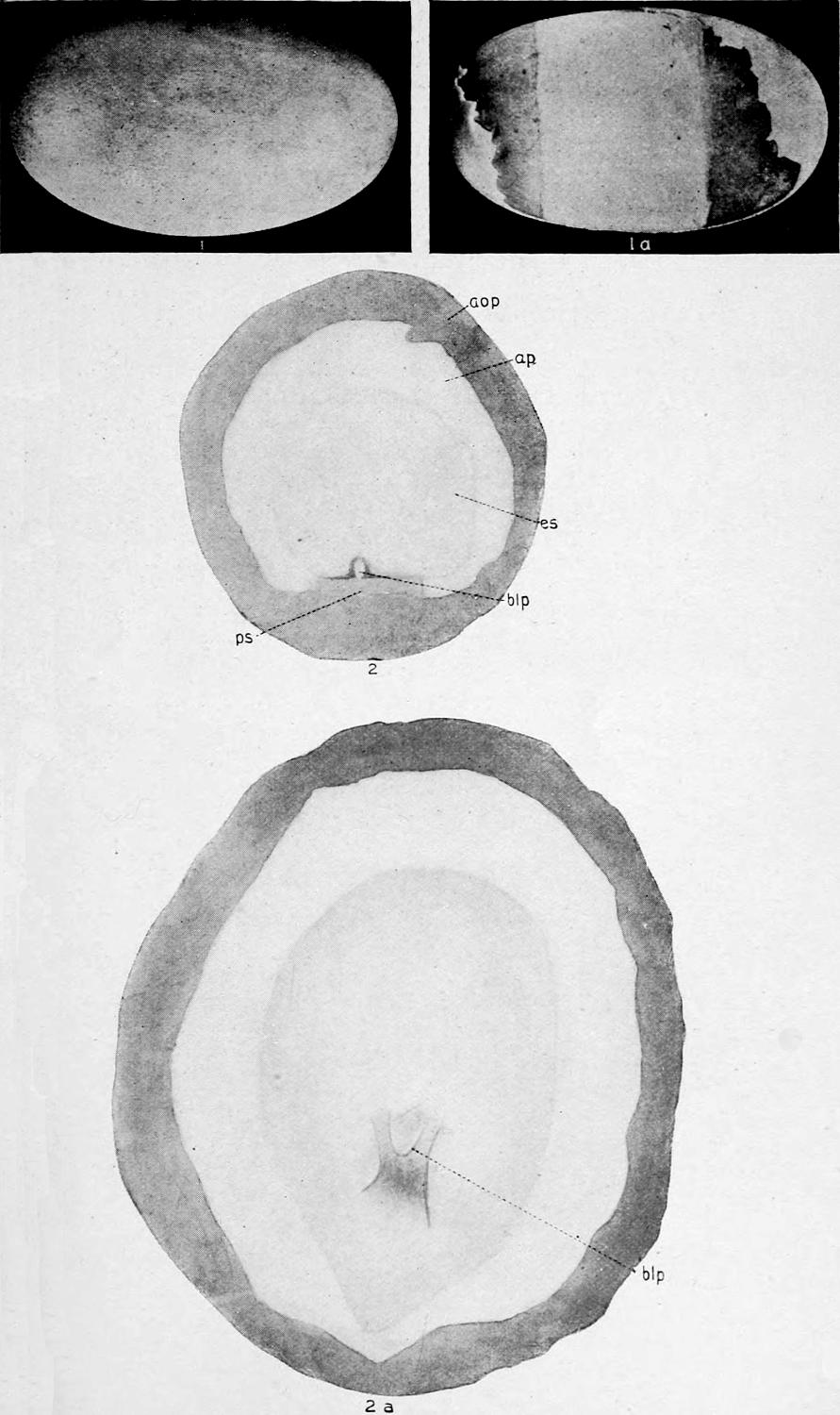
Plate VI. 1, 1a, The Egg; 2, 2a, Stage I.
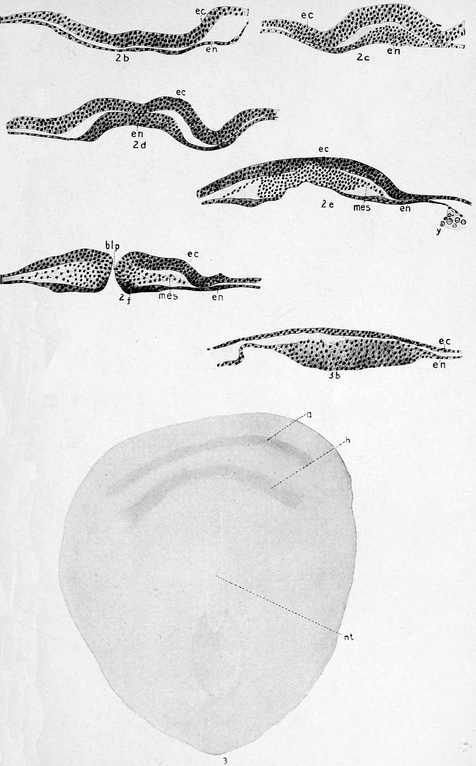
Plate VII. 2b-2f, Stage I. 3, 3b, Stage II.
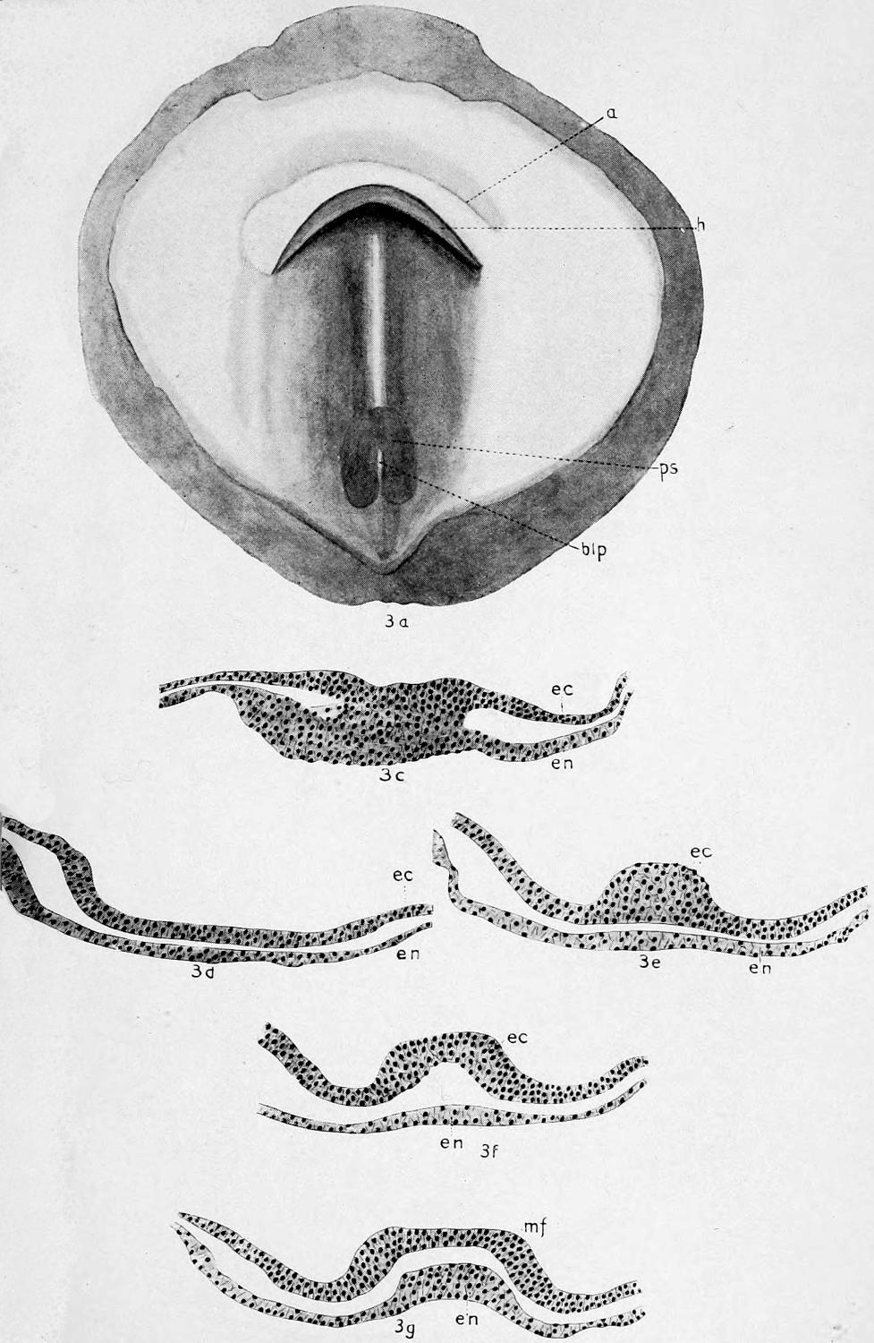
Plate VIII. 3a-3g, Stage II.
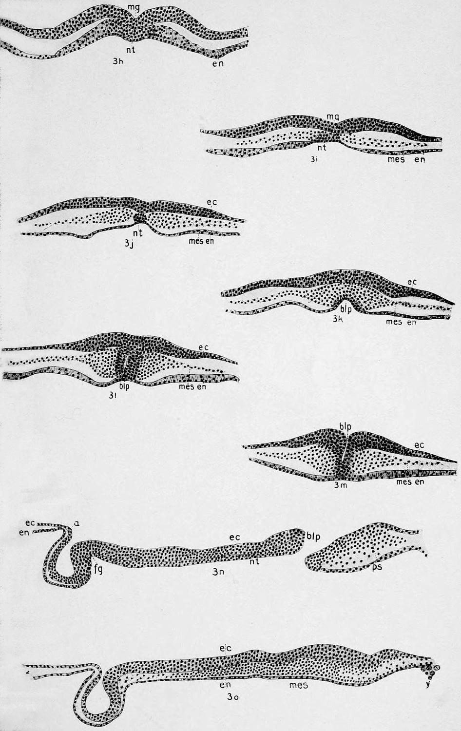
Plate IX. 3h-3o, Stage II.
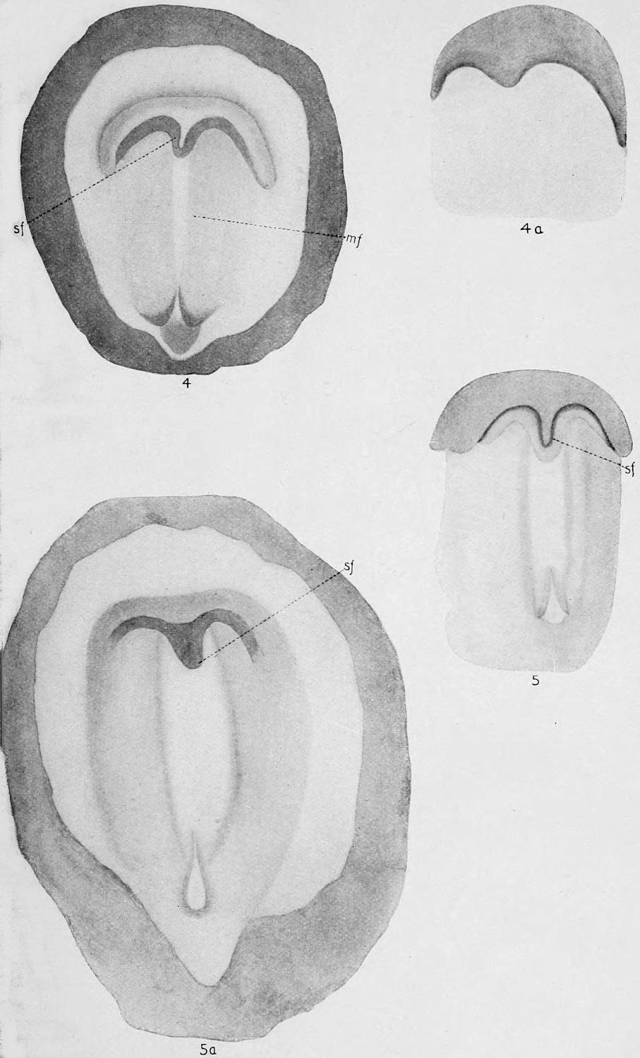
Plate X. Stage III.
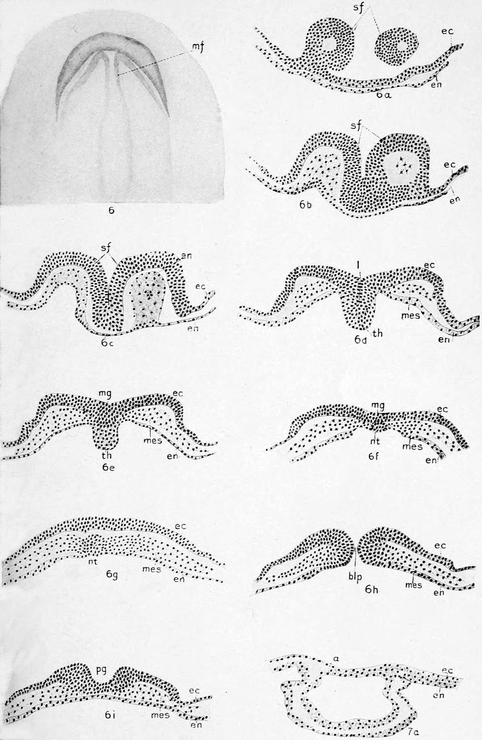
Plate XI. 6-6i, Stage III; 7a, Stage IV.
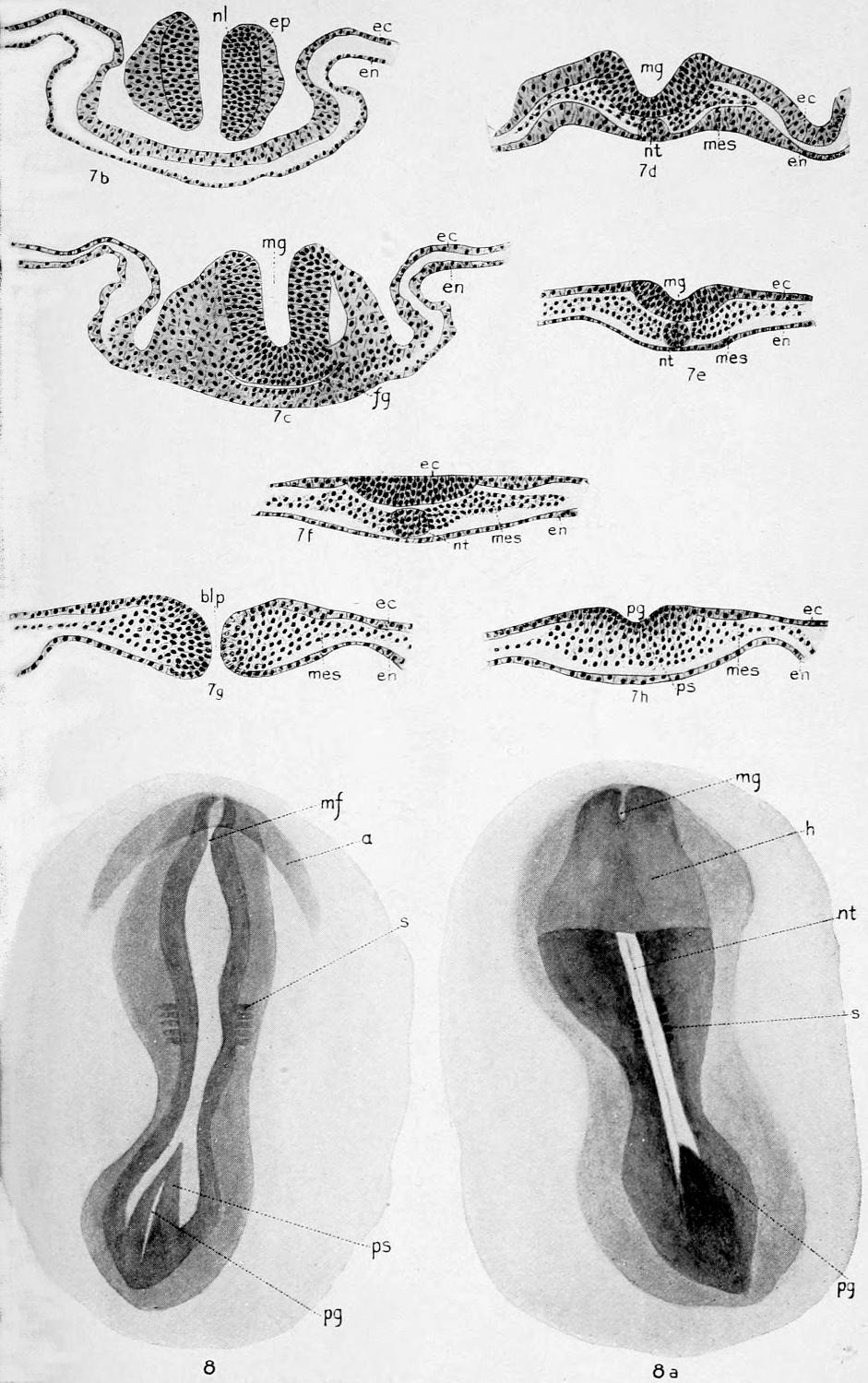
Plate XII. 7b-7h, Stage IV; 8, 8a, Stage V.
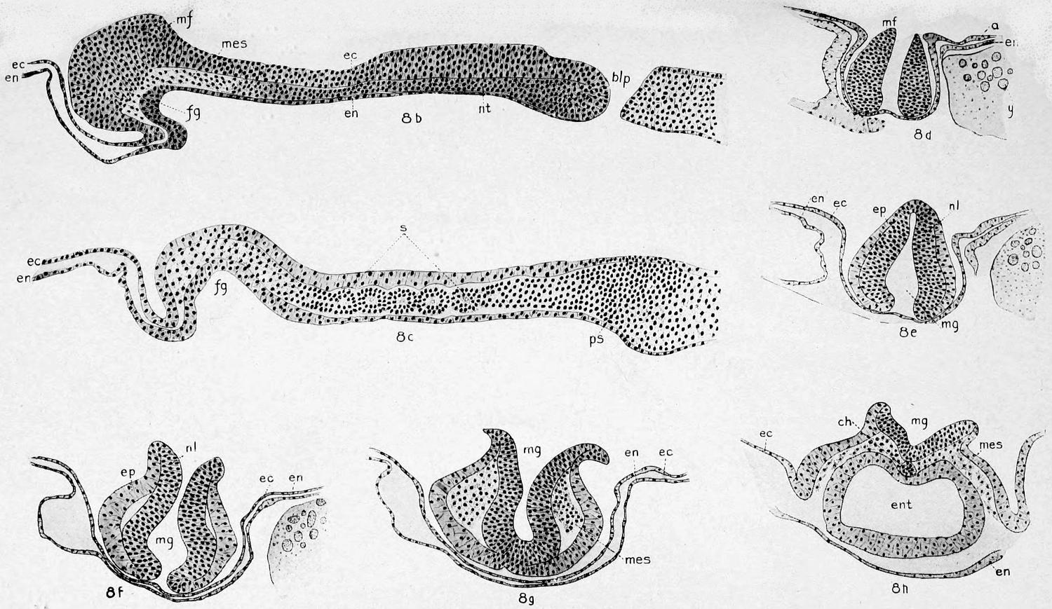
Plate XIII. Stage V.
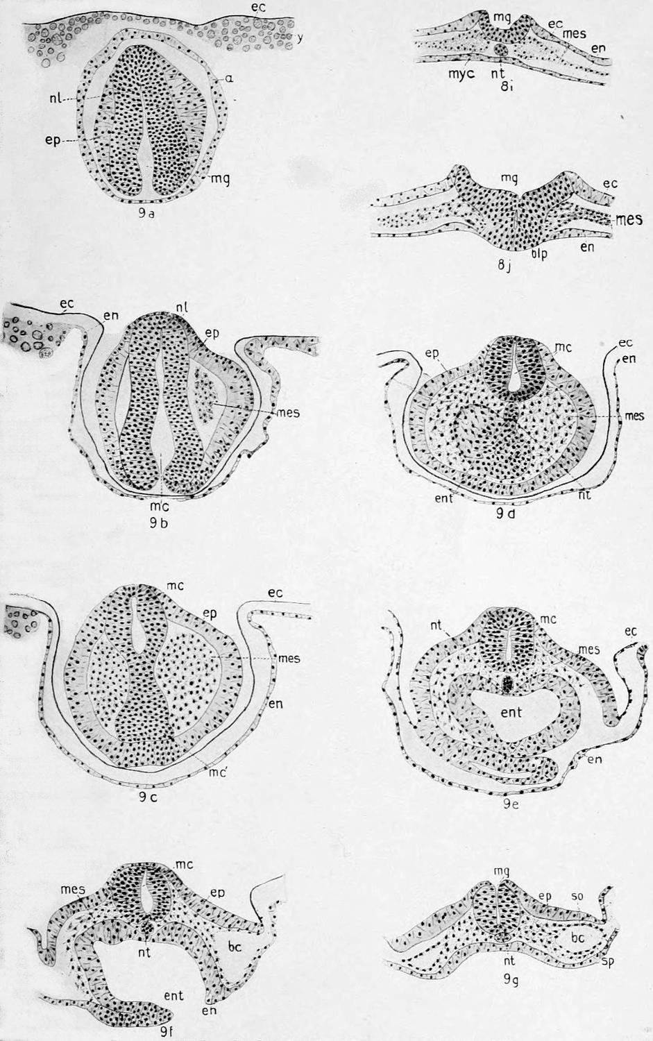
Plate XIV. 8i-8j, Stage V; 9a-9g, Stage VI.
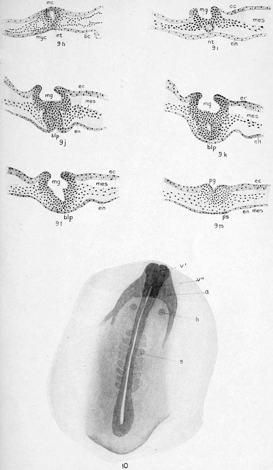
Plate XV. 9h-9m, Stage VI; 10, Stage VII.
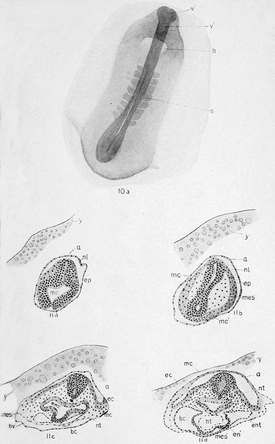
Plate XVI. 10a, Stage VII; 11a-11d, Stage VIII.
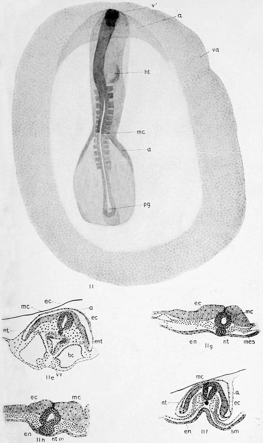
Plate XVII. Stage VIII.
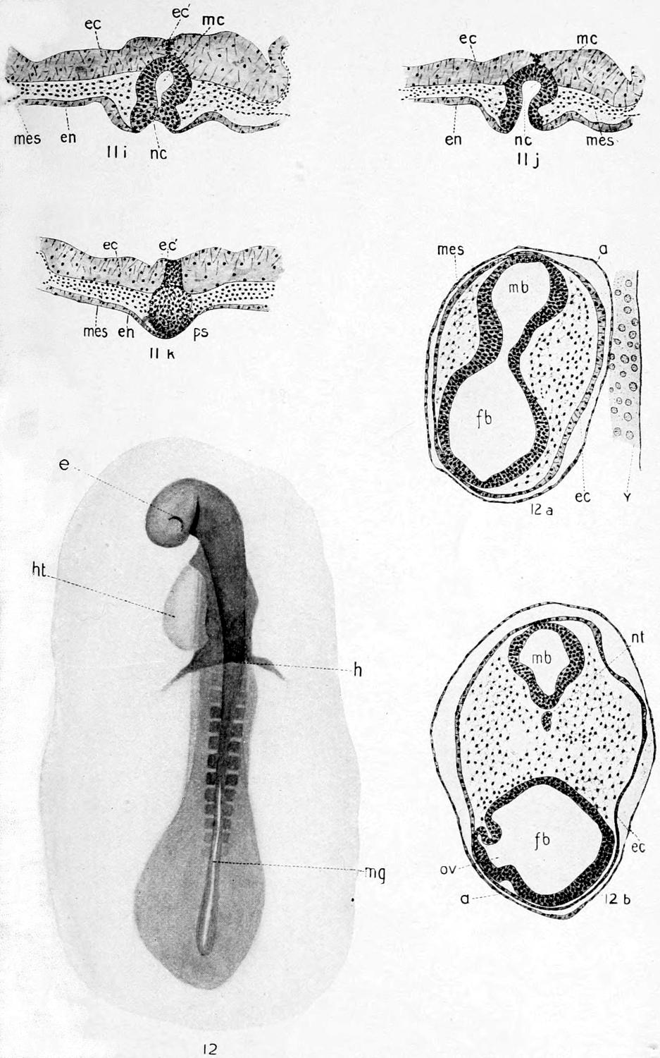
Plate XVIII. 11i-11k, Stage VIII; 12-12b, Stage IX.
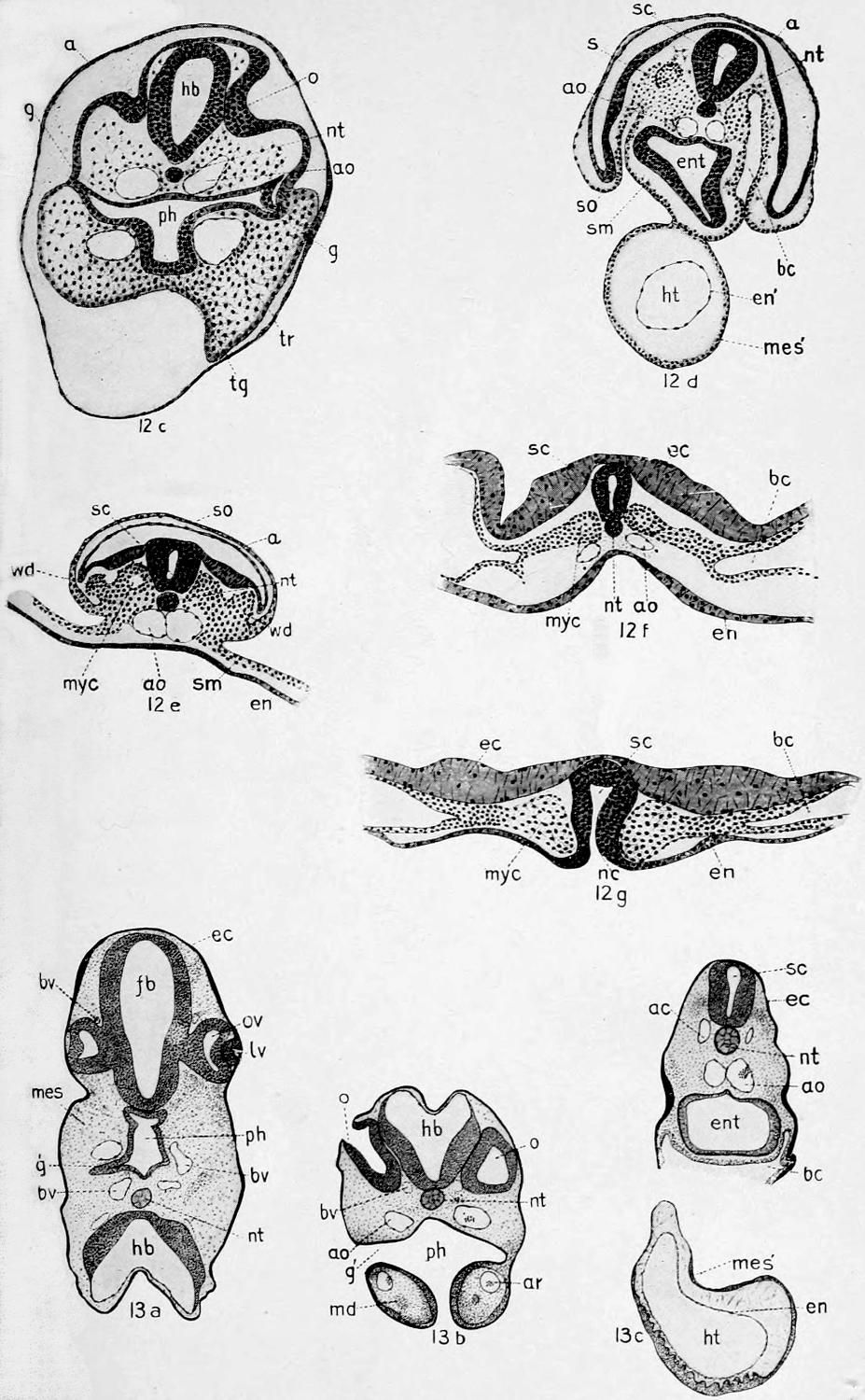
Plate XIX. 12c-12g, Stage IX; 13a-13c, Stage X.
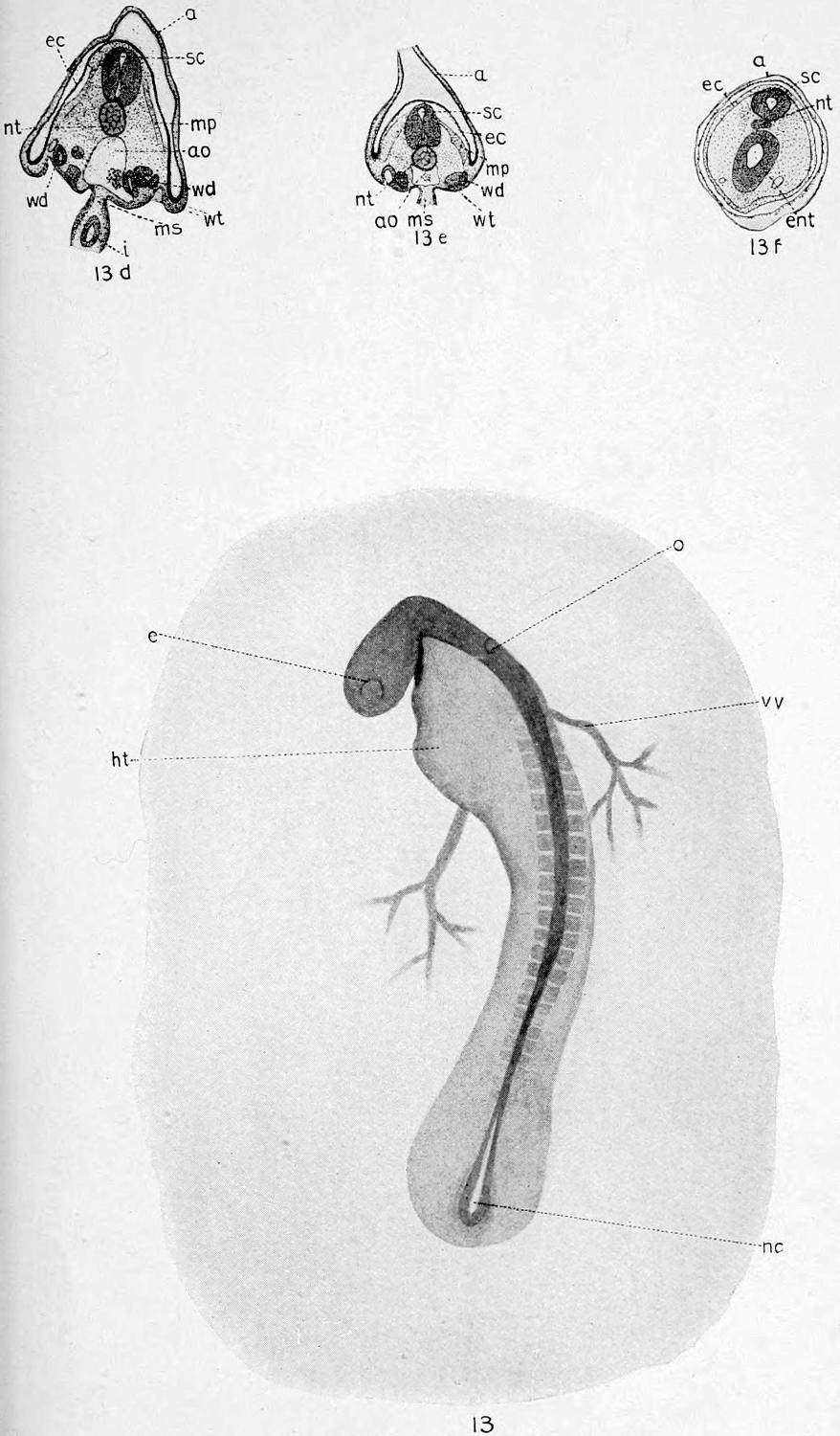
Plate XX. Stage X.
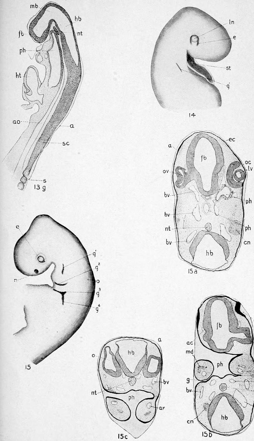
Plate XXI. 13g, Stage X; 14, Stage XI; 15-15c, Stage XII.
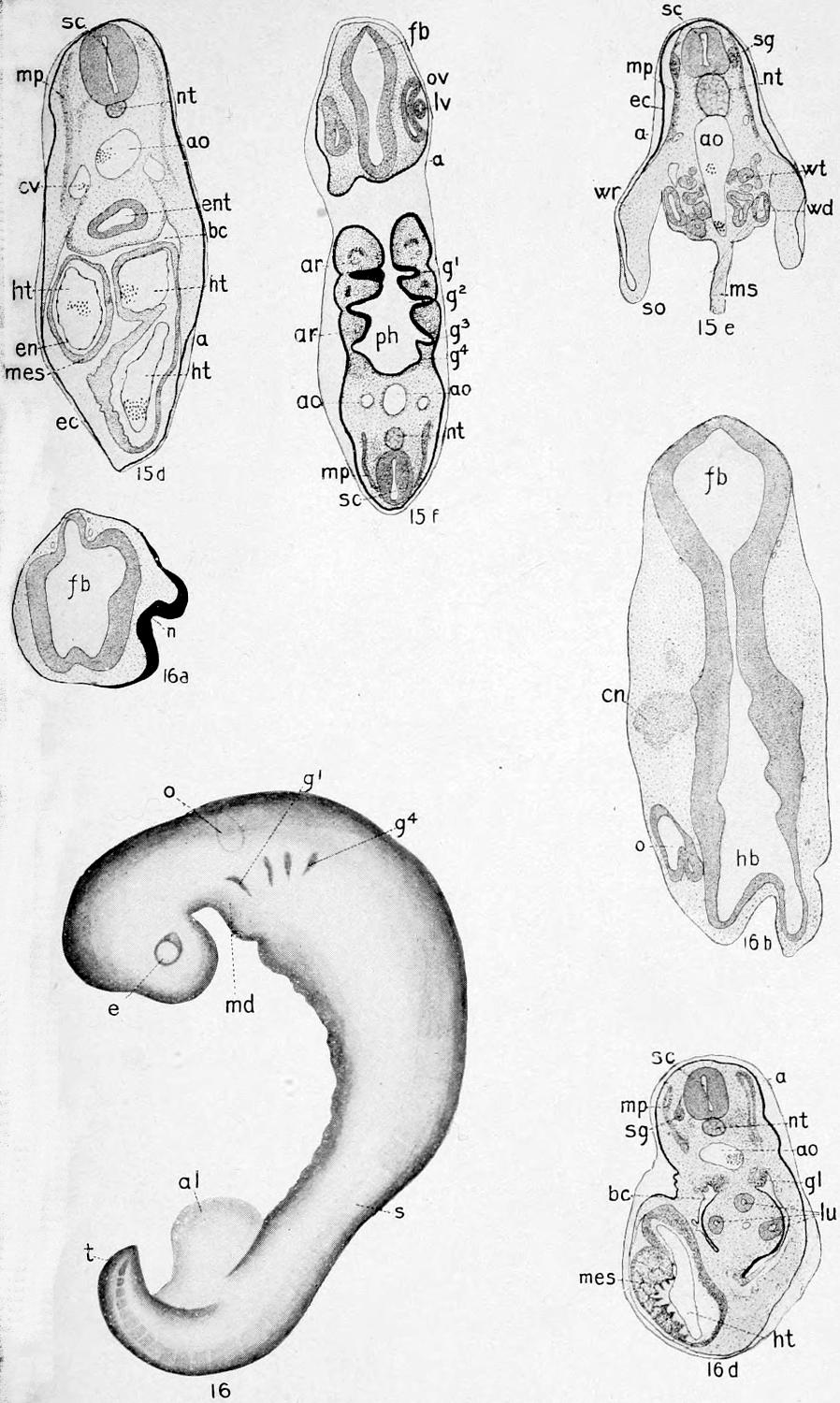
Plate XXII. 15d-15f, Stage XII; 16-16d, Stage XIII.
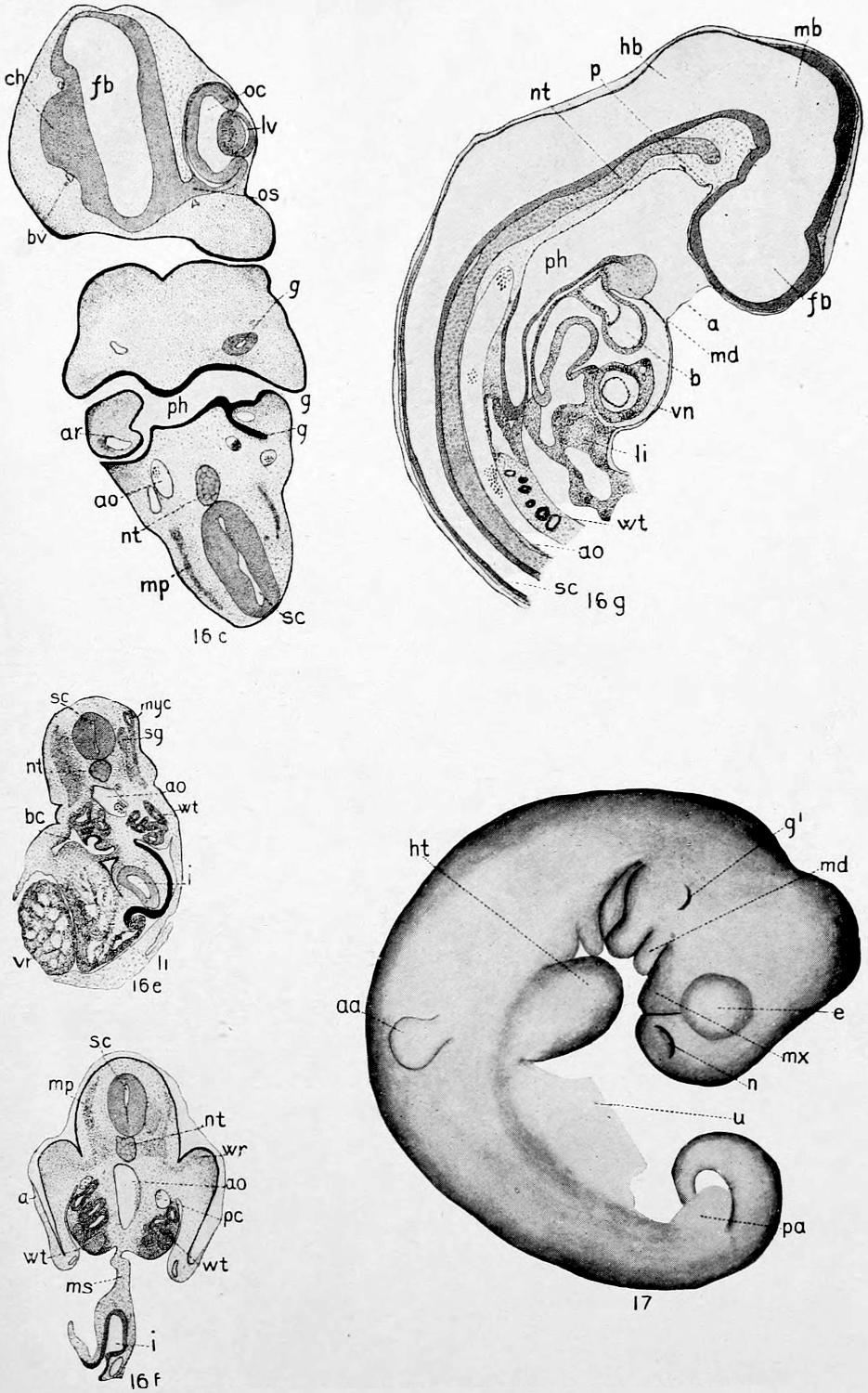
Plate XXIII. 16c-16g, Stage XIII; 17, Stage XIV.
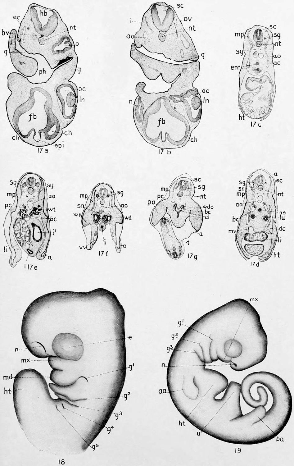
Plate XXIV. 17a-17g, Stage XIV; 18, Stage XV; 19, Stage XVI.
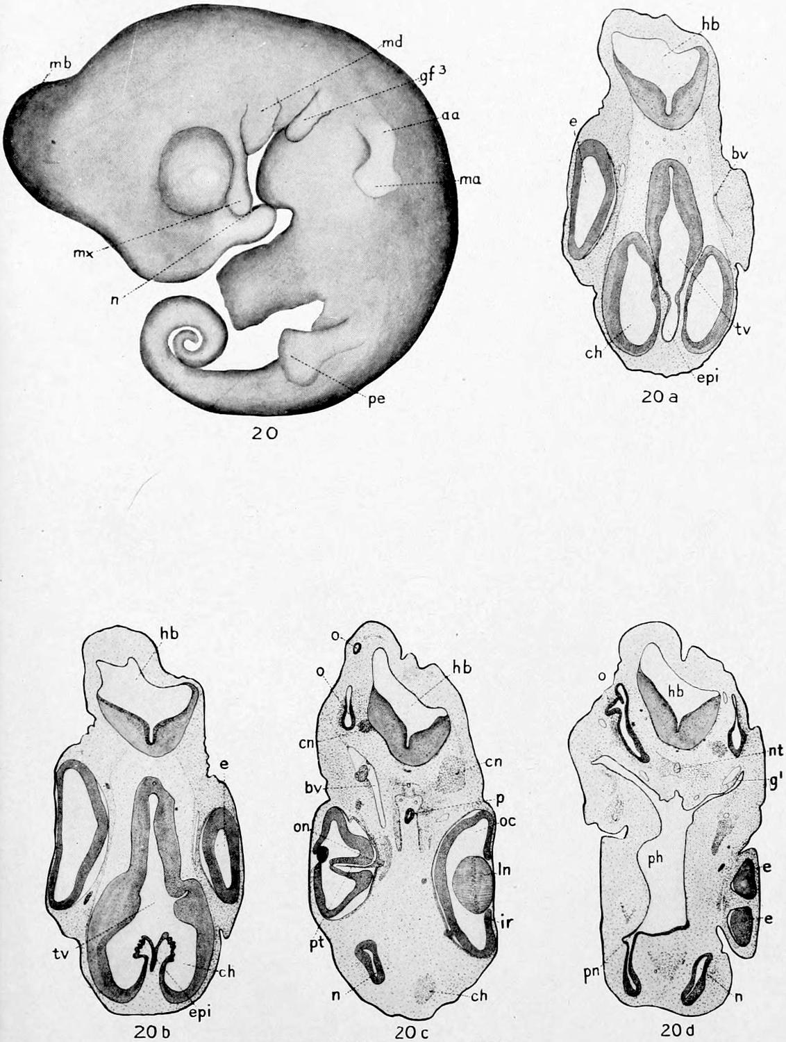
Plate XXV. Stage XVII.
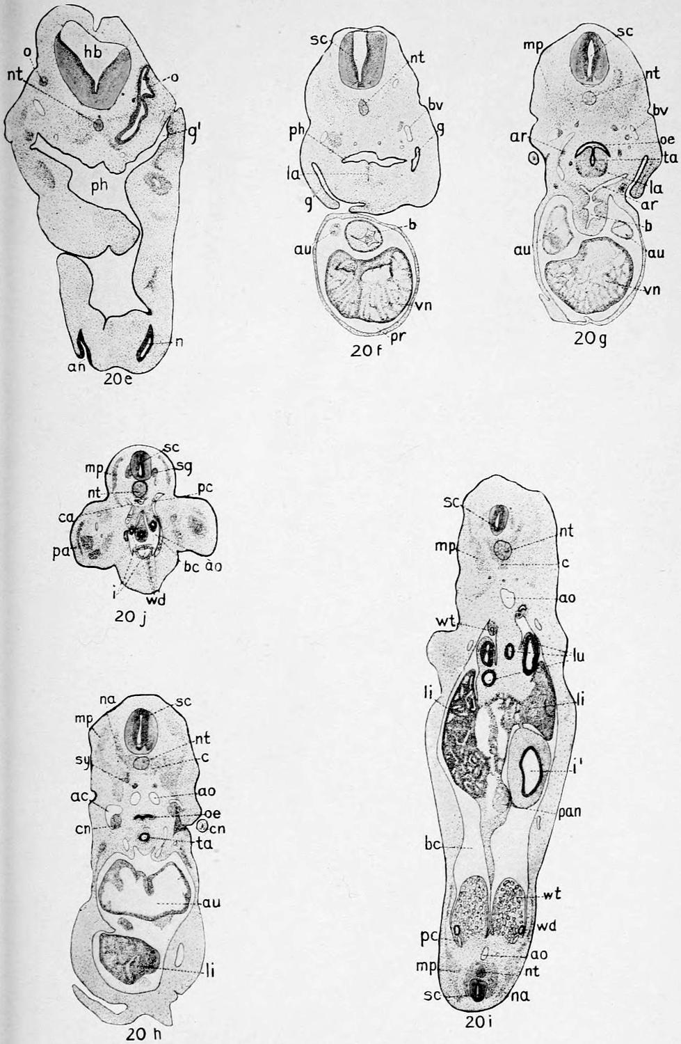
Plate XXVI. Stage XVII.
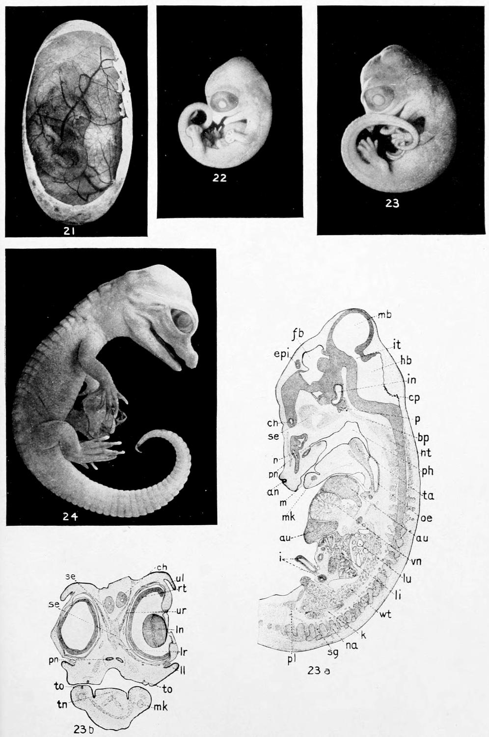
Plate XXVII. Stage XVIII; 22, Stage XIX; 23-23b, Stage XX; 24, Stage XXI.
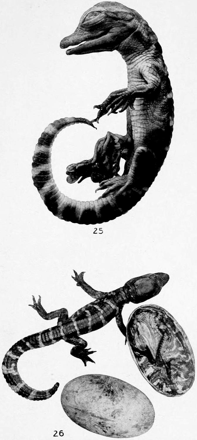
Plate XXVIII. 25, Stage XXII, Alligator Embryo; 26, Stage XXIII, Alligator Just Hatched and Relative Size of Egg.