Chapter VII
Reading edition
INJURIES TO THE HEAD AND NECK
Injuries to the head formed one of the most fruitful sources of death, both upon the battlefield and in the Field hospitals. It has been suggested that the mere fact of wounds of the head being readily visible ensured all such being at once distinguished and correctly reported, while wounds hidden by the clothing often escaped detection. When the external insignificance of many of the fatal wounds of the trunk is taken into consideration this is possible; but, on the other hand, it must be borne in mind that the head is in any attitude the most advanced, and often the most exposed, part of the body, and even when the soldier had taken 'cover,' it was frequently raised for purposes of observation. For the latter reasons I believe injury to the head fully deserved the comparative importance as a fatal accident with which it was credited.
A number of somewhat sensational immediate recoveries from serious wounds of the head have been placed upon record. Observation, however, shows that these, with but few exceptions, belonged either to certain groups of cases the relatively favourable prognosis in which is familiar to us in civil practice, or that the wounds were received from a very long range of fire, and hence the injuries were strictly localised in character.
Anatomical Lesions
Wounds of the scalp.—Nothing very special is to be recorded with regard to these; they either formed the terminals of perforating wounds, or were the result of superficial glancing shots. The glancing wounds were of the nature of furrows, varying in depth from mere grazes to wounds laying bare the bone. Their peculiarity was centred in the fact that a definite loss of substance accompanied them, the skin being actually carried away by the bullet; hence gaping was the rule. Every gradation in depth was met with, but the only situations in which wounds of considerable length could occur were the frontal region in tranverse shots, or, when the bullet passed sagitally, the sides of the head, or the flat area of the vertex.
The danger of overlooking injuries to the bone was of special importance in the short subcutaneous tracks occasionally met with at the points at which the surface of the skull makes sharp bends. In all such wounds it was a safe rule to assume a fracture of the skull until this was excluded by direct examination. In some of the gutter wounds and subcutaneous tracks crossing the forehead and sides of the head, signs of intracranial disturbance were occasionally observed in the absence of external fracture, such as transient muscular weakness, unsteadiness in movements, giddiness, diplopia, or loss of memory and intellectual clearness. In connection with such symptoms the classical injury of splintering of the internal table of the skull, the external remaining intact, had to be borne in mind, but I observed no proven instance of this accident. I am of opinion, moreover, that its occurrence with small bullets travelling at a high degree of velocity must be very rare, since little deflection is probable unless the contact has been sufficiently decided to fracture the external table; while in the cases of spent bullets the injury is unlikely, as requiring a considerable degree of force.
Injuries to the cranial bones, without evidence of gross lesion to the brain.—It may be premised that these were of the rarest occurrence, and they may be most readily described by shortly recounting the conditions observed in a few cases I noted at the time. The injuries resulted from blows with spent bullets, from bullets barely striking the skull directly, or those striking over the region of the frontal sinuses. Wounds of the mastoid process will not be considered in this connection as being of a special nature (see p. 299).
I saw only one case of escape of the internal, with depressed fracture of the external, table of the skull.
(45) In marching on Heilbron a man in the advance guard was struck by a bullet at right angles just within the margin of the hairy scalp. The regiment was at the time to all intents and purposes outside the range of rifle fire, and the patient was the only individual struck among its number. When brought into the Highland Brigade Field Hospital, a single typical entry wound was discovered; examination with the probe gave evidence of a slight depression in the external table of the frontal bone just above the temporal ridge. Although no perforation was detectible by the probe, and this was positively excluded on the raising of a flap (Major Murray, R.A.M.C.), it was considered advisable to remove a 1/4-inch trephine crown, the pin of the instrument being applied to the margin of the depression. No depression or splintering of the internal table was discovered, nor any injury to the dura, nor blood upon the surface of that membrane. The man made an uninterrupted recovery.
(46) A case of frontal injury was shown to me at Wynberg, in which a distinct furrow could be traced across the upper part of the frontal sinuses. There had been no symptoms beyond temporary diplopia, and the wound was healed; no surgical interference had been deemed necessary.
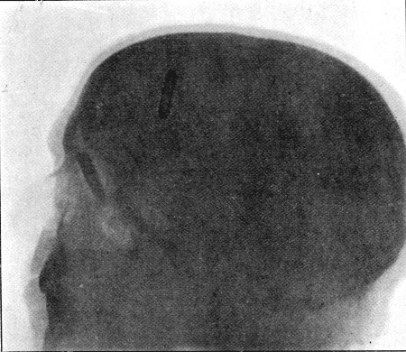
(47) In a man wounded at Poplar Grove, a single typical wound of entry was found 3/4 of an inch above the right eyebrow and the same distance from the median line. No primary symptoms were observed, but on the evening of the second day the temperature rose above 100° F., and the man seemed somewhat heavy and dull. The patient was examined by Major Fiaschi and Mr. Watson Cheyne, and it was decided to explore the wound. Mr. Cheyne removed fragments both of external and internal tables, one of the latter having made a punctiform opening, not admitting the finest probe, in the dura-mater. The bullet was traced into the nasal fossæ, where it was subsequently localised with the aid of the Roentgen rays when the patient came under my observation at Wynberg some days later (fig. 60).
Gunshot fracture of the skull with concurrent brain injury.—This was the commonest form of head injury, and possessed two main peculiarities; firstly, the large amount of brain destruction compared with the extent of the bone lesion; secondly, the fact that any region of the skull was equally open to damage. In consequence of the second peculiarity, the position and direction of secondary fissures are not so dependent on anatomical structure as in the corresponding injuries of civil practice. Thus, fractures of the base, for instance, were less constant in their course and position. The cases as a whole are best divided into four classes.
Fig. 60.—Mauser Bullet in Nasal Fossa. (Skiagram by H. Catling.) Case No. 47
1. Extensive sagittal tracks passing deeply through the brain, and vertical wounds passing from base to vertex or vice versa, in the posterior two thirds of the skull. These will be referred to as general injuries.
2. Vertical or coronal wounds in the frontal region.
3. Glancing or obliquely perforating wounds of varying depth in any part of the head.
4. Fractures of the base.
Of these classes the first was nearly uniformly fatal; the second relatively favourable, and with low degrees of velocity often accompanied by surprisingly slight immediate effects; while the third had perhaps the best prognosis of all, but this varied as to the defects that might be left, and with the region of the head affected.
1. General injuries.—Fractures of this class may be treated of almost apart. For their production the retention of a considerable degree of velocity on the part of the bullet was always necessary, and the results were consequently both extensive and severe.
The aperture of entry was comparatively small, since to take so direct and lengthy a course through the skull the impact of the bullet needed to be at nearly an exact right angle to the surface of the bone. Any disposition to assume the oval form, therefore, depended mainly upon the degree of slope of the actual area of the skull implicated. In size the aperture of entry did not greatly exceed the calibre of the bullet; in outline it was seldom exactly circular, but rather roughly four-sided, with rounded angles, slightly oval, or pear-shaped. The margin of the opening consisted of outer table alone, the inner being always considerably comminuted. Fragments of the latter, together with the majority of those corresponding to the loss of substance of the outer table, were driven through the dura mater and embedded in the brain. These bony fragments were more or less widely distributed over an area of a square inch or more, and not confined to a narrow track.
Fig. 61.
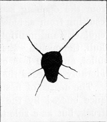
Diagram of Aperture of Entry in Occipital Bone, showing radiating fissures exact length. The exit in the frontal region was of typical explosive character. Range '100 yards'
The amount of fissuring at the aperture of entry was often not so extensive as I had been led to expect. Fig. 61 is a diagram illustrating a fairly typical instance; in some cases no fissuring existed. As a rule the nearer to the base, the greater was the amount of fissuring observed. The fissures were sometimes very extensive in this position, probably as a result of the lesser degree of elasticity in this region of the skull. Again, when the aperture of entry was near the parts of the vertex where sudden bends take place, considerable fissuring of the same nature as that seen in the superficial tracks (fig. 68) was produced in the flat portion of the skull above the point of entrance.
Radial fissuring around the aperture of entry in the skull scarcely corresponds in degree with that seen when the shafts of the long bones are struck, and is far less marked and regular than when one of these small bullets strikes a thick sheet of glass set in a frame. I saw several apertures in the thick glass of the windows of the waterworks building at Bloemfontein produced by Mauser bullets. They differed little from the opening seen in an ordinary plate-glass window resulting from a blow from a stone, except perhaps in the regularity and multiplicity of the radial fissures. As in the skull, the opening was a little larger than the calibre of the bullet, and the loss of substance on the inner aspect considerably exceeded that on the outer.
The degree of fissuring is probably affected by the resistance offered by the particular skull, or the special region struck, but as a rule the elasticity and capacity for alteration in shape possessed by the bony capsule, is opposed to the production of the extreme radial starring observed in the long bones or a fixed sheet of glass. Corroborative evidence of the influence of elasticity in the prevention of starring is seen in the limited nature of the comminution of the ribs in cases of perforating wounds of the thorax.
In the most severe cases we can only speak of the 'aperture' of exit in a limited sense in so far as the opening in the scalp is concerned; this was often comparatively small, not exceeding 3/4 of an inch in diameter. Beneath this limited opening in the soft parts, the bone of the skull was smashed in a most extensive manner. The portion exactly corresponding to the point of exit of the bullet was carried altogether away, but around this point a number of large irregularly shaped fragments of bone, from 3/4 to 1 inch in diameter, were found loose, and often so displaced as to expose a considerable area of the dura-mater. Beyond the area of these loose fragments, fissures extended into the base and vertex, in the latter case often being limited in their extent by the nearest suture.
Over extensive fractures of this nature general œdema and infiltration of the scalp, due to extravasation of blood, were present. When the exit was situated in the frontal region ecchymosis often extended to the eyelids and down the face, while in the occipital region similar ecchymosis was often seen at the back of the neck.
The opening in the dura mater at the aperture of entry was either slitlike, or more often irregular from laceration by the fragments of bone driven in by the bullet. At the point of exit a similar limited opening corresponded with the spot at which the bullet had passed, while separate rents of larger size were often seen at some little distance. The latter were the result of laceration of the outer surface of the membrane by the margins of the large loose fragments of bone above described.
Injury to the brain more than corresponded in extent to the fractures of the bone. Pulping of its tissue existed over a wide area both at the points of entrance and of exit. In the former position the amount of damage was the less, the gross changes roughly corresponding with the tissue directly implicated by the bullet itself, and the fragments of bone carried forward by it. The degree of splintering of the skull therefore in great part determined the severity of the lesion. At the exit aperture much more widespread destruction existed, while masses of brain tissue, small shreds of the membranes, fragments of bone, and débris from the scalp were found occasionally bound together by coagulated blood and protruding from an exit opening of some size. The largest masses of such débris were most often seen in instances in which the bullet had entered by the base to escape at the vertex of the skull.
The brain in the line of injury suffered comparatively slightly, but small parenchymatous hæmorrhages into its tissue indicated in lesser degree the same type of injury undergone by the mass of brain pulp and small blood-clots found at the external limits of the wound. Beyond this extensive hæmorrhages at the base of the skull were common.
With regard to the extensive character of the brain destruction in the region of the aperture of exit, it must be borne in mind that this lesion corresponds in position with one which would exist even if the injury were of a non-penetrating degree. A large proportion of the contusion and destruction is therefore explained by violent impact of the projected brain with the skull prior to the passage of the bullet, and not to the direct action of the bullet on the tissues.
These cases of 'general injury' afford a marked example of the lesions to which the term 'explosive' has been applied, and as such have an important bearing on the theories held as to the mode of production of explosive effect. The increased area of tissue damage at the aperture of exit favours the theory of direct transmission of a part of the force with which the bullet is endowed, to the molecules of tissue bounding the track made by the projectile. Thus the area of destruction corresponds with the cone-like figure which one would expect to be built up by the vibrations spreading from the primary point of impact. The exit region of the skull is subjected not alone to the force of the travelling bullet, but also to that exerted over a much wider area by the tissue to which secondary vibrations have been communicated. The brain itself is, in fact, dashed with such violence against the bone as to cause a great part of the injury.
No doubt the brain in its reaction to the bullet forms as near an approach to a fluid as any solid tissue in the human body, and experimental observation has shown how greatly its presence or absence in the skull affects the degree of comminution on the exit side; hence the fondness for the so-called hydraulic theory that has been always exhibited in the case of these injuries. The localisation of the injury in its highest degree to the neighbourhood of the exit aperture, however, shows that in any case the main wave takes a definite direction in a course corresponding to that of the bullet.
The real importance of the presence of the brain within the skull in increasing the amount of damage at the exit end of the track, is as a medium for the ready transmission of forcible vibrations. That the latter are to some extent conveyed as by a fluid is evidenced by the occasional presence of brain matter and fragments of bone in the aperture of entry, which suggests recoil or splash such as would be expected from a fluid wave.
Experience of the character of the lesions observed after severe concussion by the ordinarily somewhat coarser forms of violence common to civil life, fully explains the severity of the damage to the brain tissue met with in injuries due to bullets of small calibre. Viewing the elaborate arrangements which exist for the preservation of the central nervous system from the moderate vibration incidental to ordinary existence, it is easy to appreciate the harmfulness of such exquisite vibratory force as that transmitted by a bullet of small calibre travelling at a high rate of velocity.
Effect of ricochet in the production of severe forms of injury.—In connection with the lesions above described mention must be made of cases in which the aperture of entry reaches a large size, or a portion of the skull is actually blown away.
Examples of the former class were not uncommon; I will briefly relate one.
(48) A Highlander while lying in the prone position at Rooipoort, was struck by a bullet probably at a distance of about 1,000 yards. A large entry wound in the scalp was produced, while the defect in the skull was coarsely comminuted and was capable of admitting three fingers into a mass of pulped brain. Both brain matter and fragments of bone were found in the external wound, which was situated just anterior to the right parietal eminence. The bullet passed onwards through the base of the skull, crossing the external auditory meatus, fracturing the zygoma and probably the condyle of the mandible, and eventually lodged beneath the masseter muscle. Blood and brain matter escaped from the external auditory meatus.
The patient was brought off the field in a semi-conscious condition, the pupils moderately contracted but equal, the pulse 66, very small and irregular in beat, the respiration quiet and easy, and with paralysis of the left side of the body. The fæces had been passed involuntarily.
The wound was cleansed and bone fragments removed. The patient had to travel in a wagon for the next three days until the column halted. The progress of the case was unsatisfactory, as the wound became infected, and the man eventually died on the 14th day of general septicæmia, but with little evidence of local extension of septic inflammation.
In this instance the head was no doubt struck by a bullet which had previously made ricochet contact with the ground. I saw several such cases.
Closely connected with such injuries are those in which large portions of the skull and scalp were actually blown away. I never witnessed one of these myself, but I recall two instances described to me by officers who lay near the wounded men on the field. In one the frontal region was carried away so extensively that, to repeat the familiar description given by the officer, 'he could see down into the man's stomach through his head.' In a second case the greater part of the occipital region was blown away in a similar manner, and this was of especial interest as the wounded man was seen to sit up on the buttocks and turn rapidly round three or four times before falling apparently dead. The observation offers interesting evidence of the result of an extensive gross lesion of the cerebellum.
In the absence of exact information, it may well be that such injuries as the two latter were produced by some special form of bullet, but as both were produced while the patients were lying on the ground, and therefore especially liable to blows from ricochet bullets, I am inclined to attribute both to this cause.
In considering injuries of the above nature, one cannot help speculating on the possible influence of a head-over-heels ricochet turn on the part of the bullet while traversing the long sagittal axis of the skull. It is not uncommon for apical target ricochets to present evidence of damage to the apex and base of the mantle alone. This must depend on a rapid turn on impact, which might well be imitated in the case of the skull, and would then go far to explain the production of some of the most severe forms of explosive exit wounds met with. See cases 48, 54, 68.
Short of ricochet, the influence of simple wobbling must also be considered in shots from a long range. The entry wound may be large as a result of this condition, but as the velocity possessed by the bullet is low, the injuries would probably not be of a very severe nature.
In connection with the subject of wobbling, reference should be made to the form suggested by Nimier and Laval, in which the wobble, as the result of resistance to the apex of the revolving bullet, assumes the form of movement seen when the spin of a top is failing. This would explain a peculiarity in some wounds of entry over the skull first pointed out to me by Mr. J. J. Day. When such wounds were explored, as well as the presence of brain in the entry aperture, a number of fragments of the external table of the skull were found everted and fixed in the tissues of the scalp. As already suggested, this may be mere evidence of splash, but it may be equally well explained by a process of wobble around the axis of revolution of the bullet. This might, no doubt, also be invoked to explain the displacement of some of the fragments in fractures of the long bones, where considerable resistance to the passage of the bullet is offered.
II. Vertical or coronal wounds in the frontal region.—These injuries were common, and offered some of the most interesting illustrations of the variations in symptoms and effects following apparently exactly identical lesions, judging from the condition of the external soft parts alone; since the latter sometimes gave little indication of the force (dependent on the rate of velocity) which had been applied.
With the lower degrees of velocity simple punctured fractures of the skull resulted, without extensive lesion of the frontal lobes as evidenced by immediate symptoms. The nature of the fractures differed in no way from the punctured fractures we are familiar with in civil practice. The openings of entry in the bone were irregularly rounded, corresponding in size to the particular calibre of the bullet concerned. The margin consisted of outer table alone, while the inner table was either considerably comminuted, or a large piece was depressed, wounding the dura-mater and projecting into the brain substance (see fig. 63). The aperture of exit presented exactly the opposite characters, the splintering comminution or separation of a large fragment affecting the outer table, while the inner presented a simple perforation. The latter condition is represented in figs. 71 and 72, and I will here give short notes of four illustrative cases, as being the shortest and most satisfactory method of conveying a correct idea of the nature of such injuries.
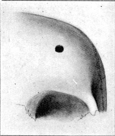
Fig. 62—Aperture of Entry in Frontal Bone. Case No. 50. 1/2
(49) Vertical perforation of frontal bone.—Wounded at Belmont, while in the prone position. Aperture of entry (Mauser), at the anterior margin of the hairy scalp on the left side; course, through the anterior part of the left frontal lobe, roof of the left orbit, cutting the optic nerve and injuring the back of the eyeball, floor of the orbit, the antrum, the hard palate, and tongue. Exit, in mid line of the submaxillary region. No cerebral symptoms were noted, and on the fifth day the man was sent to the Base hospital without operation; the pulse was then 70 and the temperature normal. The movements of the eyeball were perfect, but blindness was absolute. At the Base hospital the eye suppurated and was removed. The patient was then sent home apparently well. He has since been discharged from the service, and is now employed as a painter in Portsmouth Dockyard.
(50) Vertical perforation of frontal bone.—Wounded at Paardeberg while in the prone position. Range, 600-700 yards. Aperture of entry (Mauser), at the fore margin of the hairy scalp above the centre of the right eyebrow; course, through the anterior third of the right frontal lobe, roof of orbit, front of eyeball, margin of floor of orbit making a distinct palpable notch, and cheek; exit through the red margin of the upper lip, 1/2 an inch from the right angle of mouth. The bullet slightly grooved the lower lip.
The patient rose almost immediately after being struck, and walked about a mile, although feeling dizzy and tired. The wounds, which both bled considerably, were then dressed. After three days' stay in a Field hospital, the patient was sent in a bullock wagon three days and nights' journey to Modder River and thence to the Base.
There was anæsthesia over the area supplied by the outer branch of the supra-orbital nerve, extending from the supra-orbital notch backwards into the parietal region, but none over the area supplied by the second division of the fifth nerve.
On the tenth day there were no signs of cerebral disturbance except a pulse of 48. The eyeball was suppurating, and the temperature rose to 99° at night. The lids were still swollen and closed.
A few days later the eyeball was removed and at the same time a flap was raised and the fracture explored (Major Burton, R.A.M.C.). An opening somewhat angular, 1/3 of an inch in diameter, was found with a thin margin in the outer table of the skull (fig. 62); when this was enlarged with a Hoffman's forceps, an opening in the dura was discovered, and cerebro-spinal fluid escaped. A piece of the inner table of the skull (fig. 63), 3/4 by 1/3 an inch in size, was discovered projecting downwards vertically into the brain. This latter was removed and the wound closed. Healing by primary union followed, and no further symptoms were observed.
Fig. 63.
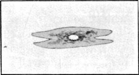
Fragment of Inner Table depending vertically from lower margin of puncture shown in fig. 62. The centre was perforated. Exact size
(51) Transverse frontal wound.—Wounded at Paardeberg. The man was sitting down at the time he was struck, in the belief that he was out of the range of fire. The entry and exit wounds were almost symmetrical, placed on the two sides of the forehead at the margin of the hairy scalp, 2¼ inches above the level of the external angular processes of the frontal bone. The patient lost consciousness for about half an hour, then rose and walked half a mile to the Field hospital. The wounds were dressed, and after a stay of three days in hospital, the man was sent the three days' journey to Modder River; during the journey he got in and out of the wagon when he wished. After two days' stay at Modder, a journey was again made by rail to De Aar (122½ miles). The wounds were healed. The man stayed at De Aar nearly a month, and then, rejoining his regiment, made a two days' march of some 22 miles on hot days. He had to fall out twice on the way by reason of headache, feeling dizzy, and 'things looking black.' He did not own to any loss of memory or intellectual trouble, but was invalided to England. This patient returned to South Africa later, and is now on active service.
(52) Transverse frontal wound.—Within a few days an almost identical symmetrical wound in the frontal region occurred in the same district, from a near range. The patient became immediately unconscious, and remained so until his death some four days later, his symptoms being in no way alleviated by operation and the removal of a quantity of bone fragments and cerebral débris. At the post-mortem examination, extensive destruction of both hemispheres of the brain was revealed, and large fissures extended into the base of the skull.
III. Glancing or oblique perforating wounds of varying depth in any portion of the cranium.—These injuries were the most common, the most highly characteristic of small-calibre bullet wounds, the most interesting from the point of view of diagnosis, prognosis, and treatment, and beyond this they formed the variety most unlike any that we meet with in civil practice.
They were met with in every region of the cranium, and in every degree of depth and severity. The lesser are best designated as gutter fractures, the deeper are perforating and gradually approximate themselves to the type of injury described as class 1.
When the bullet struck a prominent or angular spot on the skull a considerable oval-shaped fragment was occasionally carried away, leaving an exposed surface of the diploë (case 60, p. 274). Under these circumstances the apparent lesion on raising a flap was slight, but exploration often showed extensive intra-cranial mischief. Thus in the case referred to both dura and brain were wounded, and continuing hæmorrhage led to the development of progressive paralysis, relieved only by operation.
From the more deeply passing bullets a more or less oval opening resulted, in which both tables were freely comminuted and displaced. These cases differed from the typical gutter fracture only in length and outline, and the nature of the accompanying intra-cranial lesion was identical, while in the latter particular they differed much from fractures in which the impact of the bullet was direct, in spite of a near resemblance in the appearances in the osseous defect.
I saw one instance in which a circular fissure about 1½ inch from the actual opening of entry surrounded the latter, the area of bone within the circle being somewhat depressed, though radial fissures were absent.
In several of these cases fragments of lead were either found on the fractured surface of the bone or within the cranial cavity, showing that the bullets had undergone fissuring of the mantle, or had actually broken up on impact.
Gutter fractures.—The nature of the injury to the bones in these is best illustrated by a series of diagrams of sections such as are shown below.
Fig. 64.
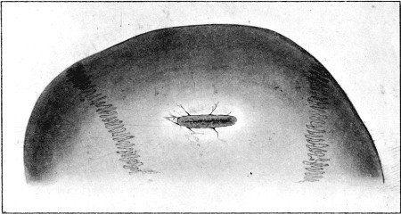
Gutter Fracture of first degree. The drawing does not show well the small fragments of bone usually carried from the margins of the depression by the bullet
In the most superficial injuries the outer table was grooved and depressed, usually with loss of substance from small fragments directly shot away: these latter had either been driven through the wound in the soft parts, or remained embedded on the deep aspect of the enveloping scalp (fig. 64). In the less common variety the scalp was slit to a length corresponding with the injury to the bone, but more often oval openings in the skin existed at either end of the track. The inner table was practically never intact, but the amount of comminution naturally varied with the depth to which the outer table was implicated (fig. 65 A, and B).
The following is an illustrative example of this degree, and also emphasises the consequences which may follow primary non-interference.
Fig. 65.
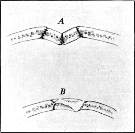
Diagrammatic transverse sections of varying condition of bones in Gutter Fractures of the first degree. A. With no loss of substance. B. With comminution
(53) Superficial gutter fracture in parietal region. Convulsive twitchings. Secondary paralysis.—Wounded at Modder River. Range, 400 yards. A scalp wound 3 inches in length ran vertically downwards, commencing 1 inch from the median line, and situated immediately over the upper third of the right fissure of Rolando. The patient was unconscious for several hours after the injury, and later suffered with severe headache, and twitchings in the left shoulder and arm.
The wound healed, but a well-marked groove was palpable in the bone beneath, and the twitchings persisted. The latter came on about every twenty minutes, and loss of power in the left upper extremity, and to a less degree in the lower, developed. The memory was defective, and the patient suffered at times with headache. The pupils were equal but sluggish in action. No changes were discovered in the fundus beyond a well-developed myopic crescent at the lower and outer part of the left disc (Mr. Hanwell).
The twitchings became more frequent and latterly were accompanied by somewhat severe muscular contractions in the upper extremity, while the loss of power in the lower extremity became more marked. Headache was also more troublesome.
The patient throughout refused any operation, saying he would rather go home first, and at the end of a month he left for England.
In the deeper injuries more and more of the outer table was cut away, and the inner became gradually more depressed, fractured, or comminuted (fig 66).
Fig. 66.
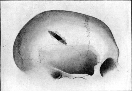
Gutter Fracture of the second degree. Perforating the skull in the centre of its course. External table alone carried away at either end
Bevelling at the expense of the outer table at both entry and exit ends of the course existed, but in either case a portion of the inner table was also detached and depressed. Sometimes the depressed portion of the inner table was mainly composed of one elongated fragment; this was either when the bullet had not implicated a great thickness of the outer table, or had passed with great obliquity through especially dense bone (see fig. 70). When the bullet had passed more deeply the inner table was comminuted into numberless fragments. I have frequently seen 50 or 60 removed. Where such tracks crossed convex surfaces of the skull, the two conditions were often combined; thus at one portion of the track, usually the centre, the comminution was extreme, while at either end a considerable elongated fragment of inner table was often found, the latter perhaps more commonly at the distal or exit extremity (fig. 67).
Fig. 67.
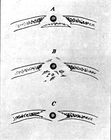
Diagrammatic transverse sections of complete Gutter Fracture. A. External table destroyed, large fragment of internal table depressed. (Low velocity or dense bone.) B. Comminution and pulverisation of both tables centre of track. C. Depression of inner table (low velocity)
The nature of the injury to the bone when the flight of the bullet actually involved the whole thickness of the calvarium was comparable to that seen in the case of the long bones when struck by a bullet travelling at a moderate rate (see plate XIX. of the tibia, or what is illustrated in the case of the pelvis in fig. 55). In point of fact, a clean longitudinal track appeared to have been cut out. The length of these tracks naturally depended upon the region of the skull struck. When a point corresponding to a sharp convexity, or a sudden bend in the surface, was implicated, an oval opening of varying length in its long axis was the result; when a flat area, as exists in the frontal or lateral portions of the skull, was the seat of injury, a long track was cut.
Superficial perforating fractures.—These formed the next degree; the chief peculiarity in them was the lifting of nearly the whole thickness of the skull at the distal margin of the entry, and the proximal edge of the exit, openings; the flatter the area of skull under which the bullet travelled the more extensive was the comminution. In some cases nearly the whole length of the bone superficial to the track would be raised; in fact, the bullet having once entered, the force is applied from within in exactly the same way that it operates on the inner table in the gutter fractures. A corresponding injury is met with in the case of the bones of the extremities (see fig. 57 of the tibia), and again the resemblance between these injuries of the skull and such perforations of the long bones as are illustrated by skiagrams Nos. III. and XXIII. of the clavicle and fibula is a close one.
Fig. 68.
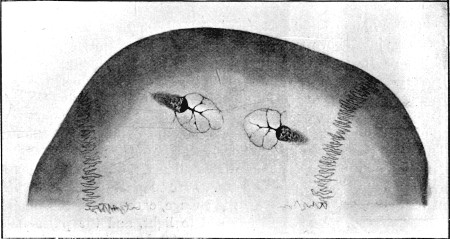
Superficial Perforating Fracture. Illustrating lifting of roof at both entry and exit openings
I will add here a case of coexistent gutter fracture and perforating wound of the skull, the conditions of the bone in which will illustrate the behaviour of the outer and inner tables respectively, when struck with moderate force.
Fig. 69.
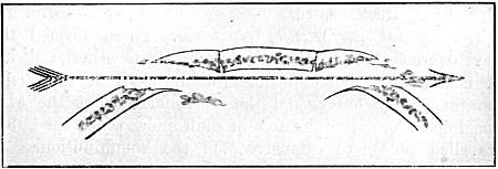
Diagrammatic longitudinal section of Fracture shown in fig. 68
Fig. 70.
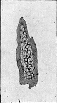
Fragment forming the main part of the floor of Gutter Fracture in the squamous portion of the temporal bone. (Low velocity, hard bone)
(54) Wounded at Thaba-nchu. Guedes bullet. Entry behind left ear, just above posterior root of zygoma; gutter fracture; bullet retained within skull. Above and corresponding to right frontal eminence there was a hæmatoma, beneath which a loose fragment of bone was readily palpable. When brought into the Field hospital, twenty-four hours after the injury, the man appeared to understand when spoken to, but made no answers to questions. The urine was passed unconsciously, the bowels were confined.
He was drowsy, the pupils widely dilated, the pulse 68, of good strength, and the temperature 104°. He slept well the following night and midday there was little change, except that the pupils acted to light, and the pulse had risen to 88, becoming dicrotic and small. The temperature was 103°, the tongue furred and dry, but he was lying with the mouth wide open.
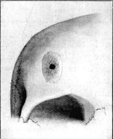
At 2 p.m. the wound was explored. The entry led down to a typical gutter fracture in the squamous portion of the temporal bone, at the point of junction of the vertical with the horizontal part; the floor of the gutter had been displaced inwards as a single fragment (fig. 70). A flap was raised in the frontal region, where a scale of outer table (fig. 71), clothed with diploic tissue, was found loose. Beneath this a puncture on the frontal bone, about corresponding in size to the bullet, was discovered. This opening was enlarged, and a bullet detected and removed. The bullet was a Guedes, with no marks of rifling, and was in no way deformed. At least a square inch of the right frontal lobe was pulped, so that the bullet lay in a cavity.
The patient improved somewhat during the next two days, and on the third took a 16 hours' journey to Bloemfontein, where Mr. Bowlby (who was present at the operation) kindly took him into the Portland Hospital. The pulse gradually rose to 112, the temperature remained on an average from 102° to 103°, the respiration rose to 36, the face became somewhat livid, and on the sixth day death occurred rather suddenly, apparently from respiratory failure. For two days before his death the patient sometimes asked for food, &c.; there was occasional twitching of the left angle of the mouth, and, when the posterior wound was manipulated, some twitching of the fingers of the left hand. When the wound was dressed on the fourth day, there were breaking-down blood-clot and signs of incipient suppuration.
Mr. Bowlby made a post-mortem examination, and found considerable pulping of the tip of the right frontal and left temporo-sphenoidal lobes, and a thick layer of hæmorrhage extending over the whole base of the brain.
Fig. 71.
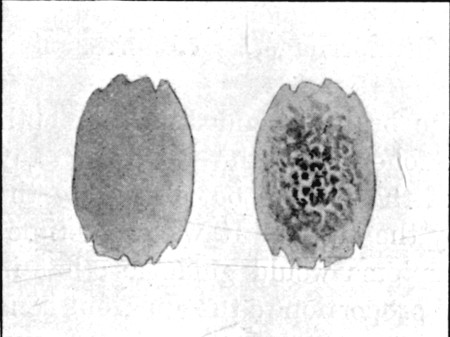
Scale of outer table of Frontal Bone and Diploë. Exact size, from fracture shown in fig. 72
Fig. 72.—Perforating Fracture of Frontal Bone from within Separation of plate outer table. (Low velocity.) 1/2
The injury to the cranial contents varied with the degree of bone injury. Hæmorrhage on the surface of the dura may in rare instances have been the sole gross lesion; I never met with such a condition, however. In all the cases in which comminution had occurred, some laceration of the dura, even if not more than surface damage or a punctiform opening, had resulted. In the more serious gutter fractures an elongated rent of some extent usually existed. In the perforating fractures two more or less irregular openings were the rule. The amount of hæmorrhage, even if the venous sinuses were implicated, was on the whole surprisingly small, when the cases were such as to survive the injury long enough to be brought to the Field hospital. I never saw a typical case of middle meningeal hæmorrhage, although many fractures crossing the line of distribution of the large branches came under observation. Case 60, p. 274, illustrated the fact that the osseous lesions of lesser apparent degree are sometimes the more to be feared in the matter of hæmorrhage, as compression is more readily developed.
The degree of injury to the brain depended on the depth of the track, the resistance offered by the bones of any individual skull, the weight of the patient, but chiefly on the degree of velocity retained by the bullet. It was sometimes slight and local as far as symptoms would guide us; but in the majority of cases out of all proportion to the apparent bone lesion, if the range was at all a short one. Cases illustrative of these injuries are included under the heading of symptoms.
It will be, of course, appreciated that the coarse brain lesions under the third heading differed in localisation and in extent alone, and in no wise in nature, from those observed in the two preceding classes. The damage consisted in direct superficial laceration and contusion, and beyond the limits of the area of actual destruction, abundant parenchymatous hæmorrhages more or less broke up the structure of the brain, such hæmorrhages decreasing both in size and number as macroscopically uninjured tissue was reached. No opportunity was ever afforded of examining a simple wound track in a case in which no obvious cerebral symptoms had been present.
IV. Fractures of the base.—In addition to the above classes, a few words ought to be added regarding the gunshot fractures of the base of the skull. These possessed some striking peculiarities; first in the fact that they might occur in any position, and hence differed from the typically coursing 'bursting' fractures we are accustomed to in civil life as the consequence of blows and falls, and consequently were often present without any of the classical symptoms by which we are accustomed to locate such fissures. Secondly, the peculiar form was not uncommon in which extensive mischief was produced from within by direct contact of a passing bullet.
As far as could be judged from clinical symptoms, indirect fractures of the base such as we are accustomed to meet in civil practice in connection with fractures of the vault were decidedly rare, and, as has already been mentioned, ocular evidence of extensive fissures extending from perforating wounds of the vertex was wanting, except in the extreme cases classed under heading I. For these reasons I am inclined to regard them as uncommon.
Direct fractures of the base, on the other hand, were of common occurrence, especially in the anterior fossa of the skull. These might be produced either from within, the most characteristic form of gunshot injury, or from without. The fractures from within were often simple punctures of the roof of the orbit or nose.
Punctured fractures of the roof of the orbit caused little trouble as far as the cranium was concerned, but the orbital structures often suffered severely. I saw one or two very severe comminutions of the roof of the orbit caused by bullets which had crossed the interior of the skull; in one case the whole roof was in small fragments, while the damage in others was not greater than chipping off some portion of the lesser wing of the sphenoid. The roof of the orbit again was sometimes very severely damaged by bullets which first traversed that cavity itself; thus in one case which recovered, the bullet passed transversely, smashing both globes, and fracturing the roof of both orbits and the cribriform plate so severely as to lacerate both dura-mater and brain, portions of the latter being found in the orbit on removal of the damaged eyes.
Fractures of the middle and posterior fossæ were met with far less frequently, partly I think because vertical wounds passing from the vertex to the base in these regions were with few exceptions rapidly fatal, and partly from the fact that the occipital region, being ordinarily sheltered from the line of fire, was rarely exposed to the danger of direct fracture from without. As an odd coincidence I may mention that in my whole experience during the war I only once saw bleeding from the ear as a sign of fracture of the base, apart from direct injuries to the tympanum or external auditory meatus.
Symptoms of fracture of the skull, with concurrent injury to the brain.—These consisted in various combinations of the groups of signs indicative of the conditions of concussion, compression, cerebral irritation, or destruction. Although the symptoms possessed no inherent peculiarities, yet certain characteristics exhibited served to illustrate the fact that, as a result of the special mechanism of causation of the injuries, the type deviated in many ways from that accompanying the corresponding injuries of civil practice.
The characters of the external wounds will be first considered, followed by some remarks concerning the symptoms attendant on the different degrees and types of lesion, the symptoms special to injuries to different regions of the head, and on the subsequent complications observed.
In the simplest injuries the type forms of entry and exit wound were found, and it has already been observed that in these, if symmetrical, considerable difficulty existed in discriminating between the two apertures. This is to be explained by the fact that the arrangement and structure of the scalp are identical in corresponding regions; hence the only difference in the conditions of production of the entry and exit wounds exists in the absence of support to the skin in the latter. The granular structure of the hairy scalp is opposed to the occurrence of the slit forms of exit, hence the openings were usually irregularly rounded. Any increase of size in the exit wound in the soft parts due to the passage of bone fragments with the bullet, was equalised in that of entry by the fact that the latter, as supported by a hard substratum, was usually larger than those met with in situations where the skin covers soft parts alone.
In some cases of gutter fracture the wounds of entry were large and irregular, as a result of upward splintering of the bone at the distal margin of the aperture of entry in the skull, and consequent laceration of the scalp. Again, on the forehead very pure types of slit exit wound were often met with in the position of the vertical or horizontal creases. With higher degrees of velocity on the part of the bullet and consequent comminution at the aperture of exit in the bone, the scalp was more extensively lacerated, and large irregular openings in the soft parts, often occupied by fragments of bone and brain pulp, were met with. It is well to repeat here, however, that the presence of brain pulp in a wound by no means necessarily indicated the aperture of exit, for it was sometimes found in the entry opening also.
In the most severe cases, such as are included in class I., the exit wound often possessed in the highest degree the so-called 'explosive' character. From an opening in the skin with everted margins two or more inches in diameter a mass of brain débris, bone fragments and particles of dura-mater, skin, and hair, bound together by coagulated blood, protruded as a primary hernia cerebri if the patient survived the first few hours after the injury. In other cases of the same class the actual opening was smaller, but the whole scalp was swollen and œdematous, sometimes crackling when touched from the presence of extravasated blood in the cellular tissue, while firm palpation often gave the impression that the head consisted of a bag of bones over a considerable area.
Gutter fractures of the scalp were sometimes situated beneath an open furrow, gaping from loss of substance, or beneath a bridge of skin; in the latter case they were usually palpable. Simple punctures were also usually palpable, but the smallness of the openings sometimes rendered their detection more difficult than might be assumed.
I never saw a case in which the skull escaped injury when the bullet struck the scalp at right angles, but the frequency with which Mauser bullets were found within the helmets of men would suggest that this must have sometimes occurred. A case of injury to the external table alone has been described (p. 243). An illustration of the next degree of injury is afforded by the following:—A bullet lodged in the centre of the forehead, the point lying within the cranial cavity, while the base projected from the surface: this patient suffered but slight immediate trouble, so little, indeed, that he merely asked his officer to remove the bullet for him, as it was inconvenient. The bullet was subsequently removed in the Field hospital.
In a few cases the bullet entered the skull and was retained, when only a single wound was found. Such cases are described in Nos. 54 and 68, where the position of the bullet was determined by palpable fractures beneath the skin. With regard to the retention of bullets, however, in small-calibre wounds, it was always necessary to examine the other parts of the body with great care, and to ascertain, if possible, the direction from which the wound was received, as an exit was often found some distance down the neck or trunk. Again the possibility of the opening having been produced by glancing contact had to be considered.
In cases which survived the injury on the field, free hæmorrhage, as in wounds of other regions, was rare, and although general evidence of loss of blood was often noted in patients brought in, progressive bleeding was seldom observed. Again, when the wounds were explored, the amount of blood, although considerable, was usually not more than sufficed to fill up the space consequent on the loss of brain tissue. This was especially striking when large venous sinuses, as the superior longitudinal, were involved in the injury. None the less, hæmorrhage at the base of the brain was, I believe, responsible for early death in many of the severe cases, especially when the wounds were near the lower regions of the skull.
Escape of cerebro-spinal fluid was not so prominent a feature as might have been expected, considering how freely the arachnoid space was opened up in many cases. I think this was usually checked by early coagulation of the blood, and later by adhesions. It must be remembered also that extensive wounds were most common on the vertex, or at any rate over the convex surface of the brain, while fractures of the middle fossa were usually rapidly fatal.
Concussion.—Cases exhibiting symptoms of pure uncomplicated concussion were distinctly rare, as would be expected from the mechanism of the injuries. On the other hand, symptoms of concussion formed the dominant feature of all severe cases.
The symptoms in many instances consisted in great part in transitory signs of the so-called 'radiation' type, such as are seen in destructive lesions where the signs of nervous damage rapidly tend to diminish and localise themselves.
As to the causation of the 'radiation' symptoms, it is difficult to discriminate the effects of neighbouring parenchymatous hæmorrhages from those of local vibratory concussion of the nervous tissue. The local character of the signs seems, however, to point to causation by molecular disturbance, resulting from the conduction of forcible mechanical vibration to the brain tissue rather than to upset in the intra-cranial pressure. Again the limited nature of the paralysis observed, sharply defines it from the general loss of power accompanying ordinary cases of concussion of the brain. The similarity of the phenomena to those described in other parts of the body under the heading of 'local shock' is sufficiently obvious.
The following instance well exemplifies the condition in question:
(55) Wounded at Spion Kop. A scalp wound 3 inches in length crossed the left parietal bone nearly transversely, starting 1½ and ending 2 inches from the median line: the centre of the wound corresponded with the position of the fissure of Rolando. The patient was struck at a distance of fifty yards while kneeling; he fell and remained unconscious an hour and a half. Right hemiplegia without aphasia followed. The wound was cleansed and sutured, and in three days both arm and leg could be moved, after which time the man improved rapidly. Three weeks later when I saw him at Wynberg there was still comparative weakness of the right side, but beyond some neuralgia of the scalp, the man considered himself well. No groove could be detected on the bone on palpation. (This case offers a good example of the ease with which bone injury may be overlooked. The man came over to England 'well;' but while on furlough, two pieces of bone came away spontaneously. He is now again on active service.)
Compression.—Equally rare was it for pure symptoms of compression to be exhibited. This depended on two circumstances: first, the rarity of injuries giving rise to meningeal hæmorrhage; secondly, the fact that in nearly every case a more or less extensive destructive lesion was present, at the margins of which less completely destroyed tissue remained, capable of giving rise to symptoms of irritation. Again, as we have seen, free hæmorrhage into, or from the walls of, the cavities produced in the brain was not a marked feature, and beyond this the large defect in the cranial parietes was calculated to render a high degree of compression impossible.
As the most serious head injuries presented a remarkable similarity in their symptoms, I will shortly summarise their common features.
Every degree of mental stupor up to complete unconsciousness was met with, but in some instances where the pulse, respiration, and general bodily condition pointed to speedy dissolution, the patients answered rationally often between moans or cries indicative of pain.
Widespread paralysis often existed, but this was seldom completely general; more commonly it was combined with extreme restlessness of the unparalysed parts, or sometimes, even when the whole of one hemisphere was tunnelled, and in all probability widely destroyed, restlessness was the only symptom. In some cases twitching of the features or the limbs or severe convulsions were superadded.
The pupils were rarely unequal, and at the stage in which these patients were first seen were usually moderately contracted. Wide dilatation was uncommon throughout.
The pulse was with very few exceptions slow, sometimes irregular. In some instances, when the wounds had been thought suitable for exploration, the slow pulse was altered after operation to a rapid one, and death usually quickly supervened.
Respiration was irregular, sometimes sighing; in the late stage often of the Cheyne-Stokes type; actual stertor was exceptional, but the respiration was often noisy.
The temperature was often raised from an early stage to 99° or 100°, and if the patient survived a day or two, it often rose to 103° or 104°. How far the secondary rise depended on sepsis it was not always easy to determine. The urine was usually retained.
Cases presenting the above characters were usually those suffering from lesions such as are described in class I., and mostly died in twenty-four to forty-eight hours. The correspondence of the train of symptoms with those due to combined brain destruction and severe concussion is at once apparent.
To illustrate the nature of the symptoms in patients suffering from the less extensive forms of injury, such as those included in classes II. and III. under the heading of anatomical lesion, the relation of a short series of histories will be advisable. I may first premise, however, that the special characteristics of these were in some instances the almost entire absence of primary symptoms of gravity; in others general symptoms of a severity out of apparent proportion to the external lesion; while in all destructive lesions, very widely distributed radiation symptoms developed, often disappearing with great rapidity.
The symptoms consisted in those of concussion, irritation, local pressure, and actual destruction.
The symptoms of concussion were either general, and then usually transient, or local paralysis of the radiation variety, which also rapidly improved.
Signs of irritation consisted in irritability of temper, drowsiness, closure of the eyes and objection to light, contracted pupils sometimes unequal, a tendency to the assumption of the flexed position at all the joints, twitchings, and sometimes convulsions. Sometimes these appeared early as a direct result of mechanical irritation from bone fragments or blood-clot; sometimes only in the course of a few days, as a result of irritation of parts recovering from the radiation effects which had prevented earlier nervous reaction. Possibly in some cases the symptoms of irritation depended upon an increase in the amount of hæmorrhage, and in others upon the development of local inflammatory changes.
Local pressure, or actual destruction of brain tissue, was evidenced by temporary paralysis in the former, permanent loss of function in the latter, condition.
Fractures of the anterior fossa of the skull were attended by very marked evidence of orbital hæmorrhage, as subconjunctival ecchymosis (rarely pure), increased tension, and proptosis.
Injuries to the cranial nerves at the base, with the single exception of lesion of the optic nerves, which was not rare, were in my experience uncommon in the hospitals—a fact pointing to the very fatal nature of direct basal injuries, except in the anterior fossa of the skull. Signs indicative of injury to the olfactory lobe were occasionally observed.
I should, perhaps, again insist here on the rarity with which acute diffuse septic infection occurred in cases of these degrees of severity, also on the fact that interference with the wounds in the way of secondary exploration, even when they were manifestly the seat of local infection, was followed almost without exception by good immediate results; and, lastly, that when suppuration did occur, it was usually strictly local in character. The influence of the climate of South Africa and our surroundings has already been discussed, but whether climate, condition of the patients, or peculiarity in the nature of causation of the wounds was responsible, in no series of cases was the absence of acute inflammatory troubles more striking than in this one of brain injuries.
Frontal injuries were those most frequently unaccompanied by primary symptoms of severity; slowing of the pulse—this often fell to 40—and occasional irregularity, were almost the only constant signs of cerebral damage. Some patients temporarily lost consciousness, others rose at once and walked to the dressing station, and in few cases was any psychical disturbance noted in the early stages.
I think, however, it may be affirmed that frontal injuries, accompanied by trivial signs, resulted without exception from the passage of bullets travelling at a low rate of velocity. Thus in several of the instances here related the patients at the time of reception of the wound were under the impression that they were entirely beyond the range of fire, and in one, in which well-marked signs of concussion followed, the bullet, which had traversed the head, retained only sufficient force to perforate the skin of the neck and bury itself in the posterior triangle without even fracturing the clavicle, against which it impinged. In men struck at a shorter range, signs of concussion, often followed by transient radiation signs of injury to the parietal lobe, were common. These signs were, I think, not as a rule due to surface hæmorrhage, since they were of a purely paralytic nature and not irritative. Several cases with partial or complete hemiplegia, hemiplegia and aphasia, or facial paralysis are recorded below.
(56) Frontal injury.—Wounded at Magersfontein. In prone position when struck, distance 700 to 800 yards. Entry (Mauser), at the margin of the hairy scalp above and to the left of the frontal eminence; course, through anterior third of left frontal lobe, roof of orbit, obliquely across line of optic nerve, inner wall of orbit, nose, right superior maxilla piercing alveolar process, and passing superficial to inferior maxilla: exit, one inch anterior to angle of jaw. The bullet again entered the posterior triangle of the neck, struck the right clavicle, and turned a somersault, so that its base lay deepest in the wound.
The patient was unconscious for a short time, suffered with general headache and giddiness, and was somewhat irritable. On the third day the pulse was 70, temperature normal, and he was sent to the Base. There was considerable proptosis, œdema and discoloration of the eyelid, and subconjunctival ecchymosis, but the movements of the eyeball could be made and light could be distinguished. The sense of smell was apparently absent. A week later the headache was gone, the pulse numbered 80 to 90, the temperature was normal, he slept well, sat up in bed and smoked, took his food well, and exhibited no cerebral symptoms. He could detect the smell of tobacco, but not as a definite odour.
No further symptoms were noted, the sense of smell returned, the swelling of the eyelid and proptosis decreased, but the upper lid could not be raised. When the lid was drawn up, there appeared to be vision at the margins of the field with a large central blind spot. The patient left for England at the end of a month apparently well.
(57) Gutter fracture of frontal bone.—Wounded at Paardeberg. Entry (Mauser), 3/4 of an inch within the margin of hairy scalp above outer extremity of right eyebrow; gutter fracture; exit, 2 inches nearer middle line, at the same distance from the margin of the hairy scalp. The patient was knocked head over heels, his main feeling being a sense of dulness in the right great toe. He sat up and got a first field dressing applied, then lay down, but as he was still under fire, he retired 1,000 yards to the collecting station; here he dressed some patients, and later mounted an ambulance wagon and was driven to the Field hospital. The next day he helped with the work of the hospital, amongst other things controlling the artery during an amputation of the arm. He then took a three days' and nights' journey to Modder River in a bullock wagon, during which journey he had a fit, which was general, the thumbs being turned in and a wedge being necessary between the teeth to prevent him biting his tongue.
On the sixth day the wound was examined, and between this and the tenth day he had several fits of the same nature as the first, accompanied by stertorous breathing and profuse sweating. On the tenth day Mr. Cheatle opened up the wound and removed numerous fragments of bone, leaving a clean gutter 2 inches by 3/4 of an inch. After the operation no further fits occurred, and eight days later he was conscious, but was excitable and talked at random. On the twentieth day he arrived at the Base after 30 hours' railway journey (623 miles). He was then quite rational, but unable to make any demands on his memory and very sensitive to noise; at times he wandered in the evenings and his temperature rose as high as 100°. The wound was open and granulating, the floor pulsating freely.
Three weeks later the wound was still open, and the skin dipped in at the lower margin. The mental condition was much improved, although attempts at giving a history of his case were obviously tiresome.
The wounds in the leather headband of this patient's helmet were interesting, the round aperture of entry in the exterior of the helmet being followed by a starred exit aperture in the leather band, the second entry opening in the leather band being again circular, and the external opening in the puggaree a transverse slit.
(58) Transverse superficial perforating frontal injury.—Wounded at Graspan. Aperture of entry (Lee-Metford), at upper and outer part of left frontal eminence; exit, at margin of hairy scalp over outer third of right eyebrow. On the second day the patient complained of giddiness and headache; the pulse was 60. He was then walking about. The wounds were explored and typical entry and exit apertures discovered in the frontal bone from which cerebral matter was protruding. Both openings were enlarged (Mr. S. W. F. Richardson) with Hoffman's forceps, and a considerable number of splinters of the inner table were removed from the aperture of entry.
The headache gradually passed off, but there was throbbing about the scar, and pulsation was visible for some three weeks, after which no further symptoms were observed.
(59) Oblique frontal gutter fracture.—Wounded at Magersfontein. Entry (Mauser), 1/2 an inch to right of median line of forehead, 3/4 of an inch from the margin of the hairy scalp; exit, about 3/4 of an inch anterior to the lower extremity of the right fissure of Rolando. Weakness of left facial muscles, especially of angle of mouth. No further motor symptoms. Wounds explored (Mr. Stewart); numerous fragments of bone and some pulped cerebral matter were removed. Patient developed no further signs; the paralysis, although improved, did not completely disappear. The man a year later was still on active duty, the paralysis almost well, and no further ill effects of the injury remained.
In the fronto-parietal or parietal regions, signs of damage to the cortical motor area were seldom absent, sometimes evanescent, at others prolonged. In some cases the signs were permanent and followed by evidence of local sclerosis.
The motor area on both sides of the brain was sometimes implicated; thus in a child shot at Kimberley the bullet entered in the right frontal region, and emerged to the left of the line connecting bregma and inion a little behind its centre. Paralysis of both lower extremities resulted, power rapidly returning in the right, while incomplete paralysis persisted in the left.
In only one instance (see case 73, p. 292) was any permanent sensory defect observed, and the mental condition of this patient would have certainly suggested a functional explanation for its presence, had it not been for the accompanying inequality in the axillary surface temperatures.
In a second case (No. 67) blunting of sensation followed a definite lesion of the inferior parietal lobule. In this instance an occipital lesion was associated with the parietal.
(60) Parietal gutter fracture.—Wounded at Magersfontein. A scalp wound 3 inches in length ran transversely across the right parietal bone at the level of the lower third of the fissure of Rolando. A second wound of entry was found crossing the third dorsal spine; the bullet was retained and was palpable over the right scapula. There was left facial paralysis, weakness and numbness of both upper extremities, especially of the left, and some difficulty in swallowing. The man was sent to the Base, where he arrived on the fourth day. The symptoms had then become much more marked, consciousness was incomplete, and articulation slow and imperfect. There was complete left hemiplegia, and deviation of the tongue to the right. The pulse was 40. An exploration (Mr. J. J. Day) showed that an oval plate of the outer table of the parietal bone had been struck off. A trephine was applied to the exposed diploë and a crown of bone removed; considerable comminution of the inner table had occurred, several large fragments having perforated the dura-mater. The latter did not pulsate; it was therefore freely incised, and many more fragments of bone and a large quantity of blood-clot removed.
The first effect of the operation was slight, but ten days later rapid improvement commenced, the first sign being acceleration of the pulse, which rose to 70. On the eighteenth day the original symptoms still remained to a diminished extent, but a fortnight later there remained traces of the facial weakness only, and there was little difference in the grip of the two hands. The patient was shortly afterwards sent home. Ten months later he returned to South Africa on active service.
(61) Fronto-parietal gutter fracture.—Wounded at Graspan. Entry (Mauser), 1 inch within the margin of the hairy scalp, 1/2 an inch to the left of the median line; exit, 3½ inches posterior in same line. Complete right-sided hemiplegia. The wounds were explored on the fourth day (Major Moffatt, R.A.M.C.) and a gutter fracture involving the frontal and parietal bones exposed. The dura-mater was lacerated and brain matter from the frontal lobe escaped freely. A large number of bone fragments were removed. On the fourth day after the operation, the patient became unconscious with right-sided twitchings, but rapidly improved, and at the end of three weeks, except for slight headache, he was well, the power of the right side being good. Ten months later he rejoined his regiment in South Africa, no apparent ill effects remaining.
(62) Fronto-parietal perforating fracture.—Wounded at Magersfontein. Entry, within the margin of the hairy scalp; exit, behind and below the left parietal eminence, the track crossing about the centre of the fissure of Rolando. Right hemiplegia, the lower half of the face only being involved. The wounds were explored and a large number of fragments of bone and a quantity of pulped cerebral matter removed. Six days later the hemiplegia persisted, speech was slow, headache was troublesome and the pulse not above 45. After this, gradual improvement took place, and a month later the lower extremity and face had regained good power. The upper extremity remained flaccid and paralysed, except for some slight power of movement of the shoulder.
(63) Fronto-parietal perforating fracture.—Wounded at Magersfontein. Entry (Mauser), 2½ inches from the median line, 3½ inches from the occipital protuberance; exit, 3/4 of an inch from the median line, 4½ inches from the glabella; sanious fluid escaped from both ears. There was left facial paralysis, complete paralysis of the left upper extremity, and partial paralysis of the left lower extremity. The patient was deaf, drowsy, and the pulse 45.
Exploration showed the entry wound to be in the parietal, the exit to involve both parietal and frontal bones. The openings were enlarged, and a number of fragments of bone, together with pulped cerebral matter and blood-clot, were removed. The wound healed, except at the front part, where a small prominence suggested a hernia cerebri.
The patient improved slowly; fourteen days after the operation he could hear well, and the flow from the ears had ceased. The facial weakness was slight, the upper extremity was still powerless, but he could move the lower and draw it up in bed. At the end of six weeks the wound had healed, and he was got up and dressed.
At the end of two months he was well enough to be sent home; there was only a trace of facial weakness; the right upper extremity, however, was powerless and slightly rigid, occasional twitchings occurring in it. Considerable power had been regained in the lower extremity, so that the patient could walk with help, but foot-drop persisted; the gait was spastic in character, the reflexes were much exaggerated, and there was marked clonus. The patient was sensible, but his manner suggested some mental weakness. Both the openings in the skull were closed by very firm material, apparently bony.
This patient became a Commissionaire some ten months later. His mental condition is normal, and loss of memory seems confined to the events immediately following the injury. The lower extremity has improved, but the upper is useless.
(64) Parietal injury: retained bullet.—Wounded at Paardeberg. Aperture of entry (Mauser), 1 inch diagonally below and anterior to left parietal eminence. No exit. The patient was trephined by the surgeons of the German ambulance at Jacobsdal.
Sixteen days later he arrived at the Base. A circular pulsating trephine opening was then to be felt beneath the flap, but no information was forthcoming as to the bullet. The patient could speak, but lost words and the gist of sentences; he could remember nothing as to himself since the day of the injury. There was right facial weakness; he could not close the right eye or whistle, but there was little apparent want of symmetry; there was weakness in the grip of both hands, more marked on the right side; both lower extremities could be moved. The reflexes were normal, although the left limb was slightly rigid. The pupils were equal, reflex normal; slight nystagmus. Pulse 72, small and regular. Temperature normal. Rapid improvement followed.
During the fourth week the temperature rose to 103°, and remained elevated for six days, but no local or general signs appeared; at the end of five weeks there was little evidence of the paralysis remaining. The patient was discharged from the service on his return home.
In the upper part of the occipital region glancing or superficial injuries were comparatively favourable; those near the base, especially if perforating, were very dangerous. Two such cases are referred to elsewhere. Case 69 is included as the only example of cerebellar injury I happened to see who lived any appreciable time after the accident.
The main interest in these cases centres in the defects produced in the area of the visual field. I am extremely indebted to my colleague, Mr. J. H. Fisher, who has kindly determined this for me in three of the following cases. It will be noted that in two instances the injury was to the left occipital lobe. In these the resulting hemianopsia was of the pure lateral homonymous character, and in both the visual symptoms were accompanied by a certain degree of amnesic aphasia (65 and 68).
In 65 the injury was definitely unilateral, and at the time of the operation I decided that at least an inch and a half of the posterior extremity of the left occipital lobe was totally destroyed.
In 68 the lesion was probably confined to the left lobe, but it is impossible to exclude slight injury to the right lobe also. In this instance amnesic aphasia was a far more marked symptom than in 65, and the position of the lesion suggested damage both to the visual and auditory word centres.
Cases 66 and 67 are instances of damage to both occipital lobes. In 66, although the wound was a glancing one, and did not perforate, it was so near the median line, and accompanied by such severe damage to the bone, that a symmetrical lesion of the cuneate and precuneate lobules of both right and left sides is to be inferred. In 67 the great longitudinal fissure was traversed by the bullet obliquely. It is of great interest to observe that in each of these cases the lesion of the visual field was a horizontal one and affected the lower half in place of assuming a lateral distribution.
In all four cases the primary effect of the occipital injury was the same—viz. absolute blindness—while the return of vision in each was of the nature of the dawning of light. I regret that I am unable to furnish any detail as to increase of the field of vision in the progress of the cases, but circumstances rendered continuous observation of the patients impossible.
In each case deafness was apparently the direct result of concussion of the ear on the side corresponding to the wound. Deafness of the opposite ear was never noted.
In case 67 some general blunting of sensation was noted in the paralysed upper extremity, and in this patient, no doubt, injury to the inferior parietal lobule accompanied the occipital lesion.
(65) Injury to left occipital lobe.—Wounded at Belmont. A single transverse wound, 2 inches in length, extended across the occipital bone, 2 inches above the level of the external protuberance. When seen on the third day the wound was gaping and pulped cerebral matter was found in it. The patient was very drowsy, lying with closed eyes, and complaining of great coronal and frontal headache. He could distinguish light and darkness, but not persons. Total blindness immediately followed the injury, persisting some three days, and the patient spoke of return of sight as of the appearance of dawn. The pupils were equal, moderately dilated and acted to light, which was unpleasant to him. He was somewhat irritable and silent, but apparently rational. Temperature 99°. Pulse 56 full. Tongue clean. No sickness, no difficulty in micturition.
Fifty-six hours after the injury the wound was opened up and cleaned, and an oval fractured opening about 3/4 by 1/2 inch was exposed 3/4 inch to the left, and 2 inches above the occipital protuberance. The margins of the opening showed several small fragments of lead attached to the bone. A 3/4-inch trephine was applied at the left extremity of the opening, and it was found that about a square inch of the internal table was comminuted and driven into the brain, together with several small fragments of lead. On introducing the finger, about 1½ square inches of the occipital lobe were found to be pulped, and the finger could be swept across the tentorium. There was no sinus hæmorrhage (nor did the history suggest that hæmorrhage had ever been severe). The cavity was carefully sponged out, and the wound closed with a drainage aperture. Little change followed in the patient's condition, and on the sixth day he was sent to the Base hospital.
Three weeks later the wound was firmly healed. The patient still complained of frontal headache, and wore a shade, as the light hurt his eyes and made them water freely. The pupils acted, but were wide; objects could be distinguished, and also persons. Otherwise, the man's condition was good: he began to get up, and at the end of six weeks returned to England.
A year later the man was earning his living as a Commissionaire porter. He complains of giddiness when he stoops, or when he looks upwards, and at times he suffers much with headache both in the region of the injury and across the temples.
There is a bony defect and slight pulsation at the site of the injury, but no prominence. When attempts are made to read the lines run together, and a dark shadow comes before his eyes. He speaks of the latter as still terribly weak. Speech is slow and somewhat simple, but he makes no mistakes as to words. Memory is bad for recent events.
Mr. Fisher makes the following report as to the eyes: Pupils and movement of eyes normal in every respect. No changes in fundi.
Vision,R.5/12with—0.55/6
L.5/9with—0.55/5
There is therefore practically full direct vision. Though the man chooses a concave glass he is not really myopic. There is typical right homonymous hemianopsia; the answers, when tested with the perimeter, are quite certain, and the fields absolutely reliable.
The man's statements confirm the condition; he is aware of his inability to see objects to his right-hand side, and is apt to collide with persons or objects on that side.
Fig. 73.
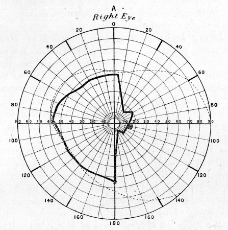
Right Visual Field, in case 65. Injury to left occipital lobe. Field for white. Test spot 10 mm. Good daylight. Right homonymous hemianopsia
Fig. 74.—Left Visual Field, case 65
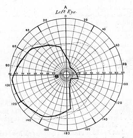
The lesion is one of the left occipital cortex in the cuneate lobe and the neighbourhood of the calcarine fissure. The speech suggests a slight degree of aphasia.
(66) Injury to occipital lobes.—Wounded at Magersfontein while in prone position. Distance, 500 yards. He says he was never unconscious, but for two days was absolutely blind. His eyesight gradually improved, but headache was very severe, and sleeplessness nearly absolute. On the eighth day the wound, which was situated over the right posterior superior angle of the parietal bone, was opened up, and a number of fragments of bone and a quantity of pulped brain removed from a depressed punctured fracture, surrounded by an annular fissure, completely encircling it, 1½ inch from the opening. The portion of brain destroyed was probably a considerable portion of the cuneate and precuneate lobules of both sides, as well as a portion of the first occipital convolution, and the superior parietal lobule of the right side. There was no evidence of injury to the superior longitudinal sinus in the way of hæmorrhage.
After the operation the patient slept better, but still complained of headache, and when he arrived at the Base, the flap became œdematous, and the stitch holes and also the central part of the wound suppurated. The temperature rose to 101°. The wound was therefore re-opened, and a number of additional fragments of bone, some as deeply situated as 2 inches from the surface, were removed. Steady improvement followed, and at the end of a further three weeks the wound was healed, the headache had ceased, and there were no abnormal symptoms, except that light was unpleasant to the right eye, and the field of vision was manifestly contracted (Mr. Pooley).
A year later the man was employed as a letter-carrier. He complains of headache at times, and on six occasions has had 'fainting fits.' He says that the latter commence with tremor, that his legs then give way and he falls. In a quarter of an hour he gets up, and feels no further inconvenience. Speech is perfect, there is no deafness. The bone defect is very nearly completely closed.
Mr. Fisher reports as follows as to the vision. There is a high degree of hypermetropia in each eye, the R. has nearly 6.0 D and the L. about 5.0 D. With correction he gets practically full direct vision with each.
Fig. 75.
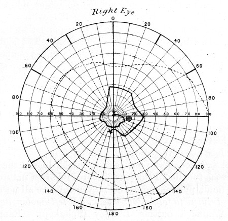
Right Visual Field, in case 66. Injury to both occipital lobes. Field for white. Test spot 10 mm. Good artificial light. Defect in field complicated by functional symptoms
Fig. 76.—Left Visual Field, in case 66. Defect in lower half of field
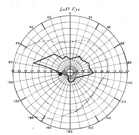
The patient has been examined before, and has been informed that his vision quite incapacitates him from further service. He began by stating that he could not see on either side of him, but only straight in front; that he is apt to collide with people in walking, was nearly knocked down by a horse, and that his acquaintances accuse him of passing them unnoticed. The fields of vision are very small, but the loss is not typically in the temporal half of either. That of the right eye which we know as the spiral field, becoming more and more contracted as the perimeter test is continued, is what is found in functional cases; that of the left, however, shows a characteristic loss of the lower part of the field of vision, and agrees with the statement of the man that he can see the upper part of my face but not the lower when he looks at me. Such a loss agrees with a lesion involving the upper part of the cuneate lobe above the calcarine fissure.
I feel satisfied that there is considerable loss in the right field also, but the functional element obscures its exact nature.
The fundi, pupils, and ocular movements are all normal.
(67) Injury to occipital lobes and left motor and sensory areas.—Wounded outside Lindley (Spitzkop). Range within 1,000 yards. Entry, one inch within the right lateral angle of the occipital bone, external wound more than 1/2 an inch in diameter; exit, 2 inches from the median line, over the upper half of the left fissure of Rolando. Behind the wound of exit comminution of the parietal bone, extending back to the lambdoid suture, existed. I attributed this to oblique lateral impact by the bullet on the inner surface of the skull.
The patient could afterwards remember being struck, but became rapidly unconscious. When brought into the Field hospital some five hours later the condition was as follows: Semi-conscious, can speak, apparently blind, pupils equal, of moderate size, do not react to light. Right hemiplegia. No sickness. Moans with pain in head. Passes water normally.
Considerable hæmorrhage had occurred from each wound, the scalp was puffy, and the bones yielded on pressure over the left parietal bone, indicating considerable comminution.
The night was so cold that no operation could be considered, so the head was partly shaved, the wounds cleansed, and a dressing applied. The next morning the Division marched at 5 a.m., and it was considered wise to leave the man at Lindley in the local hospital.
Fig. 77.—Right Visual Field, in case 67.
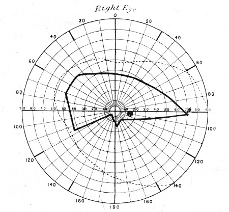
Injury to both occipital lobes. Field for white. Test spot 10 mm. Good artificial light. Defect in lower half of field
Fig. 78.—Left Visual Field, in case 67
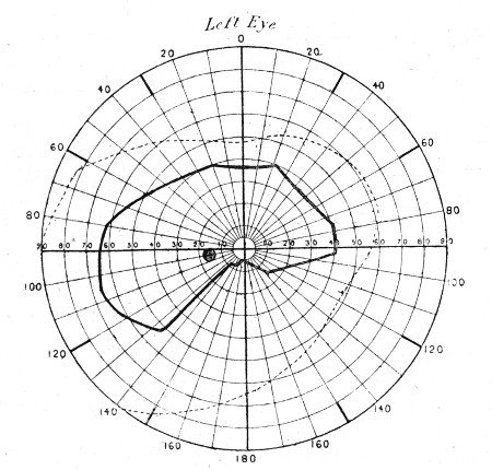
No operation was performed there, but I heard later that the man recovered full consciousness at the end of five days, and at the end of a fortnight he commenced to see again.
Six weeks later he travelled to Kroonstadt, thence to Bloemfontein, and thence to Cape Town and home to Netley. The paralytic symptoms meanwhile steadily improved.
Seven months later his condition is as follows: Scarcely a trace of facial paralysis. Slight power of movement of arm, forearm, and fingers, but grip is very weak. Little power of abduction of the shoulder or of straightening the elbow. The latter movement is made with effort and in jerks. Sensation over the back of the arm is somewhat lowered, and is 'furry' at the finger tips. There is very little wasting of the muscles noticeable.
Walks well, but with some foot-drop. Slight increase of patellar reflex. He says that he does not walk in the street with confidence, as he often feels as if omnibuses &c. were coming too near him.
He is absolutely deaf in the right ear.
The openings in the skull are closed, the occipital lies about halfway between the external auditory meatus and the external occipital protuberance, while the parietal still affords evidence of the earlier comminution, one fissure passing backwards as far as the lambda, and the whole surface is lumpy and uneven.
The track through the brain no doubt involved a considerable extent of the outer aspect of the right occipital lobe and the cuneate lobule. It must also have crossed the great longitudinal fissure, and penetrated the left Rolandic region, just above its centre, probably involving the precuneate lobule, and a portion of the internal capsular fibres as well as the cortex on the left side. The deafness was probably due to concussion of the internal ear.
Mr. Fisher has kindly furnished the following note regarding the vision. The pupils, movements, and fundi are quite healthy. There is good direct vision R. or L. 5/5 fairly, and together 5/5. The man complains he has lost his side sight, also the lower; he demonstrates the latter quite obviously with his hand, and says he has to repeatedly look down when walking. He thinks no improvement has taken place during the last month. The accompanying fields of vision show the loss quite characteristically.
(68) Injury to left occipital lobe.—Wounded at Paardeberg. Entry (Mauser), through the lambdoid suture on the right side of the mid line. Bullet retained, but a palpable prominence behind the left ear suggested its localisation.
The patient became at once unconscious and remained so for several days. He was completely blind; vision returned later, but only to a limited degree. There was complete loss of memory as to the events of the day.
When admitted at Rondebosch into No. 3 General Hospital the condition was as follows: The field of vision is limited, and examination shows right homonymous hemianopsia. When any one comes into the tent the patient sees a shadow only until his bed is reached.
When spoken to the patient 'thinks and thinks,' and then apologises for not answering, saying he will remember at some future time. He is absolutely unable to remember times, names, or localities, but places his hand to his head and appears to think deeply in the effort to recall them. Occasionally when you go into his tent he suddenly remembers something he has been trying to think of for some days, and will tell you.
A fortnight later after an attack of influenza the patient was not so well, and vision was apparently becoming more impaired.
An incision was made (Mr. J. E. Ker) so as to raise a flap the centre of the convexity of which was 2½ inches behind the left external auditory meatus. A slight prominence and a fissure was discovered in the temporal bone, and over this a trephine was applied. On removal of the crown of bone the bullet was discovered with the point turned backwards (having evidently undergone a partial ricochet turn) on the upper surface of the petrous bone, just above the lateral sinus. The dura-mater was healed but thickened, and some clot upon its surface was removed.
The wound healed per primam, and a rapid recovery was made. Ten days later a running water-tap was able to be detected 120 yards from the tent door. The hemianopsia however persisted.
The following letter, dictated by the patient to his wife, and sent to me, gives a clear account of his condition ten months later:—
I am pleased to say my memory is better than it was some time ago, though at times I am entirely lost and really forget all that I was speaking about. I also find that I often call things and places by their wrong names. I sometimes try to read a paper or book which I have to read letter by letter, sometimes calling out the wrong letter, such as B for D &c., and by the time I have read almost halfway through, I have forgotten the commencement.
My sight is about the same. There is no improvement in the right eye, and the doctor at Stoke said that the left eye was not as it ought to be and might get worse.
I ofttimes go to take up a thing, but find I am not near to it, though it appears to me so.
I have no pain to speak of in the head, though at times a shooting pain.
I have a continual noise in the left ear as if of a locomotive blowing off steam, and a deafness in the left ear which I had not before being wounded.
I am extremely indebted to my friend Mr. J. Errington Ker for the notes of the above case, so successfully treated by him.
(69) Injury to occipital lobe.—Wounded at Modder River. Scalp wound in occipital region. Two days later on arrival at the Base the patient was extremely restless and in a condition of noisy delirium. The wound was explored (Mr. J. J. Day) and a vertical gutter fracture discovered 1/2 an inch above and to the left of the occipital protuberance. The gutter was 1½ inch in length and finely comminuted, the dura wounded, and the left occipital lobe pulped. A number of fragments of bone (one lodged in the wall of, but not penetrating, the lateral sinus) and pulped brain were removed. No improvement took place in the general condition, but the patient lived twenty-two days, during which time he coughed up a large quantity of gangrenous lung tissue and foul pus.
At the post-mortem examination a wound track was found extending to the crest of the left ilium, where the bullet was lodged. The patient was no doubt lying with his head dipped into a hole scooped out in the sand (a common custom) when struck; the bullet then traversed the muscles of the neck, entered the upper opening of the thorax, where it struck the bodies of the second and third dorsal vertebræ, one third of the bodies of each of which were driven into an extensive laceration of the lung; it then grooved the inner surfaces of the eighth and ninth ribs, fractured the tenth and eleventh, and passing the twelfth traversed the deep muscles of the back to the pelvis. Beyond the injury to the occipital lobe, the cerebellum was found to be lacerated and extensively bruised and ecchymosed.
Complications.—Hernia cerebri as a primary feature has already been mentioned as one of the peculiarities of some explosive wounds. In the later stages of the cases in which primary union did not take place the development of granulation tumours was often seen, sometimes in connection with slight local suppuration, sometimes over a cerebral abscess. In some cases a wound which had once closed reopened and a hernia developed. This sequence was chiefly of prognostic significance as an indication of intra-cranial inflammation, usually of a chronic character, and affecting rather the lowly organised granulation tissue formed in the cavity than the brain itself. When primary union of the skin flap and wound failed, the process of definitive closure of the subjacent cavity was always a very prolonged one, and it was in such cases that a great proportion of the so-called herniæ developed.
Abscess of the brain.—Local abscesses formed in a considerable proportion of the cases where serious damage to the brain had occurred, in whatever region this happened to be. I never saw one develop in cases where primary union had taken place, even when bone fragments had not been removed; neither did I ever see an abscess situated at a distance from the original injury. I take it that the latter is to be explained by the early date of the suppuration, and the fact that in the great majority of small-calibre wounds the exit opening exists in the situation of the contre-coup damages of civil practice.
The main feature in the symptoms when abscesses developed was the insidious mode of their appearance, usually at the end of fourteen to twenty-one days, and their comparative mildness.
Very slight evidences of compression were observed; thus, varying degrees of headache, drowsiness, irritability of temper or depression, twitchings, or in some cases Jacksonian seizures, combined with slow pulse and slight rises of temperature. I never happened to see complete unconsciousness. The slight evidence of compression was perhaps explained in most cases by the large bony defect in the skull, which acted as a kind of safety-valve. Again the firm nature of the cicatricial tissue which formed at the periphery of the injury and extended up to the skull and there formed a more or less firm attachment, also preserved the actual brain tissue to some degree from either pressure or direct irritation. After evacuation of the pus, the usual difficulty was experienced in ensuring free drainage, and definitive healing and closure of the cavities was very slow. The following two cases will illustrate the character of the cases of cerebral abscess we met with:—
(70) Fronto-parietal abscess.—Wounded at Magersfontein (Mauser). Entry, 1¾ inch above the line from the lower margin of the orbit to the external auditory meatus, and 1¾ inch behind the external angular process; exit, a little posterior to the left parietal eminence. There was right hemiplegia. The wounds were explored, and a large number of fragments of bone and pulped brain were removed, especially from the anterior wound. No great improvement followed, and the patient was sent to the Base. At this time there was a large hernia cerebri at the anterior wound which was suppurating.
A further operation was here performed (Mr. J. J. Day). The hernia cerebri was removed, also several fragments of bone which were found deeply imbedded in the brain. The patient then improved, but a month later his temperature rose, and on exploration an abscess was discovered in the frontal lobe and drained.