A STUDY IN EARLY RENAISSANCE ANATOMY,
Reading edition
WITH A NEW TEXT:
THE ANOTHOMIA OF HIERONYMO MANFREDI (1490)
By Charles Singer
TEXT TRANSCRIBED AND TRANSLATED BY A. MILDRED WESTLAND
I.Anatomy in the Fourteenth and Fifteenth Centuries79
II.Bolognese Works on Anatomy92
III.Hieronymo Manfredi, Professor at Bologna, 1463–9397
IV.The Manuscript Anatomy of Manfredi103
V.Translation of Selected Passages from the Anothomia, with Commentary106
(a) The Brain, Cranial Nerves, &c.106
(b) The Eye118
(c) The Heart122
Italian Text of the Anothomia130
I. Anatomy in the Fourteenth and Fifteenth Centuries
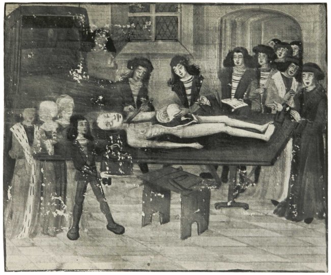
There was little or no progress in the knowledge of anatomy between the death of Mondino in 1327 and the sixteenth century. This appears the more remarkable when we recall how widespread was the practice of dissection during the period. In France, at the University of Montpellier, public dissections were decreed in the year 1377,121 and Catalonian Lerida followed suit in 1391.122 At Bologna, where dissection had long been customary, it received official recognition in the University Statutes in 1405,123 and the same event took place at Padua in 1429. Public anatomies were instituted at the University of Prague in 1460, of Paris in 1478, and of Tübingen in 1485.124 For these ‘Anatomies’ the bodies of executed criminals were usually employed, and therefore the number of subjects available varied greatly in different localities.125 In addition to these regular dissections, there was certainly a considerable amount of post-mortem examination, surreptitious (Plate XXVIII b126), or even open (Plate XXIX127), long before Benivieni published his memorable list of cases.128
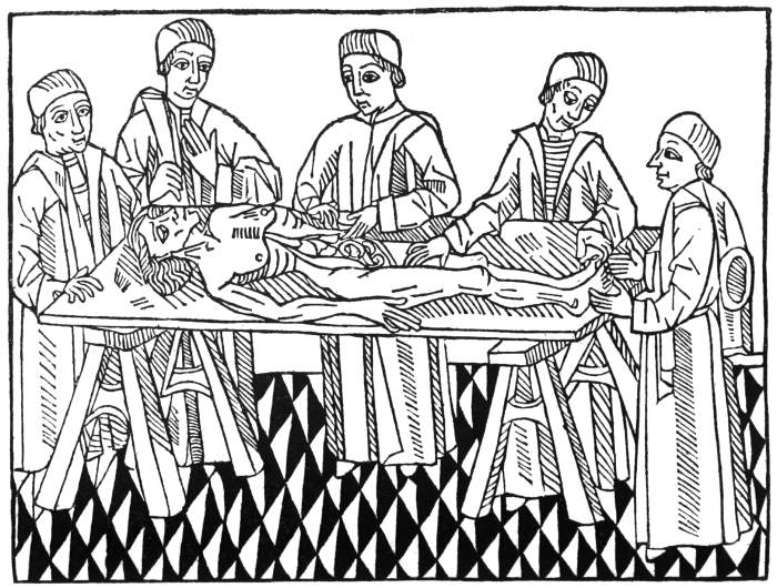
Fig. 1. From the French translation of Bartholomaeus Anglicus, Lyons, 1482. The first printed picture of dissection.
MS. fr. 184 fo. 14 r Plate XXIX. A POST-MORTEM EXAMINATION. Late XIVth Century
VATICAN MS. HISPANICE 4804 fo. 8 r Plate XXX a. A DEMONSTRATION OF SURFACE MARKINGS BRISTOL REFERENCE LIBRARY MS. fo. 25 r Plate XXX b. A DEMONSTRATION OF THE BONES TO ILLUSTRATE GUY DE CHAULIAC
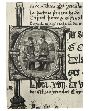
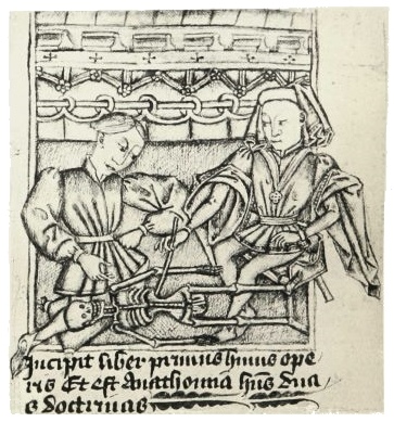
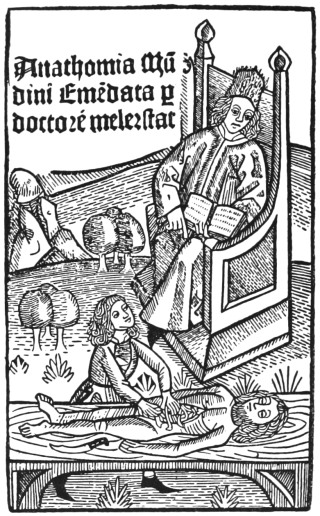
Fig. 2. Title-page of Mellerstadt’s edition of the Anatomy of Mondino, Leipzig, 1493. The scene is laid in the open air.131
That so much industry was rewarded by so small an increase in knowledge may probably be attributed to the method adopted. The so-called ‘anatomies’ were conducted in the most formal manner. Bertuccio, for example, who succeeded Mondino as professor of Surgery at Bologna, was accustomed, as we learn from his pupil Guy de Chauliac, to give short systematic anatomical demonstrations on a fixed and rigid method.129 The occupant of the chair at this period was indeed no professor in the modern sense of the word. To expound the tradition of anatomy as it had reached him was regarded as the limit of his duty. Of any attempt to extend the bounds of knowledge, of any systematic endeavour to correct or improve the anatomical views of his predecessors, we find little or no trace. Indeed, at Padua it was expressly laid down in the statutes that the exposition of anatomy should follow the very words of Mondino.130
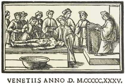
Early figures portraying the teaching of anatomy (Plate XXVII and Figs. 1–3, 5) usually show us a medical doctor sitting at a desk, well removed from the subject of dissection, and reading from his text-book the description of the part. Meanwhile an assistant, who is usually also a doctor, performs the actual work of dissection. The professor of Surgery, to whom the teaching of anatomy was entrusted, stands by with a pointer to indicate the different organs.
Sometimes the professor changes places with the reader at the desk. In some later MSS. the teacher is figured as himself handling the body and demonstrating to his pupil (Plate XXX a132 and b133), but there is evidence that the miniatures portraying this are the work of artists unfamiliar with dissection and with the teaching of anatomy.
Fig. 3. A DISSECTION SCENE From the Venice 1495 edition of ‘Ketham’ (compare Plate XXVII).
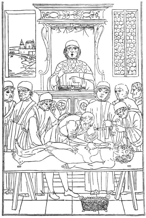
Fig. 4. From the English translation of Bartholomaeus Anglicus, printed by Wynkyn de Worde, 1495. The first picture of dissection in an English-printed book.
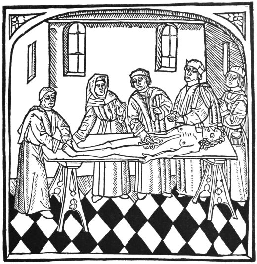
Fig. 5. A LECTURE ON ANATOMY From the 1535 Venice edition of Berengar of Carpi’s Commentary on Mondino.
The study of anatomy had to contend with two great difficulties, want of subjects for dissection, and faith in the written word.
Thus, at Bologna, where it was arranged that every medical student of over two years’ standing should attend an Anatomy once a year, no less than twenty students were admitted to see the anatomy of each man, and thirty to the anatomy of each woman.134 This was all the practical instruction received. Some other Universities had to be content with the cadaver of a single criminal per annum for the whole body of students.
In the first period during which the human body was dissected in Europe, the thirteenth century, a certain amount of progress was certainly made, despite the rarity of subjects. The rebirth of learning in the thirteenth century was not, however, as favourable to anatomical progress as might have been hoped. Galen, indeed, ceased to be a mere name, and the Latin translations of his text, or of its adumbra in the writings of the Arabians, became ever more familiar. On the other hand, with more authoritative texts in their hands, men were but the more inclined to follow the evil scholastic way, and to trust rather to the written words of the master than to the evidence of their own senses. Thus it came about that the second period, which covers the fourteenth and most of the fifteenth century, was really stationary so far as the first-hand knowledge of anatomy was concerned. With the last decade of the fifteenth century, however, there opens a new and third period in the history of our subject. From that time dates the true era of anatomical renaissance, which may be regarded as continuing until the commencement of modern anatomy with the great work of Vesalius in 1543.
Plate XXXI. From the MS. of GUY DE VIGEVANO of 1345 at CHANTILLY
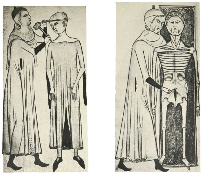
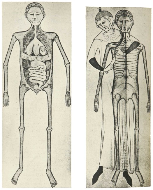
Plate XXXII. From the MS. of GUY DE VIGEVANO of 1345 at CHANTILLY
We have said that throughout the second period, the formal demonstrations based on the declaimed text of Galen or Avicenna or Mondino were practically the sole opportunities afforded to either teacher or pupil for the investigation of the minuter details of the human frame. But in making this statement concerning the arrest of anatomical progress, we must expressly exclude the products of the mighty genius of Leonardo da Vinci (1452–1519), whose anatomical researches were without influence, and remained long unnoticed.135 We must also omit evidence gathered from the work of such early Renaissance painters as Antonio Pollaiuolo (1429–98) or Andrea del Verrocchio (1435–88), for these pursued the study of anatomy in a special field and with a special object.136 Furthermore, there are a number of artists of similar date of whose anatomical studies we have no direct evidence, but who yet outlined the muscles of the nude human figure in such a way as leads us to suppose that they had investigated the superficial structures at least of flayed parts. Such is the suggestion of some of the work of Luca Signorelli (c. 1442–c. 1524), and of Andrea Mantegna (died 1506). With such reservations, however, it is probably true that no evidence is forthcoming until the last decade of the fifteenth century of any advance from the standpoint of Mondino.137
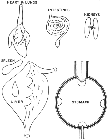
But if descriptive anatomy developed slowly in the hands of the physicians, the art of graphic representation of anatomical structures was still more backward. Several groups of anatomical drawings of mediaeval date have come down to our time, but examination of them shows that they have been drawn without direct reference to the human frame. Some of these figures are of the crude type known as the ‘five-figure series’ (Plate XXXIII), mere traditional diagrammatic sketches.138 Hardly better or more instructive are the series of dissections which illustrate certain MS. works of Henri de Mondeville (Plate XXVIII a)139 and Guido de Vigevano (Plates XXXI and XXXII), 1345.140 A few sketches representing the separate organs have also survived (Fig. 6),141 but these never suggest that the draughtsman had before him the structure which he seeks to depict, and the drawings appear to have been made in order to illustrate contemporary physiological theory rather than observed anatomical fact. Even the magnificent illuminated Dresden Codex of Galen, prepared in France or Flanders as late as the second half of the fifteenth century, betrays not the slightest first-hand knowledge of anatomy.142 Although the illustrations of this MS. are prepared with the utmost technical skill, they yet show us a teacher exhibiting to his pupils a heart of the form found on playing-cards, and other anatomical figures scarcely more faithful to the facts (Plate XXXIV).
Fig. 6. DIAGRAMS OF THE INTERNAL ORGANS After Bodleian Library MS. Ashmole 399 of about 1298, fos. 23 recto–24 recto.
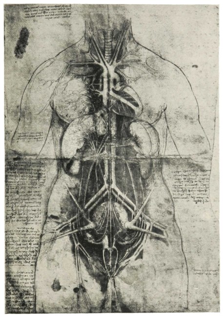
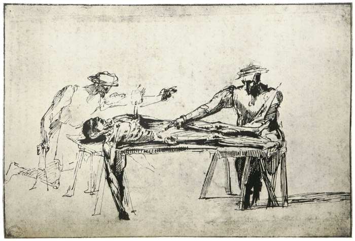
The spirit of investigation of the artist who perforce went direct to nature, dissecting with his own hands and observing with his own eyes (Plate XXXVI), showed itself indeed far more fruitful than the tedious ex cathedra methodization of the professor.143 Yet the system of the schools needed to be combined with the freedom of the artist for the production of an effective anatomical work. What the projected treatise of Marcantonio della Torre (1473–1506) might have been we may guess from the anatomical sketches of Leonardo da Vinci (Plate XXXV), who was to have been associated with him in the work.144 In the event, however, the medical schools had to wait yet another generation before the subject was placed on a sound basis by André Vesale.
The Mondino pamphlet—for it is little more—used since its author’s death in 1327 as a text-book in the schools of northern Italy, was first printed in 1478. Not until the last decade of the fifteenth century did there appear another work bearing evidence of the hand of a practical anatomist. This was an Italian translation of Ketham’s Fasciculus medicinae, impressed at Venice in the year 1493.145 The volume comprises Mondino’s pamphlet and a collection of other medical tracts that were probably put together by Giorgio di Monteferrato from the work of a writer of the previous century, for their contents are traceable to a fourteenth-century MS.146 The text is neither original nor remarkable, but the Venice volume derives its importance from certain figures which appear in it for the first time.
Two of these plates are of great interest both intrinsically and also in relation to the history of anatomy. One of them is the magnificent representation of a dissection scene, which is regarded as perhaps the finest example of book illustration produced during the first century of typography147 (Plate XXVII). This work of the ‘maître aux dauphins’, as the unknown artist is called by critics,148 is doubly interesting, for it is the subject of an experiment in colour printing, no less than four pigments being laid on by means of stencils. As early as 1457 the method of stencilling was employed for colouring the initials of a Psalter, and in 1485 Erhard Ratdolt in an astronomical work added yellow to the earlier red and black. The figure from which our plate is taken represents, however, the first attempt at a complex colour scheme and leads up to the work of Hugo da Carpi.149
In this picture the professor, a youthful figure perhaps intended to represent Mondino himself, is shown standing at a desk which hides his book. Around a corpse, laid on a trestle table before him, there cluster a number of men in doctor’s robes. Their valid faces are sufficient to convince us that the artist is here presenting us with portraits. One of the listeners has removed his robe and stands with upturned sleeves and knife in hand, ready to make the first incision on the direction of the doctor, who points to the part with a wand held in the left hand. In the impression of 1495 and in those of later date, the book appears above the desk, the attitudes of the students are somewhat changed, and many other details are altered. In all these, however, the blocks have been recut and the result is artistically inferior150 (Fig. 3).
Fig. 7. A FEMALE FIGURE LAID OPEN TO SHOW THE WOMB AND OTHER ORGANS From the 1493 Venice edition of ‘Ketham’ translated into Italian. This is the first printed anatomical figure drawn from the object.
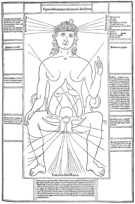
The second plate from the 1493 Ketham with which we are here concerned is the outline of a female body, in a traditional pose,151 laid open to exhibit some of the internal organs (Fig. 7). These had clearly been sketched from the object, and therefore this drawing, the first printed figure of its kind, may be said to introduce the new era for the investigation of the human frame. The anatomical renaissance had begun. Into a discussion of the full development of that age we cannot now enter. But the MS. of Manfredi, with which we have here to deal, was written at the very dawn of the new era and is itself one of its earliest documents.
II. Bolognese Works on Anatomy
An organized Medical Faculty existed at Bologna at least as early as 1156,152 though the first record of dissection there is of considerably later date. In February 1302 a certain Azzolino died under suspicious circumstances. Poison was suspected, an inquest was held and a post-mortem examination ordered. The investigation was conducted by two physicians and three surgeons, who unanimously agreed ‘that the said Azzolino assuredly met his death by no poison, but on the contrary, we assert that the quantity of blood collected in the great vein known as the vena chilis [vena cava]153 and in the veins of the liver adjacent thereunto, has prevented the due movement of the spiritus throughout the body, and has thus produced the diminution or rather extinction of the innate heat and thereby induced a rapid post-mortem discoloration. Of this condition we have assured ourselves by the evidence of our own senses and by the anatomization of the parts.’154
The first anatomical document emanating from the University of Bologna is, however, of still earlier date, and is the work of William of Saliceto (1210?–80). This writer was educated at Bologna, and it is claimed that he was the first to dissect the human body there.155 His Cyrurgia, which was completed in 1275 (editio princeps, Piacenza, 1476), is divided into five books, of which the fourth and shortest is devoted to anatomy. Its descriptions are brief and concise. They are often clearly the result of actual observation, and they show hardly any trace of the absurd and irritating teleology that the influence of the Arabians and of Galen made customary in early anatomical literature. The anatomy of Saliceto appears to us very sensible and so far as it goes practical. It betrays the method rather of the Salernitan than of the Arabian anatomical writings, and is on the whole the best European work of the kind before the Renaissance. It was, however, soon replaced by the text-book of Mondino di Luzzi (1285–1327),156 which, though inferior to that of Saliceto, held the field until the subject was revolutionized by Vesalius.
Plate XXXIII. The FIVE-FIGURE SERIES BODLEIAN MS. ASHMOLE 399, about 1292 Fos. 18 r–22 r VEINS, &c. ARTERIES NERVES BONES MUSCLES
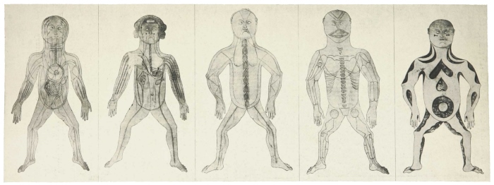
From the DRESDEN GALEN MS. Plate XXXIV. DEMONSTRATIONS OF ANATOMY Second half of XVth Century
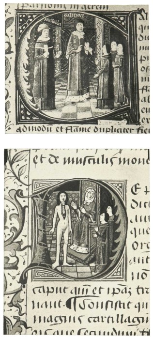
Mondino was professor at Bologna till his death in 1327. His work, easily accessible in one of its many editions, ‘is corrupted by the barbarous leaven of the Arabian schools, and his Latin defaced by the exotic nomenclature of Avicenna and Rhazes’.157 But it is not the language alone that has suffered. The schoolman’s attitude, well fitted for the classification of ideas, is an ill instrument for the investigation of Nature, and in the scholastic Mondino the very basis of scientific judgement is undermined, so that he readily accepts the views of the ancients against what must often have been the evidence of his own senses. The work, however useful to the contemporary student, was thus essentially reactionary as against the efforts of the earlier Salernitan anatomists and of William of Saliceto. This is the more remarkable because it is quite clear that he was accustomed to demonstrate on the actual body—a privilege denied to the early Salernitan school,—and he was, moreover, a popular and successful teacher. His work is a manual of dissection rather than a treatise on anatomy. This, added to its conciseness and brevity, strengthened its appeal to the ‘practical’ man—an epithet claimed then, as now, by the majority of stupid and unpractical people. The personal influence and enthusiasm of its author no doubt helped also towards the phenomenal success of this work, which for two hundred years held a position without rival as the text-book of the medical schools of Italy, where even as late as the sixteenth century Mondino ‘was still worshipped by all the students as a very god’.158
Mondino was succeeded in the chair of Surgery at Bologna by his pupil, the Lombard Bertuccio, who died in the Black Death of 1347. Bertuccio’s surviving work is unnoteworthy, but he was the anatomical teacher of Guy de Chauliac, whose Surgery159 is of great value and was very influential in standardizing practice, especially in the north and west of Europe. Nevertheless it appears to us that the anatomical section is the weakest part of Guy’s great work. The teleology that is a blot in Mondino has here become a perfect plague, and Guy’s anatomy consists of one-third description and two-thirds wearisomely reiterated reasons for the existence of imperfectly described structures. Through Guy de Chauliac the anatomical tradition of Mondino passed over into the University of Montpellier.
A later fourteenth-century Bolognese writer was Tommaso di Garbo (died 1370), who did little but comment on Avicenna. A surgeon of the next generation, however, Pietro d’Argellata, deserves to be remembered for his description of the examination of the body of Pope Alexander V, who died suddenly at Bologna on May 4, 1410. His account throws light on the customary procedure and may be rendered here.160
‘I ordered the attendants’, he says, ‘first to cut the abdomen from the pomegranate [i.e. the Adam’s apple or laryngeal cartilage161] to the os pectinis [i.e. the symphysis pubis]. Then, so that they should not rupture the intestines, I myself sought the rectum and ligatured it in two places and then cut it between. Next I removed all the intestines as far as the duodenum and dealt with them as with the rectum, and so I had the intestines clean and without fetor. After this I extracted the liver, seizing its ligaments; then the spleen and then the kidneys, and these were all placed together in a jar. I now passed to the spiritual members [i.e. the thorax] and removed lung and heart and all their ligaments. Then I ligatured the meri [the Arabian term for oesophagus] and removed the stomach. When this had been done there were some who wished to remove the tongue but knew not how. I however cut under the chin and extracted the tongue through that hole, together with trachea arteria [trachea] and meri. Then I passed to the arteria adorti [aorta] and vena chilis [vena cava]. Lastly I removed the ligatured remnant of the intestines as far as the anal margin.’
Giovanni da Concoreggio (died 1438), who was lector in Surgery at Bologna in the early part of the fifteenth century, left a few anatomical observations of little note,162 and not very much more can be said for his successors and Manfredi’s contemporaries Gabriele Gerbi (de Zerbis, died 1505) and Alessandro Achillini (1463–1512). Gerbi163 does little but repeat in the most verbose fashion the work of Mondino and of Avicenna, some of whose errors, however—e.g. the three ventricles of the heart—he omits. He wrote also an anatomy of the infant, or rather of the foetus,164 and a treatise taken mainly from Avicenna’s De generatione embryonis. Like all his work, these are in the full scholastic style of a professor of Logic, a position to which, in fact, he ultimately attained.
Achillini’s work165 is but a slight advance on that of Gerbi. It is really little else than a note-book for students, and gives the baldest directions for dissection, accompanied by a few comments taken from Avicenna. Achillini occasionally ventures to criticize Mondino, and his work has at least the advantage of brevity. He has a claim to be remembered in that he was the first to describe the duct of Wharton and is said to have been the first to describe the ear ossicles, malleus and incus. Achillini, like Gerbi, was a windy and very ‘scholastic’ disputator. He was best known to his contemporaries as a supporter of the philosophy of Averroes. In 1506, when driven from Bologna with the other supporters of Bentivoglio, he became professor of Philosophy at Padua.
Fig. 8. THE ABDOMINAL MUSCLES From Berengar of Carpi’s Commentary on Mondino, Bologna, 1521.
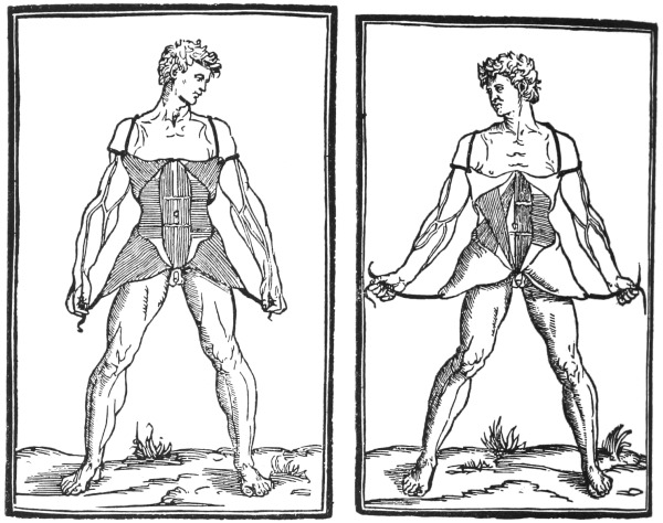
With Giacomo Berengario da Carpi we come at length to one who definitely advanced the science, and who may be regarded as the first modern anatomist, so far as printed works are concerned. He was professor of Surgery from 1502 to 1527, and during that period published his great anatomical work.166 This volume, though modestly put forward as a commentary on Mondino, is in reality an original contribution of great value. It is the earliest anatomical treatise that can properly be described as having figures illustrating the text (Fig. 8).167 Carpi does not hesitate to criticize the work on which he comments—as for instance when he denies the existence of the ‘rete mirabile’ below the brain, though descriptions of the ‘rete mirabile’ had been based on the statement of no less an authority than Galen. Furthermore he was the first to describe the vermiform appendix, and he gave the earliest correct account of several other organs, e.g. the choroid plexus and the olfactory nerves. He was an industrious dissector, and he tells us that he had examined more than a hundred bodies.
With Carpi we close our series of Bolognese anatomists. Into that group we now proceed to fit the writer with whom we are here specially concerned, Hieronymo Manfredi.
From a drawing in the Library, WINDSOR CASTLE Plate XXXV. VIEW OF THE INTERNAL ORGANS LEONARDO DA VINCI
From a Drawing in the ASHMOLEAN MUSEUM, OXFORD, attributed to BARTOLOMEO MANFREDI (1574?–1602) Plate XXXVI. THE TWO FIGURES DISSECTING ARE TRADITIONALLY SAID TO REPRESENT MICHELANGELO AND ANTONIO DELLA TORRE
III. Hieronymo Manfredi
Hieronymo Manfredi was a member of a family that had already for more than two centuries provided distinguished citizens, and especially physicians, to the city of Bologna.168 He was born about the year 1430 and was educated at the University of Bologna. Here in 1455 he was laureatus in Philosophy and Medicine, and here he became professor of the latter subject in 1463.169
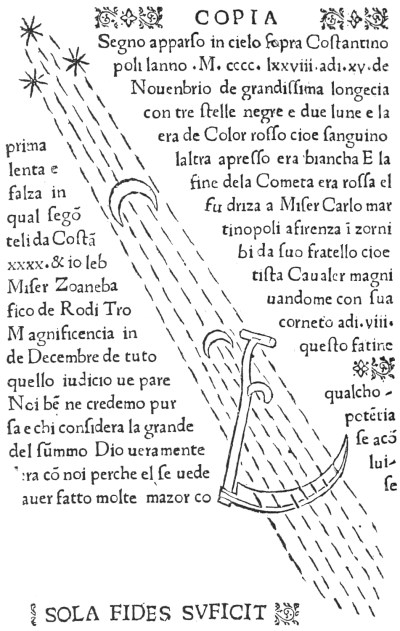
During the second half of the fifteenth century, a perfect mania for the study of astrology infected Italy and penetrated equally into the Court, the Church, and the Academy. The profession of Medicine was far from immune, and at the University of Bologna, where a chair of Astrology had long been established,170 the study was pursued with ardour and enthusiasm. Here Manfredi early devoted himself to that will-o’-the-wisp, the pursuit of which absorbed and sterilized many of the best intellects of his day. By the year 1469 he was already regarded as an authority on the vainest of studies,171 and as the years went on he seems to have devoted himself to it ever more and more. The generally credulous character of Manfredi’s astrological ideas may be gathered from the page of his Prognosticon ad annum 1479 which we here reproduce (Fig. 10).
The history of Manfredi’s connexion with the University of Bologna may be briefly told. He appears for the first time on the professorial roll in 1462, when we find him giving the ‘extraordinary’ lectures on Philosophy, a subject then regarded as under especial charge of the physicians. In 1465 he was conducting the ‘ordinary’ course in Philosophy, and at the same time giving occasional lectures on Medicine. In the following year he was called to the chair of Theoretical Medicine, and in 1469 he helped the Faculty out of a difficulty by giving lectures on ‘Astronomia’ in place of the aged professor Giovanni de Fundis. The latter died in 1474, and from that date onward Manfredi assumed responsibility for the course on ‘Astronomia’. Among the colleagues who joined him were Gabriele de Gerbi, who became lecturer on Logic in 1476, Filippo Beroaldo, who became lecturer on Rhetoric and Poetry in 1479, and Alessandro Achillini, who became lecturer on Logic in 1484.172
Such was the regard for Manfredi’s powers of astrological prediction that to all the University announcements of his course of lectures on Astronomy is added ‘cum hoc quod faciat iudicium et tachuinum’.173 In spite of his proficiency in the science, however, he was unable to foretell his own death. Giovanni Pico della Mirandola writes of him thus derisively:
‘quo anno [1493] obiit omnimoda[m] uite incolumitate[m] fuerat pollicitus Hieronymus manfredus astrologus nostra aetate singularis: a quo tamen nihil mirandum minus praeuisam aliorum mortem: qui nec suam ipse praeuiderit: nam cum proxima estate uita sit functus: in istius tame[n] anni publico uaticinio qui s[cilicet] ei fuit fatalis: multa & mira sequenti anno dicturum se non semel pollicebatur. Qui nescio oppignoratam fidem quomodo reluet: nisi forte de caelo uerius nunc terrena despiciat q[uam] de terra oli[m] caelestia suspiciebat.’174
Manfredi died in 1493 and was buried in the church of Santa Margarita in Bologna. This church no longer exists, but it contained in the eighteenth century a tomb bearing the inscription:
HIERON. MANFREDO BONON. PHILOSOPHO AC MEDICO SVAE AETATIS NEMINI SECVNDO ASTRONOMORVMQVE CITRA INVIDIAM FACILE PRIMARIO. POSVIT SVPERSTES IOAN. FILIVS SVISQVE POSTERIS. VALE ATQVE ILLVM VALERE OPTA.175
Manfredi left a widow, Anna, who was still living in 1496 with a household of ten persons in the Via S. Margarita.176 The houses on one side of this street backed on the very walls of the buildings belonging to the ‘University of Medicine’,177 and we may suppose that Hieronymo Manfredi had resided here on that account. His surviving son, Giovanni, lived hard by in the Via S. Antonio di Padoa.
It cannot be said that Manfredi’s printed works suggest great scientific attainments. All are permeated by the same astrological obsession. They comprise the following:
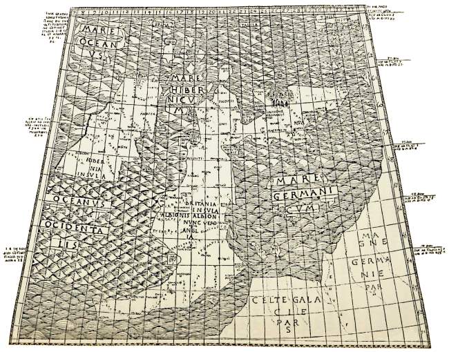
(a) The editio princeps of Ptolemy’s Cosmographia and Tabulae Cosmographiae, the best-known printed work to which Manfredi’s name is attached. He was associated in its production with the famous scholar Filippo Beroaldo, and the finely produced volume was published at Bologna in 1472 (?),178 and dedicated to the memory of Pope Alexander V (died 1410). It is interesting as containing the first printed map of England (Fig. 9). At the end of the work we read:
‘Accedit mirifica imprimendi tales tabulas ratio. Cuius inuentoris laus nihil illorum laude inferior. Qui primi litterarum imprimendarum artem pepererunt in admirationem sui studiosissimum quemque facillime conuertere potest. Opus utrumque summa adhibita diligentia duo Astrologiae peritissimi castigaueru[n]t Hieronimus Mamfredus & Petrus bonus. Nec minus curiose correxerunt summa eruditione prediti Galeottus Martius & Colla montanus. Extremam emendationis manum imposuit philippus b[e]roaldus.’
THE FIRST PRINTED MAP OF ENGLAND. From the 1472 (?) Bologna Ptolemy, edited by Manfredi and others.
(b) Liber de homine: cuius su[n]t libri duo. Primus liber de conservatione sanitatis.... [Liber secundus de causis in homine circa compositione[m] eius], Bologna, 1474. The work is in Italian, and consists of a number of paragraphs, each beginning with the word ‘perchè’. There is a servile dedicatory epistle in Latin addressed to Giovanni Bentivoglio. The first book is concerned with diet, and occupies two-thirds of the volume. The second book answers questions on the subject of physiognomy and bears resemblance in many passages to the Anatomy. It is taken in the main from the pseudo-Aristotelian Problemata. The book is without pagination or figures. It is well printed, and illuminated examples are not infrequently encountered.
This work was very popular. In 1478, during the lifetime of its author, it was audaciously pirated at Naples with the following incipit: ‘Incomenza el Libro chiamato della uita costumi natura & om[n]e altra cosa pertine[n]te tanto alla conservatione della sanita dellomo quanto alle cause et cose humane. Co[m]posto per Alberto Magno filosofo excellentissimo.’
In 1497, after Manfredi’s death, the work appeared in black-letter folio at Bologna, with its author’s original dedication slightly altered. The text in this edition commences, ‘Perchel sophio nele cose che noi viuemo: & lo indebito modo del viuere nostro: induce in noi egritudine’.
In 1507 it appeared at Venice in small black-letter quarto as Opera noua intitulata Il perche utilissima ad intendere la cagione de molte cose. By this title, Il Perchè, the work, which ran through numerous editions, has usually been known. It continued to be reprinted as late as 1668.
(c) A treatise on the Plague: Tractate degno & utile de la pestile[n]tia co[m]posto p[er] el famosissimo philosopho medico & astrologo maestro Hieronymo di manfredi da Bologna, Bologna, 1478. This was translated into Latin by the author himself in the same year. The work owes much to Avicenna, but contains some original clinical observations, and shows a certain independence of the prevailing spirit of the age by quoting opinions of contemporary as well as of ancient physicians. The remedies are similar to those recommended by John of Bourdeaux in his widely distributed tract on the plague, and are probably derived ultimately from the Regimen Sanitatis Salerni.
(d) Prognosticon ad annum 1479, Bologna, 1478. We reproduce the terminal page of this work (Fig. 10).
Fig. 10. The last page of Manfredi’s Prognosticon ad annum 1479, Bologna, 1478.
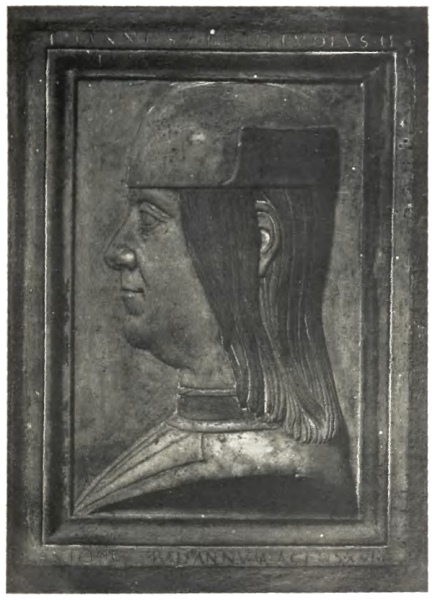
From his tomb in the Church of S. Giacomo Maggiore at Bologna Plate XXXVII. GIOVANNI BENTIVOGLIO II
BRIT. MUS. MS. ROY. 7 F VIII, fo. 50 v Plate XXXVIII a. ROGER BACON’S DIAGRAM OF THE EYE. XIIIth Century From a drawing in WINDSOR CASTLE Plate XXXVIII b. LEONARDO DA VINCI’S DIAGRAM OF THE HEART Early XVIth Century
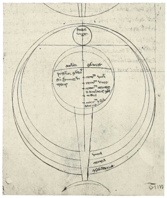
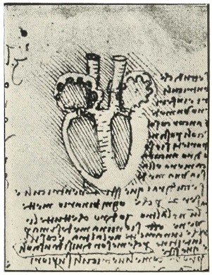
(e) Prognosticon anni 1481, in which is embodied Oratio contra turcos & hostes Christianorum, s. 1. Jan. 1481.
(f) Centilogium de medicis et infirmis, Bologna, 1488. With a dedication to Bentivoglio. This short work is wholly astrological, and consists of one hundred precepts concerning the relationship of the stars to various diseases and conditions. Reprinted Venice, 1500, and Nuremberg, 1530.
The following three works are attributed to Manfredi, but are not mentioned in Hain, Copinger, or Reichling’s lists of Incunabula; we have not seen any of them and their existence is doubtful.
(g) Ephemerides astrologicae operationes medicas spectantes, mentioned in the Biographisches Lexikon der hervorragenden Aerzte of E. Gurlt and A. Hirsch. Possibly it represents another edition of (e).
(h) Quaestiones subtilissimae super librum aphorismorum, Bologna, 1480 (?), mentioned by Haller.179 Possibly it represents another edition of (b).
(i) Chiromantia secundum naturae vires ad extra, Padua, 1484, mentioned by Haller.179
IV. The Manuscript Anatomy of Manfredi
The MS. of Manfredi’s Anatomy is in the Bodleian Library at Oxford (Canon. Ital. 237, Western 20287). It is a fairly preserved small quarto parchment, originally of forty-nine folios, of which the third and fourth are missing. The writing is in the fine Italian hand that the printed type of the period was accustomed to imitate. There are no figures or illuminations, but the titles are rubricated in burnished gold or in colours.
There is no reference to this work in any account of Manfredi, and the volume itself appears to be quite unknown. Neither the man nor his work is mentioned in Medici’s detailed history of the anatomical school at Bologna180 nor in Martinotti’s recent study on the same topic,181 nor is any MS. of Manfredi included in Mazzatinti’s monumental catalogue of the MSS. in the Italian libraries.182
Manfredi’s MS. is written in the involved Italian of the day, with sentences of inordinate length. These general characters of style are encountered also in his published works. The dedication is in Latin, of the same unpleasing quality, and is couched in the usual subservient manner. It is addressed to Giovanni Bentivoglio, and in it Manfredi relates that
‘Your illustrious lordship Johannes Bentivolus in this present year 1490 with your usual humanity condescended on one occasion to watch the dissection of a corpse.... It was then that you saw the wonderful works of Nature in the anatomy ... and you parentally urged me, Hieronymo Manfredi, to inscribe to your most noble name this work on anatomy.... I therefore extracted this work as best I might from various works of antiquity and abbreviated it. I have not followed their order, but I have so composed it that the work should be pleasing to your lordship.
‘Accept then, O great and powerful lord, this work on the anatomy of the human body inscribed to your noble name! Accept it with your customary benevolence and humanity and in a kindly and gracious spirit, for it will be pleasing to you and will delight you greatly, for it is a worthy work!’
The Giovanni Bentivoglio (Plate XXXVII), with adulation of whom Manfredi was thus accustomed to plaster his works, was the second of the name and was the son of Annibale Bentivoglio. In the year 1462 he became head of the republic of Bologna, and played there much the same rôle as did Lorenzo de’ Medici at Florence. He adorned Bologna with numerous buildings,183 and acted as patron of the arts and the sciences. The Palazzo dei Bentivogli still stands as a memorial to him and his family. A stern and high-handed tyrant, he held his position until 1506, when he was expelled and the city reverted to the papacy. He died two years later.
It is remarkable to find a man of Bentivoglio’s eminence and position taking an interest in the practical study of anatomy. Other Italian rulers, Lorenzo de’ Medici among them, encouraged and legalized the practice of dissection, but probably Bentivoglio is the only one recorded as having patronized an ‘anatomy’ in person. The interest taken in the subject by the heads of states must have been of great value to the artists whose patrons they were.
The MS. is a unique copy, and was doubtless written for presentation to Bentivoglio. That it was never printed is perhaps due to the fact that Manfredi died within a comparatively short time of its composition. It represents the most satisfactory post-mediaeval account of the human frame until the appearance of the work of Berengario da Carpi in 1521. It is more complete than the work of William of Saliceto or of Mondino or the anatomy erroneously attributed to Richardus Anglicus; it is more natural than the book of Gabriele de Gerbi, and is far superior to the crude contemporary sketches of Hundt, Peyligk, and Achillini, while it wastes less space than Guy de Chauliac on teleology, though it has none of the charm of the work of that great surgeon. In one respect at least, viz. the spirit in which it is written, Manfredi’s Anatomy is original and probably unique for its age. There is no reason to doubt the assurance of the dedication that it was composed for the edification of the tyrant of Bologna, and for the simple purpose of setting forth the wonderful structure of man’s body without thought of any medical application.
The sources of the MS. are obvious. It is in the main a rearranged and on the whole improved Mondino, but amplified by reference to translations from Galen, Rhazes, Haly Abbas, and Avicenna. Guy de Chauliac has perhaps also been used. The work gives a general impression of being the product of a practical dissector, and it provides us with a good example of early Renaissance anatomy as taught in the Italian schools before the reforms of Vesalius. It is perhaps the first complete treatise on its subject written originally in the vernacular.184 It exhibits, however, no other original features nor any considerable departures from its sources, and it may be taken to represent, with but little modification, the tradition of Mondino as developed at his own University of Bologna at the end of the fifteenth century.
Manfredi’s work, however, if not original is at least eclectic, and the variety of its sources indicates a dawning consciousness of the unwisdom of trusting to the infallibility of any one writer. The work is thus in a sense intermediate between the early printed versions of Mondino, such as that of 1478, and the edition published in 1528 by Berengario da Carpi with its frank commentary of the master. All represent stages towards the freedom of the later Renaissance investigators.
We reproduce the text in full, and the passages on the head, on the eye, and on the heart, are rendered into English. All are similar to the accounts of Mondino. We are able to illustrate them by figures from contemporary works, and thus to give an idea of the limits of the anatomical knowledge of the day.
V. Translation of Selected Passages from the Anatomy, with Commentary
(a) THE HEAD
Tractate i, Chapter 2
(folio 5 verso) There are ten layers of the head.
The first is the hair made by nature for the better protection of the head from external things, and also for beauty.
The second part is the skin, which has here to be very thick, so that the hair may be firmly embedded, having its roots thick and long; and also to be a better shield and covering for the bone and brain, since there is no muscular part here.
The third part is the flesh, developed only on the face, the temples, and about the jaws, not on the other parts.
The fourth part is an external membrane called almochatim [Arabian term for cranial periosteum] which, when the skin is raised, appears to be continuous and covers the whole cranium. And nature made this membrane firstly so that the skin which is soft should not come into contact with the hard bone, secondly that the bone of the head should have sensation through it, and thirdly that the internal membrane of the head, called dura mater, should, by means of this membrane, be attached to the bone of the cranium by certain nerves and ligaments. These, issuing through the commissures of the bones, have thus their origin in the aforesaid internal membrane, while on emerging through the bone, they weave themselves into or rather compose the external membrane called almochatim.
The fifth part is the skull. This is a bone like a cap, inside the cavity of which is located the brain. In the skull are four bones sutured together. Nature made the skull not of one but of many pieces, firstly, so that if harm should fall on one part it might not spread to the others; secondly, so that by their joints or rather sutures [Italian cusiture=sewings], the humours of the brain might be the better exhaled; and thirdly, so that when there is need of applying medicines, these might the better penetrate to the parts within.
Hence it is that four pieces of bone are sutured and joined together by nature in a denticulate fashion, so that they might be the firmer and stronger. Nor are they bound with ligaments as are the joints, for these would not have been so strong, and furthermore the bones of the head do not need to move.
These sutures are five in number, three being true and two false. The true sutures are those which pass right through the bone, while the false do not. Of the true sutures one is in the anterior part and is called coronal; it is made like the letter C, and stretches from right to left of the head, the two wings of the C being directed towards the forehead. The second true suture extends along the length of the head, beginning from the coronal and reaching the back part of the head. It is like a shaft or rather arrow that goes backwards from the brow, wherefore it is called sagittal ——(. The third true suture is in the posterior part and is called laudal, for it is made like a Λ, the letter called by the Greeks lauda. The sagittal suture extends from the coronal to the lauda 〉——(.
The false sutures are two, one on each side. They are called cortical because they do not penetrate.
Now if we consider these five sutures we shall see that there are four bones articulated together. One is the forehead bone [frontal] which begins at the coronal and ends below at another suture, which itself begins as a branch of the coronal suture and proceeds by way of the eyebrow to the corresponding branch [of the other side] Ɑ.
A second bone is behind and terminates at the laudal suture. There are two other bones which form the temples. These terminate at the false sutures which themselves begin at the laudal and end at the coronal suture.
The sixth part [of the head] consists of two membranes. One of these is called dura mater, and lies in contact with the cranium. The other is called pia mater and is in contact with and covers the brain. And nature contrived it thus, having great solicitude for this latter member, that while close to the bone, it should yet not be touched by it. Wherefore, taking due precautions, she made the one [membrane] harder than the other. Furthermore she made two membranes, so that if harm befell one of them, it might not be communicated to the underlying brain.
In the pia mater are woven certain veins by which the brain is nourished. [The brain is] everywhere covered by it except on the posterior part; because this part being dry, it has no need of this membrane, as have the anterior and middle parts. The two membranes in many places penetrate the substance of the brain, dividing it into a right and a left, a front and a back section. By this division, divers cells or rather small chambers are made therein, in which the soul (anima) performs its divers operations, for which reason it is necessary that these parts should be of different structure.
When the two membranes are raised, the seventh part of the head, namely the brain itself, appears. The brain is wrought by nature so that the vital spirit from the torrid heart should be tempered by its cold, for here it is converted into animal spirit, which is the beginning of the perceptive (cognoscitiue) and motive processes.
The brain is of a substance like marrow, white, soft, and viscous, and from it the nerves arise. The anterior part is moister, softer, and less cold than the posterior because the senses [sentimenti = senses + mental processes], which are themselves moist and soft, have here their origin. In the posterior part the motor nerves arise, and it is therefore drier and firmer.
The brain is divided into three parts or ventricles. The first ventricle or anterior part is itself divided into two, right and left, and is moreover larger than any of the other ventricles, for in this first ventricle nature has placed the two faculties subservient to perception (al cognoscere). One of these is called common sensation (senso comune); in it the external senses terminate as at a centre and deliver the images or rather species of sensible things, so that this faculty may perceive and distinguish between one sensible thing and another, and also comprehend the operations of particular senses; which two things none of these [senses of themselves can do]. The other faculty of the first ventricle is called fantasia and by some imagination; it retains and preserves the species of sensible things in the absence of the material objects themselves.
When thou examinest the first ventricle thou wilt see three things before thou comest to the second ventricle.
[a] The first is itself double, and is formed of the very substance of the brain, so that it forms the base of the anterior ventricle both right and left [= corpora striata].
[b] To the side of this is another thing like a subterranean worm, red as blood, yet tethered by certain ligaments and nervelets [= choroid plexus and taenia semicircularis]. And this worm when it lengthens itself closes these passages, and thus blocks the path between the first ventricle and the second. Nature has wrought it thus, so that when a man wills he may cease from cogitation and thought; and similarly when, on the other hand, he would think and contemplate, this worm contracts itself again and opens these passages and thus frees the way between one ventricle and another.
[c] The third structure is a little lower and is a lacuna or rounded concavity [= infundibulum]. In the middle of this is a hole which passes down towards the palate, and this lacuna provides also a direct passage which descends from the middle ventricle to its colature [= sieve-like structure, i.e. certain parts of the sphenoid bone]. And this lacuna has around it certain large round eminences which support the veins and arteries that ascend to the ventricle. This passage is wide above and narrow below, and by it the first and second ventricles purge themselves of their superfluities, but the anterior part [of the first ventricle] purges itself more by the colature of the nose [= cribriform plate]. Thus nature has made two passages to cleanse the superfluities of the brain.
When thou hast seen these three structures there will appear the second or middle ventricle which is as a passage and transit from the anterior to the posterior ventricle. Here are two faculties. One, the estimative, deduces [Italian elicere] the insensible from the sensible. The other, called the cognitive, comprehends both things sensible and things insensible, synthesizing and analysing them (componendo e dividendo). These [two] faculties in the middle ventricle minister to the intellect. Now all the other faculties described, and even the power of memory, are found in brute animals, but this [intellectual power] is encountered in man alone.
Now will appear the third ventricle in the posterior part; and it is hard, for it gives rise to the greater part of the motive nerves which are of a strong and firm nature. This ventricle is pyramidal in shape, and culminates in an apex directed upwards where images of visible things (spetie) are conserved, for these are better stored in a strait than in an ample space; but the part below is wide to receive these images, which are better received in an ample than in a strait place. This ventricle has two functions: it gives rise to the spinal cord [nucha, an Arabian term] and motor nerves; and it is also the storehouse of the memorative faculties.
From what [has been said] it will be apparent that when the back of the head is injured, the memory immediately suffers; when the middle part is injured, the estimative and cognitive faculties suffer; and when the anterior part is injured, the faculties of common sensation and of imagination (fantasia) suffer. And thus it is that the doctors have become aware of the location of these powers.
This being disposed of, thou wilt next raise the brain carefully so as not to break the nerves. Commencing now with the part in front, there will first appear two small fleshy protuberances like two nipples, of like substance to the brain in which they originate, and covered by a thin membrane, the pia mater. These are the olfactory organs, wherein is the sense of smell.
From the brain arise seven pairs of nerves. Proceed therefore farther with the anterior part, and thou wilt see the first pair of these nerves, which are large, and called the nervi optici. These have their origin in the front ventricle of the brain and proceed towards the eyes. But before they pass through the pia mater, they join together, and at their place of union there is a perforated spot. Galen maintains that these nerves only join or rather unite, but do not intersect, so that the nerve that comes from the right after union returns again towards the right, and similarly with the nerve coming from the left, which after the union returns towards the left eye.185 But Rhazes maintains the contrary,186 although the opinion of Galen is the more common. These nerves are subservient to sight, and they are united so that the images of the things received by the two eyes and conveyed by the two nerves should return in unity; so that one thing should not appear as two.
After these two nerves, raise the brain towards its middle and thou wilt see another pair of nerves, thin and firm, which also go to the eyes, to give them voluntary movement, controlling certain muscles.
Farther on thou wilt see the third pair of nerves, one part of which goes to the face to give it sensation and voluntary movement, while another part goes to give taste to the tongue. Yet a third part of these nerves mingles with the fourth187 pair of nerves, and together they descend to give sensation to the diaphragm, stomach, and other viscera. A certain part also of the fourth187 pair of nerves goes to give sensation to the palate.
Then there is the fifth pair of nerves [which] go to the petrous bone around the ear; and of these nerves there are framed in the ear-holes certain membranes, which are the organs of hearing.
Next there is the sixth pair of nerves, which divides into three parts. One part goes to the muscles of the throat, the second to the muscles of the shoulders, and the third and largest descends to the epiglottis and to the diaphragm, and spreads into the chest, the heart, and the lungs, accompanying the nerves of the third pair. From the nerves of this sixth pair which go to the epiglottis arise the nerves of the voice, called reversive.
The seventh pair of nerves arise at the back of the brain and give voluntary movement to the tongue.
Of these seven pairs of nerves, the first two pairs originate in the anterior part of the brain, the third pair originates between the anterior and posterior parts, while the remaining four pairs originate in the posterior part.
Proceeding still farther, the brain may be completely raised, and the eighth part of the head will appear, that is, the two membranes situated below the brain. When these in turn are raised there will appear the ninth part, which is a certain net called rethe mirabile, because it is composed of exceedingly strong and marvellous texture, augmented by certain very fine arteries which are branches of arteries that ascend from the heart, and are called the apoplectic arteries. In these arteries of this net is contained the vital spirit, sent from the heart to be changed to animal spirit. That the spirit may be the better modified and distributed, nature made these arteries very fine, and separated them into very small branches so that the spirit should be minutely divided. Nature placed the rethe mirabile under the brain because it was necessary to guard its site carefully, and also that the moist vapours of the brain which fall upon the net, obstructing it, should induce natural sleep.
After all these things thou wilt see the basal bone which is the tenth and last part of the head, and called basilar, because it is the base and foundation of the whole head; and it was made hard so that the superfluities which descend to it should not putrefy it. This bone can be seen to be formed of many other bones articulated together. It is divisible into the petrous bones and the bones of the nose and eyes and two other lateral bones which can only be seen by means of disarticulation. [Folio 10 verso, line 22.]
Fig. 11. From M. Hundt, Antropologium, de hominis dignitate natura et proprietatibus, Leipzig, 1501. The figure shows the ten layers of the head, the cerebral ventricles and cranial nerves, and the relation of the nerves to the senses.
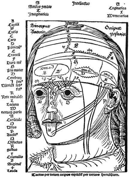
Fig. 12. THE LAYERS OF THE HEAD From the Anatomia of Johannes Dryander, Marburg, 1537.
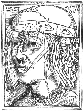
The ten parts or layers of the head are a commonplace of the anatomy of the period, taken from Avicenna. We may illustrate the division by the crude contemporary diagram of Fig. 11, which is improved in the later drawing reproduced in Fig. 12.
Manfredi’s account of the brain itself is amplified from Mondino. The division of this organ into three ventricles, each associated with a corresponding division of the mental functions, was very familiar to medical writers of the fifteenth century. The idea is found among Western writers as early as St. Augustine (354–430), and is encountered in the writings of Roger Bacon (1214–94). It had long been popularized in mediaeval psychology by the writings of Albertus Magnus (1206–80). The anatomical distinction is found in Haly Abbas, Avicenna, and Rhazes, and in some of the best MSS. of the latter writer a rough diagram of the ventricles is given.188 These writers are all clearly indebted to the anatomy of Galen,189 but on the psychological side Albertus Magnus probably drew mainly either from Ghazali190 (1059–1111), who in turn derived his inspiration from Nemesius (fourth century) and Johannes Damascenus (died 756), or else from early writers of the Salernitan tradition, such as Constantine191 (eleventh century), or Petrocello192 (twelfth century), who drew largely on Theophilus (seventh century).193
Fig. 13. From Illustrissimi philosophi et theologi domini Alberti magni compendiosum insigne ac perutile opus Philosophiae naturalis, Venice, 1496, showing the ventricles of the brain.
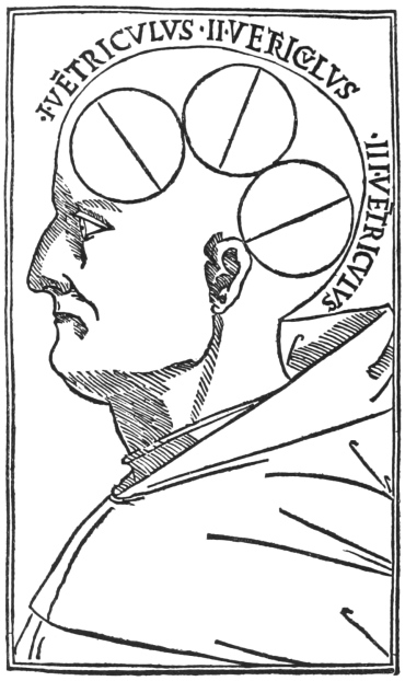
This outline of a tripartite division of the brain and its cavities was closely followed throughout the Middle Ages, as was also the curiously naïve and excessively ‘materialistic’ psychology to which it gave rise, and which Manfredi adopts. We illustrate his views of the relationship of the different parts of the brain and their parallelism in mental processes, from a series of diagrams extracted from contemporary works (Figs. 13–18).
Fig. 14. Diagram of the senses, the humours, the cerebral ventricles, and the intellectual faculties. MS. Sloane 2156, folio 11 recto, in the British Museum, being a copy written in 1428 of the De Scientia Perpectiva of Roger Bacon
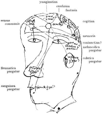
Fig. 15. From K. Peyligk’s Philosophiae naturalis compendium, Leipzig, 1489. Illustrating the general ideas on anatomy current at the Renaissance.
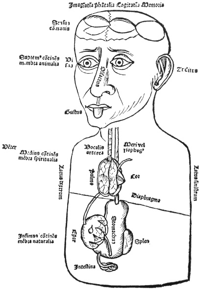
Fig. 16. The cerebral ventricles from above and from the side. According to K. Peyligk, Philosophiae naturalis compendium, Leipzig, 1489.
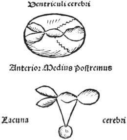
Fig. 17. The localization of cerebral functions. From the Italian edition of ‘Ketham’, Fasciculus Medicinae, Venice, 1493.
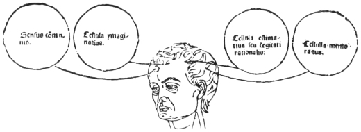
Fig. 18. From G. Reisch, Margarita philosophiae, Leipzig,? 1503. Diagram of the ventricles and the senses with their relation to the intellectual processes according to the doctrine of the Renaissance anatomists.
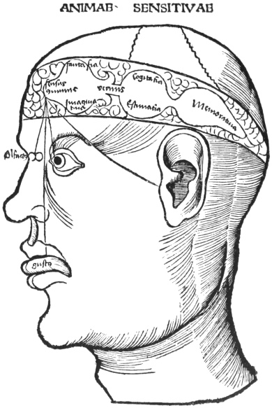
The brain was regarded by mediaeval and early Renaissance anatomists as having two channels of discharge through which the phlegm, the especial product of this organ, could be evacuated when in excess. One of these channels communicated with the anterior ventricle of the brain and poured its secretion into the nose. It may be identified with the anterior colature or cribriform plate. The second, the lacuna, led down from the second ventricle and poured its secretion into the pharynx. It may be identified with the infundibulum, pituitary body, and ‘cella turcica’. The term ‘pituitary’ which we still use is derived from its supposed association with the ‘pituita’ or phlegm. At an early date this process was connected with the four humours (Fig. 14). The rest of the description of the brain can be easily followed. The comparison of the choroid plexus to a worm is very common. The suggestion originated with Galen and was developed by the Arabians.
Comparative Table of Ancient and Modern Nomenclature of Cranial Nerves.
Mondino and Manfredi following Galen, especially in the περὶ χρείας τω̑ν ἐν ἀνθρώπου σώματι μορίων. De usu partium corporis humani.Modern usage.
Not regarded as separate nerves.I.Olfactory nerves.
I.τὰ μαλακὰ νευ̑ρα τω̑ν ὀφθαλμω̑ν.II.Optic nerves.
II.τὰ κινητικὰ τω̑ν ἀμφ᾽ αὐτοὺς μυω̑ν.III.Oculomotor nerves.
Not mentioned.IV.Trochlear nerves.
III.τρίτη συζυγία.V.Trigeminal nerves.

IV.τετάρτη συζυγία.
Mondino and Manfredi confuse Galen’s fourth pair and Galen’s sixth pair.
Not mentioned by Manfredi. By Galen probably united with II.VI.Abducent nerves.
V.πέμπτη συζυγία.VII.Facial nerves.

VIII.Auditory nerves.
VI.ἕκτη συζυγία.IX.Glossopharyngeal nerves.

X.Vagi.
XI.Accessory nerves of Willis.
VII.ἑβδόμη συζυγία.XII.Hypoglossal nerves.
The nomenclature of the cranial nerves adopted by Manfredi is taken from Mondino and is almost identical with that of Galen, whose classification is summarized above.194 Manfredi’s description of Galen’s fourth pair is confused and inadequate, but his account of Galen’s sixth pair is an improvement upon Mondino.
The ‘rete mirabile’ is an interesting survival of Galenic anatomy. This structure is hardly present in man, but is developed in the lower animals, and especially in calves, upon whose bodies Galen worked. The father of physiology regarded the ‘rete mirabile’ as the place where the psychic pneuma was elaborated.195 Galen’s findings in the lower animals were assiduously transferred to the human body, to which his descriptions are much less applicable, while his views on the pneuma lasted in more or less misunderstood form well into the seventeenth century.
(b) THE EYE
Tractate i, Chapter 3
(folio 11 recto) The socket of the eye is not over-depressed, for it has to receive the images (spetie) of visible things. Nor does it project greatly, lest it should be liable to injury from exterior violence. For the eyes of man being very soft and susceptible, nature provided eyebrows as a shield above, and eyelids as protectors in front, and made moreover the projections of the maxillae and the nose, so that the eyes should be guarded on every side. So great was the solicitude of nature for these members.
Seven are the tunics of the eye and three its humours. Three front coatings join with three coatings at the back like six shields, the edges of every pair joining each to each, the outer being larger and containing the others. The seventh tunic is largest of all, and encloses the whole eye, and therefore it is called conjunctiva because it joins and surrounds the whole eye except the place where the pupil is, and that small part [is covered] by the cornea. Now this first tunic where it covers the outside part is seen to be white.
The second tunic in its front part is called cornea because it resembles horn in its substance and colour; and this covering is transparent, so that the images of visible things may penetrate through it. And it is also solid and large and composed of four membranes, so that being near external things it should not receive hurt. With this [corneal tunic] is united posteriorly another tunic [the third] called sclerotic, i.e. hard. These two coverings have their origin in the membrane about the brain, that is in the dura mater, just as the first tunic arises from the membrane over the skull, called almochatim.
The fourth tunic as to its front part is called uvea [because] it is like a seed of a black grape, and in its midst is a hole called the pupil. Nature made this tunic opaque so that the visual spirit should be conserved and not dissipated by the light outside. Moreover nature made the opening in the tunic that the image might penetrate freely; while it is narrow, so that the visual spirit should be concentrated. Thus when the said pupil, or rather hole, dilates more than usual, either naturally or accidentally, the sight becomes imperfect. [The uveal tunic] joins posteriorly the fifth tunic, called secundina because it is made like the after-birth, i.e. the membrane in which the child is enveloped in its mother’s womb, and it arises from the pia mater.
The sixth coating in front is called arachnoid because it is formed after the manner of a spider’s web, and posteriorly it joins the seventh coating, called retina, because it is made like a net.
Between the uvea and the arachnoid anteriorly there is a humour called albugineus, like the white of an egg, to moisten the eye and to preserve the convexity of the cornea. In a dead man this humour dries up, and the cornea falls and is flattened, and then the vulgar say that there appears a curtain before the eyes which is an infallible sign of death. Also this humour holds the pupil open; therefore when it dries up the pupil contracts.
Between the two last tunics, i.e. the arachnoid and the retina, which have their origin from the optic nerve, there are two humours. These are the vitreous humour, so called from its likeness to liquified glass, and the crystalline humour, from its likeness to a crystal. This is also called the grandid, because it is like a hailstone; and it is somewhat hard and round, but flattened anteriorly where it receives the images of visible things, and posteriorly pyramidal shape and pointed. And here is completed the act of seeing. In the posterior part it is surrounded by the vitreous humours by which it is nourished. The crystalline humour is convex anteriorly and the vitreous posteriorly. And the optic nerves come to the eyes and convey the images seen by the eyes to [the seat of] common sensation and to the other internal faculties. [Folio 12 verso, line 7.]
Fig. 19. THE ANATOMY OF THE EYE From G. Reisch, Margarita philosophiae, Leipzig,? 1503. Showing the seven tunics and three humours of the eye according to the doctrines of Renaissance anatomists.196
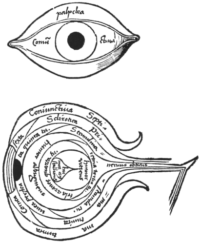
A great deal of attention was paid by the Arabians to the diseases and the structure of the eye, and the essentials of Manfredi’s description are to be found in Rhazes, Hunain ben Ishak, and Haly Abbas. The tradition presented by these writers passed early into Western science, and is reproduced, for example, in the works of Constantine Africanus and in the well-known anatomy to which the name of Richardus Anglicus (Richard of Wendover) has become attached197 (cp. Fig. 19). Avicenna’s description of the eye is somewhat different, and gave rise to the tradition reproduced in the works of John of Peckham and of Roger Bacon (Plate XXXVIII a), and it influenced the views of Leonardo and even perhaps of Vesalius (Fig. 20). The views on the anatomy of the eye expressed by Rhazes, Hunain ben Ishak, and Haly Abbas were, on the whole, more widely accepted than those of Avicenna.
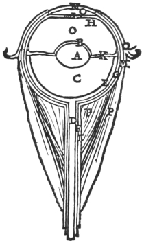
Fig. 20. THE ANATOMY OF THE EYE
From Vesalius, De humani corporis fabrica, Basel, 1543, p. 643. A, Crystalline humour; O, Albugineous humour; C, Vitreous humour; N, Cornea; Q, Conjunctiva; M, Sclerotica; G, Secundina; H, Uvea; K, Arachnoidea; E, Retina.
The treatment of the eye was always felt to be hardly within the range of the ordinary practitioner of surgery, and its structure, as we learn from Guy de Chauliac,198 was not usually treated in the general course of anatomy. The custom was rather to refer the student to special works such as those of Jesu Aly or of Alcoatim.
Manfredi’s description of the anatomy of the eye is that generally accepted at the end of the fifteenth and the beginning of the sixteenth centuries, and is unusually clear for its date. It represents a considerable advance on such writers as Henri de Mondeville (1260–1320)199 or the pseudo Richardus Anglicus, and is far superior to the descriptions of the eye dating from the fourteenth and fifteenth centuries recently brought to light by Sudhoff.200 We reproduce as illustrating Manfredi a diagram taken from the Margarita philosophica of Gregorius Reisch (died 1525). This represents the earliest printed figure of any value of the anatomy of the eye (Fig. 19).201 We give for comparison the figure from a thirteenth-century MS. of Roger Bacon (Plate XXXVIII a), representing the rival tradition of Avicenna and Alhazen that influenced Leonardo da Vinci and other contemporaries of Manfredi. These figures may be compared with that of Vesalius (1543, Fig. 20), whose description of the eye is less free from traditional bias than are most parts of his epoch-making work.
In reading any early description of the eye, it is to be remembered that until the nineteenth century the ‘emanation theory’ prevailed. Light was regarded as of the nature of a stream of particles emitted from the object seen, and the act of vision was considered as a collision of this emanation with an emission of something from the eye itself, called in mediaeval writings the ‘visual spirit’.
(c) THE HEART
Tractate ii, Chapter 3
(folio 19 verso) Then you will see in the midst of the lung the heart, covered by its membranes. [It is thus situated] that the air attracted by this lung should cool it, and that thus the heat and spirit of the heart be tempered. This member is the most important of the four [principal members], because it is the first to live and the last to die. It is of medium size compared with the other members of man, but compared with the hearts of other animals it is very large, because man, in a quantitative and not an intensive sense, has more natural heat than other animals. It is pyramidal, that is in the form of a flame; because it is of excellent warmth, therefore it is necessary that it should be of a shape resembling a flame. Its figure is also called ‘pine-shaped’, because it is wide below and narrow above, being thus formed that distinction could better be made between its cavities or ventricles; moreover, had it been made of a shape all uniform as is the lower part, it would be too heavy and ponderous.
This member is situated in the middle of the entire body, measured in every direction; that is, in the middle between the upper and lower parts: in the middle also between front and back and right and left, like a king standing in the midst of his kingdom, and this was done that it might give the strength of life equally to all the members; and although the heart as regards its foundation and base be in the middle, yet its point declines to the left below the left breast, so that it warms the left side as the liver warms the right.
This member is sustained and strengthened by a certain cartilaginous bone. For since it is continually moving, it needs some point of purchase to support it in its movements. Moreover, it has a certain fatty layer on the outside which prevents the heart from drying and keeps it moist: and there are certain veins and arteries dispersed through its substance: and it is formed also of a kind of hard flesh so that it may sustain many and forceful movements; also it is formed of longitudinal, latitudinal, and transverse fibres, so that it may have the power to attract, retain, and expel.
This member has three ventricles or chambers, like the brain. One ventricle is on the right side, the second on the left, and the third in between. The right ventricle towards the liver has two orifices. One is towards the liver and is very large. Into this there enters a vein called vena chilis, which arises in the convexity of the liver and brings the blood from the liver to the heart. In that right ventricle the blood is purified, and then sent by the heart to all the other members.
Now since the heart attracts by this orifice of the vena chilis more than it expels, therefore nature ordains that in the moment of contraction when the blood is expelled this orifice closes, and when the heart dilates it opens.
Moreover there are three little valves (hostiolitti) or doors opening from without inward, and these valves are not very depressed; so that by this same orifice only part of the purified blood is expelled to the other members, because part goes to the lungs and the remainder forms the vital spirit; therefore nature ordains that these valves do not entirely close. From the vena chilis, before it enters the cavity of the heart, there arises another vein, which surrounds the root of the heart; and from it are given off branches which disperse themselves through the substance of the heart, and from the blood of that vein the heart nourishes itself.
The right ventricle towards the lung has another orifice into which opens the arterial vein, bringing the blood from the heart to nourish the lung: in this orifice also are three valves (hostioli) opening from within outward and closing from without inward, in the opposite way to the valves of the other orifice; and this is so that they should entirely close. Hence by this orifice the heart during the period of contraction can expel, and yet during the period of its dilatation cannot attract anything through it as was done in the first orifice.
The left ventricle of the heart has its sides denser and thicker than the sides or walls of the right ventricle; and this for three reasons: Firstly, because in the right ventricle is contained the blood, which is heavy, while in the left ventricle there is spirit, which is very light; therefore in order that the heart should not be heavier and more ponderous on one side than on the other, it was necessary to compensate in this manner, that is, that the left ventricle should be thicker in its walls than the right. In the second place, the spirit being more subtil and more volatile (resolubile) than blood, it needs a stronger habitation and better supports. Thirdly, the left ventricle is much warmer than the right, because in it is generated the spirit from the blood, by a great heat which makes that blood more subtil; and heat is better preserved in a substance that is dense and thick.
In the cavity of this ventricle near its root are two orifices: one is the orifice of an artery called artharia adorti [= aorta], because it has immediate origin in the heart and because it is the source of all the others: by this artery the heart sends the generated spirit to all the members; and the very subtil blood is mixed with the spirit when the heart contracts. For which reason there are at the entrance of this orifice three valves, which close entirely from the outside inwards; and they open from the inside outwards, and this orifice is very deep.
The other orifice is that of the venal artery which conveys the air from the lung to cool the heart and transports warm vapours from the heart to the lung as has been said above; and in this orifice are two valves which do not entirely close: and they are well raised so that they can better apply themselves to the sides [edges] of the heart when it sends out the spirit: these are marvellous works of nature, as is also the central ventricle of the heart, for this ventricle has not one cavity but many; these are small but wide, and more numerous on the right than on the left; and nature contrived thus, so that the blood which goes from the right ventricle to the left to be converted continually into spirit becomes thin in these cavities.
And by this thou canst see that four things have birth in the heart. The first is the artery called adorti, the second is the vena chilis, the third is the arterial vein, and the fourth the venal artery.
Also thou wilt see in the heart certain membranous parts like auricles, or rather like small ears, able to dilate or contract: these are contrived by nature in order that when overmuch blood or spirit is generated the heart can dilate so as to contain it; and also that the heart may contract when there is no such abundance.
And it is here that Galen asks, Why did not nature make the heart so large that it could contain every increase of blood or spirit without the addition of these membranes? Galen replies that this was first because the heart would have been too large and therefore too heavy; secondly, because as it is not always generating a great quantity of blood and spirit, if the heart had been too large, its cavity would usually have been empty: but these auricles dilate with the accumulation of blood or spirit, and contract with its decrease.
The heart is surrounded by a firm and nervous membrane, like a little house in which it is placed as in a tabernacle to defend it from accidents. This capsule is very dilated, that the heart in its dilations and movement may not be impeded thereby, and therefore nature made this capsule so that it should contain a certain dewy moisture with which the heart is bathed and moistened so that in its continual movement it should not become dry. For when this water be dried up, then the heart itself is desiccated, and emaciates and dries up all the body.
The description of the heart follows Mondino closely. Occasionally a phrase or two is reminiscent of Mondeville. The trite conception of the heart as a king in its necessarily central position was very frequently repeated by writers in the Middle Ages. To Harvey, who had a certain mediaeval element in his mentality, it seems to have appealed, and he used it in his Prelectiones Anatomiae,202 and chose it to introduce his great work on the circulation of the blood.203 The heart was similarly described as ‘flame-shaped’, because it was regarded as the source of animal heat. The idea that it is the first to live and the last to die comes from Aristotle.204 The bone in the heart also comes from Aristotle.205 The idea was quite familiar to mediaeval anatomists, who frequently endeavoured to identify the bone with the firm tissue around the orifices of the aorta and pulmonary artery. The reader may be reminded that a true ‘os cordis’ is in fact to be found in some mammalia.
Mondino, followed by Manfredi, describes the action of the heart and blood-vessels mainly according to the views of Galen, but without any very clear or connected statement. The ‘third ventricle’ especially has its origin in a misunderstanding.
This mythical structure is an attempt to combine the views of Aristotle and of Galen. Aristotle, who probably never dissected a human body, derived his anatomical conceptions largely from cold-blooded animals, in some of which the heart is provided with three cavities. He considered that the heart had three chambers, the largest being on the right, the smallest on the left, and one of intermediate size between the two. As far as they can be identified, the largest was the right ventricle plus the right auricle, the smallest or left chamber was the left auricle, while the intermediate cavity appears to have been the left ventricle.206
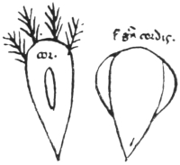
Fig. 21. THE HEART From the Roncioni MS. (Pisa 99) after Sudhoff.
Galen’s description differed altogether from that of Aristotle. He tells us expressly and somewhat contemptuously that ‘it is no marvel if Aristotle erred in many anatomical matters, a man who thought forsooth that the heart in the larger animals had three chambers’.207 Galen always describes the heart as having but two chambers, the right and left ventricles, a wholly subordinate part being assigned to the auricles. These latter were regarded as safety-valves, expanding to hold superfluous blood when the chambers of the heart to which they correspond become overfilled.
No third ventricle is described by Rhazes or Haly Abbas,208 but Avicenna, in his Canon, makes an effort to combine the views of Aristotle and Galen. Speaking of the anatomy of the heart (lib. iii, fen. xi, chap. 1) he describes the ventricular portion as follows: ‘In the heart are three cavities, two large, and a third as it were central in position. So that the heart has [a] a receptacle [the right ventricle] for the nutriment with which it nourishes itself—this nutriment is thick and firm like the substance of the heart; [b] a place where the pneuma is formed [the left ventricle], being engendered of the subtil blood; and [c], thirdly, a canal between the two.’209 A somewhat similar account is given in Constantine’s translation of Isaac.210 The idea soon crept into European medicine, for in a Pisan MS. dating from the first half of the thirteenth century211 a crude figure of a three-chambered heart is to be found (Fig. 21).
The first translator of the Canon of Avicenna, Gerard of Cremona, whose work appeared towards the end of the twelfth century, improved on his original. ‘In it [the heart] are three ventricles; two are large, and the third as it were between, which Galen called the fovea or non-ventricular meatus, so that there may be a receptaculum for the thick and strong nourishment, like to the substance of the heart, with which it is nourished, and also a storehouse for the pneuma (spiritus) generated in it from the subtil blood. And between the two are channels or meatuses.’212 Henri de Mondeville (died about 1320), by going direct to Galen, avoided some of the errors of Avicenna, with whom, however, he still describes three ventricles.213 Mondino does little but copy the Arabian, whom Manfredi also follows.
We may terminate our description of the mythical third ventricle by quoting from Bartholomew the Englishman. His encyclopaedia written about 1260 was translated into English in 1397, and printed by Thomas Berthelet214 in the 27th year of the reign of Henry VIII (1535), when Bartholomew’s work was still extremely popular. Berthelet’s rendering runs as follows:
‘And the hert hath ij holownesses, one in the left syde, that cometh sharpe: and one in the ryght side, that is within: And these two holownesses ben called the wombes of the hart. And betwene these two wombes is one hole, that some men call a veyne, other an holowe way. And this hole is brode afore the ryghte syde, and streyte afore the left syde. And that is nedefulle to make the bloode subtyll, that commeth from the ryght wombe to the lefte, and so the spirite of lyfe may be bredde the easelyer in the lefte wombe.’
In order to understand why all these authors invoked the existence of the third ventricle, regarded by some of them as a passage between the other two, we must turn to the physiological beliefs of the age. It must be recalled that before the demonstration of the circulatory movement of the blood a certain amount of communication was believed to exist between right and left ventricles. The complicated nature of the ventricular cavities and the intricacy of the columnae carneae promoted the idea of the presence of minute passages in the interventricular septum. Even so astute an observer as Leonardo da Vinci considered that ‘the ventricles are separated by a porous wall, through which the blood of the right ventricle penetrates into the left ventricle, and when the right ventricle shuts, the left opens and draws in the blood which the right one gives forth’ (Plate XXXVIII b).215
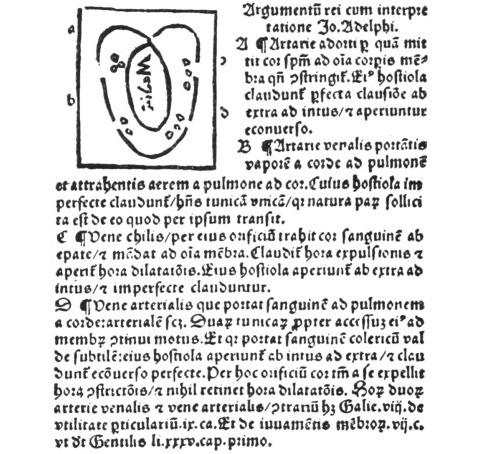
Fig. 22. From Johannes Adelphus, Mundini de omnibus humani corporis interioribus menbris Anathomia, Strassburg, 1513. The diagram shows the two lateral ventricles and the ‘central’ ventricle. By a printer’s error the letters c and d are transposed. The arteria adorti is the aorta, the arteria venalis the pulmonary vein, the vena chilis the vena cava, and the vena arterialis the pulmonary artery. The auricles are ignored, as is frequently the case in works of the period, and the pulmonary veins are represented as opening directly into the ventricles.
Although the third ventricle is described in all the twenty-five editions of Mondino, many of which are illustrated, they present no drawing of it except the wretched little diagram of J. A. Muelich (Johannes Adelphus) in 1513, which we here reproduce (Fig. 22). The confusion, however, to which the idea of a third ventricle gave rise influenced anatomy almost as late as the seventeenth century, and is illustrated in the anatomical figures of a late edition of Hans von Gersdorff (1556),216 where the trachea is actually shown opening into the left ventricle (Fig. 23). It was Vesalius who took the first great step towards the discovery of the circulation of the blood, by firmly maintaining that the interventricular septum was solid and contained neither passages nor intermediate ventricle.217
Fig. 23. From Hans von Gersdorff, Feldt und Stattbüch bewerter Wundartznei, Frankfurt, 1556. The trachea (d) is represented as opening directly into the heart.
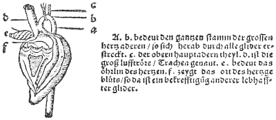
VI. Italian Text
MS. Canonici Ital. 237
Hyeronimi manfredi ad Magnificum & potentem dominum ac militem Iohannem Bentiuolum insequens opus de corporis humani anothomia exordium.
[folio 1 verso] Opportet de sapientia admirari creatoris ut XVº de utilitate particularum scribitur a Galieno. Cum enim membrorum nostri corporis admirabilem Galienus aspiceret Armoniam predictum sermonem explicauit: ut nos ad dei sublimis et gloriosi admiranda opera commoueret: Quamuis nostra cognitio a dei compraehensione deficiat: unde et Seneca XLª epistola ad Lucillum ait quid deus sit incertum est habitat in nobis: Sed deum mouemur inuocare eius sapientiam mirabiliter contemplantes. Quanta enim fuerit summi opificis in producendo res sapientia quanta eius solicitudo et prudentia opera profecto nature declarant: unde et psalmista mirabilia sunt opera tua deus, et alibi celi enarrant gloriam dei et opera manuum eius annuntiat firmamentum. Quis enim talia et tanta inspitiens creatorem suum abneget et eius potentiam? Inscipiens quidem erit hic iuxta illud psalmiste dixit inscipiens in corde suo non est deus. Sublimis autem dei multiplitia et diuersa fuere opera. Creauit enim duplitia entium genera scilicet corruptibilia et incorruptibilia; et in utrisque suam admirabilem sapientiam, suamque [folio 2 recto] infinitam potentiam ostendit. Totam enim entis latitudinem nihil prorsus de spetiebus, quas ab aeterno in mente sua retinuit obmittens perfulciuit, et eas quas ab aeterno in sua habebat essentia ad aliud esse procreauit, ut in indiuiduis esse haberent: quae in suae maiestatis lumine existebant: et uniuscuiusque spetiei modo perfecit ac uarietates per esse quod in singularibus habent (natura mediante & cum lege) imposuit. Admirantur angelorum caetus obstupent hominum intellectus tantae maiestatis opera mirabilia: ut hoc summo bono: hoc perfectissimo ente nihil melius excogitari possit. O admirabilem maiestatem, O deitatem incompraehensibilem, O inefabilem potentiam: Quis te negliget? Quis te non insequetur? Quis in operibus tuis non delectabitur?
Omnis igitur qui in operum dei gloriosi intuitu delectatur, hic prudens et non inscipiens est: hic dignus homo: hic intellectu non caret. Cum igitur tua illustris Dominatio Iohannes bentiuole magnanimis praesenti anno ex sui qua solet humanitate ad cuiusdam hominis defuncti anothomiam uno semel uidere non fuerit dedignata ob sui intellectus dignitatem qui semper alta intelligere concupiscit, cumque tu opera tam naturae miranda in anothomizato incaepisti uidere corpore tunc haec intelligendi creuit animus tua digna [folio 2 verso] creuit uoluntas: Et me hyeronimum Manfredum ad hoc opus de anothomia intitulatum materno sermone tuo dignissimo nomini inscribere concitasti: (ut omnino sicut debeor) rem gratam tuae faciam dominationi: In hoc enim tui agnoui dignitatem intellectus, tui ingenii solertiam quod in rebus naturae mirandis tuum peruoluas intellectum. Hoc enim opusculum quantum melius potui ex uariis antiquorum uoluminibus exserpsi ac id abreuiaui: nec eumdem forte tenui ordinem ut illi: et ipsum materno composui sermone ut opus hoc delectabilius tuae sit magnificentie.
Accipe igitur magnifice et potens domine hoc opus de corporis humani anothomia tuo dignisimo nomini intitulatum, ea benignitate et humanitate, qua soles: et animo illari ac gratioso id accepta: qui satis tibi erit delectabile et perplacebit quia dignum est opus: Vale miles magnanimis, et solito ama.
Finis prohemii.
[Here a folio is missing.]
[folio 3 recto] a li nerui lequale hano origine da le extremita di musculi: Unde e da sapere che li musculi sono compositi de nerui, corde, e ligamenti e carne facti da la natura a dare el moto uoluntario, Impero da le soe extrimita escono queste tale corde e uadono a membri che se debano mouere: e quando se retraheno li dicti musculi consequenter se se retraheno le lor corde: & finaliter i membri: et similiter quando se dilatano i musculi se dilatano etiam le corde & consequenter i membri.
Li ligamenti sono etiam simili a nerui facti a ligare le iuncture de le osse e non li dette la natura sentimento como fece a li nerui & a le corde acio che per el molto mouimento e fricatione de le iuncture non doleseno.
Le Artarie sono de substantia neruosa & ligamentale in longo extense o concaue: ne le quale se contene el sangue sutilissimo & depurato et el spirito uitale el quale e mandato dal core a dare uita a tuti i membri: et hano origine da esso core: & impero hebeno doe tuniche acio chel sangue sutile & el spirito uitale non usisseno fuora.
Le vene sono simile a lartarie ma sono quiete e non se moueno, ma hano origine dal figato et in esse se contene el sangue grosso cum li altri humuri che non e cusi depurato ne [folio 3 verso] cusi sutile como e el sangue de le artarie: impero non li fece senon una tunicha: per che quelo sangue non era cosi sutile chel potesse penetrare fuora ne anche non bisognandose mouere non era suspitione de rompersi como ne le artarie che era neccessario a mouerse per refrigerare el core atrahendo laiere frigido & expellendo fuora li fumi caldi da esso.
Li panniculi sono composti e texuti de fili neruosi sutilissimi che non se posseno uedere e sono questi paniculi spissi e sutili e sono de molte manerie: Alcuni forno facti a continere o coprire a Alcuni membri e custodirli ne la sua figura e substantia como sono li paniculi che copreno el cerebro e molti altri di li quali poi diremo: Alcuni altri panniculi sono facti a suspendere uno membro a laltro como li rognoni sono aligati a laschina mediante uno certo paniculo: Alcuni altri paniculi sono facti acio che alcuni membri che non hano sentimento recceuano qualche sentimento per el panniculo: nel quale sono inuolti como sono el pulmone el figato la milza & i rognoni li quali sono priuati de sentimento impero la natura aciascuno di loro li fece uno panniculo doue fusseuo inuolti per la casone dicta.
Da poi tuti questi membra hauendo la natura ordito el corpo de lhomo de [folio 4 recto] li predicti bisogno reimpire le uacuita e reimpille de carne: Fece aduncha la natura la carne per reimpire le uacuita che rimangono da lorditura de nerui uene & altri membri dicti.
Praeterea e da sapere che la natura ha dato aciascuno di li predicti membri quatro uirtu. Una e uirtu atratiua per laquale ha ad atrahere el nutrimento suo a se del quale el membro se ha a nutricare: La seconda uirtu e digegtiua per laquale el nutrimento atrato se digerisse & conuertese ne la sustantia del membro: La terza uirtu si e retentiua per laquale el nutrimento atrato se retiene debito tempo acio che la uirtu digestiua possa perficere la sua operatione circha quello: La quarta uirtu e expulsiua laquale ha expellere le superfluita che se generano dal nutrimento ne la digestione.
Anche e da sapere che la natura nel corpo de lhuomo ha facto quatro membri principali como quatro signori et aciascuno di loro li ha dato una casa o uero uno palazo a sua custodia doue habite cum certe camare o uero stantie che hano aseruirli al suo bisogno: El primo membro principale e signore e el cerebro al quale li fece la natura el capo cum le sue circumstantie per suo habitaculo e dette a questo membro che lui fusse principio e radice de tuto el sentimento e moto de tuto el [folio 4 verso] corpo: dal quale tuti li altri membri recceueno el sentire: e el mouere, & a questo membri li dette etiam cinque uirtu cognoscitiue exteriore cio e li cinque sentimenti e cinque altre uirtu cognoscitiue interiore che deserueno a lo intellecto.
El secondo membro principale e signore si e el Core alquale la natura ha dato la sua casa cio e el pecto cum le sue adiacentie: et aquesto membro li ha dato la uirtu de la uita dal quale proceda la uita in tuti li altri membri como da uno primo principio.
El terzo membro principale e signore e el Figato alquale dette la natura per suo domicilio el uentre inferiore cum li altri membri circumstanti che sono neccessarii a la sua operatione e dette a questo membro la uirtu nutritiua chel fusse principio e radice del nutricare de tuti li membri.
El quarto membro principale fu li testiculi e la sua casa e la bursa laquale li contene et aquilli deserueno piu altri membri como poi se uedera et a questi testiculi ha dato la natura la uirtu generatiua cio e de generare el sperma o uero seme el quale habia una uirtu generatiua che possa produre una cosa simile a colui dal quale se decide tale sperma: et questo fu facto per conseruare lhuomo in spetie non se possendo conseruare in indiuiduo.
Ultra questi quatro membri principali e suoi domicilii [folio 5 recto] ha facto la natura alcuni altri membri cio e el collo cum la gola che fusse uia e transito dal primo membro principale cio e cerebro ali altri membri principali et etiam a tute laltre parte & per altre utilita quale noi da poi diremo.
Item ha facto la natura le braza e le mane che hauesseno a pigliare el cibo e mandarlo al luoco conueniente et etiam per che lhuomo solo uiue per arte lequale non se possono perficere senza le braza e mano.
Item fece le cosse, gambe e piedi acio se potesse mouere da luocho a luocho secondo li soi bisogni.
Noi aduncha poneremo la Anothomia de tuti li membri e parte dicte: Comenciando per ordine dal cerebro e da la sua casa et consequenter descendendo per insino apiedi.
Capitulum secundum, tractatus primi de anothomia capitis et omnium contentorum in eo.
Fece la natura el capo ossuoso per magiore tutela del cerebro: el quale essendo inmobile non li bisogno hauere musculi: Et per che el cerebro ne lhuomo e magiore che ne li altri animali secondo la sua grandeza impero bisogno chel capo de lhuomo fusse etiamdio grande per rispecto de li altri animali: Et etiam bisogno li meati del capo ne lhuomo essere piu distincti essendo piu dedito al cognoscere.
La figura [folio 5 verso] del capo naturale e rotonda compressa da dui canti como sel fusse una cera rotonda compressa cum le mano da la parte drita e da la stancha faria doe eminentie una dinanzi e laltra de drieto e la parte drita e stancha rimaneriano piane: Bisogno fusse rotondo acio fusse pin capace et etiam che fusse piu securo e risguardato da nocumenti exteriori a li quali e molto exposito: Bisogno etiam essere facto cum quelle eminentie acio che li meati del cerebro hauesseno megliore distinctione et acio che li cinque sentimenti exteriori hauesseno origine da la eminentia anteriore.
Diece sono le parte del capo: La prima e li capilli quasi capitis pili facti da la natura a magiore tutela del capo da le cose exteriore et etiam per belleza: La seconda parte del capo e la cute la quale bisogno essere molto grossa acio che li capilli fusseno ben firmi hauendo le radice sue molte grosse e longhe et etiam che fusse megliore scuto e cooperimento de losso et del cerebro non li essendo parte musculose: La terza parte si e la carne laquale solo e ne la fronte e ne le tempie e circha le masselle e non in le altre parte: La quarta parte e uno panniculo exteriore chiamato almochatim elquale appare in continenti como e liuata su la cute e copre tuto losso del craneo de fuora: Et fece la natura questo panniculo [folio 6 recto] acio che lacute che e molle non tochasse incontinenti losso che e duro: Et etiam acio che losso del capo hauesse sentimento per questo panniculo: Et tertio anche acio che el paniculo interiore del capo chiamato Duramater mediante questo panniculo stesse suspeso a losso del craneo cum certi nerui e ligamenti che escono per le comissure del dicto osso et hano origine dal dicto panniculo interiore & uscendo fuora de losso texono o uero componeno quello panniculo exteriore dicto Almochatim: La quinta parte e el craneo cioe osso facto como uno capello nela concauita del quale glie locato el cerebro: & in questo craneo furno quatro ossa cusite insieme e la natura non fece questo osso uno ma de piu pezi acio che achadando nocumento in una parte non comunicasse a laltre parte: Et etiam acio che per quelle comissure o uero cusiture potesseno meglio exhalare fuora le fumusitade dal cerebro: Et tertio acio che bisognando la uirtu de le medicine applicate potesseno meglio penetrare ale parte dentro quisti aduncha quatro pezi de osso furno da la natura cusiti et insieme ionti in modo de denti acio fusse piu fermi e forti et non furno facti in modo che se potesseno uincare como fano le iunture per che non seriano state cusi forte: et etiam [folio 6 verso] per che non bisognaua a losso del capo mouerse: Et queste comissure sono cinque cio e tre uere e doe mendose: Le comissure uere sono quelle che passano tuto losso et le mendose non passano: De le uere comissure una si e ne la parte anteriore chiamata coronale et e facta a modo de uno C e protende da la parte drita a la stancha del capo et ha li branchi uerso la fronte. La secunda comissura uera si protende per la longheza del capo comencianda da la comissura coronale ala parte posteriore como una friza o uero sagitta che uene da larcho, impero e chiamata sagitale ——(. La terza comissura e ne la parte posteriore chiamata laudale facta a modo de uno A, per abacho chiamato dal greco lauda: e la comissura sagittale protende da la coronale a la laudale 〉——(.
Le comissure mendose sono due da ciascaduno lato una cio e dal drito e dal stanco e sono dicte corticate per che non passano.
Et se noi consideremo per queste cinque comissure hauemo quatro ossi cusiti insieme: Uno si e losso de la fronte che comenza dala comissura coronale e termina uerso la parte inferiore a una altra comissura la quale comenza da uno brancho de la comissura coronale e procede a presso le ciglie de li ochii a laltro brancho Ɑ. Laltro osso si e de drieto el [folio 7 recto] quale se termina a la comissura laudale e dui altri ossi da le tempie che se terminano da le comissure mendose le quale comenzano da la comissura laudale a la comissura coronale.
La sexta parte sono doi paniculi uno chiamato Dura mater el quale e in continenti de poi el craneo: e laltro se chiama pia mater el quale incontinente copre el cerebro e questo fece la natura hauendo grande solicitudine di questo membro acio che in continenti non fusse tocho da losso ma processe per piu mezi che uno fusse piu duro che laltro: Et anche fece dui panniculi acio che se la cadesse nocumento in uno de loro non comunicasse al cerebro in continente. Ne la pia matre sono texute certe uene per le quale se nutrisse el cerebro e si lo copre per tuto excepto la parte posteriore per che essendo quella parte sicca non bisogno di questo paniculo como la parte anteriore e meza. Questi dui panniculi in piu luochi penetrano la sustantia del cerebro et se lo diuide in parte drita e parte sinistra et in parte anteriore & parte posteriore: et per queste tale diuisione furno fabrichate nel capo diuerse celule o uero camerette ne le quale produce lanima diuerse operatione per che bisognaua che queste tale parte fuseno de diuerse complexione.