Front matter (continued 2)
Reading edition
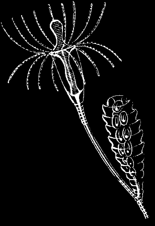
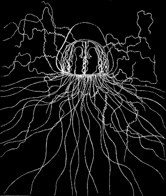
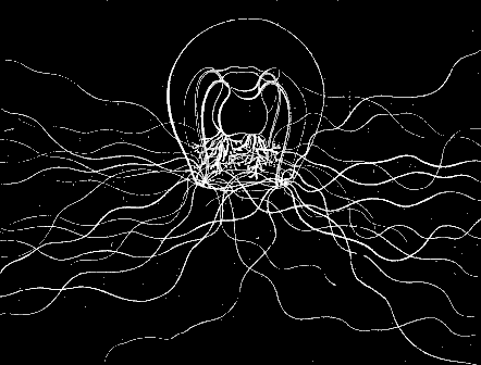
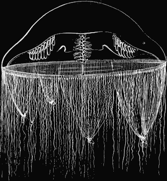
Circe. (Trachynema digitale A. Ag.)
[fig 53]
Fig. 53. Trachynema digitale; about twice the natural size.
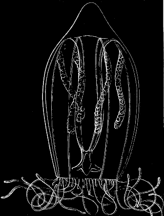
Among the Jelly-fishes, the position of which is somewhat doubtful, is the Circe (Fig. 53), differing greatly in outline from the ordinary Jelly-fishes. As may be seen in Figure 53, the bell forms but a small portion of the animal; it rises in a sharp cone on the summit, thinning out at the lower edge, to form the large cavity in which hangs the long proboscis and the eight ovaries, four of which may be seen in Fig. 53 crowded with eggs. The Circe differs in consistency, as well as in form, from other Jelly-fishes. It is hard and horny to the touch, and the veil, usually so light and filmy, is here a thick folded membrane, which at every stroke of the animal forces the water in and out of the cavity. It is very active, moving by powerful jerks, each one of which throws it far on its way. It advances usually in straight lines; or, if it wishes to change its direction, it drives the water out of the veil suddenly on one side or the other, so as to shoot off, sometimes at right angles with its former path. Four large pedunculated eyes, hidden in the figure by the tentacles, stand out prominently from the circular tube. When the animal is in motion, the tentacles are carried closely curled around the edge of the disk, as in Fig. 53, where the Circe is represented under a magnifying power of two and a half diameters. This Jelly-fish is of a delicate rose color, the tentacles assuming, however, a dark-purple tint at their extremities when contracted.
Lucernaria. (Haliclystus auricula Clark.)
[fig 54]
[fig 55]
Fig. 54. Group of Lucernaria attached to eel-grass; natural size. Fig. 55. Lucernaria seen from the mouth side.
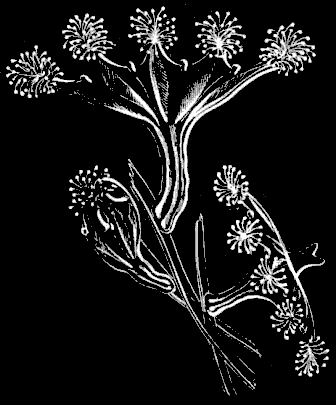
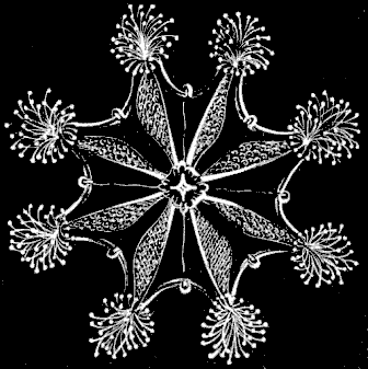
One of the prettiest and most graceful, as well as one of the most common of our Jelly-fishes, is the Lucernaria (Fig. 54). It has such an extraordinary contractility of all its parts, that it is not easy to describe it under any definite form or position, since both are constantly changing; but perhaps of all its various attitudes and outlines none are more normal to it than those given in Fig. 54. It frequently raises itself in the upright position represented here by the individual highest on the stem, spreading itself in the form of a perfectly symmetrical cup or vase, the margin of which is indented by a succession of inverted scallops, the point of junction between every two scallops being crowned by a tuft of tentacles. But watch it for a while, and the sides of this vase turn backward, spreading completely open, till they present the whole inner surface, with the edges even curved a little downward, drooping slightly, and the proboscis rising in the centre. In such an attitude one may trace with ease the shape of the mouth, the lobes surrounding it, as well as the tubes and cavities radiating from it toward the margin. A touch is, however, sufficient to make it close upon itself, shrinking together in the attitude of the third individual in Fig. 54, or even drawing its tentacles completely in, and contracting all its parts till it looks like a little ball hanging on the stem. These are but a few of its manifold changes, for it may be seen in every phase of expansion and contraction. Let us now look for a moment at the details of its structure. The resemblance to a cup or vase, as in the upper figure of the wood-cut (Fig. 54), is deceptive; for a vase is hollow, whereas the Lucernaria, though so delicate and transparent that its upper surface, when thus stretched, seems like a mere film, is nevertheless a solid gelatinous mass, traversed by certain channels, cavities, and partitions, but otherwise continuous throughout. The peduncle by which it is attached is but an extension of the floor of a gelatinous disk, corresponding to that of any Jelly-fish. Four tubes pass through the whole length of this peduncle, and open into four chambers, dividing the digestive cavity above into as many equal spaces. (Fig. 55.) These spaces are produced by folds in the upper floor of the disk, uniting it to the lower floor at given distances, and forming so many partition-walls, dividing the digestive sac into four distinct cavities. These lines of juncture between the two floors, where the partitions occur, produce the four radiating lines, running from the proboscis to the margin of the disk, on the upper surface. (Fig. 55.) The triangular figures, running from the mouth to each cluster of tentacles, are produced by the ovaries, which consist of distinct pouches or bags attached to the upper surface of the disk, and hanging down into the cavities below; every little dot within these triangular spaces represents such a bag. Each bag is crowded with eggs, which drop into the digestive cavity at the spawning season, and are passed out at the mouth. The tentacles always grow in clusters, but are nevertheless arranged according to a regular order. They are club-shaped at their extremities, but are hollow throughout, opening into the chambers of the digestive cavity, two of the clusters thus being connected with each chamber. Their chief office seems to be to catch the food and convey it to the mouth, though they may also be used as a kind of suckers, and the animal not unfrequently attaches itself by means of these appendages. Between every two clusters of tentacles will be observed a short, single appendage, of an entirely different appearance. These are the so-called auricles, and though so unlike tentacles in the adult animal, when in their earlier stages (Fig. 56) they resemble each other closely. But as their development goes on, the tentacles stretch out into longer, more delicate flexible organs, while the auricles remain short and compact throughout life. They contain a slight pigment spot representing an eye, though how far it serves the purpose of vision remains doubtful. They are chiefly used by the animal as a means of adhering to any surface upon which it may wish to fasten itself; for the Lucernaria, though usually found attached to eel-grass in shoal water, has the power of independent motion, and frequently separates from its resting-place, floating about freely in the water for a while, or attaching itself anew by means of the auricles and tentacles upon some other object. The color of this pretty Acaleph varies from a greenish hue to green, with a faint tinge of red, or to a reddish brown. One of its commonest and most exquisite tints is that of a pale aqua-marine. It may be found along our shores wherever the eel-grass grows, and as far out as this plant extends. It thrives admirably in confinement, and for this reason is especially adapted to the aquarium.
[fig 56]
Fig. 56. Young Lucernaria; magnified.
HYDROIDS.
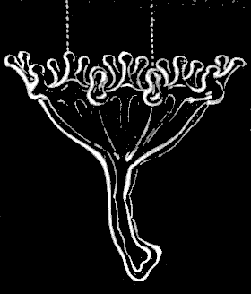
Under this order, the general character of which has already been explained in the introductory chapter on Acalephs, are included a number of groups which, whether as Hydroid communities in their earlier phases of existence, or as free swimming Medusæ in their farther development, challenge our admiration, both for their beauty of form and color, and their grace of motion. Some of them are so minute that they escape the observation of all but those who are laboriously seeking for the hidden treasures of the microscopic world, but the greater number are large enough to be readily found by the most inexperienced collector, when his attention is once drawn to them; and he may easily stock his aquarium with these pretty little communities, and even trace the development of the Jelly-fishes upon them.
To the Hydroids belong the Campanularians, the Sertularians, and the Tubularians. Some examples of each, as represented on our shores, will be found under their different heads, accompanied with full descriptions. There is another group usually considered as distinct from Hydroids, and known as a separate order among Acalephs, under the name of Siphonophoræ, but included with them here in accordance with the views of Vogt, Agassiz, and others, in whose opinion they differ from the ordinary Hydroid communities only in being free and floating, instead of fixed to the ground. Some new facts, published here for the first time, tend to sustain the accuracy of this classification.[5] With these few preliminary remarks to show the connection of the order, let us now look at some of the animals belonging to it more in detail.
Campanularians.
All the Campanularians, of which Oceania (Fig. 68), Clytia (Fig. 73), and Eucope (Fig. 61) form a part, belong among those little shrub-like communities of animals called Hydroids, from which most of our Jelly-fishes are developed. They differ in one essential feature from the Tubularians. (Fig. 93.) The whole stem, from summit to base, is enveloped in a horny sheath, extending around both the fertile and sterile individuals of the community, and forming a network at the base of the stem, which serves as a kind of foundation for the whole stock. To the naked eye such a community looks like a tiny shrub (see Fig. 57), with the branches growing in regular alternation on either side of the stems. The reproductive calycles, i.e. the protecting envelopes covering the young Medusæ, usually arise in the angles of the branches formed by a prolongation of the sheath. These calycles or bells, as they are called, assume a great variety of shapes,—elliptical, round, pear-shaped, or ringed like the Clytia. (Fig. 72.) In one such bell there may be no less than twenty or thirty Medusæ developed one below the other; when ready to hatch, the calycle bursts and allows them to escape.
Eucope. (Eucope diaphana Ag.)
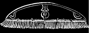
In Figs. 60 and 61 we have a representation of our little Eucope, one of the prettiest of the Jelly-fishes belonging to this group; Fig. 57 represents the Hydroid from which it arises; a single branch with the reproductive bell being magnified in Fig.58. In Fig. 59 is seen a portion of the Jelly-fish disk, with the fringe of tentacles highly magnified. The disk of the Eucope (Fig. 60) looks like a shallow bell, of which the proboscis often seems to form the handle; for the disk has such an extraordinary thinness that it turns inside out with the greatest ease, so that the inner surface may become at any moment the outer one, with the proboscis projecting from it, as in Fig. 60, while the next movement of the animal may reverse its whole position, and the proboscis then hangs down from the inside, as in other Jelly-fishes. (See Fig. 61.)
[fig 57]
[fig 58]
[fig 58]
[fig 60]
[fig 61]
[fig 62]
Fig. 57. Hydrarium of Eucope; natural size. Fig. 58. Portion of Fig. 57; magnified.
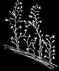
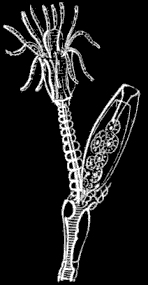
Fig. 59. Part of marginal tube and tentacles of Eucope, greatly magnified; e eye-speck, b base of tentacle., r reentering base of tentacle. Fig. 60. Young Eucope; magnified.
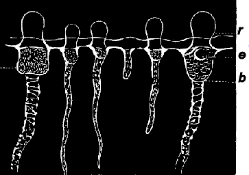
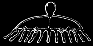
Fig. 61. Adult Eucope seen in profile; magnified. Fig. 62. Quarter disk of Fig. 60, seen from below; e e tentacles bearing eye-speck.
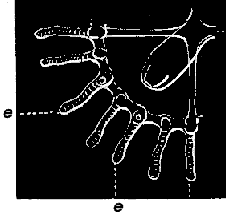
The tentacles are solid and stiff like little hairs, and two of them, in each quarter-segment of the disk, have small concretions at the base, which are no doubt eye-specks. (See Fig. 62.) Along the chymiferous tubes little swellings are developed, which increase gradually, and become either ovaries or spermaries, according to the sex of the animal. (Fig. 63.) In the adult the genital organs hang down, like elongated bags, from the chymiferous tubes. (Fig. 64.) The tentacles are numerous, multiplying to about a hundred and ninety-two in the adult, and increasing according to the numerical law to be explained in the description of the Oceania.
[fig 63]
[fig 64]
Fig. 63. Quarter-disk of young Eucope, older than Fig. 62, with a second set of tentacles (2) between the first set (1). Fig. 64. Magnified quarter-disk of adult Eucope.
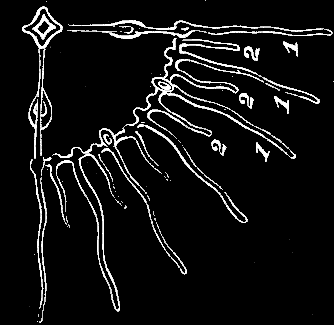
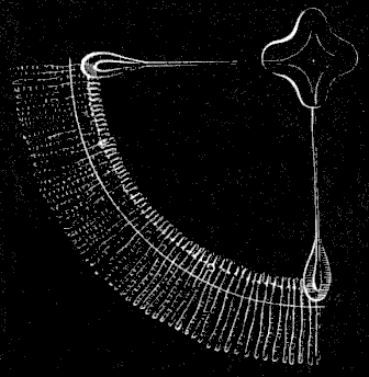
This little Jelly-fish is one of the most common in our Bay. There is not a night or day when they cannot be taken in large numbers, from the early spring till late in the autumn; and as the breeding season lasts during the whole of that period, they are found in all possible stages of growth. In consequence of this, the course of their development, and the relation between the different phases of their existence as Hydroids, and afterwards as Acalephs, are well known, though the successive steps of their growth have not been traced connectedly, as in some of the other Jelly-fishes, the Tima or Melicertum, for instance. The process is, however, so similar throughout the class of Hydroids, that, having followed it from beginning to end in some of the groups, we have the key to the history of others, whose development has not been so fully traced. The eggs laid by the Eucope in the autumn develop into planulæ, which acquire their full size as Hydroid communities toward the close of the winter, and the development of the young Medusæ upon them, as described above, begins with the opening spring.
Oceania. (Oceania languida A. Ag.)
The Oceania (Fig. 68) is so delicate and unsubstantial, that with the naked eye one perceives it only by the more prominent outlines of its structure. We may see the outline of the disk, but not the disk itself; we may trace the four faint thread-like lines produced by the radiating tubes traversing the disk from the summit to the margin; and we may perceive, with far more distinctness, the four ovaries attached to these tubes near their base; we may see also the circular tube uniting the radiating tubes, and the tentacles hanging from it, and we can detect the edge of the filmy veil that fringes the margin of the disk. But the substance connecting all these organs is not to be distinguished from the element in which it floats, and the whole structure looks like a slight web of threads in the water, without our being able to discern by what means they are held together. Under the microscope, however, the invisible presently becomes visible, and we find that this Jelly-fish, like all others, has a solid gelatinous disk.
[fig 65]
[fig 66]
[fig 67]
[fig 68]
Fig. 65. Young Oceania just escaped from its reproductive calycle; magnified. Fig. 66. The same as Fig. 65, from below, still more magnified; t long tentacles., t' rudimentary tentacle, e eye-speck on each side of base of tentacles.
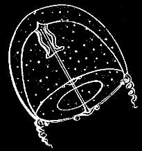
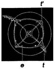
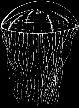
Fig. 67. Young Oceania, older than Fig. 65; magnified. Fig. 68. Adult Oceania; natural size.
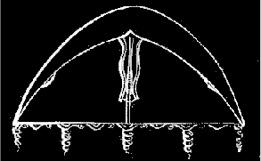
Let us begin with its earlier condition. When it first escapes from the parent Hydroid stock, the Oceania is almost spherical in form. (See Fig. 65.) The disk is divided by four chymiferous tubes, running from the summit to the margin, where they meet the circular tube in which they all unite. At this time, it has but two well-developed tentacles, opposite each other on the margin of the disk, just at the base of two of the chymiferous tubes (Fig. 66), while two others are just discernible in a rudimentary state, forming slight projections at the base of the two other tubes. Fig. 66 gives a view of the animal from below, at this stage of its growth, while Fig. 65 shows it in profile. It will be seen by the latter how very spherical is the outline of the disk at this period, while the proboscis, in which are placed the mouth and digestive cavity, is quite long, and hangs down considerably below the lower surface of the disk. As the animal advances in age the disk loses its spherical outline, and becomes much flattened, as may be seen in Fig. 67. It may be well to introduce here some explanation of the law according to which the different sets of tentacles follow each other in successive cycles of growth, since it is a law of almost universal application in Jelly-fishes and Polyps; and, owing to the smaller number and simpler arrangement of the tentacles in Oceania, it may be more easily analyzed in them than in many others, where the number and complication of the different sets of tentacles make it very difficult to trace their relation to each other during their successive growth. We have seen that the Oceania begins life with only two tentacles. These form the first set, and are marked with the number 1 in the subjoined diagram, which gives the plan of all the different sets in their regular order. The second set, marked 2, consists also of two, which are developed at equal distances between the first two, i.e. at right angles with them. The third set, however, marked 3, consists of four, as do all the succeeding sets, and they are developed between the first and second. The fourth set comes in between the first and third; the fifth between the third and second; the sixth between the first and fourth; the seventh between the fifth and second; the eighth between the third and fourth; the ninth between the fifth and third. The ultimate number of tentacles in the Oceania is thirty-two, or sometimes thirty-six, and the cycles always in twos or multiples of two. But whatever be the number included in the successive sets of tentacles, and the unit for the first set ranges from two to forty-eight, the law in different kinds of Jelly-fishes is always the same, the youngest set always forming between the oldest preceding set. Thus the fourth set comes in between the first and third, and the fifth between the second and third, the intervals occupied now by the fourth set, being limited by the first set of tentacles on one side, and by the third set on the other side, while the intervals occupied by the fifth set are bounded by the second and third sets.
[fig 67b]
Diagram of succession of tentacles.
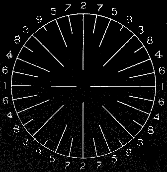
The little spheres represented between the tentacles on the margin of the disk, in Figs. 65-67, are eye-specks, and these continue to increase in number with age; in this the Oceania differs from the Eucope, in which it will be remembered there were but two eye-specks in each quarter-segment of the disk throughout life. Fig. 68 represents the adult Oceania in full size, when it averages from an inch and a half to two inches in diameter. It is slow and languid in its movements, coming to the surface only in the hottest hours of the summer days; at such times it basks in the sun, turning lazily about, and dragging its tentacles after it with seeming effort. Sometimes it remains for hours suspended in the water, not moving even its tentacles, and offering a striking contrast to its former great activity when young, and to the lively little Eucope, which darts through the water at full speed, hardly stopping to rest for a moment. If the Oceania be disturbed it flattens its disk, and folds itself up somewhat in the shape of a bale (see Fig. 69), remaining perfectly still, with the tentacles stretching in every direction. When the cause of alarm is removed, it gently expands again, resuming its natural outline and indolent attitudes. The number of these animals is amazing. At certain seasons, when the weather is favorable, the surface of the sea may be covered with them, for several miles, so thickly that their disks touch each other. Thus they remain packed together in a dense mass, allowing themselves to be gently drifted along by the tide till the sun loses its intensity, when they retire to deeper waters. Some points, not yet observed, are still wanting to complete the history of this Jelly-fish. By comparing such facts, however, as are already collected respecting it, with our fuller knowledge of the same process of growth in the Eucope, Tima, and Melicertum, we may form a tolerably correct idea of its development. It is hatched from a Campanularia.
[fig 69]
Fig. 69. Attitude assumed by Oceania when disturbed.
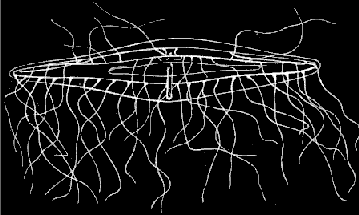
Clytia. (Clytia bicophora Ag.)
In Figs. 70-73 we have the Acalephian and Hydroid stages of the Clytia (Fig. 73), another very pretty little Jelly-fish, closely allied to the Oceania. When first hatched, like the Oceania, it is very convex, almost thimble-shaped (see Fig. 70), but a little later the disk flattens and becomes more open, as in Fig. 71. In Fig. 72, we have a branch of the Hydroid, a Campanularia, greatly magnified, with the annulated reproductive calycle attached to it, and crowded with Jelly-fishes ready to make their escape as soon as the calycle bursts. The adult Clytia (Fig. 73) is somewhat smaller and more active than the Oceania, and is easily recognized by the black base of its tentacles, at their point of juncture with the margin of the disk. It is more commonly found at night, than in the day-time, being nocturnal in its habits.
[fig 70]
[fig 71]
[fig 72]
[fig 73]
Fig. 70. Young Clytia just escaped from the reproductive calycle. Fig. 71. Clytia somewhat older than Fig. 70.
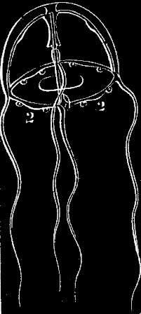
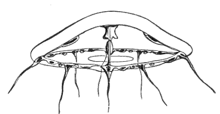
Fig. 72. Magnified portion of Hydrarium of Clytia. Fig. 73. Adult Clytia; twice natural size.
Zygodactyla. (Zygodactyla groenlandica Ag.)
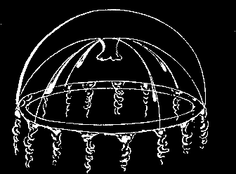
Little has been known, and still less published, of this remarkable genus of Jelly-fish (Figs. 74, 75) up to the present time. The name Zygodactyla, or Twinfinger, was given to it by Brandt, from drawings made by Mertens, who had some opportunity of studying it in his journey around the world. These drawings were published in the Transactions of the St. Petersburg Academy. In the year 1848 Professor Agassiz read a paper upon one of the species of this genus belonging to our coast, before the American Academy, in which he called it Rhacostoma, not being aware that it had already received a name, and gave some account of its extraordinary phosphorescent properties. The name Rhacostoma must of course yield to that of Zygodactyla, which has a prior claim.
[fig 74]
Fig. 74. Zygodactyla seen from above.
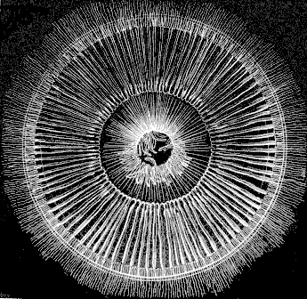
The average size of this Jelly-fish when full grown is from seven to eight inches in diameter; sometimes it may measure even ten or eleven, but this is rather rare. The light-violet colored disk is exceedingly delicate and transparent, its edge being fringed with long fibrous tentacles, tinged with darker violet at their point of juncture with the disk, and hanging down a yard and more when fully extended, though they vary in length according to the size of the specimen, and, in consequence of their contractile power, may seem much shorter at some moments than at others. The radiating tubes in this Jelly-fish are exceedingly numerous, the whole inner surface of the disk being ribbed with them. (See Figs. 74 and 75.) The ovaries follow the length of the tubes, though they do not extend quite to their extremity, where they join the circular tube around the margin of the disk; nor do they start exactly at the point where the tubes diverge from the central cavity, but a little below it. (Fig. 74.) Each ovary consists of a long, brownish, flat bag, split along the middle, so closely folded together that it seems like a flat blade attached along the length of the tube. Perhaps a better comparison would be to a pea-pod greatly elongated, with the edges split along their line of juncture, and attached to a tube of the same length. The ovaries are not perfectly straight, but slightly waving, as may be seen in Fig. 74, and these undulations are stronger when the ovaries are crowded with eggs, as is the case at the time of spawning.
[fig 75]
Fig. 75. Zygodactyla seen in profile.
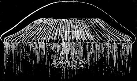
The large digestive cavity hangs from the centre of the under side of the disk (Fig. 75), terminating in the proboscis, which, in this kind of Jelly-fish, is short in proportion to the diameter of the disk, while the opening of the mouth is very large. (Fig. 74.) It is unfortunate that a variety of inappropriate names, likely to mislead rather than aid the unscientific observer, have been applied to different parts of the Jelly-fish. What we call here digestive cavity, proboscis, and mouth, are, in fact, parts of one organ. An exceedingly delicate, transparent, filmy membrane hangs from the under side of the disk; that membrane forms the outer wall of the digestive cavity, which it encloses; it narrows toward its lower margin, leaving open the circular aperture called the mouth; this narrowing of the membrane is produced by a number of folds in its lower part, while at its margin these folds spread out to form ruffles around the edge of the mouth, and these ruffles again extend into the long scalloped fringes hanging down below.
The motion of these Jelly-fishes is very slow and sluggish. Like all their kind, they move by the alternate dilatation and contraction of the disk, but in the Zygodactyla these undulations have a certain graceful indolence, very unlike the more rapid movements of many of the Medusæ. It often remains quite motionless for a long time, and then, if you try to excite it by disturbing the water in the tank, or by touching it, it heaves a slow, lazy sigh, with the whole body rising slightly as it does so, and then relapses into its former inactivity. Indeed, one cannot help being reminded, when watching the variety in the motions of the different kinds of Jelly-fishes, of the difference of temperament in human beings. There are the alert and active ones, ever on the watch, ready to seize the opportunity as it comes, but missing it sometimes from too great impatience; and the slow, steady people, with very regular movements, not so quick perhaps, but as successful in the long run; and the dreamy, indolent characters, of which the Zygodactyla is one, always floating languidly about, and rarely surprised into any sudden or abrupt expression. One would say, too, that they have their aristocratic circles; for there is a delicate, high-bred grace about some of them quite wanting in the coarser kinds. The lithe, flexible form of the greyhound is not in stronger contrast to the heavy, square build of the bull dog, than are some of the lighter, more frail species of Jelly-fish to the more solid and clumsy ones. Among these finer kinds we would place the Tima. (Fig. 76.)
Tima. (Tima formosa Ag.)
One's vocabulary is soon exhausted in describing the different degrees of consistency in the substance of Jelly-fishes. Delicate and transparent as is the Tima, it has yet a certain robustness and solidity beside the Oceania, described above. In fact, all are gelatinous, all are more or less transparent, and it is not easy to describe the various shades of solidity in jelly. Perhaps they may be more accurately represented by the impression made upon the touch than upon the sight. If, for instance, you place your hand upon a Zygodactyla, you feel that you have come in contact with a substance that has a positive consistency; but if you dip your finger into a bowl where a Tima is swimming, and touch its disk, you will feel no difference between it and the water in which it floats, and will not be aware that you have reached it till the animal shrinks away from the contact.
[fig 76]
[fig 77]
Fig. 76. Tima; half natural size. Fig. 77. One of the lips of the mouth at the extremity of the long proboscis; m mouth, d digestive cavity, c chymiferous tube.
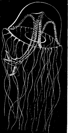
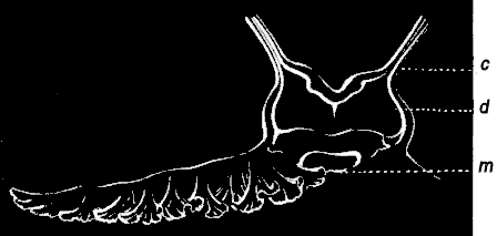
The adult Tima, represented in Fig. 76, is not more than an inch and a half or two inches in diameter. Instead of countless tubes diverging from the digestive cavity to the margin of the disk, as in the Zygodactyla, there are but four. The digestive cavity in the Tima is much smaller than in the Zygodactyla, and is placed at the end of the proboscis, which is long, and hangs down far below the disk. This removal of the digestive cavity to the extremity of the proboscis gives to the tubes arising from it a very different and much sharper curve than they have in the Zygodactyla. In the Tima they start from the end of the proboscis, as may be seen in the wood-cut (Fig. 76), and then turn abruptly off, when they arrive at the under surface of the disk, to reach its margin. The disk has, as usual, its veil and its fringe of tentacles; the tentacles in the full-grown Tima are few,—seven in all the four intermediate spaces between the tubes, with one at the base of each tube, making thirty-two in all. The ovaries, which are milk-white, follow the line of the tubes, as in the Zygodactyla, and have very undulating folds when full of eggs. The tubes meet in the digestive cavity, the margin of which spreads out to form four ruffled edges that hang down from it. One of these ruffles, considerably magnified, is represented in Fig. 77. In Fig. 78 we have a portion of the Hydroid stock from which this Jelly-fish arises, also greatly magnified. The Tima is very active, yet not abrupt in its motions; but when in good condition it is constantly moving about, rising to the surface by the regular pulsations of the disk, or swimming from side to side, or poising itself quietly in the water, giving now and then a gentle undulation to keep itself in position.
[fig 78]
Fig. 78. Magnified head of Hydrarium of Tima.
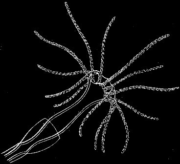
Though not a very frequent visitor of our shores, the appearance of the Tima is not limited by the seasons, since they are found at all times of the year. It is a fact, unexplained as yet, that the Tima and many other Jelly-fishes are never seen except when full grown. What may be the haunts and habits of these animals from the time of their hatching till they make their appearance again in the adult condition, is not known, though it is probable that they remain at the bottom during this period, and only come to the surface to spawn. This impression is confirmed by the observations made upon a very young Cyanea which was kept for a long time in confinement; but a question of this kind cannot of course be settled by a single experiment.[6]
[fig 79]
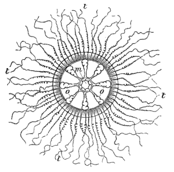
Fig. 79. Melicertum campanula seen from above; m mouth, o o ovaries, t t tentacles. (Agassiz.)
Melicertum. (Melicertum campanula Pér. et Les.)
A pretty Medusa, smaller and far more readily obtained than the Tima, is the Melicertum. (Fig. 80.) Its disk has a yellowish hue, and from its margin hangs a heavy row of yellow tentacles, while the eight ovaries (Fig. 79) are of a darker shade of the same color. This little golden-tinted Jelly-fish, moving through the water with short, quick throbs, produced by the rapid rise and fall of the disk, is a very graceful object. Its bright color, made particularly prominent by the darker undulating lines of the ovaries, which become very marked near the spawning season, renders it more conspicuous in the water than one would suppose from its size; for it does not measure more than an inch in height when full grown. (See Fig. 80.)
Development of Melicertum and Tima.
[fig 80]
Fig. 80. Melicertum seen in profile; natural size.
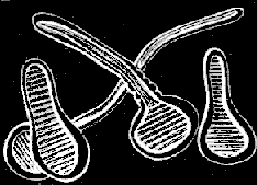
In the Melicertum and Tima we have had the good fortune to trace the process by which the eggs are changed into Hydroid communities. If any one has a curiosity to follow for themselves this singular history of alternate generations, the Melicertum is a good subject for the experiment, as it thrives well in confinement. After keeping a number of them in a large glass jar for a couple of days at the time of spawning, it will be found that the ovaries, which were at first quite full of eggs, are emptied, and that a number of planulæ; are swimming about near the bottom of the vessel. After a day or two the outline of these planulæ, spherical at first, becomes pear-shaped (see Fig. 81), and presently they attach themselves by the blunt end to the bottom of the jar. (Fig. 82.) Thus their Hydroid life begins; they elongate gradually, the horny sheath is formed around them, tentacles arise on the upper end, short and stunted at first, but tapering rapidly out into fine flexible feelers, the stem branches, and we have a little Hydroid community (Fig. 83), upon which, in the course of the following spring, the reproductive calycles containing the Medusæ buds will be developed, as in the case of the Eucope and Clytia. The Tima passes through exactly the same process, though the shape of the planulæ and the appearance of the young differ from that of the Melicertum, as may be seen in Fig. 78, where a single head of the Tima Hydroid, greatly magnified, is represented. By combining the above observations upon the development of the Hydroids of the Melicertum and Tima with those previously mentioned upon the young Medusa arising from reproductive calycles in the Eucope and Clytia, we get a complete picture of all the changes through which any one of these Hydroid Medusæ passes, from its Hydroid condition to the moment when it enters upon an independent existence as a free Jelly-fish.
[fig 81]
[fig 82]
[fig 83]
Fig. 81. Planula of Melicertum; magnified. Fig. 83. Young Hydrarium developed from planulæ; magnified.
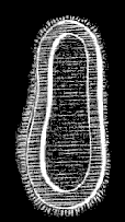
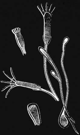
Fig. 82. Cluster of planulæ just attached to the ground.
(Laomedea amphora Ag.)
The Medusæ of the Campanularians are not all free. On the contrary, in many of the species they always remain attached to the Hydroid, never attaining so high a development as the free Medusæ, and withering on the stem after having laid their eggs. Such is the Laomedea amphora, quite common on all the bridges connecting Boston with the country, where, on account of the large amount of food brought down from the sewers by the river, they thrive wonderfully, growing to a great size, sometimes measuring from a foot to eighteen inches in height.
[fig 84]
[fig 85]
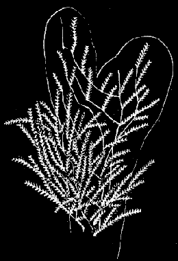
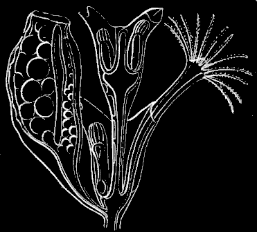
Fig. 84. Colony of Dynamena pumila; natural size. Fig. 85. Magnified portion of Fig. 84.
Sertularians.
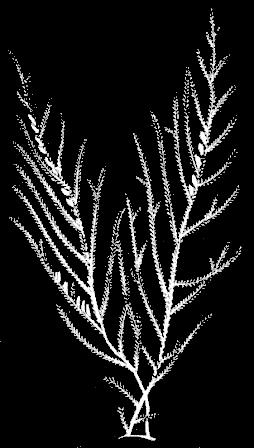
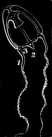
The Sertularians form another group of Hydroids closely allied to the Campanularians, though differing from them in the arrangement of the sterile Hydræ upon the stem. Among these one of the most numerous is the Dynamena (Dynamena pumila Lamx., Fig. 84), which hangs its yellowish fringes from almost every sea-weed above low-water-mark. It is especially thick and luxuriant on the fronds of our common Fucus vesiculosus. The color is usually of a pale yellow, though sometimes it is nearly white, and when first taken from the water it has a glittering look, such as a white frost leaves on a spray of grass. Fig. 84 represents such a cluster in natural size, while Fig. 85 shows a piece of the stem highly magnified, with a reproductive calycle attached to the side of a sterile Hydra stem. Many of these Sertularian Hydroids assume the most graceful forms, hanging like long pendent streamers from the Laminaria, or in other instances resembling miniature trees. One of these tree-like Sertularians (Dyphasia rosacea Ag.), abundant on all rocks in sheltered places immediately below low-water-mark, is represented in Fig. 86. In both these Sertularians the Medusæ wither on the stock, never becoming free. The free Medusæ of the Sertularians are only known in their adult condition in a single genus, which is closely allied to Melicertum, and which is produced from a Hydroid genus called Lafoea. Fig. 87 represents one of these young Sertularian Medusæ (Lafoea cornuta Lamx.).
[fig 86]
[fig 87]
Fig. 86. Dyphasia rosacea, natural size. Fig. 87. Medusa of Lafoea.
Tubularians.
In the Sertularian and Campanularian Hydroids we have found that the communities consist generally of a large number of small individuals, so small, indeed, that it is hardly possible at first glance to distinguish the separate members of these miniature societies. Among the Tubularians, on the contrary, the communities are usually composed of a small number of comparatively large individuals; and indeed these Hydroids may even grow singly, as in the case of the Hybocodon (Fig. 104), which attains several inches in height. There is also another general feature in which the Tubularians differ from both the other groups of Hydroids. In the latter, the horny sheath which encloses the stem extends to form a protecting calycle around the Hydra heads. This protecting calycle is wanting round the heads of the Tubularians, though their stems are surrounded by a sheath.
[fig 88]
[fig 89]
[fig 90]
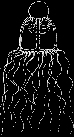
Fig. 88. Colony of Coryne; natural size. (Agassiz.) Click on image to view larger size. Fig. 90. Free Medusa of Coryne. (Agassiz.)
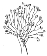
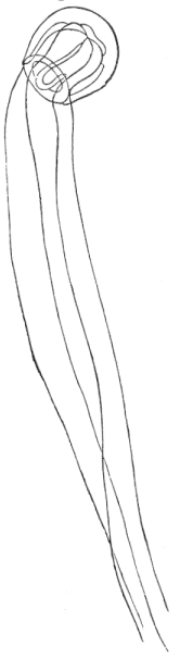
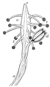
Fig. 89. Magnified head of Coryne; a stem, t tentacles, o mouth, v body, d Medusa. (Agassiz.) Click on image to view larger size.
Sarsia. (Coryne mirabilis Ag.)
Among the most common of our Tubularians is a small, mossy Hydroid (Fig. 88), covering the rocks between tides, in patches of several feet in diameter. Fig. 89 represents a single head from this little mossy tuft greatly magnified, in which is seen the medusa bud arising from the stem by the process already described in the other Hydroids. In Fig. 90 we have the little Jelly-fish in its adult condition, about the size of a small walnut, with a wide circular opening, through which passes the long proboscis, hanging from the under surface of the disk to a considerable distance below its margin. The four tentacles are of an immense length when compared to the size of the animal. As a general thing, the tentacles are less numerous in the Tubularian Medusæ than in those arising from other Hydroids; they want also the singular limestone concretions found at the base of the tentacles in the Campanularian Medusæ. In Fig. 91 we have one of the Tubularian Medusæ (Turris vesicaria A. Ag.) which lifts a rather larger number of tentacles than is usual among these Jelly-fishes. We never find the tentacles multiplying almost indefinitely in them, as in Zygodactyla and Eucope. The little Jelly-fish described above is known as Sarsia, while its Hydroid is called Coryne. These names having been given to the separate phases of its existence before their connection was understood, and when they were supposed to represent two distinct animals. They are especially interesting with reference to the history of Hydroids in general, because they were among the first of these animals in whom the true relation between the different phases of their existence was discovered. Lesson named the Sarsia after the great Norwegian naturalist, Sars, to whom we owe so large a part of what is at present known respecting this curious subject of alternate generations.
[fig 91]
Fig. 91. Turris vesicaria; natural size.
Bougainvillia. (Bougainvillia superciliaris Ag.)
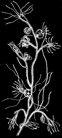
The Bougainvillia (Fig. 92), is one of our most common Jelly-fishes, frequenting our wharves as well as our sea-shore during the spring. The tentacles are arranged in four bunches or clusters at the junction of the radiating tubes with the circular tube, from which they may be seen extending in every direction whenever these animals remain quietly suspended in the water,—a favorite attitude with them, and one which they retain sometimes for days, seeming to make no effort beyond that of gently playing their tentacles to and fro (Fig. 92). These tentacles are capable of immense extension, sometimes to ten or fifteen times the diameter of the bell. The proboscis is not simple as in the Sarsia, but looks like a yellow urn suspended at its four corners from the chymiferous tubes. The oral opening is entirely concealed by clusters of shorter tentacles surrounding the mouth in a close wreath, on which the eggs are supported. A highly magnified branch of the Hydroid stock from which this Medusa arises is represented in Fig. 93. There we see the little Jelly-fishes in different degrees of development on the stem, while in Figs. 94-97 they are given separately and still more enlarged. In Fig. 94 the outline of the Jelly-fish is still oval, the proboscis is but just formed, and the tentacles appear only as round swellings or knobs. In Fig. 95 a depression has taken place at the upper end, presently to be an opening, the proboscis is enlarged, and the tentacles lengthened, but still turned inward. In Fig. 96 the appendages of the proboscis are quite conspicuous, the tentacles are turned outward, and the Jelly-fish is almost ready to break from its attachment, having assumed its ultimate outline. Fig. 97 represents it just after it has separated from the stem, when it has only two tentacles at each cluster and simple knobs around the mouth, instead of the complicated branching tentacles of the adult.
[fig 92]
[fig 93]
Fig. 92. Bougainvillia; magnified. Fig. 93. Hydrarium of Bougainvillia; magnified.
[fig 94]
[fig 95]
[fig 96]
[fig 97]
Fig. 94 Fig. 95
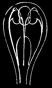
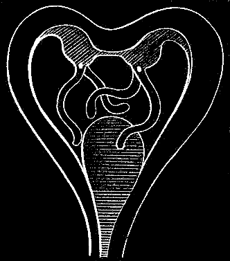
Fig. 96 Fig. 97
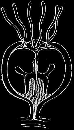
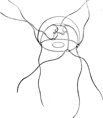
Figs. 94, 95, 96. Medusæ buds of Fig. 93, in different degrees of development. Fig. 97. Young Medusa just freed from the Hydroid; magnified.
Tubularia. (Tubularia Couthouyi Ag.)
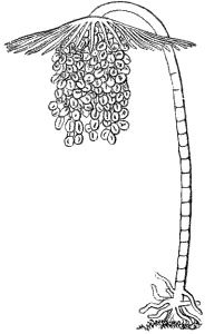
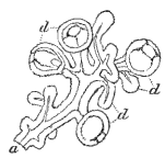
There are several other Tubularians common in our waters which should not be passed over without mention, although as this little book is by no means intended as a complete text-book, but rather as a volume of hints for amateur collectors, we would avoid as much as possible encumbering it with many names, or with descriptions already given in more comprehensive works. This Tubularia is interesting, however, from the fact that the Medusæ buds are never freed from the stem, and do not develop into full-grown Jelly-fishes, but always remain abortive. Fig. 98 represents one head of such a Hydroid with the Medusæ buds pendent from it in a thick cluster, while in Fig. 99 we have a few of them sufficiently magnified to show that, though presenting the four chymiferous tubes, they are otherwise exceedingly simple in structure, as compared with the free Jelly-fishes.
[fig 98]
[fig 99]
Fig. 98. Tubularia; magnified. (Agassiz.) Fig. 99. Part of cluster of Medusæ of Fig. 98; magnified. (Agassiz.)
Hydractinia. (Hydractinia polyclina Ag.)
This is another Tubularian, covering the surface of rocks in tide-pools, or attaching itself upon shells inhabited by hermit crabs. Indeed it was upon these shells that the Hydractinia was first noticed, and it was long supposed that the wanderings to which the little colony was thus subjected were necessary for its healthy development. But subsequent observations have shown that it attaches itself quite as frequently to the solid rock as to these nomadic shells. It has a rosy color, and, being very small, it looks, until one examines it closely, more like a thick red carpet of soft moss, than like a colony of animals. These communities are distinct in sex, the fertile individuals in each being either all male or all female. In Fig. 100 we have a portion of a female colony, representing one fertile head, in which the buds are crowded with Medusæ; one sterile head, surrounded by its wreath of tentacles; and still another member of the society whose office is not fully understood, unless it be that of a kind of purveyor, catching food for the rest. Fig. 101 represents the corresponding individuals taken from a male colony. The sex makes little difference in the appearance of the reproductive heads. All the individuals of a Hydractinia colony are connected at the base by a horny network, rising occasionally into points of a conical or cylindrical shape. This polymorphism among the Tubularians is another evidence of the relation between the Siphonophoræ, or floating Hydroids, and the fixed Hydroids.
[fig 100]
[fig 101]
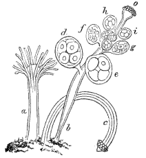
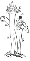
Fig. 100. Female colony of Hydractinia; a sterile individual, b fertile individual producing female Medusæ, c fertile individual with globular tentacles without Medusæ, d e f g h i Medusæ in different stages of growth, o mouth tentacles. (Agassiz.) Fig. 101. Male colony; a a sterile individuals, b fertile individuals producing male Medusæ, d; o globular tentacles, t slender tentacles of sterile individual. (Agassiz.)
Hybocodon. (Hybocodon prolifer Ag.)
Among our Medusæ derived from a Tubularian stock is the Hybocodon, viz. the hunchbacked Medusa (Fig. 102), a singular little Jelly-fish, odd and unsymmetrical in shape, as its name indicates, and interesting from its relations to one of our floating communities, the Nanomia, presently to be described. Instead of the evenly proportioned bell of the ordinary Medusæ, the Hybocodon has a one-sided outline (Fig. 102), one large tentacle only being fully developed, while the others remain always abortive, so that the whole weight of the structure is thrown on one half of the bell. Upon this large tentacle small Jelly-fishes, similar to the original, are produced by budding, this process going on till ten or twelve such Jelly-fishes (Fig. 103) may be seen suspended from the tentacle. Up to this time it has remained connected with the Hydroid from which it arises, a rather large Tubularian, usually growing singly (Fig. 104), and of a deep orange-red in color. But at this stage of its existence it frees itself, and leads an independent life hereafter, swimming about with a quick, darting motion. In the account of the Nanomia, the homology between its scale, or abortive Medusa, and the Hybocodon, is traced in detail, and I need only allude to it here. Though this Medusa is so peculiar in appearance, the Tubularian from which it is derived is very like the Tubularia Couthouyi, already described. This is one of the instances before alluded to, in which closely allied forms give rise to very dissimilar ones, or, as in many cases, the very reverse of this takes place, and closely allied forms arise from very dissimilar ones.
[fig 102]
[fig 103]
[fig 104]
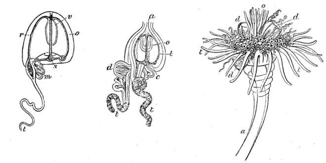
Fig. 102. Unsymmetrical free Medusa of Hybocodon; r o chymiferous tubes, v proboscis, s circular tube, m young Medusæ at base of long tentacle t. (Agassiz.)
Fig. 103. Medusa bud of Hybocodon; a base of attachment, o proboscis, c circular tube, d young Medusæ at base of long tentacle t. (Agassiz.)
Fig. 104. Single head of Hybocodon Hydroid; a stem, d Medusæ buds, o tentacles round mouth. (Agassiz.)
Dysmorphosa. (Dysmorphosa fulgurans A. Ag.)
Besides the budding at the base of the tentacle, as in Hybocodon, we find another mode of development among Hydroid Medusæ, viz. that of budding from the proboscis. One of our most common little Jelly-fishes, the Dysmorphosa (Fig. 105), to which we owe the occasional blue phosphorescence of the sea, so brilliant at times, buds in this manner. Fig. 105 represents an adult Dysmorphosa, on the proboscis of which may be seen three small buds in different stages of development. In Fig. 106 the proboscis is more enlarged, showing one of the little Jelly-fishes similar to the parent, just ready to drop off. We need not wonder at the immense number of these animals, with which the sea actually swarms at times, when we know that as fast as they are dropped, and it takes but a few days to complete their development, they each begin the same process; so that in the course of a week or ten days one such Medusa, supposing it to have produced six buds only, will have given rise to forty-two Jelly-fishes, thirty-six of which may be equally prolific in the same short period. These Medusæ budding thus, and swimming about, carrying their young with them, bear such a close resemblance to the floating communities of Hydroids formerly known as Siphonophoræ, that did we not know that some of them arise from Tubularians, it would be natural to associate them with the Siphonophoræ.
[fig 105]
[fig 106]
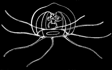
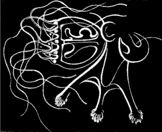
Fig. 105. Dysmorphosa seen in profile; magnified. Fig. 106. Magnified proboscis of Dysmorphosa with young Medusæ budding from it.
Nanomia. (Nanomia cara A. Ag.)
The Nanomia (Fig. 115), our free floating Hydroid, consists, when first formed, of a single Hydra containing an oblong oil bubble (Fig. 107). The whole organisation of such a Hydra is limited to a simple digestive cavity; it has, in fact, but one organ, and one function, and consists of an alimentary sac resembling the proboscis of a Medusa (Fig. 107); the oil bubble is separated from it by a transverse partition, and has no connection with the cavity. Presently, between the oil bubble and the cavity arise a number of buds of various character (Fig. 108), which we will describe one by one, beginning with those nearest the oil bubble, since these upper members of the little swimming community bear a very important part in its history. The infant community (Fig. 108) passes rapidly into the stage represented in Fig. 109, and then through all the stages intermediate between this and the adult, shown in its natural size in Fig. 115. The upper buds enlarge gradually, and soon take upon themselves a perfect Medusa structure (Fig. 110), with the exception of the proboscis, the absence of which is easily understood, when we find that these Medusæ, serve the purpose of locomotion only, having no share in the function of feeding the community, so that a digestive apparatus would be quite superfluous for them. In every other respect they are perfect Medusæ, attached to the Hydra as the Medusa buds always are when first formed, having the (four) chymiferous tubes, characteristic of all Hydroid Medusæ, radiating from the centre to the periphery; two of these tubes are very winding, as may be seen in Fig. 110, while the other pair are straight. The Medusæ themselves are heart-shaped in form, depressed at the centre of the upper surface, and bulging on either side into wing-like expansions, where they join the stem. These expansions interlock with one another, crossing nearly at right angles. The Medusæ-like buds are the swimming bells; by their contractions, alternately taking in and throwing out the water, they impel the whole community forward, so that it seems rather to move like one animal, than like a combination of individuals.
[fig 107]
[fig 108]
[fig 109]
[fig 110]
Fig. 107. Young Nanomia; magnified. Fig. 108. Young Nanomia with rudimentary Medusæ. Fig. 109. Young Nanomia, older than Fig. 108.
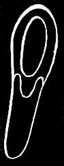
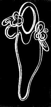
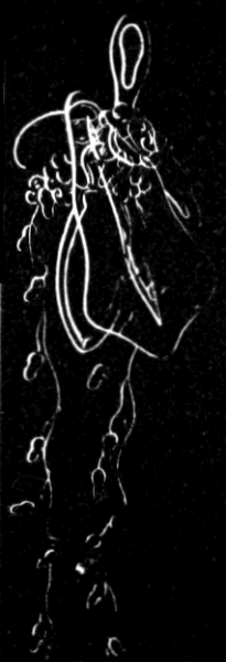
Fig. 110. Heart-shaped swimming bell of Nanomia; magnified.
[fig 111]
Fig. 111. Cluster of Medusæ with tentacles having pendent knobs.
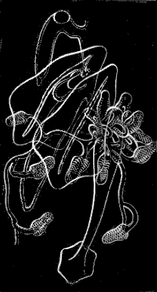
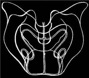
Besides these locomotive members, the community contains three kinds of Hydræ arising as buds from the primitive Hydra below the swimming bells, the latter remaining always nearest the oil bubble at the top, while the first Hydra, the founder of the community, in proportion as the new individuals are added, is gradually pushed downward, and remains always at the end of the string, the stem of which is formed by the elongated neck of the primitive Hydra. All the three sets of Hydræ have certain features in common, while they have other distinguishing characteristics marking them as distinct individuals. They are all accompanied by triangular shields (Fig. 111), arising with them at the same point on the parent stem, and all are furnished with tentacles hanging down from the summit of the Hydra at the side opposite the shield. These facts are important to remember, since we shall presently perceive, upon analyzing their parts, that these Hydræ have a close homology to the Hybocodon. The tentacles differ in structure as well as in number for each kind of Hydra. Having shown in what characters they agree, let us now take each set individually, and see what differences they present.
In the first set which we will examine the Hydra is open-mouthed. Like the original Hydra, it is only a digestive tube, similar in all respects to the proboscis of a Medusa-disk. Its only function is that of feeding, and it shows a laudable fidelity to its calling, being very constantly and earnestly engaged in the work. Let us add, however, that in this instance the occupation is not a wholly selfish one, since the cavity of every Hydra communicates with that of the stem, and the food taken in at these over-gaping mouths, is at once circulated through all parts of the community, with the exception of the oil bubble, from which it is excluded by the transverse partition dividing it from all the lower members of the stock. The shields share in this general nourishment of the compound body by means of chymiferous tubes extending toward the outer surface, and opening into the cavity of the stem. The mouth of this Hydra is very flexible (Fig. 111), expanding and contracting at the will of the animal, and sometimes acting as a sucker, fastening itself, leech-like, on the object from which it seeks to draw its sustenance. (See Fig. 111.) The tentacles attached to this set of Hydræ are exceedingly long and delicate. They arise in a cluster at the upper and inner edge of the Hydra, just at its point of juncture with the stem, and being extremely flexible and contractile, their long tendril-like sprays are thrown out in an endless variety of attitudes. (See Fig. 115.) Along the whole length of this kind of tentacle are attached little pendent knobs at even distances; Fig. 112 represents such a knob greatly magnified, and absolutely paved with lasso-cells, the inner and smaller ones being surrounded by a row of larger ones.
[fig 112]
[fig 113]
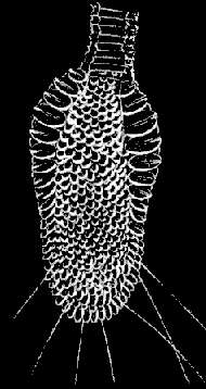
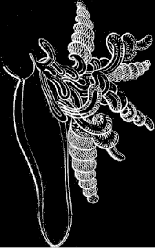
Fig. 112. Magnified pendent knob. Fig. 113. Medusa with corkscrew shaped tentacles.
The second set of Hydræ (Fig. 113), are also open-mouthed, corresponding with those described above, in everything except the tentacles, which are both shorter and thicker, and are coiled in a corkscrew-like spiral. These are thickly studded for their whole length with lasso-cells. (See Fig. 113.)
In the third and last set of Hydræ (Fig. 114), the mouth is closed; they have, therefore, no share in feeding the community, but receive their nourishment from the cavity of the stem into which they open. They differ also from the others in having a single tentacle instead of a cluster, and on this tentacle the lasso-cells are scattered at uneven distances (Fig. 114). The special function of these closed Hydræ is yet to be explained; they have oil bubbles at their upper end (see Fig. 111, the top Hydra), and though we have never seen them drop off, it seems natural to suppose that they do separate from the parent stock, and found new communities similar to those from which they arise.
[fig 114]
[fig 115]
Fig. 114. Medusa with a simple thread-like tentacle. Fig. 115. Adult Nanomia, natural size, at rest.
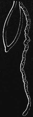
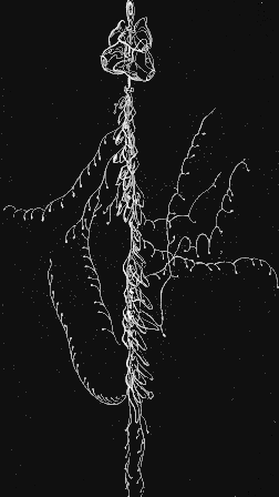
The intricate story of this singular compound existence does not end here. There is still another set of individuals whose share in maintaining the life of the community is by no means the least important. Little bunches of buds, of a different character from any described above, may be seen at certain distances along the lower part of the stem. These are the reproductive individuals. They are clusters of imperfect sexual Medusæ, resembling the rudimentary Medusæ of Tubularia (Fig. 99), which are never freed from the parent stem, but discharge their contents at the breeding season. Like many other compound Hydroids, the sexes are never combined, in one of these communities; they are always either male or female, and as those with female buds have not yet been observed, we can only judge by inference of their probable character. Front what is already known, however, of Hydroid communities of a like description, we suppose that the process of reproduction must be the same in these, and that the female stocks of Nanomia give birth to small Jelly-fishes, the eggs of which become oil bubbles, similar to that with which our little community began. (Fig. 108.)
[fig 116]
Fig. 116. Oil float of Nanomia; greatly magnified.
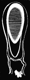
By the time all these individuals have been added along the length of the stem, the stem itself has grown to be about three inches long (Fig. 115), though the tentacles hanging from the various members of the community give to the whole an appearance of much greater length. The motion of this little string of living beings is most graceful. The oil bubble (Fig. 116) at the upper end is their float; the swimming bells immediately below it (Fig. 110), by the convulsive contractions of which they move along, are their oars. The water is not taken in and expelled again by all the bells at once, but first from all the bells on one side, beginning at the lower one, and then from all those on the opposite side, beginning also at the lower one; this alternate action gives to their movements a swinging, swaying character, expressive of the utmost freedom and grace. Whether such a little community darts with a lightning-like speed through the water, or floats quietly up and down, for its movements are both rapid and gentle, it always sways in this way from side to side. Its beauty is increased by the spots of bright red scattered along the length of the stock at the base and tips of the Hydræ, as well as upon the tentacles. The movements and attitudes of the tentacles are most various. Sometimes they shoot them out in straight lines on either side, and then the aspect of the whole thing reminds one of a tiny chandelier in which the coral drops make the pendants, or they may be caught up in a succession of loops or floating in long streamers; indeed, there is no end to the fantastic forms they assume, ever astonishing you by some new combination of curves. The prevailing hue of the whole community is rosy, with the exception of the oil bubble or float, which looks a bright garnet color when seen in certain lights.
Let us now compare one of the Hydræ hanging from the stem (Fig. 113) with the Hybocodon (Fig. 102). The reader will remember the unsymmetrical bell of this singular Medusa, one half of its disk more largely developed than the other, with the proboscis hanging from the centre, and the cluster of tentacles from one side. Let us now split the bell so as to divide it in two halves with the proboscis hanging between them; next enlarge the side where there are no tentacles, and give it a triangular outline; then contract the opposite side so as to draw up the cluster of tentacles to meet the base of the proboscis, and what have we? The proboscis now corresponds to the Hydra of our Nanomia, with the cluster of tentacles attached to its upper edge (Fig. 113), while the enlarged half of the bell represents the shield. If this homology be correct it shows that the Nanomia is not, as some naturalists have supposed all the Siphonophores to be, a single animal, its different parts being a mere collection of organs endowed with special functions, as feeding, locomotion, reproduction, &c., but that it is indeed a community of distinct individuals corresponding exactly to the polymorphous Hydroids, whose stocks are attached, such as Hydractinia, and differing from them only in being free and floating.
[fig 117]
Fig. 117. Physalia; a b air sac with crest c, m bunches of individuals, n central tentacles, t t expanded tentacles. (Agassiz.)
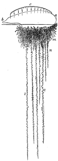
The homologies of the Siphonophoræ or floating Hydroids, with many of the fixed Hydroids, is perhaps more striking when we compare the earlier stages of their growth. Suppose, for instance, that the planula of our Melicertum (See Fig. 81) should undergo its development without becoming attached to the ground,—what should we then have? A floating community (Fig. 83), including on the same stock like the Nanomia, both sterile and fertile Hydræ, from the latter of which Medusæ bells are developed. The little Hydractinia community (Fig. 100), in which we have no less than four distinct kinds of individuals, each performing a definite distinct function, affords a still better comparison.
Physalia. (Physalia Arethusa Til.)
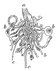
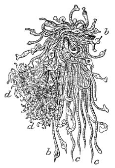
Among the most beautiful of the Siphonophores, is the well-known Physalia or Portuguese man-of-war, represented in Fig. 117. The float above is a sort of crested sac or bladder, while the long streamers below consist of a number of individuals corresponding in their nature and functions to those composing a Hydroid community. Among them are the fertile and sterile Hydræ (Fig. 118), the feeders and Medusæ bells (Fig. 119). The Physalia properly belongs to tropical waters, but sometimes floats northward, in the warm current of the Gulf Stream, and is stranded on Cape Cod. When found so far from their home, however, they have usually lost much of their vividness of color; to judge of their beauty one should see them in the Gulf of Mexico, sailing along with their brilliant float fully expanded, their crest raised, and their long tentacles trailing after them.
[fig 118]
[fig 119]
Fig. 118. Bunch of Hydræ; a base of attachment, b b b single Hydræ, c c tentacles. (Agassiz.) Fig. 119. Bunch of Hydræ; cluster of Medusæ; b b Hydræ with tentacles, c d bunches of Medusæ (Agassiz.)
Velella. (Velella mutica Bosc.)
Another very beautiful floating Hydroid, occasionally caught in our waters, though its home is also far to the south, is the Velella (Fig. 120). It is bright blue in color, and in form not unlike a little flat boat with an upright sail. Its Medusa (Fig. 121) resembles so much that of some of our Tubularians, that it has actually been removed on this account from the old group of Siphonophoræ, and placed next the Tubularians; another evidence of the close affinity between the former and the Hydroids.
[fig 120]
[fig 121]
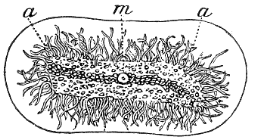
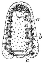
Fig. 120. Velella; m so-called mouth, a tentacles. (Agassiz.) Fig. 121. Free Medusa of Velella; a proboscis, b chymiferous tube, c circular tube. (Agassiz.)
MODE OF CATCHING JELLY-FISHES.
[fig 122]
Fig. 122. Ptychogena, natural size.
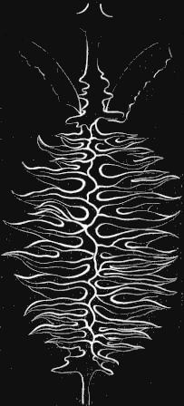
Not the least attractive feature in the study of these animals, is the mode of catching them. We will suppose it to be a warm, still morning at Nahant, in the last week of August, with a breath of autumn in the haze that softens the outlines of the opposite shore, and makes the horizon line a little dim. It is about eleven o'clock, for few of the Jelly-fishes are early risers; they like the warm sun, and at an earlier hour they are not to be found very near the surface. The sea is white and glassy, with a slight swell but no ripple, and seems almost motionless as we put off in a dory from the beach near Saunders's Ledge. We are provided with two buckets, one for the larger Jelly-fishes, the Zygodactyla, Aurelia, &c., the other for the smaller fry, such as the various kinds of Ctenophoræ, the Tima, Melicertum, &c. Beside these, we have two nets and glass bowls, in which to take up the more fragile creatures that cannot bear rough handling. A bump or two on the stones before we are fairly launched, a shove of the oar to keep the boat well out from the rocks along which we skirt for a moment, and now we are off. We pull around the point to our left and turn toward the Ledge, filling our buckets as we go. Now we are crossing the shallows that make the channel between the inner and outer rocks of Saunders's Ledge. Look down,—how clear the water is and how lovely the sea-weeds, above which we are floating, dark brown and purple fronds of the Ulvæ, and the long blades of the Laminaria with mossy green tufts between. As we issue from this narrow passage we must be on the watch, for the tide is rising, and may come laden with treasures, as it sweeps through it. A sudden cry from the oarsman at the bow, not of rocks or breakers ahead, but of "A new Jelly-fish astern!" The quick eye of the naturalist of the party pronounces it unknown to zoölogists, un-described by any scientific pen. Now what excitement! "Out with the net!—we have passed him! he has gone down! no, there he is again! back us a bit." Here he is floating close by us; now he is within the circle of the net, but he is too delicate to be caught safely in that way, so, while one of us moves the net gently about, to keep him within the space enclosed by it, another slips the glass bowl under him, lifts it quickly, and there is a general exclamation of triumph and delight,—we have him. And now we look more closely; yes, decidedly he is a novelty as well as a beauty. (See Fig. 122, Ptychogena lactea A. Ag.) Those white mossy tufts for ovaries are unlike anything we have found before (Fig. 123), and not represented in any published figures of Jelly-fishes. We float about here for a while, hoping to find more of the same kind, but no others make their appearance, and we keep on our way to East Point, where there is a capital fishing ground for Medusæ of all sorts. Here two currents meet, and the Jelly-fishes are stranded as it were along the line of juncture, able to move neither one way nor the other. At this spot the sea actually swarms with life; one cannot dip the net into the water without bringing up Pleurobrachia, Bolina, Idyia, Melicertum, &c., while the larger Zygodactyla and Aurelia float about the boat in numbers. These large Jelly-fishes produce a singular effect as one sees them at some depth beneath the water; the Aureliæ, especially, with their large white disks, look like pale phantoms wandering about far below the surface; but they constantly float upward, and if not too far out of reach, one may bring them up by stirring the water under them with the end of the oar.
[fig 123]
Fig. 123. Ovary of Ptychogena; magnified.
When we have passed an hour or so floating about just beyond East Point, and have nearly filled our buckets with Jelly-fishes of all sizes and descriptions, we turn and row homeward. The buckets look very pretty as they stand in the bottom of the boat with the sunshine lighting up their living contents. The Idyia glitters and sparkles with ever-changing hues, the Pleurobrachiæ dart about, trailing their long graceful tentacles after them, the golden Melicerta are kept in constant motion by their quick, sudden contractions, and the delicate transparent Tima floats among them all, not the less beautiful because so colorless. There is an unfortunate Idyia, who, by some mistake, has got into the wrong bucket with the larger Jelly-fish, where a Zygodactyla has entangled it among his tentacles and is quietly breakfasting upon it.
During our row the tide has been rising, and as we near the channel of Saunders's Ledge, it is running through more strongly than before, and at the entrance of the shallows a pleasant surprise is prepared us; no less than half a dozen of our new friends (the Ptychogena as he has been baptized), come to look for their lost companion perhaps, await us there, and are presently added to our spoils. We reach the shore heavily laden with the fruits of our morning's excursion.
The most interesting part of the work for the naturalist is still to come. On our return to the Laboratory, the contents of the buckets are poured into separate glass bowls and jars; holding them up against the light, we can see which are our best and rarest specimens; these we dip out in glass cups and place by themselves. If any small specimens are swimming about at the bottom of the jar, and refuse to come within our reach, there is a very simple mode of catching them. Dip a glass tube into the water, keeping the upper end closed with your finger, and sink it till the lower end is just above the animal you want to entrap; then lift your finger, and as the air rushes out the water rushes in, bringing with it the little creature you are trying to catch. When the specimens are well assorted, the microscope is taken out, and the rest of the day is spent in studying the new Jelly-fishes, recording the results, making notes, drawings, &c.
Still more attractive than the rows by day are the night expeditions in search of Jelly-fishes. For this object we must choose a quiet night, for they will not come to the surface if the water is troubled. Nature has her culminating hours, and she brings us now and then a day or night on which she seems to have lavished all her treasures. It was on such a rare evening, at the close of the summer of 1862, that we rowed over the same course by Saunders's Ledge and East Point described above. The August moon was at her full, the sky was without a cloud, and we floated on a silver sea; pale streamers of the aurora quivered in the north, and notwithstanding the brilliancy of the moon, they too cast their faint reflection in the ocean. We rowed quietly along past the Ledge, past Castle Rock, the still surface of the water unbroken, except by the dip of the oars and the ripple of the boat, till we reached the line off East Point, where the Jelly-fishes are always most abundant, if they are to be found at all. Now dip the net into the water. What genie under the sea has wrought this wonderful change? Our dirty, torn old net is suddenly turned to a web of gold, and as we lift it from the water heavy rills of molten metal seem to flow down its sides and collect in a glowing mass at the bottom. The truth is, the Jelly-fishes, so sparkling and brilliant in the sunshine, have a still lovelier light of their own at night; they give out a greenish golden light as brilliant as that of the brightest glow-worm, and on a calm summer night, at the spawning season, when they come to the surface in swarms, if you do but dip your hand into the water it breaks into sparkling drops beneath your touch. There are no more beautiful phosphorescent animals in the sea than the Medusæ; it would seem that the expression, "rills of molten metal" could hardly apply to anything so impalpable as a Jelly-fish, but, although so delicate in structure, their gelatinous disks give them a weight and substance; and at night, when their transparency is not perceived, and their whole mass is aglow with phosphorescent light, they truly have an appearance of solidity which is most striking, when they are lifted out of the water and flow down the sides of the net.
The various kinds present very different aspects; wherever the larger Aureliæ and Zygodactylæ float to the surface, they bring with them a dim spreading halo of light, the smaller Ctenophoræ become little shining spheres, while a thousand lesser creatures add their tiny lamps to the illumination of the ocean; for this so-called phosphorescence of the sea is by no means due to the Jelly-fishes alone, but is also produced by many other animals, differing in the color as well as the intensity of their light, and it is a curious fact that they seem to take possession of the field by turns. You may row over the same course, which a few nights since glowed with a greenish golden light wherever the surface of the water was disturbed, and though equally brilliant, the phosphorescence has now a pure white light. On such an evening, be quite sure that when you empty your buckets on your return and examine their contents you will find that the larger part of your treasures are small crustacea (little shrimps). Of course there will be other phosphorescent creatures, Jelly-fishes, &c., among them, but the predominant color is given by these little crustacea. On another evening the light will have a bluish tint, and then the phosphorescence is principally due to the Dysmorphosa (Fig. 105).
Notwithstanding the beauty of a moonlight row, if you would see the phosphorescence to greatest advantage you must choose a dark night, when the motion of your boat sets the sea on fire around you, and a long undulating wave of light rolls off from your oar as you lift it from the water. On a brilliant evening this effect is lost in a great degree, and it is not until you dip your net fairly under the moonlit surface of the sea, that you are aware how full of life it is. Occasionally one is tempted out by the brilliancy of the phosphorescence, when the clouds are so thick that water, sky, and land become one indiscriminate mass of black, and the line of rocks can be discerned only by the vivid flash of greenish golden light, when the breakers dash against them. At such times there is something wild and weird in the whole scene, which at once fascinates and appalls the imagination; one seems to be rocking above a volcano, for the surface around is intensely black, except where fitful flashes or broad waves of light break from the water under the motion of the boat or the stroke of the oars. It was on a night like this, when the phosphorescence was unusually brilliant, and the sea as black as ink, the surf breaking heavily and girdling the rocky shore with a wall of fire, that our collector was so fortunate as to find in the rich harvest he brought home the entirely new and exceedingly pretty little floating Hydroid, described under the name of Nanomia (Fig. 115). It was in its very infancy (Fig. 108), a mere bubble, not yet possessed of the various appendages which eventually make up its complex structure; but it was nevertheless very important to have seen it in this early stage of its existence, since, when a few full-grown specimens were found in the autumn, which lived for some days in confinement and quietly allowed their portraits to be taken (see Fig. 115), it was easy to connect the adult animal with the younger phase of its own life and thus make a complete history.
Marine phosphorescence is no new topic, and we have dwelt too long, perhaps, upon a phenomenon that every voyager has seen, and many have described. Its effect is very different, when seen from the deck of a vessel, from its appearance as one floats through its midst, distinguishing the very creatures that produce it, and any account of the Medusæ which did not include this most characteristic feature would be incomplete.
ECHINODERMS.
Our illustrations and descriptions of Echinoderms are scanty in comparison to those of the preceding class; for while, in consequence, perhaps, of the combined influence of the Gulf Stream and the cold arctic current on the New England shore, Acalephs are largely represented in our waters, our marine fauna is meagre in Echinoderms. But although we have few varieties, those which do establish themselves on a coast seemingly so ungenial for others of their kind, such as the Echinus, and our common Star-fish, for instance, thrive well and are very abundant. The class of Echinoderms includes five orders, viz. Crinoids, Ophiurans, Star-fishes, Sea-urchins, and Holothurians. The animals composing these orders differ so widely in appearance that it was very long before their true relations were detected, and it was seen that all their external differences were united under a common plan. Let us compare, for instance, the worm-like Holothurians (Figs. 124, 126, 127) with all the host of Star-fishes (Figs. 142, 146, 147) and Sea-urchins (Figs. 131, 139), or compare the radiating form of the Star-fish, its arms spreading in every direction, with the close spherical outline of the Sea-urchin, or the Crinoid floating at the end of a stem (Fig. 152) with either of these, and we shall cease to wonder that naturalists failed to find at once a unity of idea under all these varieties of execution. And yet the fundamental structure of the class of Echinoderms is represented as distinctly by any one of its five orders as by any other, and is absolutely identical in all. They differ only by trifling modifications of development.
In Echinoderms as a class, the body presents three regions differing in structure, and on the greater or less development of these regions or systems, as we may call them, their chief differences are based. Take, for instance, the dorsal system, the nature of which is explained by the name, indicating of course the back of the animal, though it does not necessarily imply the upper side of the body, since some of the Echinoderms, as the stemmed Crinoids, for example, carry the dorsal side downward, while the Star-fishes and Sea-urchins carry it upward, and the Holothurians, moving with the mouth forward, have the dorsal system at the opposite end of the body. Whatever the natural attitude of the animal, however, and the consequent position of the dorsal region, it exists alike in all the five orders, though it has not the same extent and importance in each. But in all it is made up of similar parts, bears the same relation to the rest of the body, has the same share in the general economy of the animal. And though when we compare the spreading back of a Star-fish with the small area on the top of a Sea-urchin, where all the zones unite, we may not at once see the correspondence between them, yet a careful comparison of all their structural details shows that they are both built with the same elements and represent the same region, though it is stretched to the utmost in the one case, and greatly contracted in the other.
This being true of the dorsal system, let us look at another equally important structural feature in this class. All Echinoderms have locomotive organs peculiar to themselves, a kind of suckers which may be more or less numerous, larger or smaller, in different species, but are always appendages of the same character. These are variously distributed over the body, but always with a certain regularity occupying definite spaces, shown by investigation to be homologous in all. For instance, the rays of the Star-fish correspond in every detail on their under side, along which the locomotive suckers run, with the zones on the Sea-urchin, from end to end of which the suckers are arranged; and the same is equally true of the distribution of the suckers on the Holothurians, Ophiurans, and Crinoids, though, as most persons are less familiar with these orders than with the other two, it might not be so easy to point out the coincidence to our readers. These suckers are called the ambulacra, the lines along which they run are called the ambulacral rows or zones, while the system of locomotion as a whole is known as the ambulacral system. Since these organs are thus regularly distributed over the body in distinct zones or rows, it follows that the latter must be divided by intervening spaces. These intervals are called the interambulacral spaces; but while in some orders they are occupied by larger plates and prominent spines, as in the Sea-urchin and Star-fish, in others they are either comparatively insignificant or completely suppressed, as in the Crinoids and Ophiurans. Such are the three regions or systems which by their greater or less development introduce an almost infinite variety of combinations into this highest class of Radiates. It may not be amiss before proceeding further to compare the five orders with reference to this point, and see which of these three systems has the preponderance in each one.
Taking the orders in their rank and beginning with the lowest, we find in the Crinoids that the dorsal system preponderates, being composed of highly complicated plates, and developed to such a degree as to form in many instances a stem by which the animal is attached to the ground, while the ambulacral system is limited to a comparatively small area, and the interambulacral system is wanting. The order of Crinoids has diminished so much in modern geological times that we must consult its fossil forms in order to understand fully the peculiar adaptation of the Echinoderm plan in this group.
In the Ophiurans, the dorsal system is still large, and though it no longer stretches out to form a stem, it folds over on the under side of the animal so as to enclose entirely the ambulacral system, forming a kind of shield for the arms. Here also the interambulacral system is wanting.
In the Star-fishes the dorsal system encroaches less upon the structure of the animal. The back and oral side here correspond exactly in size, and though the flat leathery upper surface of the animal, covered with spines, serves as a protection to the delicate ambulacral suckers which find their way between the rows of small plates along the under side of the arms, yet it does not enfold them as in the Ophiurans. On the contrary, in the Star-fishes the ambulacral rows are protected on either side by a row of the so-called interambulacral plates, through which no suckers pass.
In the Sea-urchin, the dorsal system is contracted to a minimum, forming a small area on the top of the animal, the rows of interambulacral plates which are separated and lie on either side of the ambulacra in the Star-fish being united in the Sea-urchin, and both the ambulacral and the interambulacral systems bent upward, meeting in the small dorsal area above, so as to form a spherical outline. Here the ambulacral and interambulacral systems have taken a great preponderance over the dorsal system, and the same is the case with the Holothurians, in which the same structure is greatly elongated, the dorsal system being thus pushed out as it were to the end of a cylinder, while the ambulacral and interambulacral systems run along its whole length. All Echinoderms without exception have ambulacral tubes, even though in some there are no external ambulacral suckers connected with them.
There is one organ peculiar to the class of Echinoderms, the general structure of which may be described here, since it is common to them all, with the exception of the Crinoids, the anatomy of which is, however, so imperfectly understood, that we are hardly justified in assuming that it does not exist even in that order. This organ is known as the madreporic body; it is a small sieve or limestone filter opening into a tube or canal; by means of this tube, which connects with the ambulacral system, the water from without, first filtered through the madreporic body and thus freed from any impurities, is conveyed to the ambulacra. In the more detailed account of the different orders we shall see what is the position of this singular organ in each group, and how it is adapted in them all to their special structure. The development of Echinoderms forms one of the most wonderful chapters in the annals of Natural History. Marvellous as is the embryonic history of the Acalephs, including all the different aspects they assume in the cycle of their growth, it is thrown into the shade by the transformations which Echinoderms undergo before assuming their adult condition. This singular mode of development, although it has features recalling the development of Jelly-fishes from Hydroids, is nevertheless entirely distinct from it, and is known only in the class of Echinoderms. As the whole story is given at length in the chapter on the embryology of the Echinoderms, we need only allude to it here in general terms. We owe the discovery of this remarkable process to Johannes Müller, one of the greatest anatomists of this century.
HOLOTHURIANS.
[fig 124]
Fig. 124. Synapta, natural size.
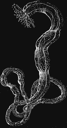
Synapta. (Synapta tenuis Ayres.)
This is one of the most curious of the Holothurians, and easily observed on account of its transparency, which allows us to see its internal structure. It has a long cylindrical body (Fig. 124) along the length of which run the five rows of ambulacra, which are in this instance closed tubes without any projecting suckers or locomotive organs of any kind attached to them, so that the name is retained only on account of their correspondence in position, and not from any similarity of function to the ambulacra in Star-fishes and Sea-urchins. But though the ambulacra in Synapta are in fact mere water-tubes like the vertical tubes in the Ctenophoræ, by means of which the water, first filtered through the madreporic body, circulates along the skin, they are as organs perfectly homologous with the ambulacra in all other Echinoderms. The mouth has a circular tube around the aperture, and a wreath of branching tentacles encircling it. The habits of these animals are singular. They live in very coarse mud, but they surround themselves with a thin envelope of finer sand, which they form by selecting the small particles with their tentacles, and making a ring around their anterior extremity. This ring they then push down along the length of the body, and continue this process, adding ring after ring, till they have entirely encircled themselves with a sand tube. They move the rings down partly by means of contractions of the body, but also by the aid of innumerable appendages over the whole surface. To the naked eye these appendages appear like little specks on the skin; but under the microscope they are seen to be warts projecting from the surface, each one containing a little anchor with the arms turned upward (Fig. 125). Around the mouth [fig 125] Fig. 125. Anchor of Synapta; a anchor, w plate upon which anchor is attached; greatly magnified. these warts are larger, but do not contain any anchors. It will be seen hereafter that these appendages are homologous with certain organs in other Holothurians, the warts with the anchors corresponding to the limestone pavement covering or partially covering the surface of the Cuvieria, for instance, while those without anchors correspond to the so-called false ambulacra in Pentacta. By means of these appendages, though aided also by the contractions of the body, the Synaptæ move through the mud and collect around themselves the sand tube in which they are encased. Their food is very coarse for animals so delicate in structure. When completely empty of food they are white, perfectly transparent, and the spiral tube forming the digestive cavity may be seen wound up and hanging loosely in the centre for the whole length of the body. In such a condition it is of a pale yellow color. But look at one that is gorged with food. The whole length of the alimentary canal is then crowded with sand, pebbles, and shells, distinctly seen through the transparent skin, and giving a dark gray color to the whole body. They swallow the sand for the sake of the nutritious substance it contains, and having assimilated and digested this, they then eject the harder materials. The motion of the body in consequence of its contractions is much like that of leeches, and on this account these Synaptæ were long supposed to be a transition type between the Radiates and worms. The body grows to a great length, often half a yard and more, but constantly drops large portions from its posterior part, by means of its own contractions, or breaks itself up by the expulsion of the intestines, which are very readily cast out. The tentacles are hollow, consisting of a central rib with branches from either side. In the Synaptæ, as in all the Holothurians, the madreporic body is placed near the mouth, between two of the ambulacra, and opposite the fifth or odd one. The tube, connecting with the central tube around the mouth, by means of which it communicates with the ambulacral tubes, is very short.
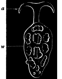
Caudina. (Caudina arenata Stimps.)
[fig 126]
Fig. 126. Caudina arenata; natural size.
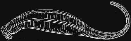
Several other Holothurians are frequently met with on our shores. Among them is the Caudina arenata (Fig. 126), a small Holothurian, yellowish in color, and thick in texture, by no means so pretty as the white transparent Synapta; the tentacles are short, resembling a crown of cloves around the mouth. It lives in the sand, and may be found in great numbers on the sandy beaches after a storm.
Cuvieria. (Cuvieria squamata D. & K.)
[fig 127]
Fig. 127. Cuvieria; natural size.
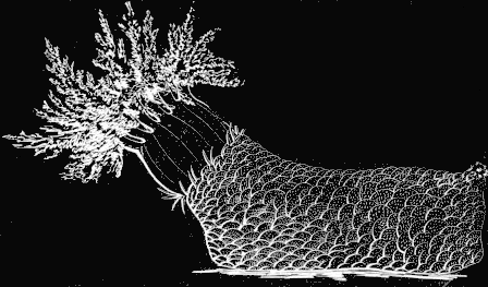
The Holothurian of our coast, excelling all the rest in beauty, is the Cuvieria. (Fig. 127.) As it lies on the sand, a solid red lump, with neither grace of form nor beauty of color, even the vividness of its tint growing dull and dead when it is removed from its native element, certainly no one could suspect that it possessed any hidden charm; but place it in a glass bowl with fresh sea-water; the dull red changes to deep vivid crimson, the tentacles creep out (Fig. 127) softly, and slowly, till the mouth is surrounded by a spreading wreath, comparable for richness of tint, and for delicate tracery, to the most beautiful sea-weeds. These tentacles, when fully expanded, are as long as the body itself. A limestone pavement composed of numerous pieces covers almost the whole surface of the animal; this apparatus corresponds, as we have already mentioned, to the warts containing anchors in the Synapta; but in the latter, the limestone particles are smaller, whereas in the Cuvieria they are developed to a remarkable extent. This animal is very sluggish, the ambulacral suckers, found only on three of the tubes, being arranged in such a way as to form a sort of sole on which they creep; the sole is tough and leathery in texture, but free from the limestone pavement described above. The young (Figs. 128, 129) are very common, swimming freely about, and more readily found than the adult; they are of a bright vermilion color, but the tentacles hardly branch at that age, nor is the limestone pavement formed, which gives such a peculiar aspect to the full-grown animal. The young Cuvieria, somewhat older than that represented in Fig. 129, are found in plenty under stones at low-water mark, just after they have given up their nomadic habits, and when the limestone pavement begins to be developed.
[fig 128]
[fig 129]
Fig. 128. Young Cuvieria, much enlarged; l body, g tentacles. Fig. 129. Somewhat older Cuvieria; l body, g tentacle round mouth, g' testaete of sole, b madreporic tentacle.
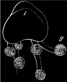
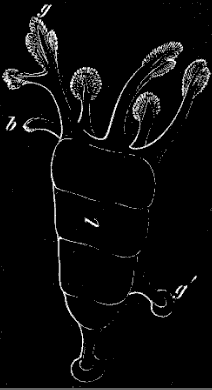
Pentacta. (Pentacta frondosa Jäg.)
The highest of our Holothurians in structure, is the Pentacta. (Fig. 130) It is very rare on our beaches, though occasionally found under stones at low-water mark; farther north, in Maine, and at Grand Manan, it is very common, covering all the rocks near low-water mark. It is a chocolate brown in color, and measures, when fully expanded, some fifteen to eighteen inches in length. Unlike the Cuvieria, the ambulacral suckers are evenly distributed and almost equally developed on all the tubes; between the five rows of ambulacral suckers are scattered irregularly certain appendages resembling suckers, but found on examination not to be true locomotive suckers, and called on that account false ambulacra. These are the organs corresponding to the warts around the mouth of the Synapta. Although the ambulacral suckers are, as we have said, equally developed on all the tubes, yet the Pentacta does not use them indiscriminately as locomotive organs. In Pentacta, as well as in all Holothurians, whether provided with ambulacral suckers, or, like the Synapta and Caudina, deprived of them, the odd ambulacrum, viz. the one placed opposite the madreporic body, is always used to creep upon, and forms the under surface of the animal.