MALARIA.
Reading edition
By JAMES H. SALISBURY, A.M., M.D.
PRIZE ESSAY OF THE ALBANY MEDICAL COLLEGE ALUMNI ASSOCIATION, FEB., 1882.
VII.
I have made careful microscopic examinations of the blood in several cases of Panama fever I have treated, and find in all severe cases many of the colorless corpuscles filled more or less with spores of ague vegetation and the serum quite full of the same spores (see Fig. N, Plate VIII.).
Mr. John Thomas. Panama fever. Vegetation in blood and colorless corpuscles. (Fig N, Plate VIII.) Vegetation, spores of, in the colorless corpuscles of the blood. Spores in serum of blood adhering to fibrin filaments.
Mr. Thomas has charge of the bridge building on the Tehuantepec Railroad. Went there about one year ago. Was taken down with the fever last October. Returned home in February last, all broken down. Put him under treatment March 15, 1882. Gained rapidly (after washing him out with hot water, and getting his urine clear and bowels open every day) on two grains of quinia every day, two hours, till sixteen doses were taken. After an interval of seven days, repeated the quinia, and so on. This fever prevails on all the low lands, as soon as the fresh soil is exposed to the drying rays of the sun. The vegetation grows on the drying soil, and the spores rise in the night air, and fall after sunrise. All who are exposed to the night air, which is loaded with the spores, suffer with the disease. The natives of the country suffer about as badly as foreigners. Nearly half of the workmen die of the disease. The fever is a congestive intermittent of a severe type.
Henry Thoman. Leucocythæmia. Spleen 11 inches in diameter, two white globules to one red. German. Thirty-six years of age. Weight, 180 pounds. Colorless corpuscles very large and varying much in size, as seen at N. Corpuscles filled--many of them--with the spores of ague vegetation. Also spores swimming in serum.
This man has been a gardener back of Hoboken on ague lands, and has had ague for two years preceding this disease.
I will now introduce a communication made to me by a medical gentleman who has followed somewhat my researches for many years, and has taken great pains of time and expense to see if my researches are correct.
REPORT ON THE CAUSE OF AGUE.--BY DR. EPHRAIM CUTTER, TO THE WRITER
At your request I give the evidence on which I base my opinion that your plan in relation to ague is true.
From my very start into the medical profession, I had a natural intense interest in the causes of disease, which was also fostered by my father, the late Dr. Cutter, who honored his profession nearly forty years. Hence, I read your paper on ague with enthusiasm, and wrote to you for some of the plants of which you spoke. You sent me six boxes containing soil, which you said was full of the gemiasmas. You gave some drawings, so that I should know the plants when I saw them, and directed me to moisten the soil with water and expose to air and sunlight. In the course of a few days I was to proceed to collect. I faithfully followed the instructions, but without any success. I could detect no plants whatever,
This result would have settled the case ordinarily, and I would have said that you were mistaken, as the material submitted by yourself failed as evidence. But I thought that there was too much internal evidence of the truth of your story, and having been for many years an observer in natural history, I had learned that it is often very difficult for one to acquire the art of properly making examinations, even though the procedures are of the simplest description. So I distrusted, not you, but myself, and hence, you may remember, I forsook all and fled many hundred miles to you from my home with the boxes you had sent me. In three minutes after my arrival you showed me how to collect the plants in abundance from the very soil in the boxes that had traveled so far backward and forward, from the very specimens on which I had failed to do so.
The trouble was with me--that I went too deep with my needle. You showed me it was simply necessary to remove the slightest possible amount on the point of a cambric needle; deposit this in a drop of clean water on a slide cover with, a covering glass and put it under your elegant 1/5 inch objective, and there were the gemiasmas just as you had described.
I have always felt humbled by this teaching, and I at the time rejoiced that instead of denouncing you as a cheat and fraud (as some did at that time), I did not do anything as to the formation of an opinion until I had known more and more accurately about the subject.
I found all the varieties of the palmellæ you described in the boxes, and I kept them for several years and demonstrated them as I had opportunity. You also showed me on this visit the following experiments that I regarded as crucial:
1st. I saw you scrape from the skin of an ague patient sweat and epithelium with the spores and the full grown plants of the Gemiasma verdans.
2d. I saw you take the sputa of a ague patient and demonstrate the spores and sporangia of the Gemiasma verdans.
3d. I saw you take the urine of a female patient suffering from ague (though from motives of delicacy I did not see the urine voided--still I believe that she did pass the urine, as I did not think it necessary to insult the patient), and you demonstrated to me beautiful specimens of Gemiasma rubra. You said it was not common to find the full development in the urine of such cases, but only in the urine of the old severe cases. This was a mild case.
4th. I saw you take the blood from the forearm of an ague patient, and under the microscope I saw you demonstrate the gemiasma, white and bleached in the blood. You said that the coloring matter did not develop in the blood, that it was a difficult task to demonstrate the plants in the blood, that it required usually a long and careful search of hours sometimes, and at other times the plants would be obtained at once.
When I had fully comprehended the significance of the experiments I was filled with joy, and like the converts in apostolic times I desired to go about and promulgate the news to the profession. I did so in many places, notably in New York city, where I satisfactorily demonstrated the plants to many eminent physicians at my room at the Fifth Avenue Hotel; also before a medical society where more than one hundred persons were present. I did all that I could, but such was the preoccupation of the medical gentlemen that a respectful hearing was all I got. This is not to be wondered at, as it was a subject, now, after the lapse of nearly a decade and a half, quite unstudied and unknown. After this I studied the plants as I had opportunity, and in 1877 made a special journey to Long Island, N.Y., for the purpose of studying the plants in their natural habitat, when they were in a state of maturity. I have also examined moist soils in localities where ague is occasionally known, with other localities where it prevails during the warm months.
Below I give the results, which from convenience I divide into two parts: 1st. Studies of the ague plants in their natural habitat. 2d. Studies of the ague plants in their unnatural habitat (parasitic). I think one should know the first before attempting the second.
First--Studies to find in their natural habitat the palmellæ described as the Gemiasma rubra, Gemiasma verdans, Gemiasma plumba, Gemiasma alba, Protuberans lamella.
Second--Outfit.--Glass slides, covers, needles, toothpicks, bottle of water, white paper and handkerchief, portable microscope with a good Tolles one inch eyepiece, and one-quarter inch objective.
Wherever there was found on low, marshy soil a white incrustation like dried salt, a very minute portion was removed by needle or toothpick, deposited on a slide, moistened with a drop of water, rubbed up with a needle or toothpick into a uniformly diffused cloud in and through the water. The cover was put on, and the excess of water removed by touching with a handkerchief the edge of the cover. Then the capillary attraction held the cover in place, as is well known. The handkerchief or white paper was spread on the ground at my feet, and the observation conducted at once after the collection and on the very habitat. It is possible thus to conduct observations with the microscope besides in boats on ponds or sea, and adding a good kerosene light in bed or bunk or on lounge.
August 11, 1877.--Excursion to College Point, Flushing, Long Island:
Observation 1. 1:50 P.M. Sun excessively hot. Gathered some of the white incrustation on sand in a marsh west of Long Island Railroad depot. Found some Gemiasma verdans, G. rubra; the latter were dry and not good specimens, but the field swarmed with the automobile spores. The full developed plant is termed sporangia, and seeds are called spores.
Observation 2. Another specimen from same locality, not good; that is, forms were seen but they were not decisive and characteristic.
Observation 3. Earth from Wallabout, near Naval Hospital, Brooklyn, Rich in spores (A) with automobile protoplasmic motions, (B) Gemiasma rubra, (C) G. verdans, very beautiful indeed. Plants very abundant.
Observation 4. Walking up the track east of L. I. R.R. depot, I took an incrustation near creek; not much found but dirt and moving spores.
Observation 5. Seated on long marsh grass I scraped carefully from the stalks near the roots of the grass where the plants were protected from the action of the sunlight and wind. Found a great abundance of mature Gemiasma verdans very beautiful in appearance.
Notes.--The time of my visit was most unfavorable. The best time is when the morning has just dawned and the dew is on the grass. One then can find an abundance, while after the sun is up and the air is hot the plants disappear; probably burst and scatter the spores in billions, which, as night comes on and passes, develop into the mature plants, when they may be found in vast numbers. It would seem from this that the life epoch of a gemiasma is one day under such circumstances, but I have known them to be present for weeks under a cover on a slide, when the slide was surrounded with a bandage wet with water, or kept in a culture box. The plants may be cultivated any time in a glass with a water joint. A, Goblet inverted over a saucer; B, filled with water; C, D, specimen of earth with ague plants.
Observation 6. Some Gemiasma verdaus; good specimens, but scanty. Innumerable mobile spores. Dried.
Observation 7. Red dust on gray soil. Innumerable mobile spores. Dried red sporangia of G. rubra.
Observation 8. White incrustation. Innumerable mobile spores. No plants.
Observation 9. White incrustation. Many minute algæ, but two sporangia of a pale pink color; another variety of color of gemiasma. Innumerable mobile spores.
Observation 10. Gemiasma verdans and G. rubra in small quantities. Innumerable mobile spores.
Observation 11. Specimen taken from under the shade of short marsh grass. Gemiasma exceedingly rich and beautiful. Innumerable mobile spores.
Observation 12. Good specimens of Gemiasma rubra. Innumerable spores present in all specimens.
Observation 13. Very good specimens of Protuberans lamella.
Observation 14. The same.
Observation 15. Dead Gemiasma verdans and rubra.
Observation 16. Collection very unpromising by macroscopy, but by microscopy showed many spores, mature specimens of Gemiasma rubra and verdans. One empty specimen with double walls.
Observation 17. Dry land by the side of railroad. Protuberans not abundant.
Observation 18. From side of ditch. Filled with mature Geraiasma verdans.
Observation 19. Moist earth near a rejected timber of the railroad bridge. Abundance of Gemiasma verdans, Sphærotheca Diatoms.
Observation 20. Scrapings on earth under high grass. Large mature specimens of Gemiasma rubra and verdans. Many small.
Observation 21. Same locality. Gemiasma rubra and verdans; good specimens.
Observation 22. A dry stem of a last year's annual plant lay in the ditch not submerged, that appeared as if painted red with iron rust. This redness evidently made up of Gemiasma rubra dried.
Observation 23. A twig submerged in a ditch was scraped. Gemiasma verdans found abundantly with many other things, which if rehearsed would cloud this story.
Observation 24. Scrapings from the dirty end of the stick (23) gave specimens of the beautiful double wall palmellæ and some empty G. verdans.
Observation 25. Stirred up the littoral margins of the ditch with stick found in the path, and the drip showed Gemiasma rubra and verdans mixed in with dirt, debris, other algae, fungi, infusoria, especially diatoms.
Observation 26. I was myself seized with sneezing and discharge running from nostrils during these examinations. Some of the contents of the right nostril were blown on a slide, covered, and examined morphologically. Several oval bodies, round algae, were found with the characteristics of G. verdans and rubra. Also some colorless sporangia, and spores abundantly present. These were in addition to the normal morphological elements found in the excretions.
Observation 27. Dried clay on margin of the river showed dry G. verdans.
Observation 28. Saline dust on earth that had been thrown out during the setting of a new post in the railroad bridge showed some Gemiasma alba.
Observation 29. The dry white incrustation found on fresh earth near railroad track entirely away from water, where it appeared as if white sugar or sand had been sprinkled over in a fine dust, showed an abundance of automobile spores and dry sporangia of G. rubra and verdans. It was not made up of salts from evaporation.
Observation 30. Some very thick, long, green, matted marsh grass was carefully separated apart like the parting of thick hair on the head. A little earth was taken from the crack, and the Protuberans lamella, the Gemiasma rubra and verdans found were beautiful and well developed.
Observation 31. Brooklyn Naval Hospital, August 12, 1877, 4 A.M. Called up by the Quartermaster. With Surgeon C. W. White, U.S.N., took (A) one five inch glass beaker, bottomless, (B) three clean glass slides, (C) chloride of calcium solution, [symbol: dra(ch)m] i to [symbol: ounce] i water. We went, as near as I could judge in the darkness, to about that portion of the wall that lies west of the hospital, southeast corner (now all filled up), where on the 10th of August previously I had found some actively growing specimens of the Gemiasma verdans, rubra, and protuberans. The chloride of calcium solution was poured into a glass tumbler, then rubbed over the inside and outside of the beaker. It was then placed on the ground, the rim of the mouth coming on the soil and the bottom elevated on an old tin pan, so that the beaker stood inclined at an angle of about forty-five degrees with the horizon. The slides were moistened, one was laid on a stone, one on a clod, and a third on the grass. Returned to bed, not having been gone over ten minutes.
At 6 A.M. collected and examined for specimens the drops of dew deposited. Results: In every one of the five instances collected the automobile spores, and the sporangia of the gemiasmas and the protuberans on both sides of slides and beaker. There were also spores and mycelial filaments of fungi, dirt, and zoospores. The drops of dew were collected with capillary tubes such as were used in Edinburgh for vaccine virus. The fluid was then preserved and examined in the naval laboratory. In a few hours the spores disappeared.
Observation 32. Some of the earth near the site of the exposure referred to in Observation 31, was examined and found to contain abundantly the Gemiasma verdans, rubra, Protuberans lamella, confirmed by three more observations.
Observation 33. In company with Surgeon F. M. Dearborne, U.S.N., in charge of Naval Hospital, the same day later explored the wall about marsh west of hospital. Found the area abundantly supplied with palmellæ, Gemiasma rubra, verdans, and Protuberans lamella, even where there was no incrustation or green mould. Made very many examinations, always finding the plants and spores, giving up only when both of us were overcome with the heat.
Observation 34. August, 1881. Visited the Wallabout; found it filled up with earth. August 17. Visited the Flushing district; examined for the gemiasma the same localities above named, but found only a few dried up plants and plenty of spores. With sticks dug up the earth in various places near by. Early in September revisited the same, but found nothing more; the incrustation, not even so much as before. The weather was continuously for a long time very dry, so much so that vegetables and milk were scarce.
The grass and grounds were all dried up and cracked with fissures.
There must be some moisture for the development of the plants. Perhaps if I had been able to visit the spots in the early morning, it would have been much better, as about the same time I was studying the same vegetation on 165th Street and 10th Avenue, New York, and found an abundance of the plants in the morning, but none scarcely in the afternoon.
Should any care to repeat these observations, these limits should be observed and the old adage about "the early bird catching the worm," etc. Some may object to this directness of report, and say that we should report all the forms of life seen. To this I would say that the position I occupy is much different from yours, which is that of discoverer. When a detective is sent out to catch a rogue, he tumbles himself but little with people or things that have no resemblance to the rogue. Suppose he should return with a report as to the houses, plants, animals, etc., he encountered in his search; the report might be very interesting as a matter of general information, but rather out of place for the parties who desire the rogue caught. So in my search I made a special work of catching the gemiasmas and not caring for anything else. Still, to remove from your mind any anxiety that I may possibly not have understood how to conduct my work, I will introduce here a report of search to find out how many forms of life and substances I could recognize in the water of a hydrant fed by Croton water (two specimens only), during the present winter (1881 and 1882) I beg leave to subjoin the following list of species, not individuals, I was able to recognize. In this list you will see the Gemiasma verdans distinguished from its associate objects. I think I can in no other way more clearly show my right to have my honest opinion respected in relation to the subject in question.
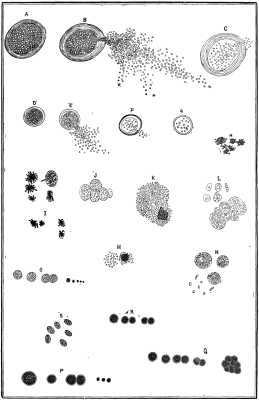
MALARIA PLANTS COLLECTED SEPT. 10, 1882, AT WASHINGTON HEIGHTS, 176TH STREET, NEAR 10TH AVENUE, NEW YORK CITY, ETC. PLATE VIII.--A, B, C, Large plants of Gemiasma verdans. A, Mature plant. B, Mature plant discharging spores and spermatia through a small opening in the cell wall. C, A plant nearly emptied. D, Gemiasma rubra; mature plant filled with microspores. E, Ripe plant discharging contents. F, Ripe plant, contents nearly discharged; a few active spermatia left behind and escaping. G, nearly empty plant. H, Vegetation in the SWEAT of ague cases during the paroxysm of sweating. I, Vegetation in the BLOOD of ague. J, Vegetation in the urine of ague during paroxysm. K, L, M, Vegetation in the urine of chronic cases of severe congestive type. N, Vegetation in BLOOD of Panama fever; white corpuscles distended with spores of Gemiasma. O, Gemiasma alba. P, Gemiasma rubra. Q, Gemiasma verdans. R, Gemiasma alba. O, P, Q, R, Found June 28,1867, in profusion between Euclid and Superior Streets, near Hudson, Cleveland, O. S, Sporangia of Protuberans.
List of objects found in the Croton water, winter of 1881 and 1882. The specimens obtained by filtering about one barrel of water:
1. Acineta tuberosa. 2. Actinophrys sol. 3. Amoeba proteus. 4. " radiosa. 5. " verrucosa. 6. Anabaina subtularia. 7. Ankistrodesmus falcatus. 8. Anurea longispinis. 9. " monostylus. 10. Anguillula fluviatilis. 11. Arcella mitrata. 12. " vulgaris. 13. Argulus. 14. Arthrodesmus convergens. 15. Arthrodesmus divergens. 16. Astrionella formosa. 17. Bacteria. 18. Bosmina. 19. Botryiococcus. 20. Branchippus stagnalis. 21. Castor. 22. Centropyxis. 23. Chetochilis. 24. Chilomonads. 25. Chlorococcus. 26. Chydorus. 27. Chytridium. 28. Clatbrocystis æruginosa. 29. Closterium lunula. 30. " didymotocum. 31. " moniliferum. 32. Coelastrum sphericum. 33. Cosmarium binoculatum. 34. Cyclops quad. 35. Cyphroderia amp. 36. Cypris tristriata. 37. Daphnia pulex. 38. Diaptomas castor. 39. " sull. 40. Diatoma vulgaris. 41. Difflugia cratera. 42. " globosa. 43. Dinobryina sertularia. 44. Dinocharis pocillum. 45. Dirt. 46. Eggs of polyp. 47. " entomostraca. 48. " plumatella. 49. " bryozoa. 50. Enchylis pupa. 51. Eosphora aurita. 52. Epithelia, animal. 53. " vegetable. 54. Euastrum. 55. Euglenia viridis. 56. Euglypha. 57. Eurycercus lamellatus. 58. Exuvia of some insect. 59. Feather barbs. 60. Floscularia. 61. Feathers of butterfly. 62. Fungu, red water. 63. Fragillaria. 64. Gemiasma verdans. 65. Gomphospheria. 66. Gonium. 67. Gromia. 68. Humus. 69. Hyalosphenia tinctad. 70. Hydra viridis. 71. Leptothrix. 72. Melosira. 73. Meresmopedia. 74. Monactina. 75. Monads. 76. Naviculæ. 77. Nitzschia. 78. Nostoc communis. 79. OEdogonium. 80. Oscillatoriaceæ. 81. Ovaries of entomostraca. 82. Pandorina morum. 83. Paramecium aurelium. 84. Pediastrum boryanum. 85. " incisum. 86. " perforatum. 87. " pertusum. 88. " quadratum. 89. Pelomyxa. 90. Penium. 91. Peredinium candelabrum. 92. Peredinium cinc. 93. Pleurosigma angulatum. 94. Plumatella. 95. Plagiophrys. 96. Playtiptera polyarthra. 97. Polycoccus. 98. Pollen of pine. 99. Polyhedra tetraëtzica. 100. " triangularis. 101. Polyphema. 102. Protococcus. 103. Radiophrys alba. 104. Raphidium duplex. 105. Rotifer ascus. 106. " vulgaris. 107. Silica. 108. Saprolegnia. 109. Scenedesmus acutus. 110. " obliquus. 111. " obtusum. 112. " quadricauda. 113. Sheath of tubelaria. 114. Sphærotheca spores. 115. Spirogyra. 116. Spicules of sponge. 117. Starch. 118. Staurastrum furcigerum. 119. " gracile. 120. Staurogenum quadratum. 121. Surirella. 122. Synchoeta. 123. Synhedra. 124. Tabellaria. 125. Tetraspore. 126. Trachelomonas. 127. Trichodiscus. 128. Uvella. 129. Volvox globator. 130. " sull. 131. Vorticel. 132. Worm fluke. 133. Worm, two tailed. 134. Yeast.
More forms were found, but could not be determined by me. This list will give an idea of the variety of forms to be met with in the hunt for ague plants; still, they are as well marked in their physical characters as a potato is among the objects of nature. Although I know you are perfectly familiar with algæ, still, to make my report more complete, in case you should see fit to have it pass out of your hands to others, allow me to give a short account of the Order Three of Algæ, namely, the Chlorosporeæ or Confervoid Algæ, derived from the Micrographic Dictionary, this being an accessible authority.
Algae form a class of the thallophytes or cellular plants in which the physiological functions of the plant are delegated most completely to the individual cell. That is to say, the marked difference of purpose seen in the leaves, stamens, seeds, etc., of the phanerogams or flowering plants is absent here, and the structures carrying on the operations of nutrition and those of reproduction are so commingled, conjoined, and in some cases identified, that a knowledge of the microscopic anatomy is indispensable even to the roughest conception of the natural history of these plants; besides, we find these plants so simple that we can see through and through them while living in a natural condition, and by means of the microscope penetrate to mysteries of organism, either altogether inaccessible, or only to be attained by disturbing and destructive dissection, in the so called higher forms of vegetation. We say "so-called" advisedly, for in the Algæ are included the largest forms of plant life.
The Macrocystis pyrifera, an Algæ, is the largest of all known plants. It is a sea weed that floats free and unattached in the ocean. Covers the area of two square miles, and is 300 feet in depth (Reinsch). At the same time its structure on examination shows it to belong to the same class of plants as the minute palmellæ which we have been studying. Algæ are found everywhere in streams, ditches, ponds, even the smallest accumulations of water standing for any time in the open air, and commonly on walls or the ground, in all permanently damp situations. They are peculiarly interesting in regard to morphological conditions alone, as their great variety of conditions of organization are all variations, as it were, on the theme of the simple vegetable cell produced by change of form, number, and arrangement.
The Algæ comprehend a vast variety of plants, exhibiting a wonderful multiplicity of forms, colors, sizes, and degrees of complexity of structure, but algologists consider them to belong to three orders: 1. Red spored Algæ, called Rhodosporeæ or florideæ. 2. The dark or black spored Algæ, or Melanosporeæ or Fucoideæ. 3. The green spored Algæ, or Chlorosporeæ or Confervoideæ. The first two classes embrace the sea-weeds. The third class, marine and aquatic plants, most of which when viewed singly are microscopic. Of course some naturalists do not agree to these views. It is with order three, Confervoideæ, that we are interested. These are plants growing in sea or fresh water, or on damp surfaces, with a filamentous, or more rarely a leaf-like pulverulent or gelatinous thallus; the last two forms essentially microscopic. Consisting frequently of definitely arranged groups of distinct cells, either of ordinary structure or with their membrane silicified--Diatomaceæ. We note three forms of fructification: 1. Resting spores produced after fertilization either by conjugation or impregnation. 2. Spermatozoids. 3. Zeospores; 2, 4, or multiciliated active automobile cells--gonidia--discharged from the mother cells or plants without impregnation, and germinating directly. There is also another increase by cell division.
SYNOPSIS OF THE FAMILIES.
1. Lemaneæ.--Frond filamentous, inarticulate, cartilaginous, leathery, hollow, furnished at irregular distances with whorls or warts, or necklace shaped. Fructification: tufted, simple or branched, necklace shaped filaments attached to the inner surface of the tubular frond, and finally breaking up into elliptical spores. Aquatic.
2. Batrachospermeæ--Plants filamentous, articulated, invested with gelatine. Frond composed of aggregated, articulated, longitudinal cells, whorled at intervals with short, horizontal, cylindrical or beaded, jointed ramuli. Fructification: ovate spores and tufts of antheridial cells attached to the lateral ramuli, which consist of minute, radiating, dichotomous beaded filaments. Aquatic.
3. Chaetophoraceæ.--Plants growing in the sea or fresh water, coated by gelatinous substance; either filiform or a number of filaments being connected together constituting gelatinous, definitely formed, or shapeless fronds or masses. Filaments jointed, bearing bristle-like processes. Fructification: zoospores produced from the cell contents of the filaments; resting spores formed from the contents of particular cells after impregnation by ciliated spermatozoids produced in distinct antheridial cells. Coleochætæ.
4. Confervaceæ.--Plants growing in the sea or in fresh water, filamentous, jointed, without evident gelatine (forming merely a delicate coat around the separate filaments) Filaments very variable in appearance, simple or branched; the cells constituting the articulations of the filaments more or less filled with green, or very rarely brown or purple granular matter; sometimes arranged in peculiar patterns on the walls, and convertible into spores or zoospores. Not conjugating.
5. Zygnemaceæ.--Aquatic filamentous plants, without evident gelatine, composed of series of cylindrical cells, straight or curved. Cell contents often arranged in elegant patterns on the walls. Reproduction resulting from conjugation, followed by the development of a true spore, in some genera dividing into four sporules before germinating.
6. OEdogoniaceæ.--Simple or branched aquatic filamentous plants attached without gelatine. Cell contents uniform, dense, cell division accompanied by circumscissile debiscence of the parent cell, producing rings on the filaments. Reproduction by zoospores formed of the whole contents of a cell, with a crown of numerous cilia; resting spores formed in sporangial cells after fecundation by ciliated spermatozoids formed in antheridial cells.
7. Siphonaceæ--Plants found in the sea, fresh water, or on damp ground; of a membranous or horny byaline substance, filled with green or colorless granular matter. Fronds consisting of continuous tubular filaments, either free or collected into spongy masses of various shapes. Crustaceous, globular, cylindrical, or flat. Fructification: by zoospores, either single or very numerous, and by resting spores formed in sporangial cells after the contents have been impregnated by the contents of autheridial cells of different forms.
8 Oscillatoriaceæ.--Plants growing either in the sea, fresh water, or on damp ground, of a gelatinous substance and filamentous structure. Filaments very slender, tubular, continuous, filled with colored, granular, transversely striated substance; seldom blanched, though often cohering together so as to appear branched; usually massed together in broad floating or sessile strata, of a very gelatinous nature; occasionally erect and tufted, and still more rarely collected into radiating series bound together by firm gelatine and then forming globose lobed or flat crustaceous fronds. Fructification: the internal mass or contents separating into roundish or lenticular gonidia.
9. Nostochacæ.--Gelatinous plants growing in fresh water, or in damp situations among mosses, etc.; of soft or almost leathery substance, consisting of variously curled or twisted necklace-shaped filaments, colorless or green, composed of simple, or in some stages double rows of cells, contained in a gelatinous matrix of definite form, or heaped together without order in a gelatinous mass. Some of the cells enlarged, and then forming either vesicular empty cells or densely filled sporangial cells. Reproduction: by the breaking up of the filaments, and by resting spores formed singly in the sporanges.
10. Ulvaceæ.--Marine or aquatic algae consisting of membranous, flat, and expanded tubular or saccate fronds composed of polygonal cells firmly joined together by their sides.
Reproduced by zoospores formed from the cell contents and breaking out from the surface, or by motionless spores formed from the whole contents.
11. Palmellaceæ.--Plants forming gelatinous or pulverulent crusts on damp surfaces of stone, wood, earth, mud, swampy districts, or more or less regular masses of gelatinous substance or delicate pseudo-membranous expansion or fronds, of flat, globular, or tubular form, in fresh water or on damp ground; composed of one or many, sometimes innumerable, cells, with green, red, or yellowish contents, spherical or elliptical form, the simplest being isolated cells found in groups of two, four, eight, etc., in course of multiplication. Others permanently formed of some multiple of four; the highest forms made up of compact, numerous, more or less closely joined cells. Reproduction: by cell division, by the conversion of the cell contents into zoospores, and by resting spores, formed sometimes after conjugation; in other cases, probably, by fecundation by spermatozoids. All the unicellular algæ are included under this head.
12. Desmidiaceæ.--Microscopic gelatinous plants, of a screen color, growing in fresh water, composed of cells devoid of a silicious coat, of peculiar forms such as oval, crescentic, shortly cylindrical, cylindrical, oblong, etc., with variously formed rays or lobes, giving a more or less stellate form, presenting a bilateral symmetry, the junction of the halves being marked by a division of the green contents; the individual cells being free, or arranged in linear series, collected into fagot-like bundles or in elegant star like groups which are embedded in a common gelatinous coat. Reproduced by division and by resting spores produced in sporangia formed after the conjugation of two cells and union of their contents, and by zoospores formed in the vegetative cells or in the germinating resting spores.
13. Diatomaceæ.--Microscopic cellular bodies, growing in fresh, brackish, and sea water: free or attached, single, or embedded in gelatinous tubes, the individual cells (frustules) with yellowish or brown contents, and provided with a silicious coat composed of two usually symmetrical valves variously marked, with a connecting band or hoop at the suture. Multiplied by division and by the formation of new larger individuals out of the contents of individual conjugated cells; perhaps also by spores and zoospores.
14. Volvocineæ.--Microscopic cellular fresh water plants, composed of groups of bodies resembling zoospores connected into a definite form by their enveloping membranes. The families are formed either of assemblages of coated zoospores united in a definite form by the cohesion of their membranes, or assemblages of naked zoospores inclosed in a common investing membrane. The individual zoospore-like bodies, with two cilia throughout life, perforating the membranous coats, and by their conjoined action causing a free co-operative movement of the whole group. Reproduction by division, or by single cells being converted into new families; and by resting spores formed from some of the cells after impregnation by spermatozoids formed from the contents of other cells of the same family.
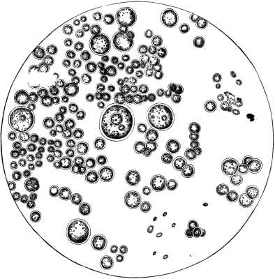
MALARIA PLANTS COLLECTED AT 165TH STREET, EAST OF 10TH AVENUE, OCT., 1881. Plate IX.--Large group of malaria plants, Gemiasma verdans, collected at 165th Street, east of 10th Avenue, New York, in October, 1881, by Dr. Ephraim Cutter, and projected by him with a solar microscope. Dr. Cuzner--the artist--outlined the group on the screen and made the finished drawing from the sketch. He well preserved the grouping and relative sizes. The pond hole whence they came was drained in the spring of 1882, and in August was covered with coarse grass and weeds. No plants were found there in satisfactory quantity, but those figured on Plate VIII. were found half a mile beyond. This shows how draining removes the malaria plants.
From the description I think you have placed your plants in the right family. And evidently they come in the genera named, but at present there is in the authorities at my command so much confusion as to the genera, as given by the most eminent authorities, like Nageli, Kutzing, Braun Rabenht, Cohn, etc., that I think it would be quite unwise for me to settle here, or try to settle here, questions that baffle the naturalists who are entirely devoted to this specialty. We can safely leave this to them. Meantime let us look at the matter as physicians who desire the practical advantages of the discovery you have made. To illustrate this position let us take a familiar case. A boy going through the fields picks and eats an inedible mushroom. He is poisoned and dies. Now, what is the important part of history here from a physician's point of view? Is it not that the mushroom poisoned the child? Next comes the nomenclature. What kind of agaricus was it? Or was it one of the gasteromycetes, the coniomycetes, the hyphomycetes, the ascomycetes, or one of the physomycetes? Suppose that the fungologists are at swords' points with each other about the name of the particular fungus that killed the boy? Would the physicians feel justified to sit down and wait till the whole crowd of naturalists were satisfied, and the true name had been settled satisfactorily to all? I trow not; they would warn the family about eating any more; and if the case had not yet perished, they would let the nomenclature go and try all the means that history, research, and instructed common sense would suggest for the recovery.
This leads me here to say that physicians trust too much to the simple dicta of men who may be very eminent in some department of natural history, and yet ignorant in the very department about which, being called upon, they have given an opinion. All everywhere have so much to learn that we should be very careful how we reject new truths, especially when they come from one of our number educated in our own medical schools, studied under our own masters. If the subject is one about which we know nothing, we had better say so when asked our opinion, and we should receive with respect what is respectfully offered by a man whom we know to be honest, a hard worker, eminent in his department by long and tedious labors. If he asks us to look over his evidence, do so in a kindly spirit, and not open the denunciations of bar room vocabularies upon the presenter, simply because we don't see his point. In other words, we should all be receptive, but careful in our assimilation, remembering that some of the great operations in surgery, for example, came from laymen in low life, as the operation for stone, and even the operation of spaying came from a swineherd.
It is my desire, however, to have this settled as far as can be among scientists, but for the practical uses of practicing physicians I say that far more evidence has been adduced by you in support of the cause of intermittent fever than we have in the etiology of many other diseases. I take the position that so long as no one presents a better history of the etiology of intermittent fever by facts and observations, your theory must stand. This, too, notwithstanding what may be said to the contrary.
Certainly you are to be commended for having done as you have in this matter. It is one of the great rights of the profession, and duties also, that if a physician has or thinks he has anything that is new and valuable, to communicate it, and so long as he observes the rules of good society the profession are to give him a respectful hearing, even though he may have made a mistake. I do not think you had a fair hearing, and hence so far as I myself am concerned I indorse your position, and shall do so till some one comes along and gives a better demonstration. Allow me also to proceed with more evidence.
Observation at West Falmouth, Mass., Sept 1, 1877. I made five observations in like manner about the marshes and bogs of this town, which is, as it were, situated on the tendo achillis of Cape Cod, Mass. In only one of these observations did I find any palmellæ like the ague plants, and they were not characteristic.
Chelsea, Mass., near the Naval Hospital, September 5, 1877. Three sets of observations. In all spores were found and some sporangia, but they were not the genuine plants as far as I could judge. They were Protococcaceæ. It is not necessary to add that there are no cases of intermittent fever regarded as originating on the localities named. Still, the ancient history of New England contains some accounts of ague occurring there, but they are not regarded as entirely authentic.
Observation. Lexington, Mass, September 6, 1877. Observation made in a meadow. There was no saline incrustation, and no palmellæ found. No local malaria.
Observation. Cambridge, Mass. Water works on the shore of Fresh Pond. Found a few palmellæ analogous to, but not the ague palmellæ.
Observation. Woburn, Mass, September 27, 1877, with Dr. J. M. Moore. Found some palmellæ, but scanty. Abundance of spores of cryptogams.
Observation. Stonington, Conn., August 15, 1877. Examined a pond hole nearly opposite the railroad station on the New York Shore Line. Found abundantly the white incrustation on the surface of the soil. Here I found the spores and the sporangias of the gemiasmas verdans and rubra.
Observation 2. Repetition of the last.
Observation 3. I examined some of an incrustation that was copiously deposited in the same locality, which was not white or frosty, but dark brown and a dirty green. Here the spores were very abundant, and a few sporangias of the Gemiasma rubra. Ague has of late years been noted in Connecticut and Rhode Island.
Observations in Connecticut. Middlefield near Middletown, summer of 1878. Being in this locality, I heard that intermittent fever was advancing eastward at the rate of ten miles a year. It had been observed in Middlefield. I was much interested to see if I could find the gemiasmas there. On examining the dripping of some bog moss, I found a plenty of them.
Observations in Connecticut. New Haven. Early in the summer of 1881 I visited this city. One object of my visit was to ascertain the truth of the presence of intermittent fever there, which I had understood prevailed to such an extent that my patient, a consumptive, was afraid to return to his home in New Haven. At this time I examined the hydrant water of the city water works, and also the east shore of the West River, which seemed to be too full of sewage. I found a plenty of the Oscillatoreaceæ, but no Palmellæ.
In September I revisited the city, taking with me a medical gentleman who, residing in the South, had had a larger experience with the disease than I. From the macroscopical examination he pronounced a case we examined to be ague, but I was not able to detect the plants either in the urine or blood. This might have been that I did not examine long enough. But a little later I revisited the city and explored the soil about the Whitney Water Works, whence the city gets its supply of water, and I had no difficulty in finding a good many of the plants you describe as found by you in ague cases. At a still later period my patient, whom I had set to use the microscope and instructed how to collect the ague plants, set to work himself. One day his mother brought in a film from off an ash pile that lay in the shade, and this her son found was made up of an abundance of the ague plants. By simply winding a wet bandage around the slide, Mr. A. was enabled to keep the plants in good condition until the time of my next visit, when I examined and pronounced them to be genuine plants.
I should here remark that I had in examining the sputa of this patient sent to me, found some of the ague plants. He said that he had been riding near the Whitney Pond, and perceived a different odor, and thought he must have inhaled the miasm. I told him he was correct in his supposition, as no one could mistake the plants; indeed, Prof. Nunn, of Savannah, Ga., my pupil recognized it at once.
This relation, though short, is to me of great importance. So long as I could not detect the gemiasmas in New Haven, I was very skeptical as to the presence of malaria in New Haven, as I thought there must be some mistake, it being a very good cloak to hide under (malaria). There is no doubt but that the name has covered lesions not belonging to it. But now the positive demonstrations above so briefly related show to my mind that the local profession have not been mistaken, and have sustained their high reputation.
I should say that I have examined a great deal of sputa, but, with the exception of cases that were malarious, I have not encountered the mature plants before. Of course I have found them as you did, in my own excretions as I was traveling over ague bogs.
[To be continued.]