Front matter
Reading edition
REPORT
ON THE
SCIENTIFIC RESULTS
OF THE
VOYAGE OF H.M.S. CHALLENGER
DURING THE YEARS 1873-76
UNDER THE COMMAND OF
Captain GEORGE S. NARES, R.N., F.R.S
AND THE LATE
Captain FRANK TOURLE THOMSON, R.N
PREPARED UNDER THE SUPERINTENDENCE OF
THE LATE
Sir C. WYVILLE THOMSON, Knt., F.R.S., &c
REGIUS PROFESSOR OF NATURAL HISTORY IN THE UNIVERSITY OF EDINBURGH
DIRECTOR OF THE CIVILIAN SCIENTIFIC STAFF ON BOARD
AND NOW OF
JOHN MURRAY
ONE OF THE NATURALISTS OF THE EXPEDITION
Zoology—Vol. XVIII.
PLATES
Published by Order of Her Majesty's Government
PRINTED FOR HER MAJESTY'S STATIONERY OFFICE
AND SOLD BY
LONDON:—EYRE & SPOTTISWOODE, EAST HARDING STREET, FETTER LANE
EDINBURGH:—ADAM & CHARLES BLACK
DUBLIN:—HODGES, FIGGIS, & CO
1887
Price (in Two Parts, with a Volume of Plates) £5, 10s.
CONTENTS.
Report on the Radiolaria collected by H.M.S. Challenger during the years 1873-1876.
By Ernst Haeckel, M.D., Ph.D., Professor of Zoology in the University of Jena.
PLATES.
CONTENTS.
PLATES 1-50. SPUMELLARIA.
" 51-98. NASSELLARIA.
" 99-128. PHÆODARIA.
" 129-140. ACANTHARIA.
MAP, showing the Geographical Distribution of the Radiolaria.
PLATE 1.
Legion SPUMELLARIA.
Order COLLOIDEA.
Family Thalassicollida.
PLATE 1. Thalassicollida. Diam. Page. Fig. 1. Actissa princeps, n. sp., × 300 13 The entire living Spumellarium. c, The spherical central capsule containing finely granulated protoplasm, which is radially striated in the cortical zone; v, spherical vacuoles enclosed by the protoplasm; n, the spherical nucleus in the centre; l, the concentric nucleolus; f, the radial pseudopodia which pierce the calymma or the (yellowish) jelly-envelope of the central capsule and arise from the granular sarcomatrix. Fig. 1a. Half of the central capsule of another specimen, in which the original central nucleus is cleft into numerous small nuclei, × 400 Fig. 1b. Half of the central capsule of another specimen, filled up by flagellate spores, × 400 Fig. 1c. Eight isolated flagellate spores, × 800 Fig. 2. Thalassolampe maxima, n. sp., × 8 17 The entire living Spumellarium. c, The big spherical central capsule; a, the large alveoles filling the central capsule and surrounding a central nucleus; f, the pseudopodia piercing the extracapsular calymma. Fig. 2a. The nucleus alone, with numerous nucleoli, × 30 Fig. 3. Thalassopila cladococcus, n. sp., × 20 17 c, The big central capsule; a, numerous large alveoles contained in the central capsule; k, oil globules, many of which are placed in the radially striped cortical zone; the nucleus placed centrally, is covered with numerous radial apophyses or cæcal sacs. f, The radially striped calymma. Fig. 4. Thalassicolla maculata, n. sp., × 100 21 c, The central capsule; v, vacuoles filling this capsule; n, the central nucleus; l, the concentric nucleolus; g, the voluminous calymma, a small radial piece of which is only represented; a, the large alveoles; b, peculiar exoplasmatic bodies; p, black pigment in the inner zone; f, the retracted pseudopodia in the outer zone. Fig. 4a. An exoplasmatic body, × 300 Fig. 4b, Vacuoles in the endoplasm, × 300 Fig. 5. Thalassicolla melacapsa, n. sp., × 300 21 n, The large nucleus; l, numerous small nucleoli inside the nucleus; v, the vacuoles filling up the central capsule and separated by black pigment; a, large alveoles in the calymma; k, oil globules; b, exoplasmatic bodies; f, the retracted pseudopodia in the outer zone of the calymma. Fig. 5a. An endoplasmatic vacuole, resembling a cell, × 600 Fig. 5b. A piece of the central capsule, × 600
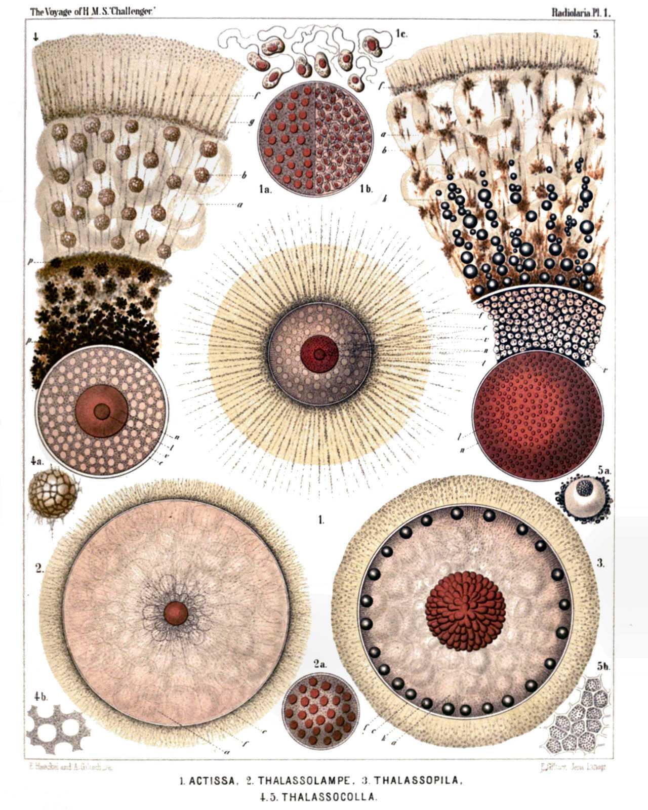
PLATE 1.
Thalassicollida.
Diam. Page.
Fig. 1. Actissa princeps, n. sp., × 300 13
The entire living Spumellarium. c, The spherical central capsule containing finely granulated protoplasm, which is radially striated in the cortical zone; v, spherical vacuoles enclosed by the protoplasm; n, the spherical nucleus in the centre; l, the concentric nucleolus; f, the radial pseudopodia which pierce the calymma or the (yellowish) jelly-envelope of the central capsule and arise from the granular sarcomatrix.
Fig. 1a. Half of the central capsule of another specimen, in which the original central nucleus is cleft into numerous small nuclei, × 400
Fig. 1b. Half of the central capsule of another specimen, filled up by flagellate spores, × 400
Fig. 1c. Eight isolated flagellate spores, × 800
Fig. 2. Thalassolampe maxima, n. sp., × 8 17
The entire living Spumellarium. c, The big spherical central capsule; a, the large alveoles filling the central capsule and surrounding a central nucleus; f, the pseudopodia piercing the extracapsular calymma.
Fig. 2a. The nucleus alone, with numerous nucleoli, × 30
Fig. 3. Thalassopila cladococcus, n. sp., × 20 17
c, The big central capsule; a, numerous large alveoles contained in the central capsule; k, oil globules, many of which are placed in the radially striped cortical zone; the nucleus placed centrally, is covered with numerous radial apophyses or cæcal sacs. f, The radially striped calymma.
Fig. 4. Thalassicolla maculata, n. sp., × 100 21
c, The central capsule; v, vacuoles filling this capsule; n, the central nucleus; l, the concentric nucleolus; g, the voluminous calymma, a small radial piece of which is only represented; a, the large alveoles; b, peculiar exoplasmatic bodies; p, black pigment in the inner zone; f, the retracted pseudopodia in the outer zone.
Fig. 4a. An exoplasmatic body, × 300
Fig. 4b, Vacuoles in the endoplasm, × 300
Fig. 5. Thalassicolla melacapsa, n. sp., × 300 21
n, The large nucleus; l, numerous small nucleoli inside the nucleus; v, the vacuoles filling up the central capsule and separated by black pigment; a, large alveoles in the calymma; k, oil globules; b, exoplasmatic bodies; f, the retracted pseudopodia in the outer zone of the calymma.
Fig. 5a. An endoplasmatic vacuole, resembling a cell, × 600
Fig. 5b. A piece of the central capsule, × 600
PLATE 2.
Legion SPUMELLARIA.
Order BELOIDEA.
Family Thalassosphærida.
PLATE 2. Thalassosphærida. Diam. Page. Fig. 1. Lampoxanthium pandora, n. sp., × 120 38 The central capsule exhibits distinct pore-canals in its membrane, and a clear interval between this and the coagulated and vacuolated protoplasm. The central nucleus contains numerous dark nucleoli. The spicula are scattered throughout the alveolate calymma. Fig. 2. Thalassoplancta brevispicula, n. sp. (vel Lampoxanthium brevispiculum), × 120 36 The central capsule contains numerous clear vacuoles, and in the cortical zone a layer of large oil-globules. The central nucleus includes numerous dark nucleoli. The calymma is alveolate. The spicula lie only in the cortical zone. Fig. 3. Thalassoxanthium cervicorne, n. sp., × 300 33 The central capsule is filled up by clear vacuoles and contains a large central nucleus, with a single nucleolus. The spicula surround the thin calymma. Fig. 4. Thalassoxanthium cervicorne, n. sp., × 600 33 A single spiculum. Fig. 5. Thalassoxanthium medusinum, n. sp., × 120 32 The central capsule is filled up by clear vacuoles and contains on its cortical zone a layer of large oil-globules. The central nucleus contains numerous dark nucleoli. The calymma is radially striped, contains numerous small xanthellæ, and is surrounded by the spicula. Fig. 6. Thalassoxanthium octoceras, n. sp., × 400 34 Three isolated spicula.
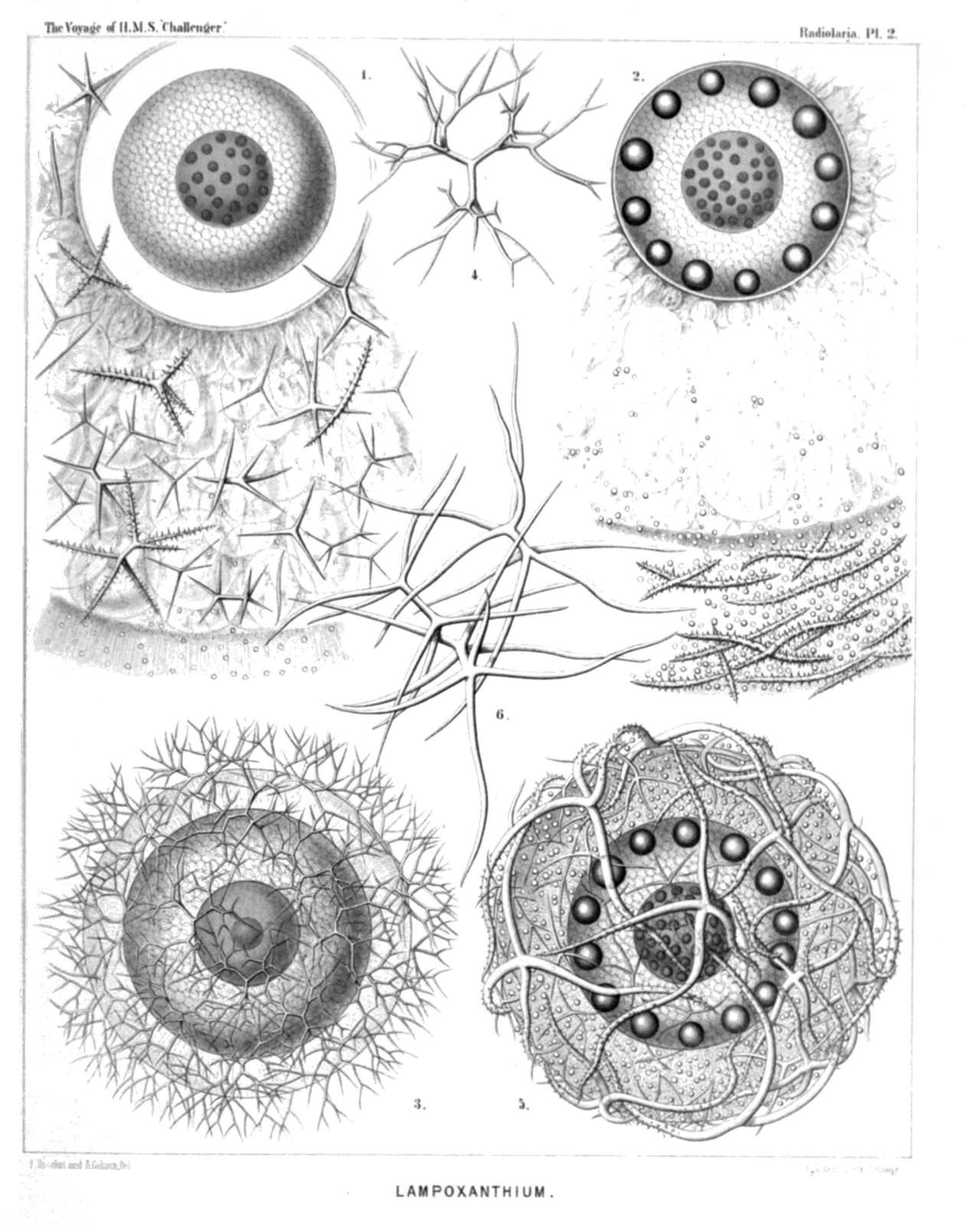
PLATE 2.
Thalassosphærida.
Diam. Page.
Fig. 1. Lampoxanthium pandora, n. sp., × 120 38
The central capsule exhibits distinct pore-canals in its membrane, and a clear interval between this and the coagulated and vacuolated protoplasm. The central nucleus contains numerous dark nucleoli. The spicula are scattered throughout the alveolate calymma.
Fig. 2. Thalassoplancta brevispicula, n. sp. (vel Lampoxanthium brevispiculum), × 120 36
The central capsule contains numerous clear vacuoles, and in the cortical zone a layer of large oil-globules. The central nucleus includes numerous dark nucleoli. The calymma is alveolate. The spicula lie only in the cortical zone.
Fig. 3. Thalassoxanthium cervicorne, n. sp., × 300 33
The central capsule is filled up by clear vacuoles and contains a large central nucleus, with a single nucleolus. The spicula surround the thin calymma.
Fig. 4. Thalassoxanthium cervicorne, n. sp., × 600 33
A single spiculum.
Fig. 5. Thalassoxanthium medusinum, n. sp., × 120 32
The central capsule is filled up by clear vacuoles and contains on its cortical zone a layer of large oil-globules. The central nucleus contains numerous dark nucleoli. The calymma is radially striped, contains numerous small xanthellæ, and is surrounded by the spicula.
Fig. 6. Thalassoxanthium octoceras, n. sp., × 400 34
Three isolated spicula.
PLATE 3.
Legion SPUMELLARIA.
Order COLLOIDEA.
Family Collozoida.
PLATE 3. Collozoida. Diam. Page. Fig. 1. Collozoum serpentinum, n sp (vel Collophidium serpentinum, Hkl), × 10 26 A living cœnobium, with expanded pseudopodia. The spherical calymma (or the common jelly-mass of the colony) is alveolate and contains numerous cylindrical, serpentine, central capsules. Numerous yellow cells or xanthellæ are scattered between the radial pseudopodia in the periphery. Fig. 2. Collozoum serpentinum, n. sp., × 50 26 An isolated, cylindrical, worm-shaped, central capsule, with an axial series of oil-globules; the red points are nuclei. Fig. 3. Collozoum serpentinum, n. sp., × 150 26 An isolated, cylindrical, serpentine, central capsule. k, Oil-globules forming an axial series; n, densely placed, red-coloured nuclei;, c, the capsule membrane under which are scattered small black pigment spots in the colourless cortical zone of the endoplasm; a, extracapsular alveoles; x, xanthellæ or "yellow cells." Fig. 4. Collozoum amœboides, n. sp., × 100 28 A spherical cœnobium or jelly-colony. Each amœboid central capsule contains an oil-globule; the small red points are nuclei. Fig. 5. Collozoum amœboides, n. sp., × 400 28 c, A single isolated central capsule; n, nuclei; k, oil-globule. Fig. 6. Collozoum vermiforme, n. sp., × 30 27 g, A spherical cœnobium or jelly-colony; a, large alveoles, forming a cortical zone; c, central capsules; k, oil-globules. Fig. 7. Collozoum vermiforme, n. sp., × 100 27 c, A single isolated central capsule; x, xanthellæ surrounding this central capsule; k, oil-globules; n, nuclei. Fig. 8. Collozoum ellipsoides, n. sp., × 2 26 A spherical colony; the red points are central capsules. Fig. 9. Collozoum ellipsoides, n. sp., × 150 26 c, A single isolated central capsule; k, oil-globules; n, nuclei. Fig. 10. Collozoum inerme, Hkl., × 2 25 An old, cylindrical, articulated cœnobium; the red points are centralcapsules. Fig. 11. Collozoum inerme, Hkl., × 2 25 A young cylindrical cœnobium; the red points are central capsules. Fig. 12. Collozoum inerme, Hkl., × 400 25 A piece of a young colony with eight small central capsules, without oil-globules. n, The central nucleus in different stages of division. Two capsules are also dividing. x, Xanthellæ in the jelly-like calymma (blue), which also contains numerous vacuoles.
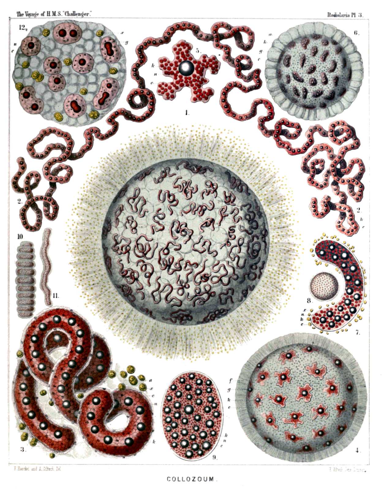
PLATE 3.
Collozoida.
Diam. Page.
Fig. 1. Collozoum serpentinum, n sp (vel Collophidium serpentinum, Hkl), × 10 26
A living cœnobium, with expanded pseudopodia. The spherical calymma (or the common jelly-mass of the colony) is alveolate and contains numerous cylindrical, serpentine, central capsules. Numerous yellow cells or xanthellæ are scattered between the radial pseudopodia in the periphery.
Fig. 2. Collozoum serpentinum, n. sp., × 50 26
An isolated, cylindrical, worm-shaped, central capsule, with an axial series of oil-globules; the red points are nuclei.
Fig. 3. Collozoum serpentinum, n. sp., × 150 26
An isolated, cylindrical, serpentine, central capsule. k, Oil-globules forming an axial series; n, densely placed, red-coloured nuclei;, c, the capsule membrane under which are scattered small black pigment spots in the colourless cortical zone of the endoplasm; a, extracapsular alveoles; x, xanthellæ or "yellow cells."
Fig. 4. Collozoum amœboides, n. sp., × 100 28
A spherical cœnobium or jelly-colony. Each amœboid central capsule contains an oil-globule; the small red points are nuclei.
Fig. 5. Collozoum amœboides, n. sp., × 400 28
c, A single isolated central capsule; n, nuclei; k, oil-globule.
Fig. 6. Collozoum vermiforme, n. sp., × 30 27
g, A spherical cœnobium or jelly-colony; a, large alveoles, forming a cortical zone; c, central capsules; k, oil-globules.
Fig. 7. Collozoum vermiforme, n. sp., × 100 27
c, A single isolated central capsule; x, xanthellæ surrounding this central capsule; k, oil-globules; n, nuclei.
Fig. 8. Collozoum ellipsoides, n. sp., × 2 26
A spherical colony; the red points are central capsules.
Fig. 9. Collozoum ellipsoides, n. sp., × 150 26
c, A single isolated central capsule; k, oil-globules; n, nuclei.
Fig. 10. Collozoum inerme, Hkl., × 2 25
An old, cylindrical, articulated cœnobium; the red points are centralcapsules.
Fig. 11. Collozoum inerme, Hkl., × 2 25
A young cylindrical cœnobium; the red points are central capsules.
Fig. 12. Collozoum inerme, Hkl., × 400 25
A piece of a young colony with eight small central capsules, without oil-globules. n, The central nucleus in different stages of division. Two capsules are also dividing. x, Xanthellæ in the jelly-like calymma (blue), which also contains numerous vacuoles.
PLATE 4.
Legion SPUMELLARIA.
Orders BELOIDEA.
Families Sphærozoida.
PLATE 4. Sphærozoida Diam. Page. Fig. 1. Sphærozoum trigeminum, n. sp., × 50 43 An annular colony. The main mass of the jelly-colony is filled up by large alveoles; the entire surface is densely covered with spicula, and beyond this skeleton-cover lie the spherical central capsules, each with an oil-globule. This species is by mistake not mentioned in the text. Fig. 2. Sphærozoum alveolatum, n. sp., × 50 43 Section through a spherical colony; displaying the inside of a hemisphere. All the central capsules lie in a single stratum on the surface of the jelly-sphere, each being surrounded by a thick-walled alveole. The spicula lie between the alveole and the capsule, which includes a central oil-globule. Fig. 3. Sphærozoum alveolatum, n. sp., × 400 43 A single central capsule, filled up by crystal-spores. Numerous geminato-radiate spicula and spherical xanthellæ lie between the capsule and the including thick-walled alveole. In the jelly-calymma, between the capsule and the alveole, numerous thin ramified pseudopodia are expanded. Fig. 4. Sphærozoum geminatum, n. sp., × 400 45 A single central capsule, with a central oil-globule, surrounded by numerous spicula and spherical xanthellæ. The jelly-substance of the calymma is expanded between the points of the spicula. Fig. 5. Sphærozoum variabile, n. sp., × 300 45 Three isolated spicula. Fig. 6. Sphærozoum pandora, n. sp. (vel Rhaphidozoum pandora), × 300 49 A group of various spicula. Fig. 7. Sphærozoum verticillatum, n. sp., × 300 44 A single spiculum. Fig. 8. Sphærozoum arborescens, n. sp., × 300 44 A single spiculum. Fig. 9. Sphærozoum armatum, n. sp., × 300 43 A single spiculum.
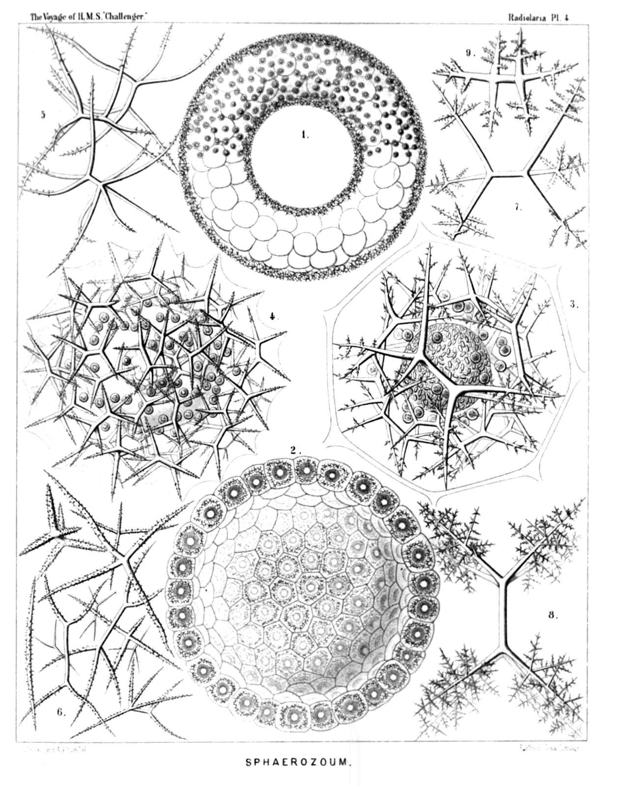
PLATE 4.
Sphærozoida
Diam. Page.
Fig. 1. Sphærozoum trigeminum, n. sp., × 50 43
An annular colony. The main mass of the jelly-colony is filled up by large alveoles; the entire surface is densely covered with spicula, and beyond this skeleton-cover lie the spherical central capsules, each with an oil-globule. This species is by mistake not mentioned in the text.
Fig. 2. Sphærozoum alveolatum, n. sp., × 50 43
Section through a spherical colony; displaying the inside of a hemisphere. All the central capsules lie in a single stratum on the surface of the jelly-sphere, each being surrounded by a thick-walled alveole. The spicula lie between the alveole and the capsule, which includes a central oil-globule.
Fig. 3. Sphærozoum alveolatum, n. sp., × 400 43
A single central capsule, filled up by crystal-spores. Numerous geminato-radiate spicula and spherical xanthellæ lie between the capsule and the including thick-walled alveole. In the jelly-calymma, between the capsule and the alveole, numerous thin ramified pseudopodia are expanded.
Fig. 4. Sphærozoum geminatum, n. sp., × 400 45
A single central capsule, with a central oil-globule, surrounded by numerous spicula and spherical xanthellæ. The jelly-substance of the calymma is expanded between the points of the spicula.
Fig. 5. Sphærozoum variabile, n. sp., × 300 45
Three isolated spicula.
Fig. 6. Sphærozoum pandora, n. sp. (vel Rhaphidozoum pandora), × 300 49
A group of various spicula.
Fig. 7. Sphærozoum verticillatum, n. sp., × 300 44
A single spiculum.
Fig. 8. Sphærozoum arborescens, n. sp., × 300 44
A single spiculum.
Fig. 9. Sphærozoum armatum, n. sp., × 300 43
A single spiculum.
PLATE 5.
Legion SPUMELLARIA.
Order SPHÆROIDEA.
Family Collosphærida.
PLATE 5. Collosphærida. Diam. Page. Fig. 1. Trypanosphæra transformata, n. sp., × 150 111 A living colony. The centre of the spherical cœnobium contains a large alveole, surrounded by a net of sarcode. The entire calymma is filled up by smaller, thin-walled alveoles. Its inner part contains numerous small, young, central capsules (each with an oil-globule) without shells; in the cortical zone of the calymma lie larger capsules, each of which is enclosed by a fenestrated shell with from two to four or more dentated tubes. Between the radiant pseudopodia very numerous small yellow cells (xanthellæ), which are scattered everywhere. Fig. 2. Trypanosphæra transformata, n. sp., × 300 111 A single shell. Fig. 3. Trypanosphæra coronata, n. sp., × 300 110 Fig. 4. Trypanosphæra trepanata, n. sp., × 300 110 Fig. 5. Odontosphæra monodon, n. sp., × 300 102 Fig. 6. Odontosphæra cyrtodon, n. sp., × 300 102 Fig. 7. Acrosphæra inflata, n. sp., × 300 101 Fig. 8. Mazosphæra hippotis, n. sp., × 400 108 Fig. 9. Mazosphæra lagotis, n. sp., × 300 108 Fig. 10. Pharyngosphæra stomodæa, n. sp., × 400 98 Fig. 11. Buccinosphæra invaginata. n. sp., × 500 99 Each shell contains numerous larger and smaller crystals. Fig. 12. Tribonosphæra centripetalis, n. sp., × 500 98 Each shell contains numerous large crystals. Fig. 13. Collosphæra polygona, n. sp., × 200 96
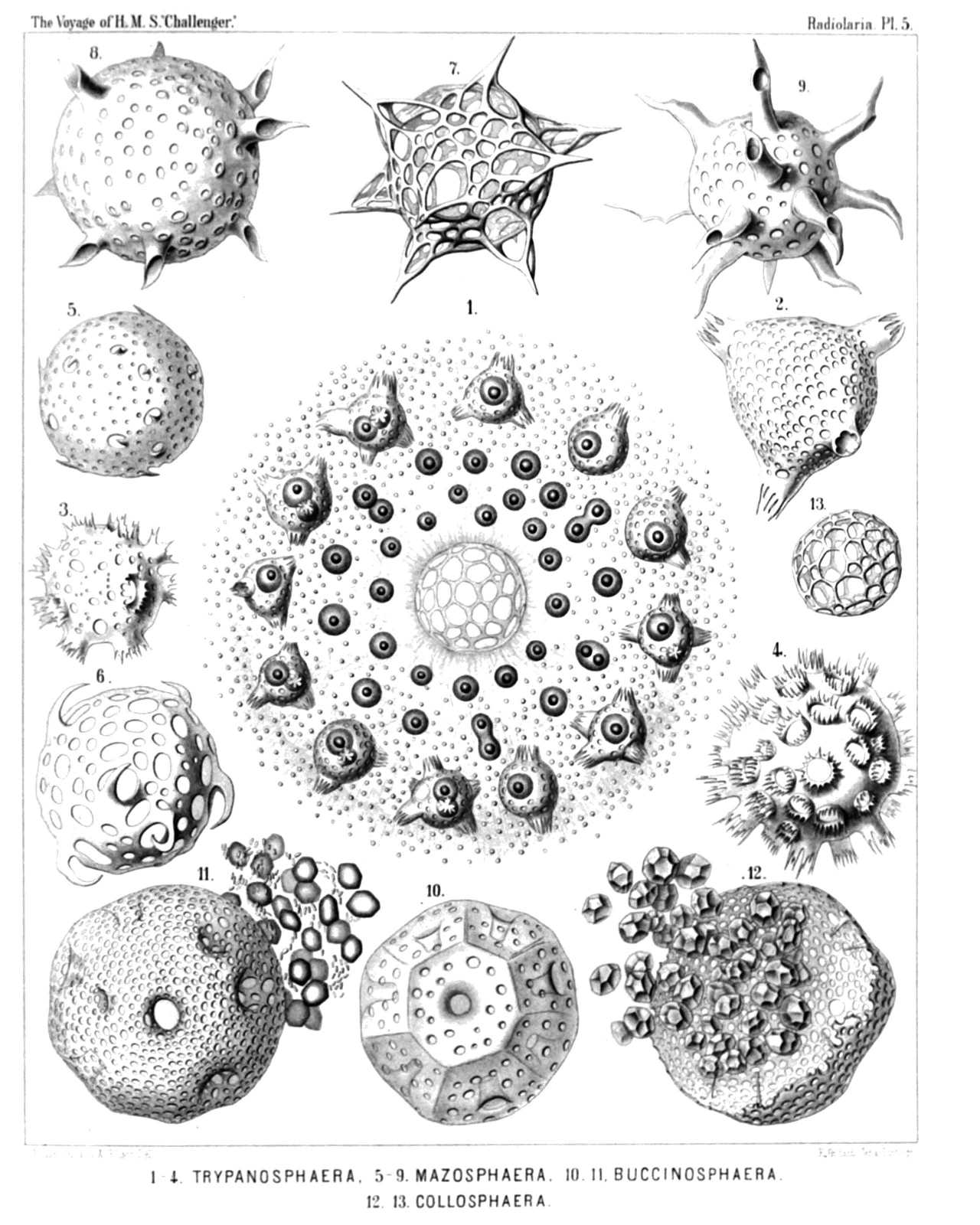
PLATE 5.
Collosphærida.
Diam. Page.
Fig. 1. Trypanosphæra transformata, n. sp., × 150 111
A living colony. The centre of the spherical cœnobium contains a large alveole, surrounded by a net of sarcode. The entire calymma is filled up by smaller, thin-walled alveoles. Its inner part contains numerous small, young, central capsules (each with an oil-globule) without shells; in the cortical zone of the calymma lie larger capsules, each of which is enclosed by a fenestrated shell with from two to four or more dentated tubes. Between the radiant pseudopodia very numerous small yellow cells (xanthellæ), which are scattered everywhere.
Fig. 2. Trypanosphæra transformata, n. sp., × 300 111
A single shell.
Fig. 3. Trypanosphæra coronata, n. sp., × 300 110
Fig. 4. Trypanosphæra trepanata, n. sp., × 300 110
Fig. 5. Odontosphæra monodon, n. sp., × 300 102
Fig. 6. Odontosphæra cyrtodon, n. sp., × 300 102
Fig. 7. Acrosphæra inflata, n. sp., × 300 101
Fig. 8. Mazosphæra hippotis, n. sp., × 400 108
Fig. 9. Mazosphæra lagotis, n. sp., × 300 108
Fig. 10. Pharyngosphæra stomodæa, n. sp., × 400 98
Fig. 11. Buccinosphæra invaginata. n. sp., × 500 99
Each shell contains numerous larger and smaller crystals.
Fig. 12. Tribonosphæra centripetalis, n. sp., × 500 98
Each shell contains numerous large crystals.
Fig. 13. Collosphæra polygona, n. sp., × 200 96
PLATE 6.
Legion SPUMELLARIA.
Order SPHÆROIDEA.
Family Collosphærida.
PLATE 6. Collosphærida. Diam. Page. Fig. 1. Siphonosphæra socialis, n. sp., × 500 106 A small piece of the surface of a living cœnobium, seen from the surface. Only four individuals are visible, the central capsule of which contains numerous small nuclei and a central oil-globule. The including spherical lattice-shell is provided with a few (one to four) larger apertures, which are prolonged into short cylindrical tubules. Through these latter radiate bundles of fine pseudopodia, branching and anastomosing, and forming a fine sarcode network between the alveoles of the calymma. On the surface of the alveolated jelly-sphere the pseudopodia form a dense radiating zone. Xanthella or yellow cells are everywhere scattered. Fig. 2. Siphonosphæra socialis, n. sp., × 300 106 A small cœnobium or colony in the state of alveolation, forming a jelly-sphere, composed of a great number of capsulated individuals, densely aggregated. Each central capsule contains an oil-globule, and is enclosed by a spherical lattice-shell, which bears a few (one to four) short cylindrical tubules. Each shell is again enveloped by a membranous polyhedral alveole and separated from it by structureless jelly. The thick cortical jelly-envelope, which surrounds the whole spherical colony, exhibits a fine radial striation, produced by radiating pseudopodia; many xanthellæ or yellow cells are scattered in the calymma. Fig. 3. Siphonosphæra pipetta, n. sp., × 300 108 Fig. 4. Siphonosphæra tubulosa, J. Müller, × 300 105 The central capsule, enclosed in the cavity of the shell, has a central oil-globule, and is surrounded by a few xanthella. Fig. 5. Siphonosphæra chonophora, n. sp., × 300 107 Fig. 6. Siphonosphæra serpula, n. sp., × 300 107 Fig. 7. Siphonosphæra patinaria, n sp., × 300 105 The central capsule, enclosed in the cavity of the shell, contains a central oil-globule, and is surrounded by a few xanthella. Fig. 8. Siphonosphæra patinaria, n. sp., × 300 105 Fig. 9. Siphonosphæra conifera, n. sp., × 300 106 Fig. 10. Siphonosphæra cyathina, n. sp., × 300 105
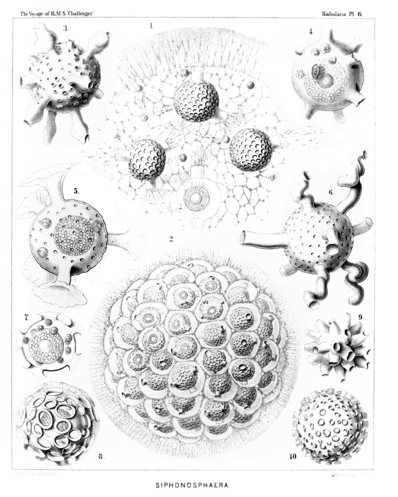
PLATE 6.
Collosphærida.
Diam. Page.
Fig. 1. Siphonosphæra socialis, n. sp., × 500 106
A small piece of the surface of a living cœnobium, seen from the surface. Only four individuals are visible, the central capsule of which contains numerous small nuclei and a central oil-globule. The including spherical lattice-shell is provided with a few (one to four) larger apertures, which are prolonged into short cylindrical tubules. Through these latter radiate bundles of fine pseudopodia, branching and anastomosing, and forming a fine sarcode network between the alveoles of the calymma. On the surface of the alveolated jelly-sphere the pseudopodia form a dense radiating zone. Xanthella or yellow cells are everywhere scattered.
Fig. 2. Siphonosphæra socialis, n. sp., × 300 106
A small cœnobium or colony in the state of alveolation, forming a jelly-sphere, composed of a great number of capsulated individuals, densely aggregated. Each central capsule contains an oil-globule, and is enclosed by a spherical lattice-shell, which bears a few (one to four) short cylindrical tubules. Each shell is again enveloped by a membranous polyhedral alveole and separated from it by structureless jelly. The thick cortical jelly-envelope, which surrounds the whole spherical colony, exhibits a fine radial striation, produced by radiating pseudopodia; many xanthellæ or yellow cells are scattered in the calymma.
Fig. 3. Siphonosphæra pipetta, n. sp., × 300 108
Fig. 4. Siphonosphæra tubulosa, J. Müller, × 300 105
The central capsule, enclosed in the cavity of the shell, has a central oil-globule, and is surrounded by a few xanthella.
Fig. 5. Siphonosphæra chonophora, n. sp., × 300 107
Fig. 6. Siphonosphæra serpula, n. sp., × 300 107
Fig. 7. Siphonosphæra patinaria, n sp., × 300 105
The central capsule, enclosed in the cavity of the shell, contains a central oil-globule, and is surrounded by a few xanthella.
Fig. 8. Siphonosphæra patinaria, n. sp., × 300 105
Fig. 9. Siphonosphæra conifera, n. sp., × 300 106
Fig. 10. Siphonosphæra cyathina, n. sp., × 300 105
PLATE 7.
Legion SPUMELLARIA.
Order SPHÆROIDEA.
Family Collosphærida.
PLATE 7. Collosphærida. Diam. Page. Fig. 1 Caminosphæra dendrophora, n. sp., × 300 112 Fig. 2. Caminosphæra dichotoma, n. sp., × 300 112 Fig. 3. Coronosphæra diadema, n. sp., × 300 117 Fig. 4. Coronosphæra calycina, n. sp., × 300 117 Fig. 5. Otosphæra auriculata, n. sp., × 300 116 Fig. 6. Otosphæra polymorpha, n. sp., × 300 116 Fig. 7. Solenosphæra serpentina, n. sp., × 300 114 Fig. 8. Solenosphæra cornucopia, n. sp., × 300 115 Fig. 9. Solenosphæra ascensionis, n. sp., × 300 115 Fig. 10. Solenosphæra pandora, n. sp., × 300 113 Fig. 11. Solenosphæra pandora, n. sp., × 100 113 An entire spherical cœnobium. The shells of the colony bear a variable number of fenestrated radial tubes and are densely crowded in the jelly-sphere of the calymma, the cortical zone of which is radially striped.
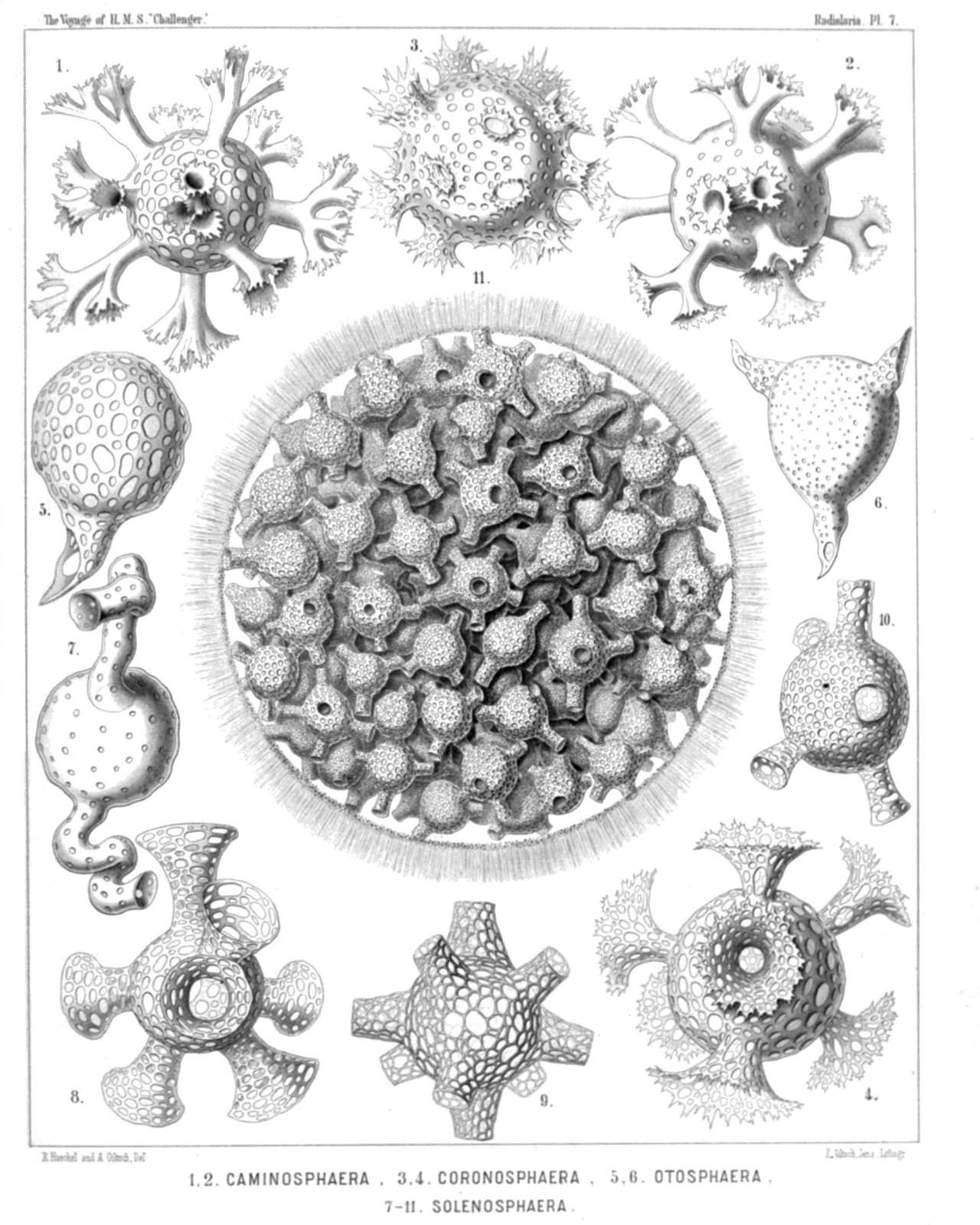
PLATE 7.
Collosphærida.
Diam. Page.
Fig. 1 Caminosphæra dendrophora, n. sp., × 300 112
Fig. 2. Caminosphæra dichotoma, n. sp., × 300 112
Fig. 3. Coronosphæra diadema, n. sp., × 300 117
Fig. 4. Coronosphæra calycina, n. sp., × 300 117
Fig. 5. Otosphæra auriculata, n. sp., × 300 116
Fig. 6. Otosphæra polymorpha, n. sp., × 300 116
Fig. 7. Solenosphæra serpentina, n. sp., × 300 114
Fig. 8. Solenosphæra cornucopia, n. sp., × 300 115
Fig. 9. Solenosphæra ascensionis, n. sp., × 300 115
Fig. 10. Solenosphæra pandora, n. sp., × 300 113
Fig. 11. Solenosphæra pandora, n. sp., × 100 113
An entire spherical cœnobium. The shells of the colony bear a variable number of fenestrated radial tubes and are densely crowded in the jelly-sphere of the calymma, the cortical zone of which is radially striped.
PLATE 8.
Legion SPUMELLARIA.
Order SPHÆROIDEA.
Family Collosphærida.
PLATE 8. Collosphærida. Diam. Page. Fig. 1. Acrosphæra echinoides, n. sp., × 400 100 Fig. 2. Acrosphæra collina, n. sp., × 300 101 Fig. 3. Chænicosphæra nassiterna, n. sp., × 400 103 Fig. 4. Chænicosphæra murrayana, n. sp., × 300 102 Fig. 5. Chænicosphæra flammabunda, n. sp., × 300 103 Fig. 6. Clathrosphæra circumtexta, n. sp., × 400 118 Fig. 7. Clathrosphæra arachnoides, n. sp., × 300 119 Fig. 8. Clathrosphæra lamellosa, n. sp., × 300 119 Fig. 9. Xanthiosphæra erinacea, n. sp., × 400 120 Fig. 10. Xanthiosphæra lappacea, n. sp., × 300 120 Fig. 11. Xanthiosphæra lappacea, n. sp., × 100 120 A complete spherical cœnobium. The associated central capsules (each with a double shell) are densely crowded in the central part of the calymma, whilst its peripheral part is occupied by a layer of large alveoles. Numerous xanthellæ or yellow cells an scattered in the calymma.
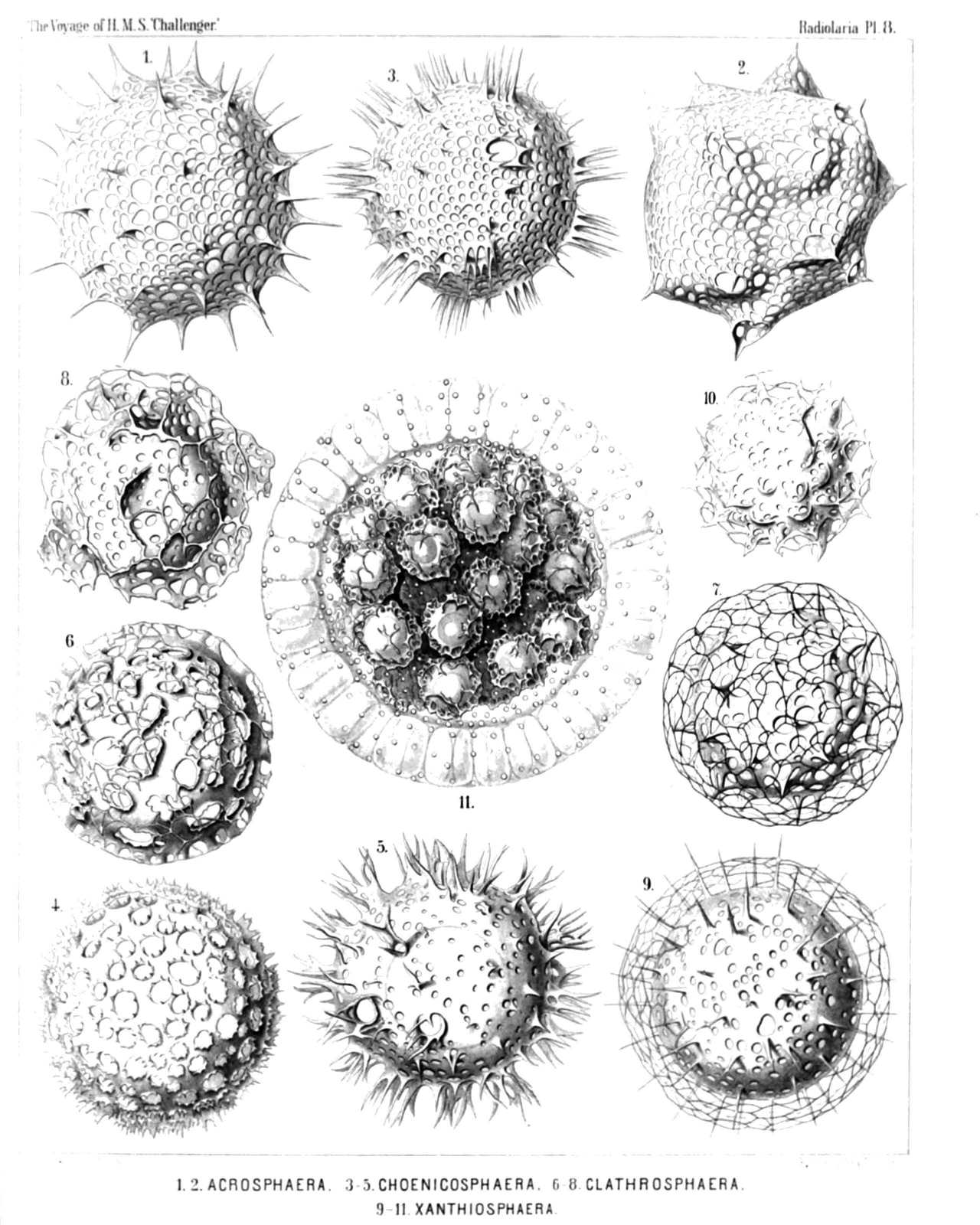
PLATE 8.
Collosphærida.
Diam. Page.
Fig. 1. Acrosphæra echinoides, n. sp., × 400 100
Fig. 2. Acrosphæra collina, n. sp., × 300 101
Fig. 3. Chænicosphæra nassiterna, n. sp., × 400 103
Fig. 4. Chænicosphæra murrayana, n. sp., × 300 102
Fig. 5. Chænicosphæra flammabunda, n. sp., × 300 103
Fig. 6. Clathrosphæra circumtexta, n. sp., × 400 118
Fig. 7. Clathrosphæra arachnoides, n. sp., × 300 119
Fig. 8. Clathrosphæra lamellosa, n. sp., × 300 119
Fig. 9. Xanthiosphæra erinacea, n. sp., × 400 120
Fig. 10. Xanthiosphæra lappacea, n. sp., × 300 120
Fig. 11. Xanthiosphæra lappacea, n. sp., × 100 120
A complete spherical cœnobium. The associated central capsules (each with a double shell) are densely crowded in the central part of the calymma, whilst its peripheral part is occupied by a layer of large alveoles. Numerous xanthellæ or yellow cells an scattered in the calymma.
PLATE 9.
Legion SPUMELLARIA.
Order LARCOIDEA.
Family Pylonida.
PLATE 9. Pylonida. Diam. Page. Fig. 1. Monozonium alatum, n. sp., × 300 633 Dorsal view. Fig. 1a. Apical view. Fig. 2. Dizonium pleuracanthum, n. sp., × 400 636 Fig. 3. Dizonium stauracanthum, n. sp., × 300 636 Fig. 4. Trizonium tricinctum, n. sp., × 300 637 Dorsal view. Fig. 4a. Lateral view. Fig. 4b. Apical view. Fig. 5. Amphipyle tetraceros, n. sp., × 400 642 Dorsal view. Fig. 6. Amphipyle callizona, n. sp., × 300 644 Dorsal view. Fig. 7. Amphipyle amphiptera, n. sp., × 300 642 Dorsal view. Fig. 7a. Lateral view. Fig. 8. Tetrapyle circularis, n. sp., × 300 645 Dorsal view. Fig. 9. Tetrapyle pleuracantha, n. sp., × 400 646 Dorsal view. The lentelliptical central capsule is visible between medullary and cortical shell. Fig. 10. Tetrapyle turrita, n. sp., × 400 649 Oblique view, half dorsal, half lateral. Fig. 11. Octopyle stenozona, n. sp., × 400 652 Dorsal view. Fig. 12. Octopyle sexangulata, n. sp., × 300 653 Dorsal view. Fig. 13. Octopyle decastyle, n. sp., × 300 654 Dorsal view. Fig. 13a. Lateral view. Fig. 14. Pylonium quadricorne, n. sp., × 400 655 Dorsal view. Fig. 15. Tetrapylonium quadrangulare, n. sp., × 300 658 Dorsal view. Fig. 16. Pylozonium octacanthum, n. sp., × 300 660 Dorsal view.
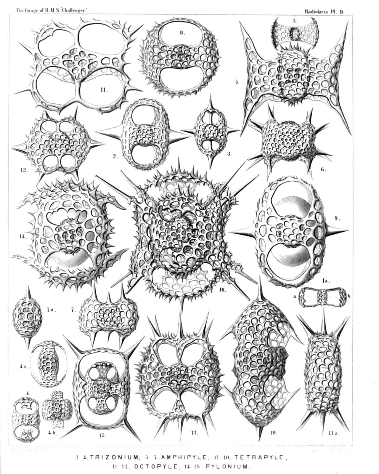
PLATE 9.
Pylonida.
Diam. Page.
Fig. 1. Monozonium alatum, n. sp., × 300 633
Dorsal view.
Fig. 1a. Apical view.
Fig. 2. Dizonium pleuracanthum, n. sp., × 400 636
Fig. 3. Dizonium stauracanthum, n. sp., × 300 636
Fig. 4. Trizonium tricinctum, n. sp., × 300 637
Dorsal view.
Fig. 4a. Lateral view.
Fig. 4b. Apical view.
Fig. 5. Amphipyle tetraceros, n. sp., × 400 642
Dorsal view.
Fig. 6. Amphipyle callizona, n. sp., × 300 644
Dorsal view.
Fig. 7. Amphipyle amphiptera, n. sp., × 300 642
Dorsal view.
Fig. 7a. Lateral view.
Fig. 8. Tetrapyle circularis, n. sp., × 300 645
Dorsal view.
Fig. 9. Tetrapyle pleuracantha, n. sp., × 400 646
Dorsal view. The lentelliptical central capsule is visible between medullary and cortical shell.
Fig. 10. Tetrapyle turrita, n. sp., × 400 649
Oblique view, half dorsal, half lateral.
Fig. 11. Octopyle stenozona, n. sp., × 400 652
Dorsal view.
Fig. 12. Octopyle sexangulata, n. sp., × 300 653
Dorsal view.
Fig. 13. Octopyle decastyle, n. sp., × 300 654
Dorsal view.
Fig. 13a. Lateral view.
Fig. 14. Pylonium quadricorne, n. sp., × 400 655
Dorsal view.
Fig. 15. Tetrapylonium quadrangulare, n. sp., × 300 658
Dorsal view.
Fig. 16. Pylozonium octacanthum, n. sp., × 300 660
Dorsal view.
PLATE 10.
Legion SPUMELLARIA.
Order LARCOIDEA.
Family Tholonida.
PLATE 10. Tholonida. Diam. Page. Fig. 1. Tholartus tricolus, n. sp., × 200 664 Fig. 2. Tholodes cupula, n. sp., × 500 665 Fig. 3. Amphitholus artiscus, n. sp., × 400 666 Fig. 4. Amphitholus panicium, n. sp., × 500 668 Fig. 5. Amphitholus acanthometra, n. sp., × 300 667 Fig. 6. Amphitholus acanthometra, n. sp., × 300 667 Frontal section of the shell. Fig. 7. Amphitholonium tricolonium, n. sp., × 300 669 Fig. 8. Staurotholus tetrastylus, n. sp., × 300 673 Fig. 9. Staurotholus dodecastylus, n. sp., × 400 674 Fig. 10. Tholoma quadrigeminum, n. sp., × 200 672 Fig. 11. Staurotholonium octodoronium, n. sp., × 300 676 Fig. 12. Tholocubus tessellatus, n. sp., × 200 677 Fig. 13. Tholoma metallasson, n. sp., × 300 672 Fig. 14. Cubotholus regularis, n. sp., × 200 680 Fig. 15. Cubotholonium ellipsoides, n. sp., × 300 682 Fig. 16. Tholocubus tesseralis, n. sp., × 400 678 Fig. 17. Tholonium hexonium, × 400 679
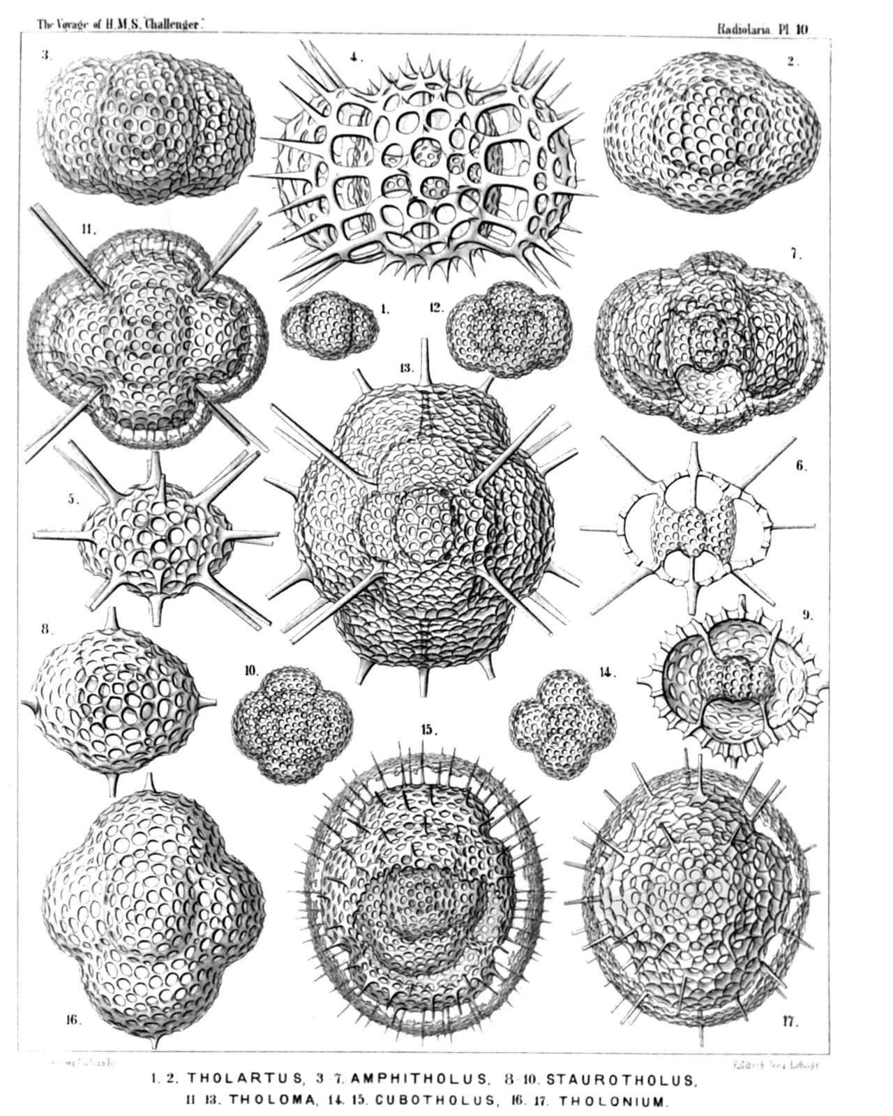
PLATE 10.
Tholonida.
Diam. Page.
Fig. 1. Tholartus tricolus, n. sp., × 200 664
Fig. 2. Tholodes cupula, n. sp., × 500 665
Fig. 3. Amphitholus artiscus, n. sp., × 400 666
Fig. 4. Amphitholus panicium, n. sp., × 500 668
Fig. 5. Amphitholus acanthometra, n. sp., × 300 667
Fig. 6. Amphitholus acanthometra, n. sp., × 300 667
Frontal section of the shell.
Fig. 7. Amphitholonium tricolonium, n. sp., × 300 669
Fig. 8. Staurotholus tetrastylus, n. sp., × 300 673
Fig. 9. Staurotholus dodecastylus, n. sp., × 400 674
Fig. 10. Tholoma quadrigeminum, n. sp., × 200 672
Fig. 11. Staurotholonium octodoronium, n. sp., × 300 676
Fig. 12. Tholocubus tessellatus, n. sp., × 200 677
Fig. 13. Tholoma metallasson, n. sp., × 300 672
Fig. 14. Cubotholus regularis, n. sp., × 200 680
Fig. 15. Cubotholonium ellipsoides, n. sp., × 300 682
Fig. 16. Tholocubus tesseralis, n. sp., × 400 678
Fig. 17. Tholonium hexonium, × 400 679
PLATE 11.
Legion SPUMELLARIA.
Order SPHÆROIDEA.
Family Astrosphærida.
PLATE 11. Astrosphærida. Diam. Page. Fig. 1. Lychnosphæra regina, n. sp., × 200 277 The entire shell and the central capsule. Numerous club-shaped radial apophyses or cœcal sacs arise from the pink central capsule and are protruded through the pores of the medullary shell, which is completely hidden by them. The sarcomatrix in the calymma, surrounding the central capsule, exhibits a fine radial striation. Numerous retracted pseudopodia, bearing red granules, arise from the sarcomatrix and pierce the calymma radially. The interval between the two concentric shells is filled up by the hyaline calymma. Fig. 2. Lychnosphæra regina, n. sp., × 400 277 A part of the cortical shell, with a radial spine. Fig. 3. Lychnosphæra regina, n. sp., × 400 277 The medullary shell and the basal parts of the radial spines arising from it. Fig. 4. Lychnosphæra regina, n. sp., × 400 277 Distal end of a radial spine. Fig. 5. Rhizoplegma lychnosphæra, n. sp., × 200 276 The central capsule and the enclosed parts of the skeleton. The protoplasm is radially striped. The central nucleus (red) sends out numerous radial apophyses, which are protruded through the pores of the medullary shell.
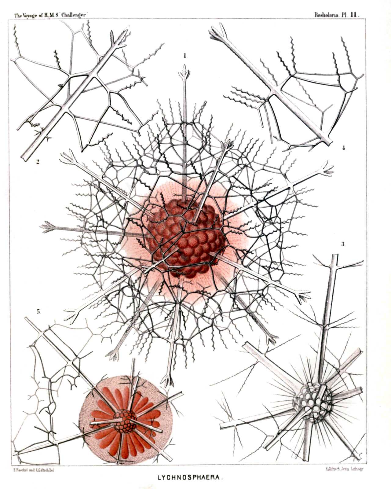
PLATE 11.
Astrosphærida.
Diam. Page.
Fig. 1. Lychnosphæra regina, n. sp., × 200 277
The entire shell and the central capsule. Numerous club-shaped radial apophyses or cœcal sacs arise from the pink central capsule and are protruded through the pores of the medullary shell, which is completely hidden by them. The sarcomatrix in the calymma, surrounding the central capsule, exhibits a fine radial striation. Numerous retracted pseudopodia, bearing red granules, arise from the sarcomatrix and pierce the calymma radially. The interval between the two concentric shells is filled up by the hyaline calymma.
Fig. 2. Lychnosphæra regina, n. sp., × 400 277
A part of the cortical shell, with a radial spine.
Fig. 3. Lychnosphæra regina, n. sp., × 400 277
The medullary shell and the basal parts of the radial spines arising from it.
Fig. 4. Lychnosphæra regina, n. sp., × 400 277
Distal end of a radial spine.
Fig. 5. Rhizoplegma lychnosphæra, n. sp., × 200 276
The central capsule and the enclosed parts of the skeleton. The protoplasm is radially striped. The central nucleus (red) sends out numerous radial apophyses, which are protruded through the pores of the medullary shell.
PLATE 12.
Legion SPUMELLARIA.
Orders PHÆOSPHÆRIA et SPHÆROIDEA.
Families Orosphærida, Astrosphærida et Liosphærida.
PLATE 12. Orosphærida, Astrosphærida et Liosphærida. Diam. Page. Fig. 1. Orosphæra huxleyii, n. sp. (vel Oroscena huxleyii), × 50 1599 Fig. 1a. A piece of the network, the bars of which contain partly an axial canal, × 200 1599 Fig. 2. Conosphæra orthoconus, n. sp., × 200 221 Fig. 3. Conosphæra platyconus, n. sp., × 300 221 Fig. 4. Conosphæra plagioconus, n. sp., × 300 222 Fig. 5. Ethmosphæra conosiphonia, n. sp., × 400 69 Fig. 5a. Vertical section through the wall. Fig. 6. Ethmosphæra polysiphonia, n. sp., × 400 70 Fig. 7. Cenosphæra compacta, n. sp., × 300 65 Fig. 8. Cenosphæra elysia, n. sp., × 300 64 Fig. 8a. Vertical section through the wall. Fig. 9. Cenosphæra mellifica, n. sp., × 300 62 Fig. 10. Cenosphæra favosa, n. sp., × 300 62 Fig. 10a. Vertical section through the wall. Fig. 11. Cenosphæra vesparia, n. sp., × 300 62 Fig. 11a. Vertical section through the wall.
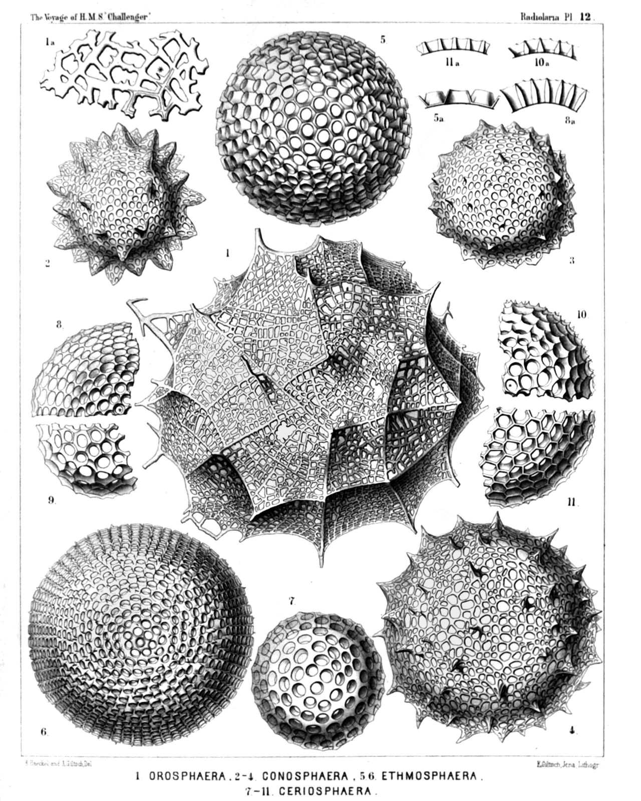
PLATE 12.
Orosphærida, Astrosphærida et Liosphærida.
Diam. Page.
Fig. 1. Orosphæra huxleyii, n. sp. (vel Oroscena huxleyii), × 50 1599
Fig. 1a. A piece of the network, the bars of which contain partly an axial canal, × 200 1599
Fig. 2. Conosphæra orthoconus, n. sp., × 200 221
Fig. 3. Conosphæra platyconus, n. sp., × 300 221
Fig. 4. Conosphæra plagioconus, n. sp., × 300 222
Fig. 5. Ethmosphæra conosiphonia, n. sp., × 400 69
Fig. 5a. Vertical section through the wall.
Fig. 6. Ethmosphæra polysiphonia, n. sp., × 400 70
Fig. 7. Cenosphæra compacta, n. sp., × 300 65
Fig. 8. Cenosphæra elysia, n. sp., × 300 64
Fig. 8a. Vertical section through the wall.
Fig. 9. Cenosphæra mellifica, n. sp., × 300 62
Fig. 10. Cenosphæra favosa, n. sp., × 300 62
Fig. 10a. Vertical section through the wall.
Fig. 11. Cenosphæra vesparia, n. sp., × 300 62
Fig. 11a. Vertical section through the wall.
PLATE 13.
Legion SPUMELLARIA.
Orders SPHÆROIDEA et PRUNOIDEA.
Families Stylosphærida et Ellipsida.
PLATE 13. Stylosphærida et Ellipsida. Diam. Page. Fig. 1. Ellipsostylus aquila, n. sp., × 300 300 Fig. 2. Ellipsostylus hirundo, n. sp., × 300 301 Fig. 3. Ellipsostylus columba, n. sp., × 300 300 Fig. 4. Xiphostylus alcedo, n. sp., × 400 127 Fig. 5. Xiphostylus edolius, n. sp., × 400 130 Fig. 6. Ellipsostylus psittacus, n. sp., × 400 300 Fig. 7. Stylostaurus caudatus, n. sp., × 400 157 Fig. 8. Ellipsostylus ciconia, n. sp., × 300 300 Fig. 9. Xiphostylus phasianus, n. sp., × 400 127 Fig. 10. Xiphostylus trochilus, n. sp., × 300 129 Fig. 11. Xiphostylus emberiza, n. sp., × 300 131 Fig. 12. Saturnalis circoideus, n. sp., × 400 132 Not fully developed. Fig. 13. Xiphostylus alca, n. sp., × 300 130 Fig. 14. Xiphostylus falco, n. sp., × 300 130 Fig. 15. Saturnalis rotula, n. sp., × 400 133 Fig. 16. Saturnalis annularis, n. sp., × 400 132
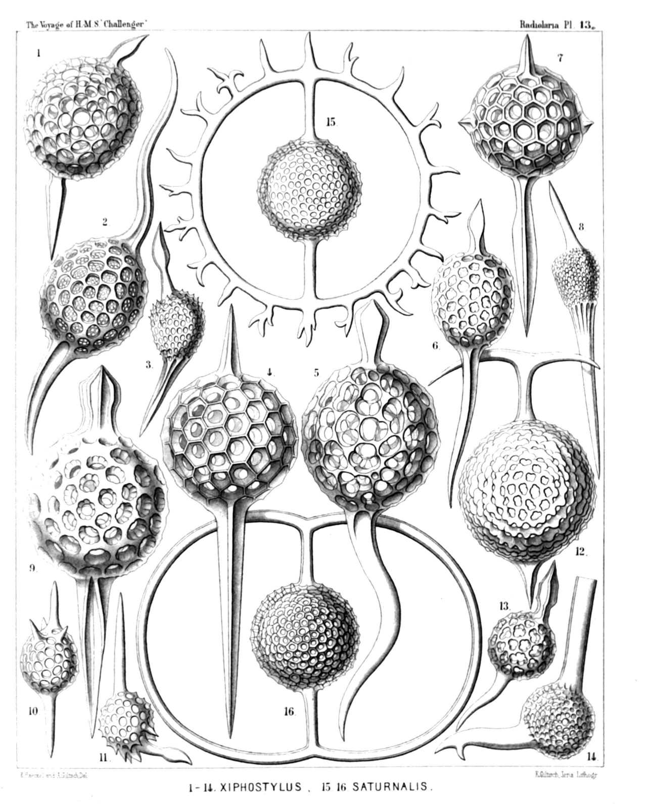
PLATE 13.
Stylosphærida et Ellipsida.
Diam. Page.
Fig. 1. Ellipsostylus aquila, n. sp., × 300 300
Fig. 2. Ellipsostylus hirundo, n. sp., × 300 301
Fig. 3. Ellipsostylus columba, n. sp., × 300 300
Fig. 4. Xiphostylus alcedo, n. sp., × 400 127
Fig. 5. Xiphostylus edolius, n. sp., × 400 130
Fig. 6. Ellipsostylus psittacus, n. sp., × 400 300
Fig. 7. Stylostaurus caudatus, n. sp., × 400 157
Fig. 8. Ellipsostylus ciconia, n. sp., × 300 300
Fig. 9. Xiphostylus phasianus, n. sp., × 400 127
Fig. 10. Xiphostylus trochilus, n. sp., × 300 129
Fig. 11. Xiphostylus emberiza, n. sp., × 300 131
Fig. 12. Saturnalis circoideus, n. sp., × 400 132
Not fully developed.
Fig. 13. Xiphostylus alca, n. sp., × 300 130
Fig. 14. Xiphostylus falco, n. sp., × 300 130
Fig. 15. Saturnalis rotula, n. sp., × 400 133
Fig. 16. Saturnalis annularis, n. sp., × 400 132
PLATE 14.
Legion SPUMELLARIA.
Orders SPHÆROIDEA et PRUNOIDEA.
Families Stylosphærida et Ellipsida.
PLATE 14. Stylosphærida et Ellipsida. Diam. Page. Fig. 1. Ellipsoxiphus atractus, n. sp., × 300 298 Fig. 2. Xiphosphæra venus, n. sp., × 300 123 Fig. 3. Ellipsoxiphus claviger, n. sp., × 300 297 Fig. 4. Xiphosphæra pallas, n. sp., × 400 124 Fig. 5. Xiphosphæra gæa, n. sp., × 400 123 Fig. 6. Xiphosphæra vesta, n. sp., × 300 126 Fig. 7. Ellipsoxiphus elegans, n. sp., var. palliatus, × 400 296 Fig. 8. Lithapium halicapsa, n. sp., × 300 303 Fig. 9. Lithapium pyriforme, n. sp., × 300 303 Fig. 10. Lithapium monocyrtis, n. sp., × 300 304 Fig. 11. Ellipsoxiphus bipolaris, n. sp., × 600 297 Fig. 12. Xiphostylus trogon, n. sp., × 400 129 Fig. 13. Xiphostylus picus, n. sp., × 300 129 Fig. 14. Lithomespilus flammabundus, n. sp., × 400 303 Fig. 15. Xiphostylus alauda, n. sp., × 400 128 Fig. 16. Lithomespilus phloginus, n. sp., × 600 302 Fig. 17. Lithomespilus phlogoides, n. sp., × 600 302
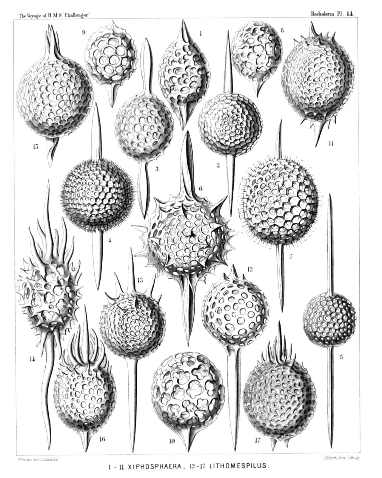
PLATE 14.
Stylosphærida et Ellipsida.
Diam. Page.
Fig. 1. Ellipsoxiphus atractus, n. sp., × 300 298
Fig. 2. Xiphosphæra venus, n. sp., × 300 123
Fig. 3. Ellipsoxiphus claviger, n. sp., × 300 297
Fig. 4. Xiphosphæra pallas, n. sp., × 400 124
Fig. 5. Xiphosphæra gæa, n. sp., × 400 123
Fig. 6. Xiphosphæra vesta, n. sp., × 300 126
Fig. 7. Ellipsoxiphus elegans, n. sp., var. palliatus, × 400 296
Fig. 8. Lithapium halicapsa, n. sp., × 300 303
Fig. 9. Lithapium pyriforme, n. sp., × 300 303
Fig. 10. Lithapium monocyrtis, n. sp., × 300 304
Fig. 11. Ellipsoxiphus bipolaris, n. sp., × 600 297
Fig. 12. Xiphostylus trogon, n. sp., × 400 129
Fig. 13. Xiphostylus picus, n. sp., × 300 129
Fig. 14. Lithomespilus flammabundus, n. sp., × 400 303
Fig. 15. Xiphostylus alauda, n. sp., × 400 128
Fig. 16. Lithomespilus phloginus, n. sp., × 600 302
Fig. 17. Lithomespilus phlogoides, n. sp., × 600 302
PLATE 15.
Legion SPUMELLARIA.
Orders SPHÆROIDEA et PRUNOIDEA.
Families Staurosphærida et Druppulida.
PLATE 15. Staurosphærida et Druppulida. Diam. Page. Fig. 1. Cromyatractus tetracelyphus, n. sp., × 300 335 Fig. 1a. The two inner medullary shells. Fig. 2. Cromyatractus tetraphractus, n. sp., × 300 335 Fig. 3. Cromyatractus cepicius, n. sp., × 300 336 The spongy distal part of a polar spine. Fig. 4. Cromyatractus ceparius, n. sp. (vel Caryostylus ceparius), × 300 336 Fig. 5. Staurolonche pertusa, n. sp., × 300 159 Fig. 5a. Its medullary shell. Fig. 6. Staurosphæra philippi, n. sp., × 300 154 Fig. 7. Stauroxiphus gladius, n. sp., × 400 163 Fig. 8. Staurocaryum arborescens, n. sp., × 300 167 Fig. 9. Rhizoplegma radicatum, n. sp., × 200 276 Fig. 9a. The medullary shell, which is completely hidden in fig. 9 by the numerous club-shaped apophyses of the central capsule.
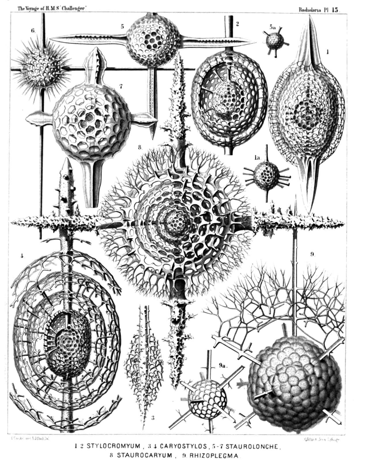
PLATE 15.
Staurosphærida et Druppulida.
Diam. Page.
Fig. 1. Cromyatractus tetracelyphus, n. sp., × 300 335
Fig. 1a. The two inner medullary shells.
Fig. 2. Cromyatractus tetraphractus, n. sp., × 300 335
Fig. 3. Cromyatractus cepicius, n. sp., × 300 336
The spongy distal part of a polar spine.
Fig. 4. Cromyatractus ceparius, n. sp. (vel Caryostylus ceparius), × 300 336
Fig. 5. Staurolonche pertusa, n. sp., × 300 159
Fig. 5a. Its medullary shell.
Fig. 6. Staurosphæra philippi, n. sp., × 300 154
Fig. 7. Stauroxiphus gladius, n. sp., × 400 163
Fig. 8. Staurocaryum arborescens, n. sp., × 300 167
Fig. 9. Rhizoplegma radicatum, n. sp., × 200 276
Fig. 9a. The medullary shell, which is completely hidden in fig. 9 by the numerous club-shaped apophyses of the central capsule.
PLATE 16.
Legion SPUMELLARIA.
Orders SPHÆROIDEA et PRUNOIDEA.
Families STYLOSPHÆRIDA et Druppulida.
PLATE 16. STYLOSPHÆRIDA et Druppulida. Diam. Page. Fig. 1. Stylosphæra melpomene, n. sp., × 300 135 Fig. 2. Lithatractus jugatus, n. sp., (vel Stylosphæra jugata), × 400 323 Fig. 3. Lithatractus fragilis, n. sp. (vel Stylosphæra fragilis), × 400 319 Fig. 4. Stylosphæra lithatractus, n. sp., × 300 The entire shell. Fig. 5. Stylosphæra lithatractus, n. sp., × 300 The greater part of the cortical shell and the two spines taken off. The description of Stylosphæra lithatractus (intermediate between Stylosphæra jugata and Stylosphæra terpsichore, p. 137) is by mistake not given in the text. Fig. 6. Stylosphæra calliope, n. sp., × 400 134 Fig. 7. Stylosphæra clio, n. sp., × 400 134 Fig. 8. Druppatractus ostracion, n. sp., × 300 326 The entire shell. Fig. 9. Druppatractus ostracion, n. sp., × 300 326 The anterior half of the cortical shell has been removed. Fig. 10. Druppatractus hippocampus, n. sp., × 300 324 The entire shell. Fig. 11. Druppatractus hippocampus, n. sp., × 300 324 The greater part of the cortical shell has been removed. Fig. 12. Stylosphæra nana, n. sp., × 300 136 The entire shell Fig. 13. Stylosphæra nana, n. sp., × 300 136 The greater part of the cortical shell taken off. Fig. 14. Sphærostylus ophidium, n. sp., × 300 140 The entire shell. Fig. 15. Sphærostylus ophidium, n. sp., × 300 140 The medullary shell alone. Fig. 16. Saturnulus ellipticus, n. sp., × 400 141 Fig. 17. Saturnulus planetes, n. sp., × 400 142
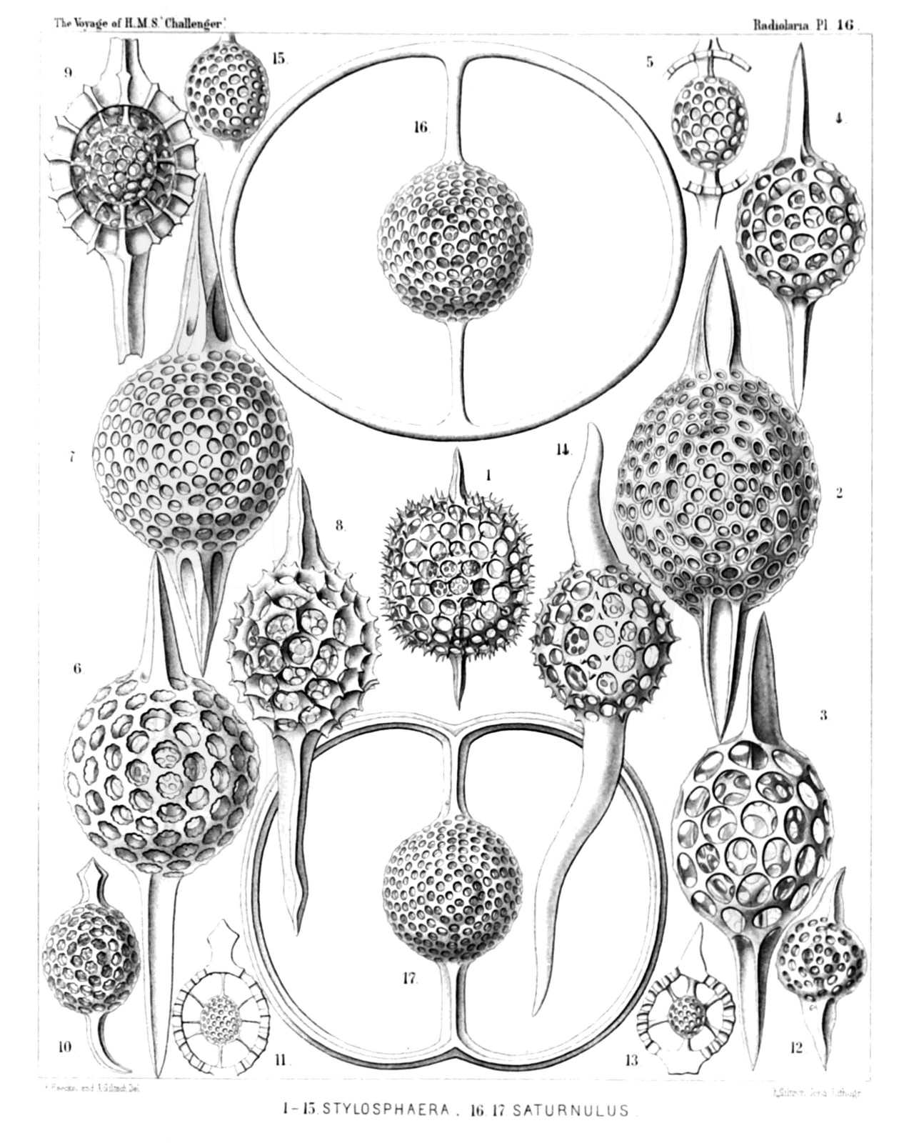
PLATE 16.
STYLOSPHÆRIDA et Druppulida.
Diam. Page.
Fig. 1. Stylosphæra melpomene, n. sp., × 300 135
Fig. 2. Lithatractus jugatus, n. sp., (vel Stylosphæra jugata), × 400 323
Fig. 3. Lithatractus fragilis, n. sp. (vel Stylosphæra fragilis), × 400 319
Fig. 4. Stylosphæra lithatractus, n. sp., × 300
The entire shell.
Fig. 5. Stylosphæra lithatractus, n. sp., × 300
The greater part of the cortical shell and the two spines taken off.
The description of Stylosphæra lithatractus (intermediate between Stylosphæra jugata and Stylosphæra terpsichore, p. 137) is by mistake not given in the text.
Fig. 6. Stylosphæra calliope, n. sp., × 400 134
Fig. 7. Stylosphæra clio, n. sp., × 400 134
Fig. 8. Druppatractus ostracion, n. sp., × 300 326
The entire shell.
Fig. 9. Druppatractus ostracion, n. sp., × 300 326
The anterior half of the cortical shell has been removed.
Fig. 10. Druppatractus hippocampus, n. sp., × 300 324
The entire shell.
Fig. 11. Druppatractus hippocampus, n. sp., × 300 324
The greater part of the cortical shell has been removed.
Fig. 12. Stylosphæra nana, n. sp., × 300 136
The entire shell
Fig. 13. Stylosphæra nana, n. sp., × 300 136
The greater part of the cortical shell taken off.
Fig. 14. Sphærostylus ophidium, n. sp., × 300 140
The entire shell.
Fig. 15. Sphærostylus ophidium, n. sp., × 300 140
The medullary shell alone.
Fig. 16. Saturnulus ellipticus, n. sp., × 400 141
Fig. 17. Saturnulus planetes, n. sp., × 400 142
PLATE 17.
Legion SPUMELLARIA.
Orders SPHÆROIDEA et PRUNOIDEA.
Families Stylosphærida, Druppulida et Spongurida.
PLATE 17. Stylosphærida, Druppulida et Spongurida. Diam. Page. Fig. 1. Stylatractus giganteus, n. sp. (vel Amphistylus giganteus), × 300 329 Fig. 2. Stylatractus sethoporus, n. sp., × 400 330 The greater part of the cortical shell taken off. Fig. 3. Stylatractus sethoporus, n. sp., × 400 330 The entire cortical shell. Fig. 4. Stylatractus compactus, n. sp., × 400 329 Fig. 5. Amphisphæra cronos, n. sp. (vel Amphistylus cronos), × 400 144 Fig. 6. Stylatractus neptunus, n. sp. (vel Amphisphæra neptunus), × 300 328 Fig. 7. Amphisphæra pluto, n. sp., × 300 144 The entire cortical shell. Fig. 8. Amphisphæra pluto, n. sp., × 300 144 Meridional section through the three concentric shells. Fig. 9. Xiphatractus glyptodon, n. sp., × 400 334 The entire cortical shell. Fig. 10. Xiphatractus glyptodon, n. sp., × 400 334 The greater part of the cortical shell taken off. Fig. 11. Xiphatractus armadillo, n. sp., × 400 332 Fig. 12. Spongoxiphus prunococcus, n. sp., × 300 354 The spongy cortical shell. Fig. 13. Spongoxiphus prunococcus, n. sp., × 600 354 The two concentric latticed medullary shells.
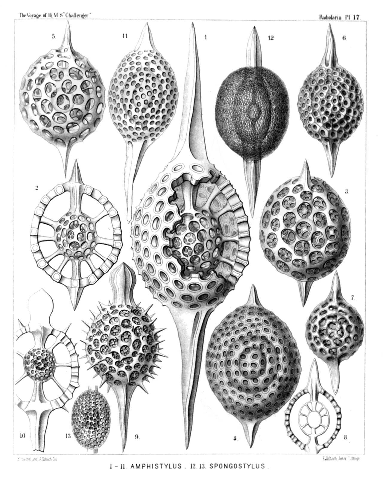
PLATE 17.
Stylosphærida, Druppulida et Spongurida.
Diam. Page.
Fig. 1. Stylatractus giganteus, n. sp. (vel Amphistylus giganteus), × 300 329
Fig. 2. Stylatractus sethoporus, n. sp., × 400 330
The greater part of the cortical shell taken off.
Fig. 3. Stylatractus sethoporus, n. sp., × 400 330
The entire cortical shell.
Fig. 4. Stylatractus compactus, n. sp., × 400 329
Fig. 5. Amphisphæra cronos, n. sp. (vel Amphistylus cronos), × 400 144
Fig. 6. Stylatractus neptunus, n. sp. (vel Amphisphæra neptunus), × 300 328
Fig. 7. Amphisphæra pluto, n. sp., × 300 144
The entire cortical shell.
Fig. 8. Amphisphæra pluto, n. sp., × 300 144
Meridional section through the three concentric shells.
Fig. 9. Xiphatractus glyptodon, n. sp., × 400 334
The entire cortical shell.
Fig. 10. Xiphatractus glyptodon, n. sp., × 400 334
The greater part of the cortical shell taken off.
Fig. 11. Xiphatractus armadillo, n. sp., × 400 332
Fig. 12. Spongoxiphus prunococcus, n. sp., × 300 354
The spongy cortical shell.
Fig. 13. Spongoxiphus prunococcus, n. sp., × 600 354
The two concentric latticed medullary shells.
PLATE 18.
Legion SPUMELLARIA.
Order SPHÆROIDEA.
Families Liosphærida et Astrosphærida.
PLATE 18. Liosphærida et Astrosphærida. Diam. Page. Fig. 1. Centrocubus cladostylus, n. sp., × 100 278 Fig. 2. Octodendron spathillatum, n. sp., × 300 280 The entire inner shell, but a small part only of the outer shell is represented. Fig. 3. Octodendron cubocentron, n. sp., × 400 279 The central capsule (somewhat irregular by compression?) exhibits a large excentric nucleus (probably dislocated artificially?). Fig. 4. Octodendron spathillatum, n. sp., × 800 280 Free distal end of a radial spine, with the spathillæ on the end of the branches. Fig. 5. Rhizosphæra serrata, n. sp., × 300 284 Fig. 6. Rhizosphæra serrata, n. sp., × 300 284 Medullary shell. Fig. 7. Rhizosphæra serrata, n. sp., × 600 284 A single radial spine. Fig. 8. Plegmosphæra exodictyon, n. sp., × 200 89 The central shell-cavity encloses the spherical central capsule and the concentric nucleus. Fig. 9. Spongodrymus elaphococcus, n. sp., × 150 272 The entire inner shell, but only a small part of the outer spongy envelope is represented.
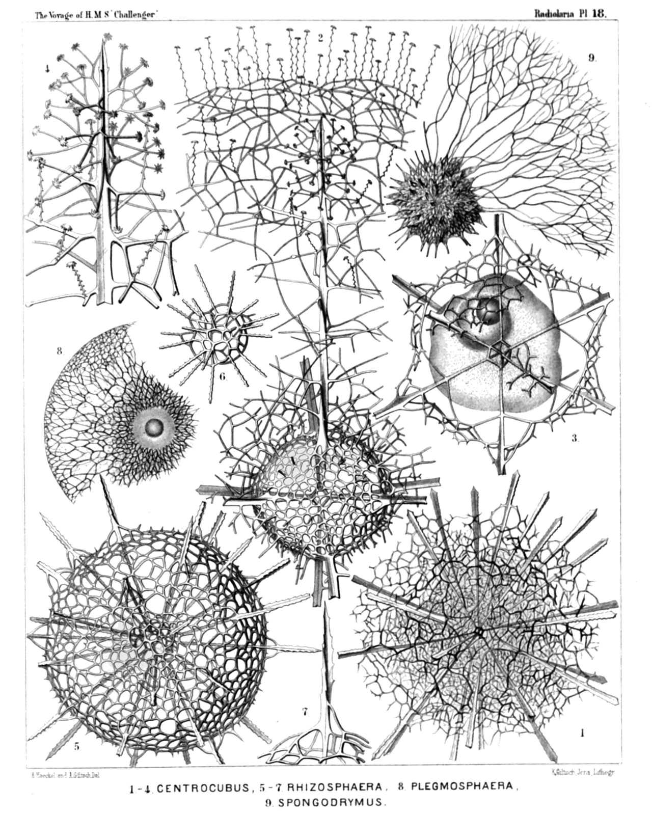
PLATE 18.
Liosphærida et Astrosphærida.
Diam. Page.
Fig. 1. Centrocubus cladostylus, n. sp., × 100 278
Fig. 2. Octodendron spathillatum, n. sp., × 300 280
The entire inner shell, but a small part only of the outer shell is represented.
Fig. 3. Octodendron cubocentron, n. sp., × 400 279
The central capsule (somewhat irregular by compression?) exhibits a large excentric nucleus (probably dislocated artificially?).
Fig. 4. Octodendron spathillatum, n. sp., × 800 280
Free distal end of a radial spine, with the spathillæ on the end of the branches.
Fig. 5. Rhizosphæra serrata, n. sp., × 300 284
Fig. 6. Rhizosphæra serrata, n. sp., × 300 284
Medullary shell.
Fig. 7. Rhizosphæra serrata, n. sp., × 600 284
A single radial spine.
Fig. 8. Plegmosphæra exodictyon, n. sp., × 200 89
The central shell-cavity encloses the spherical central capsule and the concentric nucleus.
Fig. 9. Spongodrymus elaphococcus, n. sp., × 150 272
The entire inner shell, but only a small part of the outer spongy envelope is represented.
PLATE 19.
Legion SPUMELLARIA.
Order SPHÆROIDEA.
Family Astrosphærida.
PLATE 19. Astrosphærida. Diam. Page. Fig. 1. Drymosphæra polygonalis, n. sp., × 200 249 Fig. 2. Leptosphæra hexagonalis, n. sp., × 200 244 Showing the central capsule (forming numerous club-shaped protuberances) and the simple spherical nucleus in its centre. The skeleton is nearly the same as in Diplosphæra hexagonalis (fig. 3). Fig. 3. Diplosphæra hexagonalis, n. sp., × 200 246 The spherical central capsule, with radially striped protoplasm, is enclosed in the inner shell, and exhibits in its centre the clear spherical nucleus. Fig. 4. Astrosphæra hexagonalis, n. sp., × 300 250 Fig. 5. Astrosphæra stellata, n. sp., × 300 251 The central capsule, enclosed in the inner shell, exhibits a distinct radial striation of the protoplasm, and in the centre a clear spherical nucleus. Fig. 6. Haliomma rhodococcus, n. sp. (vel Sethosphæra rhodococcus), × 400 237 The greater part of the outer shell is removed.
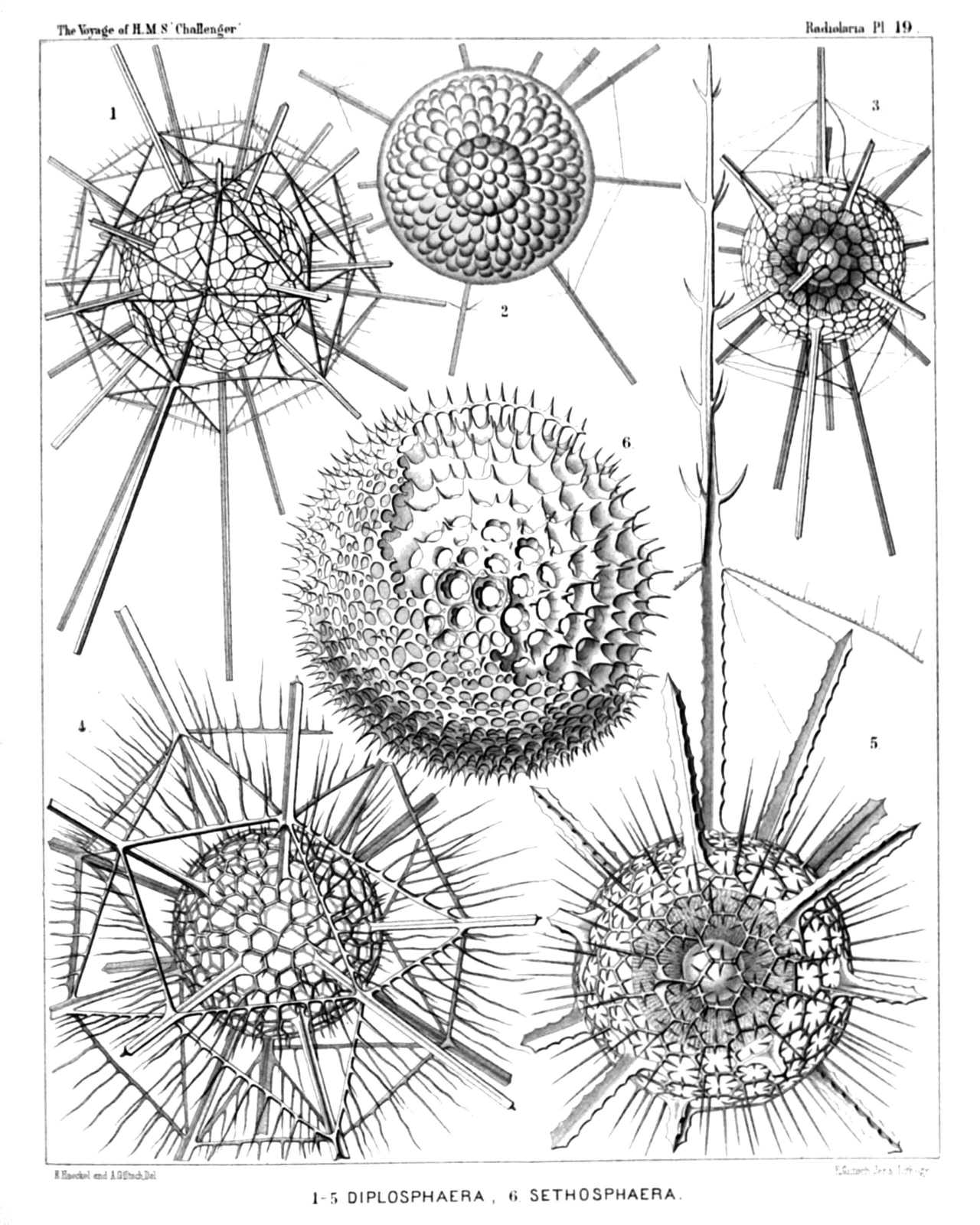
PLATE 19.
Astrosphærida.
Diam. Page.
Fig. 1. Drymosphæra polygonalis, n. sp., × 200 249
Fig. 2. Leptosphæra hexagonalis, n. sp., × 200 244
Showing the central capsule (forming numerous club-shaped protuberances) and the simple spherical nucleus in its centre. The skeleton is nearly the same as in Diplosphæra hexagonalis (fig. 3).
Fig. 3. Diplosphæra hexagonalis, n. sp., × 200 246
The spherical central capsule, with radially striped protoplasm, is enclosed in the inner shell, and exhibits in its centre the clear spherical nucleus.
Fig. 4. Astrosphæra hexagonalis, n. sp., × 300 250
Fig. 5. Astrosphæra stellata, n. sp., × 300 251
The central capsule, enclosed in the inner shell, exhibits a distinct radial striation of the protoplasm, and in the centre a clear spherical nucleus.
Fig. 6. Haliomma rhodococcus, n. sp. (vel Sethosphæra rhodococcus), × 400 237
The greater part of the outer shell is removed.
PLATE 20.
Legion SPUMELLARIA.
Order SPHÆROIDEA.
Families Liosphærida et Astrosphærida.
PLATE 20. Liosphærida et Astrosphærida. Diam. Page. Fig. 1. Drymosphæra dendrophora, n. sp., × 300 249 Fig. 1a. Meridional section through the central capsule. In the centre the large spherical nucleus is visible. The protoplasm around it is distinctly radiate. From the central capsule arise numerous club-shaped apophyses or cæcal sacs, which are protruded through the meshes of the inner shell, × 300 Fig. 1b. Basal part of a single radial spine, and its connection with the network of the two shells, × 400 Fig. 2. Liosphæra polypora, n. sp., × 300 78 The greater part of the outer shell is removed. Fig. 3. Liosphæra hexagonia, n. sp., × 400 76 Fig. 4. Carposphæra melitomma, n. sp. (vel Melitomma melittosphæra), × 400 73
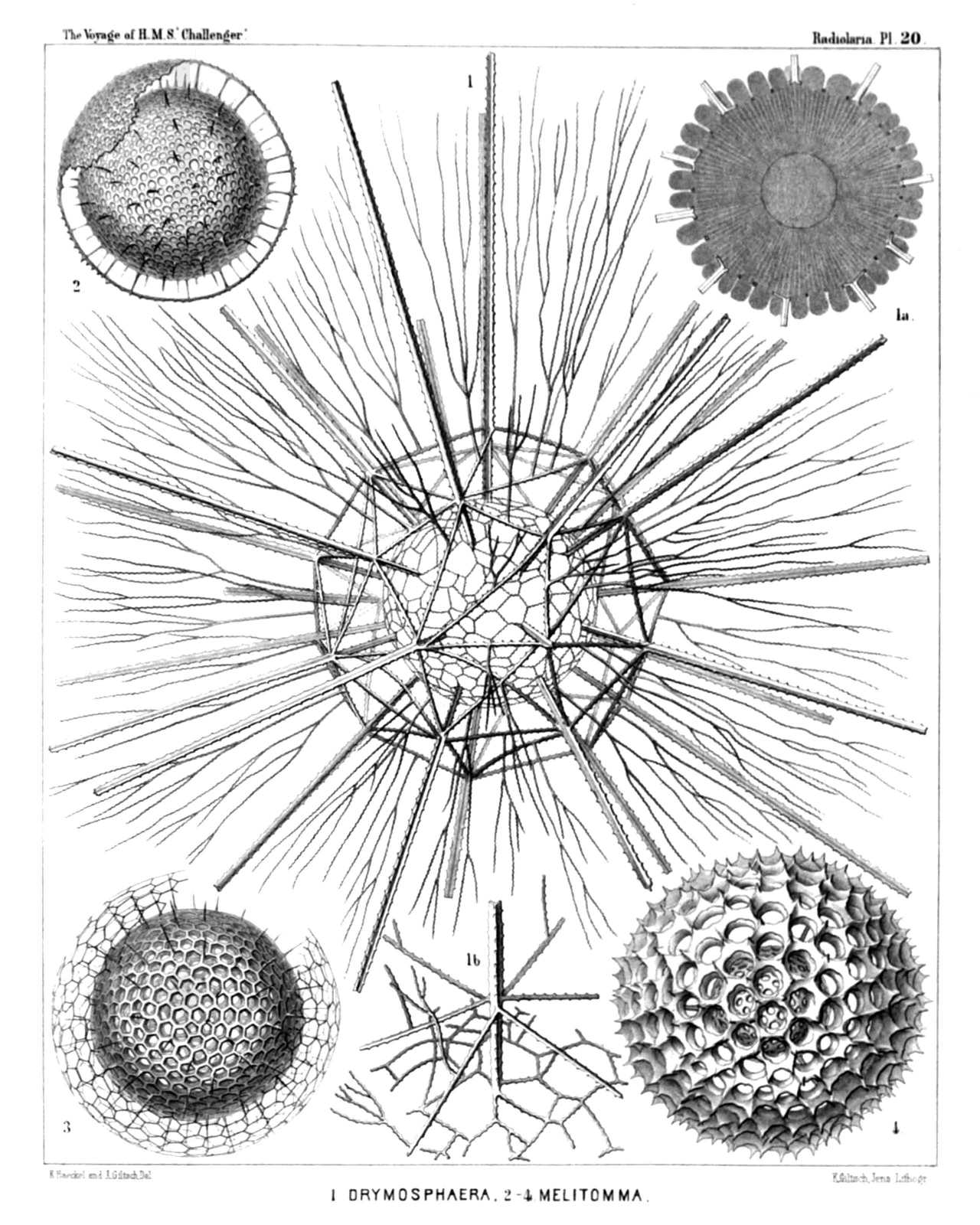
PLATE 20.
Liosphærida et Astrosphærida.
Diam. Page.
Fig. 1. Drymosphæra dendrophora, n. sp., × 300 249
Fig. 1a. Meridional section through the central capsule. In the centre the large spherical nucleus is visible. The protoplasm around it is distinctly radiate. From the central capsule arise numerous club-shaped apophyses or cæcal sacs, which are protruded through the meshes of the inner shell, × 300
Fig. 1b. Basal part of a single radial spine, and its connection with the network of the two shells, × 400
Fig. 2. Liosphæra polypora, n. sp., × 300 78
The greater part of the outer shell is removed.
Fig. 3. Liosphæra hexagonia, n. sp., × 400 76
Fig. 4. Carposphæra melitomma, n. sp. (vel Melitomma melittosphæra), × 400 73
PLATE 21.
Legion SPUMELLARIA.
Order SPHÆROIDEA.
Family Cubosphærida.
PLATE 21. Cubosphærida. Diam. Page. Fig. 1. Hexastylus cochleatus, n. sp., × 400 174 From the central capsule, enclosed in the shell, numerous delicate radial pseudopodia arise, which are protruded through the pores of the shell. Fig. 2. Hexastylus triaxonius, n. sp., × 400 175 Fig. 3. Hexastylus phænaxonius, n. sp., × 300 171 Fig. 4. Hexastylus thaletis, n. sp., × 400 172 Fig. 5. Hexastylus minimus, n. sp., × 400 172 Fig. 6. Hexastylus dimensivus, n. sp., × 400 175 Fig. 7. Hexastylus spiralis, n. sp., × 400 177 Fig. 8. Hexastylus dictyotus, n. sp., × 400 176 Fig. 9. Hexastylus dictyotus, n. sp., × 400 176 Central capsule with concentric nucleus and nucleolus; the protoplasm is radially striped. Fig. 10. Hexastylus marginatus, n. sp., × 400 176 Fig. 10a. Radial section through the shell-wall. Fig. 11. Hexastylus solonis, n. sp., × 400 173 Fig. 12. Hexastylus contortus, n. sp., × 300 177
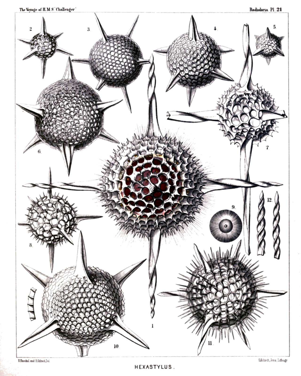
PLATE 21.
Cubosphærida.
Diam. Page.
Fig. 1. Hexastylus cochleatus, n. sp., × 400 174
From the central capsule, enclosed in the shell, numerous delicate radial pseudopodia arise, which are protruded through the pores of the shell.
Fig. 2. Hexastylus triaxonius, n. sp., × 400 175
Fig. 3. Hexastylus phænaxonius, n. sp., × 300 171
Fig. 4. Hexastylus thaletis, n. sp., × 400 172
Fig. 5. Hexastylus minimus, n. sp., × 400 172
Fig. 6. Hexastylus dimensivus, n. sp., × 400 175
Fig. 7. Hexastylus spiralis, n. sp., × 400 177
Fig. 8. Hexastylus dictyotus, n. sp., × 400 176
Fig. 9. Hexastylus dictyotus, n. sp., × 400 176
Central capsule with concentric nucleus and nucleolus; the protoplasm is radially striped.
Fig. 10. Hexastylus marginatus, n. sp., × 400 176
Fig. 10a. Radial section through the shell-wall.
Fig. 11. Hexastylus solonis, n. sp., × 400 173
Fig. 12. Hexastylus contortus, n. sp., × 300 177
PLATE 22.
Legion SPUMELLARIA.
Order SPHÆROIDEA.
Family Cubosphærida.
PLATE 22. Cubosphærida. Diam. Page. Fig. 1. Hexalonche pythagoræa, n. sp., × 300 185 Fig. 2. Hexalonche conicornis, n. sp., × 300 181 Fig. 3. Hexalonche aristarchi, n. sp., × 400 185 Fig. 4. Hexalonche philosophica, n. sp., × 400 186 Fig. 5. Hexalonche anaximandri, n. sp., × 400 182 Fig. 6. Hexalonche octocolpa, n. sp., × 300 183 Fig. 6a. The inner shell alone. Fig. 7. Hexalonche heracliti, n. sp., × 300 187 Fig. 8. Hexalonche octahedra, n. sp., × 400 181 Fig. 8a. The inner shell alone. Fig. 9. Hexancistra tricuspis, n. sp., × 300 188 Fig. 10. Hexancistra triserrata, n. sp., × 300 188 Fig. 11. Hexancistra quadricuspis, n. sp., × 300 189
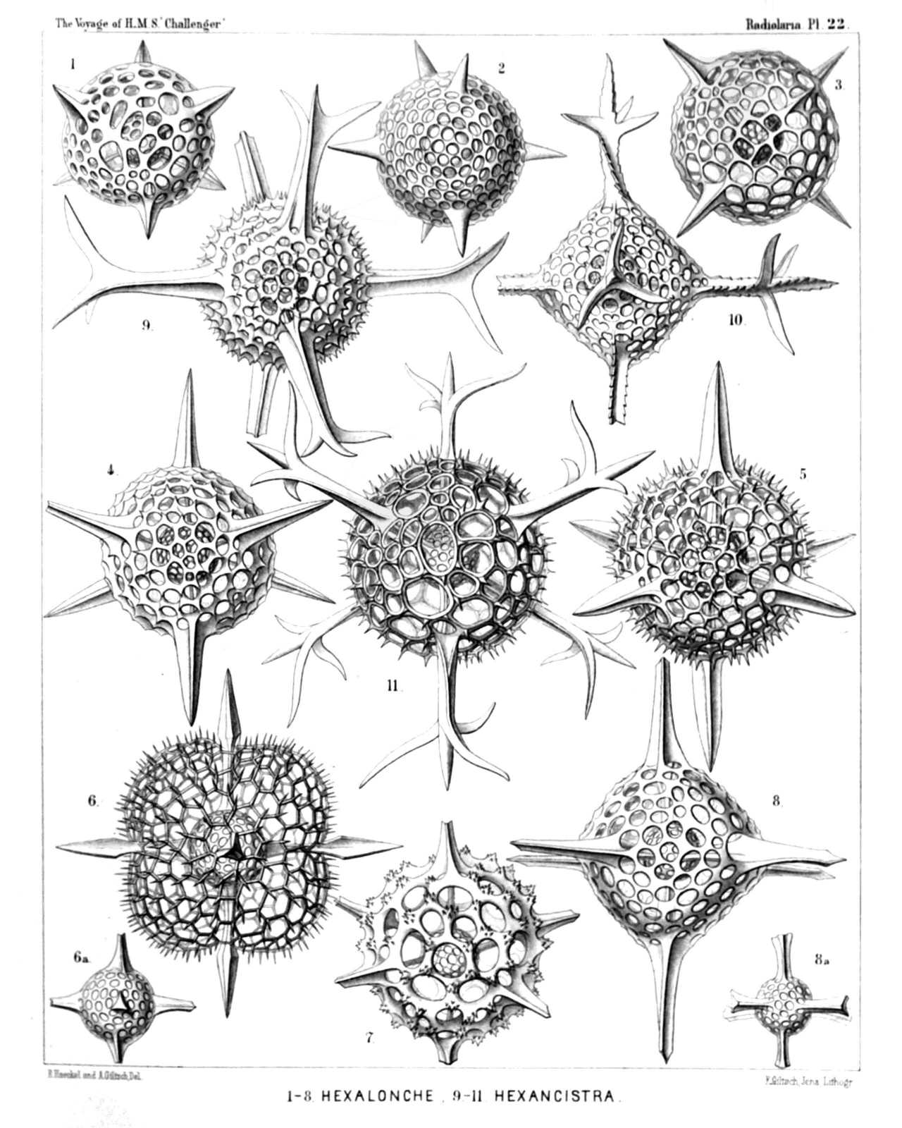
PLATE 22.
Cubosphærida.
Diam. Page.
Fig. 1. Hexalonche pythagoræa, n. sp., × 300 185
Fig. 2. Hexalonche conicornis, n. sp., × 300 181
Fig. 3. Hexalonche aristarchi, n. sp., × 400 185
Fig. 4. Hexalonche philosophica, n. sp., × 400 186
Fig. 5. Hexalonche anaximandri, n. sp., × 400 182
Fig. 6. Hexalonche octocolpa, n. sp., × 300 183
Fig. 6a. The inner shell alone.
Fig. 7. Hexalonche heracliti, n. sp., × 300 187
Fig. 8. Hexalonche octahedra, n. sp., × 400 181
Fig. 8a. The inner shell alone.
Fig. 9. Hexancistra tricuspis, n. sp., × 300 188
Fig. 10. Hexancistra triserrata, n. sp., × 300 188
Fig. 11. Hexancistra quadricuspis, n. sp., × 300 189
PLATE 23.
Legion SPUMELLARIA.
Order SPHÆROIDEA.
Family Cubosphærida.
PLATE 23. Cubosphærida. Diam. Page. Fig. 1. Hexadendron bipinnatum, n. sp., × 400 200 Fig. 2. Hexacromyum octahedrum, n. sp., × 400 202 Fig. 3. Hexancistra mirabilis, n. sp. (= Hexapitys mirabilis), × 400 189 The spherical central capsule encloses the concentric spherical inner shell (which is filled up by the nucleus), and is surrounded by the octahedral outer shell. The latter is enveloped by the octahedral calymma, which is radially striated and contains numerous xanthellæ. Fig. 4. Hexacaryum arborescens, n. sp., × 400 203 Fig. 5. Hexacontium clavigerum, n. sp., × 300 19
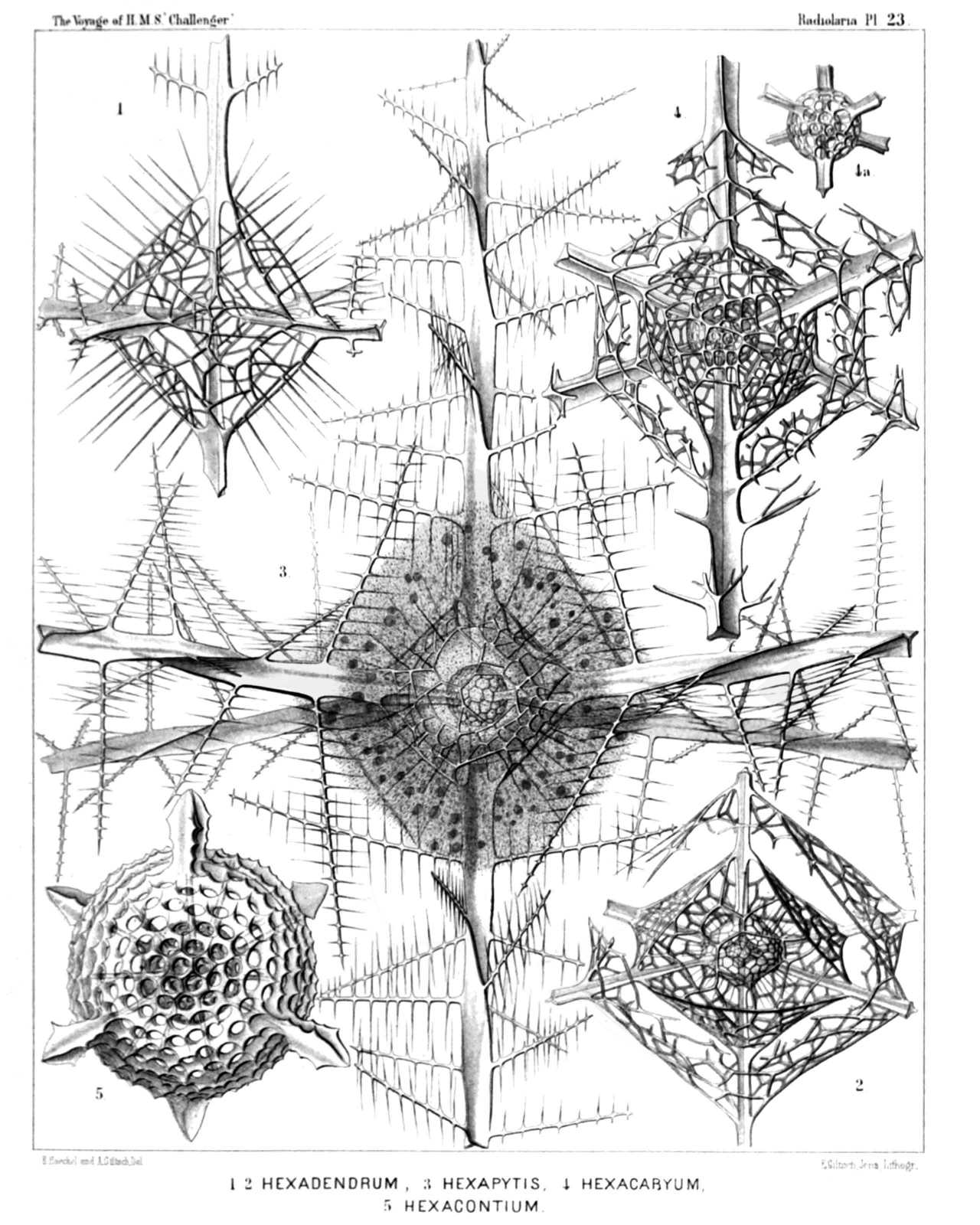
PLATE 23.
Cubosphærida.
Diam. Page.
Fig. 1. Hexadendron bipinnatum, n. sp., × 400 200
Fig. 2. Hexacromyum octahedrum, n. sp., × 400 202
Fig. 3. Hexancistra mirabilis, n. sp. (= Hexapitys mirabilis), × 400 189
The spherical central capsule encloses the concentric spherical inner shell (which is filled up by the nucleus), and is surrounded by the octahedral outer shell. The latter is enveloped by the octahedral calymma, which is radially striated and contains numerous xanthellæ.
Fig. 4. Hexacaryum arborescens, n. sp., × 400 203
Fig. 5. Hexacontium clavigerum, n. sp., × 300 19
PLATE 24.
Legion SPUMELLARIA.
Order SPHÆROIDEA.
Family Cubosphærida.
PLATE 24. Cubosphærida. Diam. Page. Fig. 1. Hexacontium sceptrum, n. sp., × 400 194 Fig. 1a. The two medullary shells. Fig. 2. Hexacontium favosum, n. sp., × 400 194 Fig. 2a. The two medullary shells. Fig. 3. Hexacontium axotrias, n. sp., × 300 192 The six lattice-plates, which form the cortical shell, are not yet fully developed. Fig. 4. Hexacontium floridum, n. sp., × 300 195 Fig. 4a. The two medullary shells. Fig. 5. Hexacontium papillosum, n. sp., × 400 197 Fig. 5a. The two medullary shells. Fig. 6. Hexacontium lævigatum, n. sp., × 400 193 The contours of the two medullary shells are visible in the centre. Fig. 7. Hexacontium prionacanthum, n. sp., × 400 195 Fig. 7a. The two medullary shells, connected with a fragment of the cortical shell. Fig. 8. Cubosphæra cubaxonia, n. sp., × 400 203 Fig. 8a. A single radial spine. Fig. 9. Hexacromyum elegans, n. sp., × 400 201 A part of the two cortical shells is broken off.
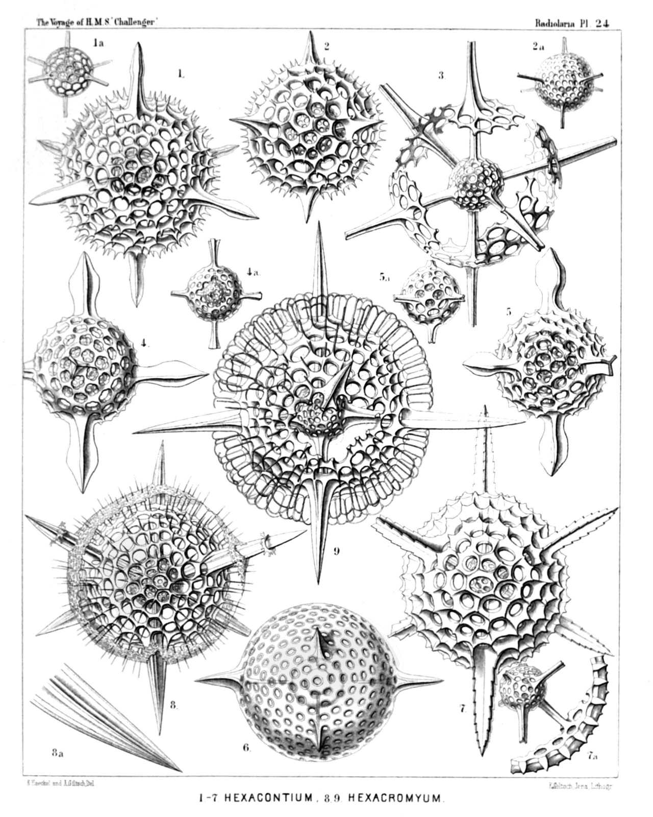
PLATE 24.
Cubosphærida.
Diam. Page.
Fig. 1. Hexacontium sceptrum, n. sp., × 400 194
Fig. 1a. The two medullary shells.
Fig. 2. Hexacontium favosum, n. sp., × 400 194
Fig. 2a. The two medullary shells.
Fig. 3. Hexacontium axotrias, n. sp., × 300 192
The six lattice-plates, which form the cortical shell, are not yet fully developed.
Fig. 4. Hexacontium floridum, n. sp., × 300 195
Fig. 4a. The two medullary shells.
Fig. 5. Hexacontium papillosum, n. sp., × 400 197
Fig. 5a. The two medullary shells.
Fig. 6. Hexacontium lævigatum, n. sp., × 400 193
The contours of the two medullary shells are visible in the centre.
Fig. 7. Hexacontium prionacanthum, n. sp., × 400 195
Fig. 7a. The two medullary shells, connected with a fragment of the cortical shell.
Fig. 8. Cubosphæra cubaxonia, n. sp., × 400 203
Fig. 8a. A single radial spine.
Fig. 9. Hexacromyum elegans, n. sp., × 400 201
A part of the two cortical shells is broken off.
PLATE 25.
Legion SPUMELLARIA.
Order SPHÆROIDEA.
Family Cubosphærida.
PLATE 25. Cubosphærida. Diam. Page. Fig. 1. Hexadoridium streptacanthum, n. sp., × 400 206 Fig. 1a. The two concentric medullary shells. Fig. 2. Hexalonche amphisiphon, n. sp., × 300 182 Fig. 2a. Medullary shell connected with a fragment of the cortical shell. Fig. 2b. Vertical section through the wall of the cortical shell. (Below the centre of the Plate, also lettered 3a by mistake.) Fig. 3. Hexalonche rosetta, n. sp., × 400 180 Fig. 3a. Medullary shell. Fig. 3b. Vertical section through the wall of the cortical shell. Fig. 4. Hexalonche curvicornis, n. sp., × 300 181 Outer shell not yet complete, or partly broken off (?). Fig. 5. Hexalonche anaximenis, n. sp., × 400 183 Fig. 6. Hexalonche hystricina, n. sp., × 300 187 Fig. 7. Hexacontium circumtextum, n. sp., × 400 193 Fig. 7a. Vertical section through the double wall of the cortical shell. Fig. 8. Hexacontium gladiatum, n. sp., × 400 198 A part of the two outer shells and of the radial spines is broken off.
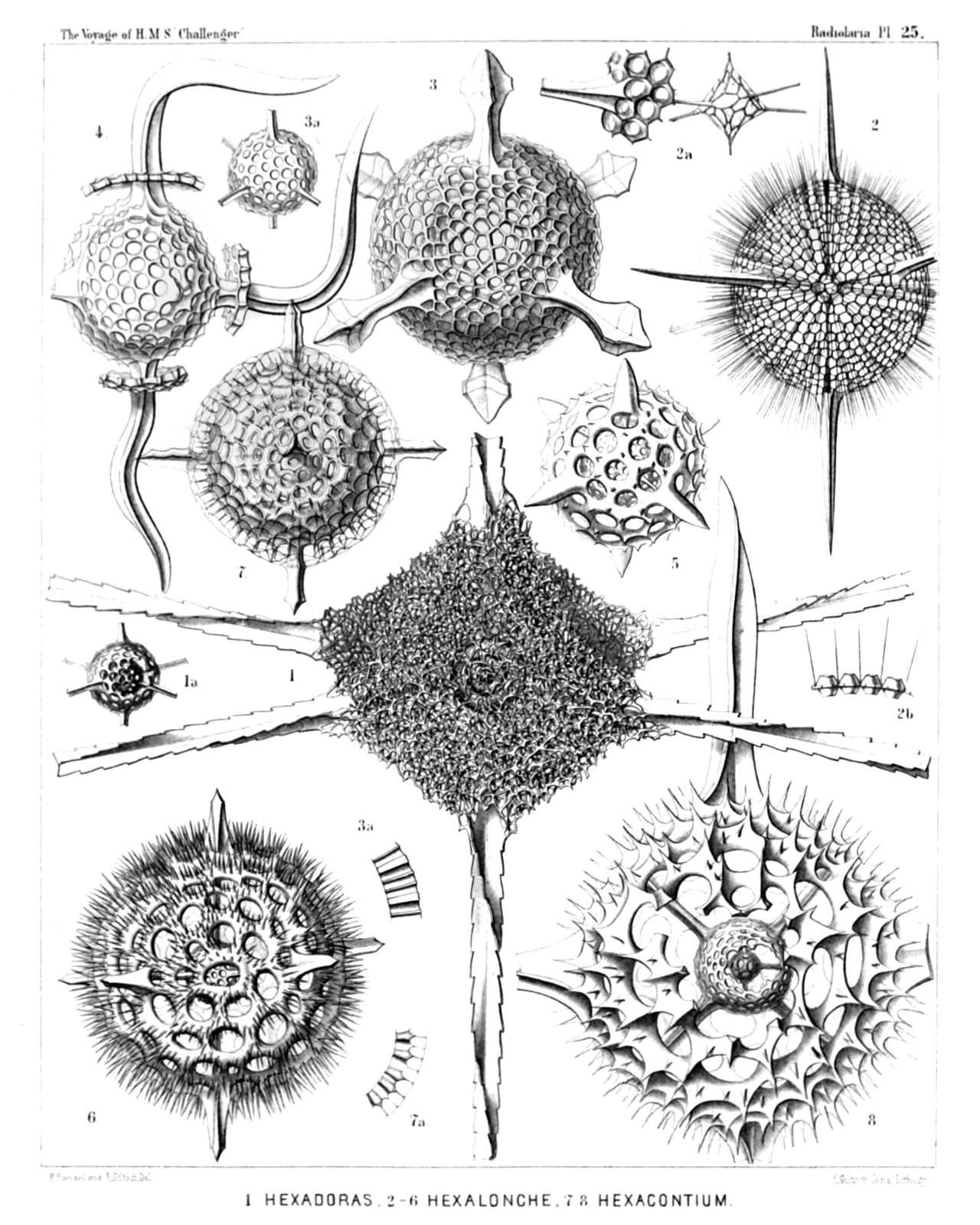
PLATE 25.
Cubosphærida.
Diam. Page.
Fig. 1. Hexadoridium streptacanthum, n. sp., × 400 206
Fig. 1a. The two concentric medullary shells.
Fig. 2. Hexalonche amphisiphon, n. sp., × 300 182
Fig. 2a. Medullary shell connected with a fragment of the cortical shell.
Fig. 2b. Vertical section through the wall of the cortical shell. (Below the centre of the Plate, also lettered 3a by mistake.)
Fig. 3. Hexalonche rosetta, n. sp., × 400 180
Fig. 3a. Medullary shell.
Fig. 3b. Vertical section through the wall of the cortical shell.
Fig. 4. Hexalonche curvicornis, n. sp., × 300 181
Outer shell not yet complete, or partly broken off (?).
Fig. 5. Hexalonche anaximenis, n. sp., × 400 183
Fig. 6. Hexalonche hystricina, n. sp., × 300 187
Fig. 7. Hexacontium circumtextum, n. sp., × 400 193
Fig. 7a. Vertical section through the double wall of the cortical shell.
Fig. 8. Hexacontium gladiatum, n. sp., × 400 198
A part of the two outer shells and of the radial spines is broken off.
PLATE 26.
Legion SPUMELLARIA.
Order SPHÆROIDEA.
Families Liosphærida et Astrosphærida.
PLATE 26. Liosphærida et Astrosphærida. Diam. Page. Fig. 1. Coscinomma amphisiphon, n. sp., × 300 222 Fig. 1a. A piece of the lattice-shell, × 600 Fig. 1b. Vertical section through the shell-wall, × 600 Fig. 2. Heliosphæra hexagonaria, n. sp., × 300 217 Fig. 3. Acanthosphæra castanea, n. sp., × 400 211 Fig. 4. Acanthosphæra angulata, n. sp., × 300 216 Fig. 5. Acanthosphæra reticulata, n. sp., × 300 217 Fig. 6. Heliosphæra coronata, n. sp., × 400 219 Fig. 6a. A single pore with its coronal, × 300 Fig. 7. Acanthosphæra mucronata, n. sp., × 400 212 Fig. 8. Acanthosphæra clavata, n. sp., × 400 212 Fig. 9. Heliosphæra pectinata, n. sp., × 400 218 Fig. 10. Cenosphæra perforata, n. sp., × 400 66 Fig. 11. Cenosphæra coronata, n. sp., × 400 67
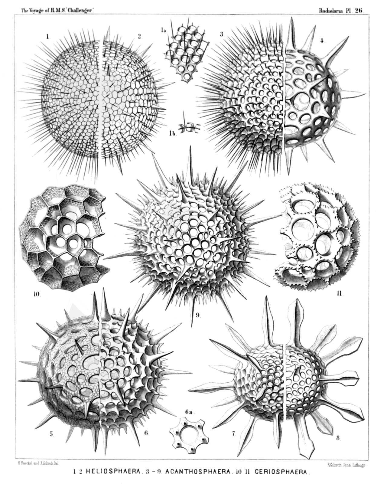
PLATE 26.
Liosphærida et Astrosphærida.
Diam. Page.
Fig. 1. Coscinomma amphisiphon, n. sp., × 300 222
Fig. 1a. A piece of the lattice-shell, × 600
Fig. 1b. Vertical section through the shell-wall, × 600
Fig. 2. Heliosphæra hexagonaria, n. sp., × 300 217
Fig. 3. Acanthosphæra castanea, n. sp., × 400 211
Fig. 4. Acanthosphæra angulata, n. sp., × 300 216
Fig. 5. Acanthosphæra reticulata, n. sp., × 300 217
Fig. 6. Heliosphæra coronata, n. sp., × 400 219
Fig. 6a. A single pore with its coronal, × 300
Fig. 7. Acanthosphæra mucronata, n. sp., × 400 212
Fig. 8. Acanthosphæra clavata, n. sp., × 400 212
Fig. 9. Heliosphæra pectinata, n. sp., × 400 218
Fig. 10. Cenosphæra perforata, n. sp., × 400 66
Fig. 11. Cenosphæra coronata, n. sp., × 400 67
PLATE 27.
Legion SPUMELLARIA.
Order SPHÆROIDEA.
Family Astrosphærida.
PLATE 27. Astrosphærida. Diam. Page. Fig. 1. Cladococcus pinetum, n. sp., × 300 226 Fig. 2. Cladococcus scoparius, n. sp., × 300 225 Fig. 3. Cladococcus abietinus, n. sp., × 300 226 The central capsule, enclosed originally in the shell, sends out numerous club-shaped apophyses through the pores of the lattice-sphere. The central spherical nucleus fills up half the shell-cavity. Fig. 4. Cladococcus stalactites, n. sp., × 300 227 Fig. 5. Cladococcus dendrites, n. sp., × 200 227
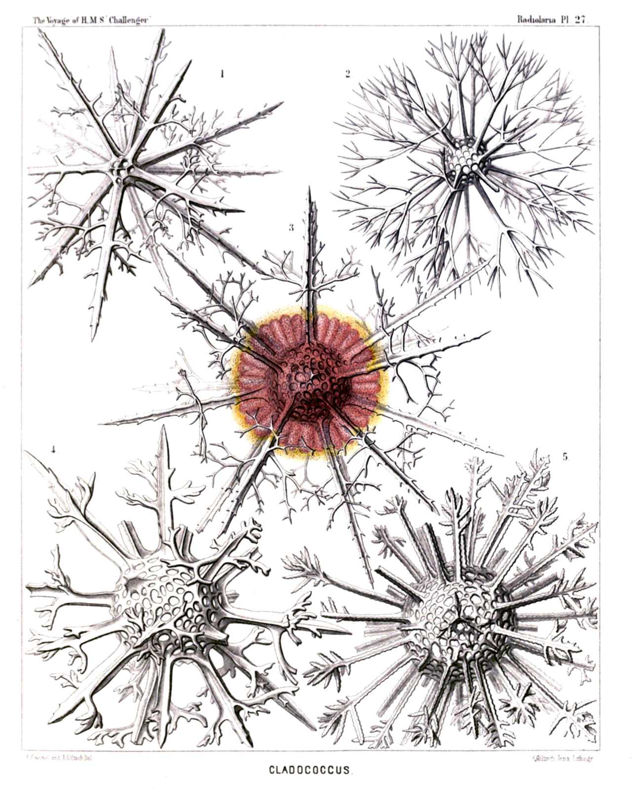
PLATE 27.
Astrosphærida.
Diam. Page.
Fig. 1. Cladococcus pinetum, n. sp., × 300 226
Fig. 2. Cladococcus scoparius, n. sp., × 300 225
Fig. 3. Cladococcus abietinus, n. sp., × 300 226
The central capsule, enclosed originally in the shell, sends out numerous club-shaped apophyses through the pores of the lattice-sphere. The central spherical nucleus fills up half the shell-cavity.
Fig. 4. Cladococcus stalactites, n. sp., × 300 227
Fig. 5. Cladococcus dendrites, n. sp., × 200 227
PLATE 28.
Legion SPUMELLARIA.
Order SPHÆROIDEA.
Families Liosphærida et Astrosphærida.
PLATE 28. Liosphærida et Astrosphærida. Diam. Page. Fig. 1. Haliomma lirianthus, n. sp., × 300 232 Fig. 1a. Medullary shell, × 300 Fig. 1b. Three pores of the cortical shell, × 900 Fig. 2. Carposphæra nodosa, n. sp., × 300 76 Fig. 2a. The medullary shell is visible, the upper half of the cortical shell being taken off, × 300 Fig. 3. Heliosoma radians, n. sp., × 300 240 Fig. 3a. Medullary shell, × 300 Fig. 4. Heliosoma hastatum, n. sp., × 400 241 Fig. 5. Haliomma compactum, n. sp., × 400 239 Fig. 5a. The upper half of the cortical shell is removed, × 300 Fig. 6. Haliomma macrodoras, n. sp., × 400 238 Fig. 7. Haliomma circumtextum, n sp., × 400 233 Fig. 8. Elatomma juniperinum, n. sp., × 400 243 Fig. 8a. Medullary shell, × 400
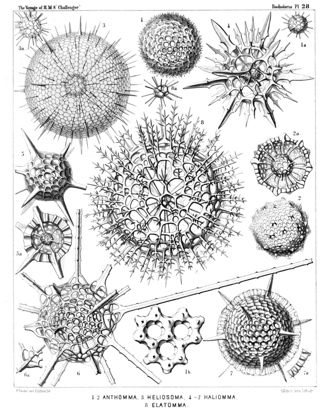
PLATE 28.
Liosphærida et Astrosphærida.
Diam. Page.
Fig. 1. Haliomma lirianthus, n. sp., × 300 232
Fig. 1a. Medullary shell, × 300
Fig. 1b. Three pores of the cortical shell, × 900
Fig. 2. Carposphæra nodosa, n. sp., × 300 76
Fig. 2a. The medullary shell is visible, the upper half of the cortical shell being taken off, × 300
Fig. 3. Heliosoma radians, n. sp., × 300 240
Fig. 3a. Medullary shell, × 300
Fig. 4. Heliosoma hastatum, n. sp., × 400 241
Fig. 5. Haliomma compactum, n. sp., × 400 239
Fig. 5a. The upper half of the cortical shell is removed, × 300
Fig. 6. Haliomma macrodoras, n. sp., × 400 238
Fig. 7. Haliomma circumtextum, n sp., × 400 233
Fig. 8. Elatomma juniperinum, n. sp., × 400 243
Fig. 8a. Medullary shell, × 400
PLATE 29.
Legion SPUMELLARIA.
Order SPHÆROIDEA.
Family Astrosphærida.
PLATE 29. Astrosphærida. Diam. Page. Fig. 1. Echinomma toxopneustes, n. sp., × 400 259 Fig. 2. Echinomma sphærechinus, n. sp., × 400 258 Fig. 3. Actinomma denticulatum, n. sp., × 400 254 Fig. 4. Actinomma pachyderma, n. sp., × 400 254 The half of the cortical shell is removed. Fig. 5. Actinomma pachyderma, n. sp., × 400 254 Fig. 6. Actinomma capillaceum, n. sp., × 300 255 Fig. 7. Actinomma arcadophorum, n. sp., × 400 255 A part of the two outer shells is removed. Fig. 8. Actinomma arcadophorum, n. sp., × 400 255 Inner medullary shell. Fig. 9. Pityomma drymodes, n. sp., × 300 260 A part of the two outer shells is removed.
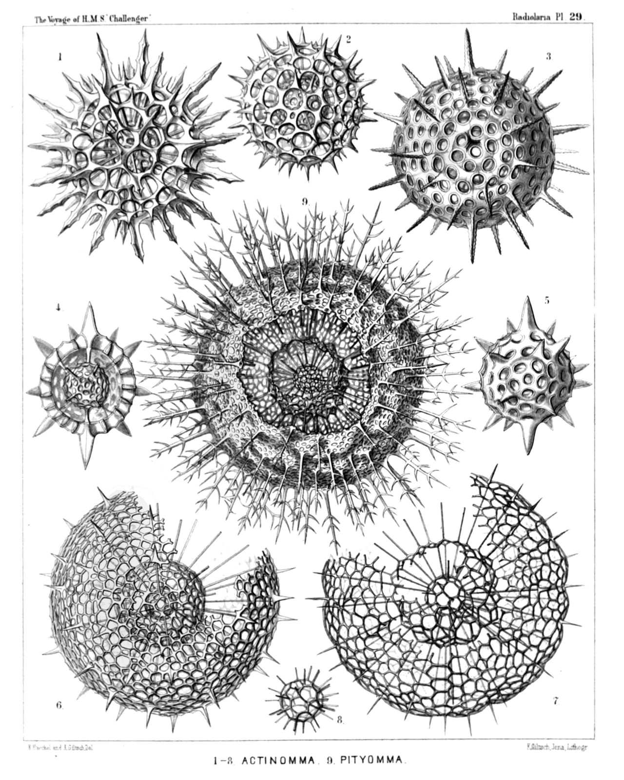
PLATE 29.
Astrosphærida.
Diam. Page.
Fig. 1. Echinomma toxopneustes, n. sp., × 400 259
Fig. 2. Echinomma sphærechinus, n. sp., × 400 258
Fig. 3. Actinomma denticulatum, n. sp., × 400 254
Fig. 4. Actinomma pachyderma, n. sp., × 400 254
The half of the cortical shell is removed.
Fig. 5. Actinomma pachyderma, n. sp., × 400 254
Fig. 6. Actinomma capillaceum, n. sp., × 300 255
Fig. 7. Actinomma arcadophorum, n. sp., × 400 255
A part of the two outer shells is removed.
Fig. 8. Actinomma arcadophorum, n. sp., × 400 255
Inner medullary shell.
Fig. 9. Pityomma drymodes, n. sp., × 300 260
A part of the two outer shells is removed.
PLATE 30.
Legion SPUMELLARIA.
Order SPHÆROIDEA.
Families Liosphærida et Astrosphærida.
PLATE 30. Liosphærida et Astrosphærida. Diam. Page. Fig. 1. Cromyechinus icosacanthus, n. sp., × 300 263 Fig. 2. Cromyomma villosum, n. sp., × 300 261 Fig. 3. Cromyechinus dodecacanthus, n. sp., × 400 264 Fig. 3a. The innermost shells. Fig. 4. Cromyomma circumtextum, n. sp., × 300 262 Fig. 5. Cromyomma mucronatum, n. sp., × 200 263 Fig. 5a. The innermost shells. Fig. 6. Cromyodrymus abietinus, n. sp., × 300 265 Fig. 7. Cromyodrymus quadricuspis, n. sp., × 400 264 Fig. 7a. The inner concentric shells. Fig. 8. Cromyomma perspicuum, n. sp., × 300 262 Fig. 9. Cromyosphæra quadruplex, n. sp., × 300 84 Fig. 9a. The innermost shells.
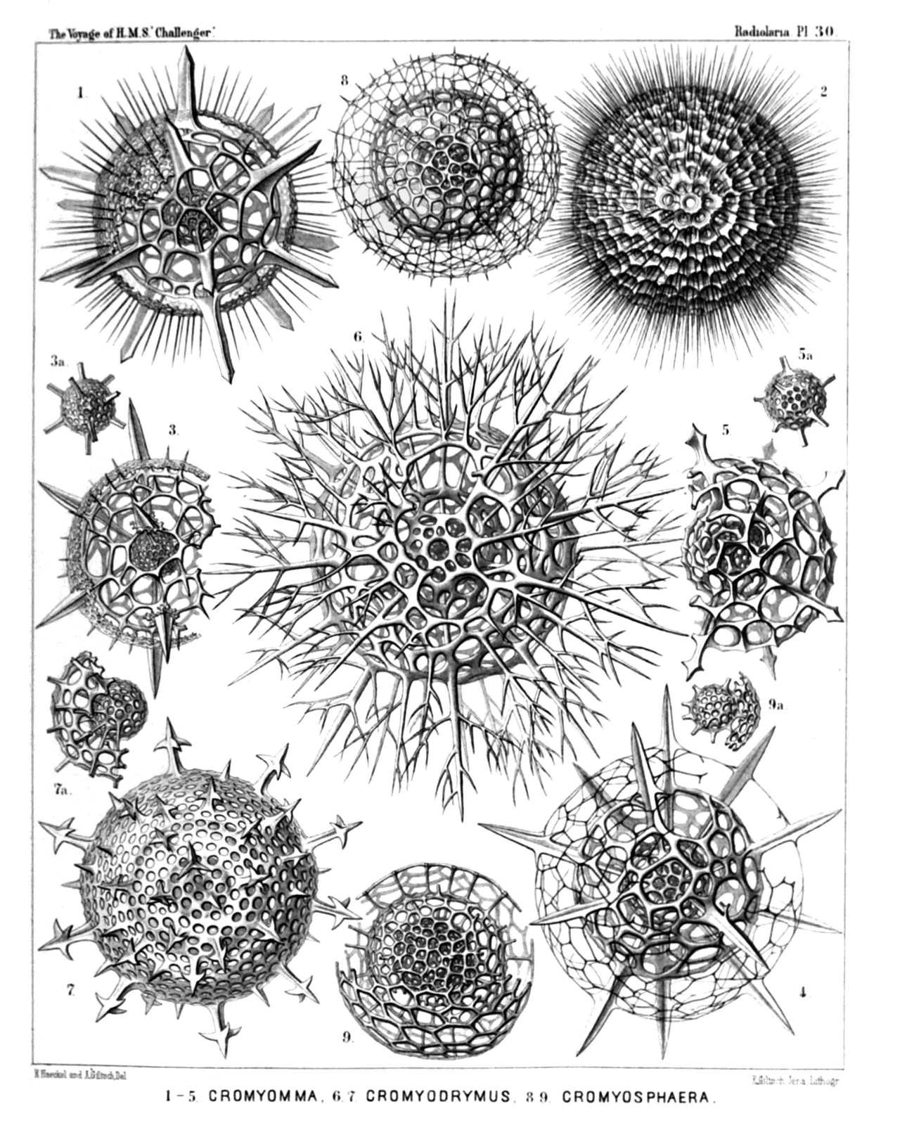
PLATE 30.
Liosphærida et Astrosphærida.
Diam. Page.
Fig. 1. Cromyechinus icosacanthus, n. sp., × 300 263
Fig. 2. Cromyomma villosum, n. sp., × 300 261
Fig. 3. Cromyechinus dodecacanthus, n. sp., × 400 264
Fig. 3a. The innermost shells.
Fig. 4. Cromyomma circumtextum, n. sp., × 300 262
Fig. 5. Cromyomma mucronatum, n. sp., × 200 263
Fig. 5a. The innermost shells.
Fig. 6. Cromyodrymus abietinus, n. sp., × 300 265
Fig. 7. Cromyodrymus quadricuspis, n. sp., × 400 264
Fig. 7a. The inner concentric shells.
Fig. 8. Cromyomma perspicuum, n. sp., × 300 262
Fig. 9. Cromyosphæra quadruplex, n. sp., × 300 84
Fig. 9a. The innermost shells.
PLATE 31.
Legion SPUMELLARIA.
Order DISCOIDEA.
Families Cenodiscida et Phacodiscida.
PLATE 31. Cenodiscida et Phacodiscida. Diam. Page. Fig. 1. Sethostaurus orthostaurus, n. sp., × 300 433 Fig. 2. Sethostaurus orthostaurus, n. sp., × 300 433 Vertical section through the centrum. Fig. 3. Sethostaurus recurvatus, n. sp., × 100 434 Optical section through the equatorial plane. Fig. 4. Sethostaurus rhombostaurus, n. sp., × 100 434 Optical section through the equatorial plane. Fig. 5. Sethostaurus cruciatus, n. sp. (vel Heliostaurus cruciatus), × 300 434 Fig. 6. Phacostaurus oceanidum, n. sp., × 300 435 Fig. 7. Phacostaurus magnificus, n. sp., × 400 436 Fig. 8. Phacostaurus magnificus, n. sp., × 200 436 Vertical section through the centrum. Fig. 9. Sethostylus distyliscus, n. sp., × 400 428 Fig. 10. Sethostylus dicylindrus, n. sp., × 300 428 Marginal view. Fig. 11. Stylodiscus endostylus, n. sp. (vel Sethostylus endostylus), × 300 413 Fig. 12. Phacostylus amphistylus, n. sp., × 300 430
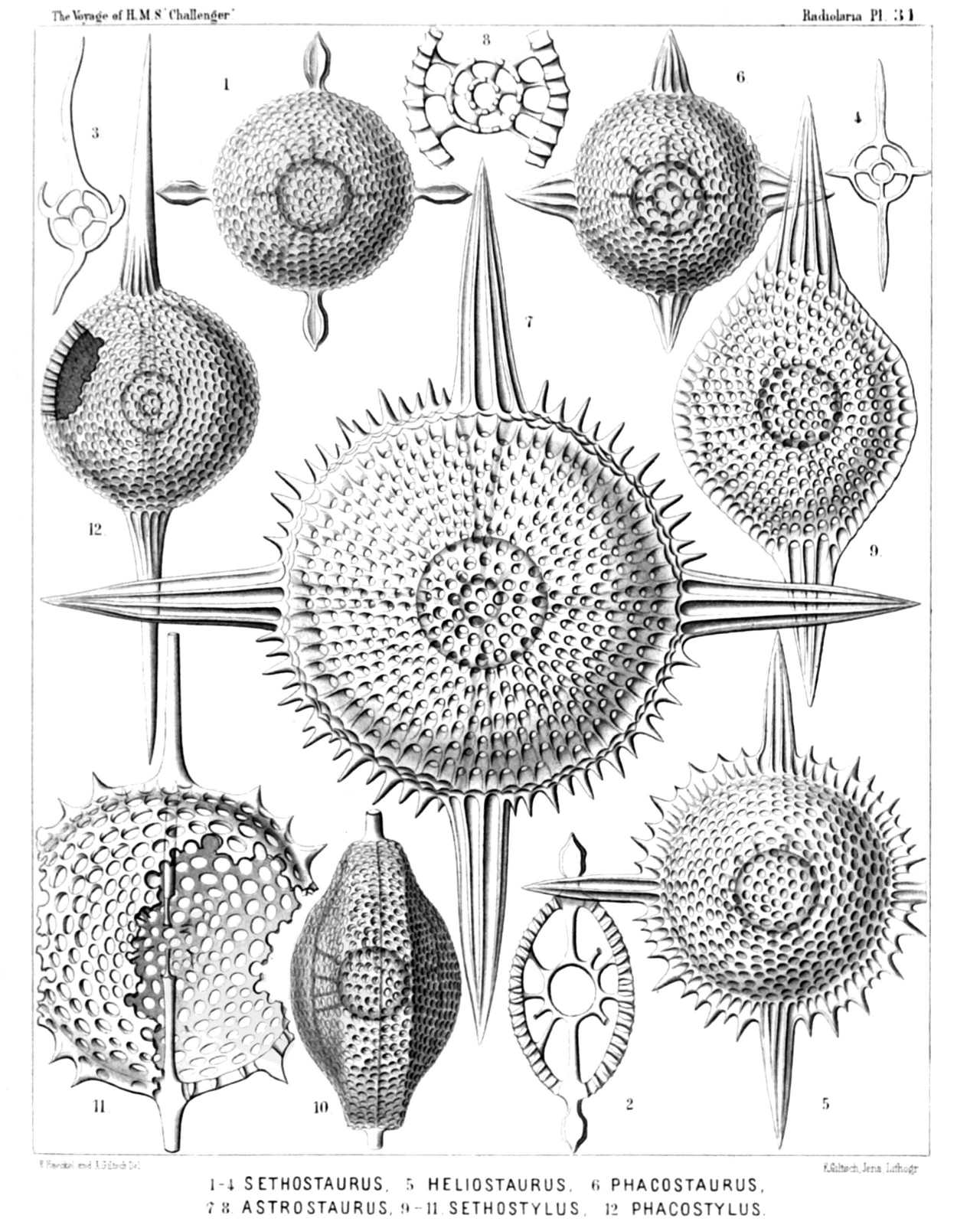
PLATE 31.
Cenodiscida et Phacodiscida.
Diam. Page.
Fig. 1. Sethostaurus orthostaurus, n. sp., × 300 433
Fig. 2. Sethostaurus orthostaurus, n. sp., × 300 433
Vertical section through the centrum.
Fig. 3. Sethostaurus recurvatus, n. sp., × 100 434
Optical section through the equatorial plane.
Fig. 4. Sethostaurus rhombostaurus, n. sp., × 100 434
Optical section through the equatorial plane.
Fig. 5. Sethostaurus cruciatus, n. sp. (vel Heliostaurus cruciatus), × 300 434
Fig. 6. Phacostaurus oceanidum, n. sp., × 300 435
Fig. 7. Phacostaurus magnificus, n. sp., × 400 436
Fig. 8. Phacostaurus magnificus, n. sp., × 200 436
Vertical section through the centrum.
Fig. 9. Sethostylus distyliscus, n. sp., × 400 428
Fig. 10. Sethostylus dicylindrus, n. sp., × 300 428
Marginal view.
Fig. 11. Stylodiscus endostylus, n. sp. (vel Sethostylus endostylus), × 300 413
Fig. 12. Phacostylus amphistylus, n. sp., × 300 430
PLATE 32.
Legion SPUMELLARIA.
Order DISCOIDEA.
Family Phacodiscida.
PLATE 32. Phacodiscida. Diam. Page. Fig. 1. Astrophacus solaris, n. sp., × 300 453 Fig. 2. Astrophacus apollinis, n. sp., × 300 455 Fig. 3. Astrophacus phacodiscus, n. sp., × 300 454 Vertical section through the centrum. Fig. 4. Astrosestrum ephyra, n. sp., × 300 442 Fig. 4a. Transverse section through the double medullary shell, × 300 442 Fig. 5. Astrosestrum nauphanta, n. sp., × 300 442 Fig. 6. Phacostylus caudatus, n. sp. (vel Astrosestrum caudatum), × 200 431 Fig. 7. Perizona scutella, n. sp., × 400 427 Fig. 8. Perizona pterygota, n. sp., × 400 427 Fig. 8a. Medullary shells and radial beams connecting them with the disk, × 300 427
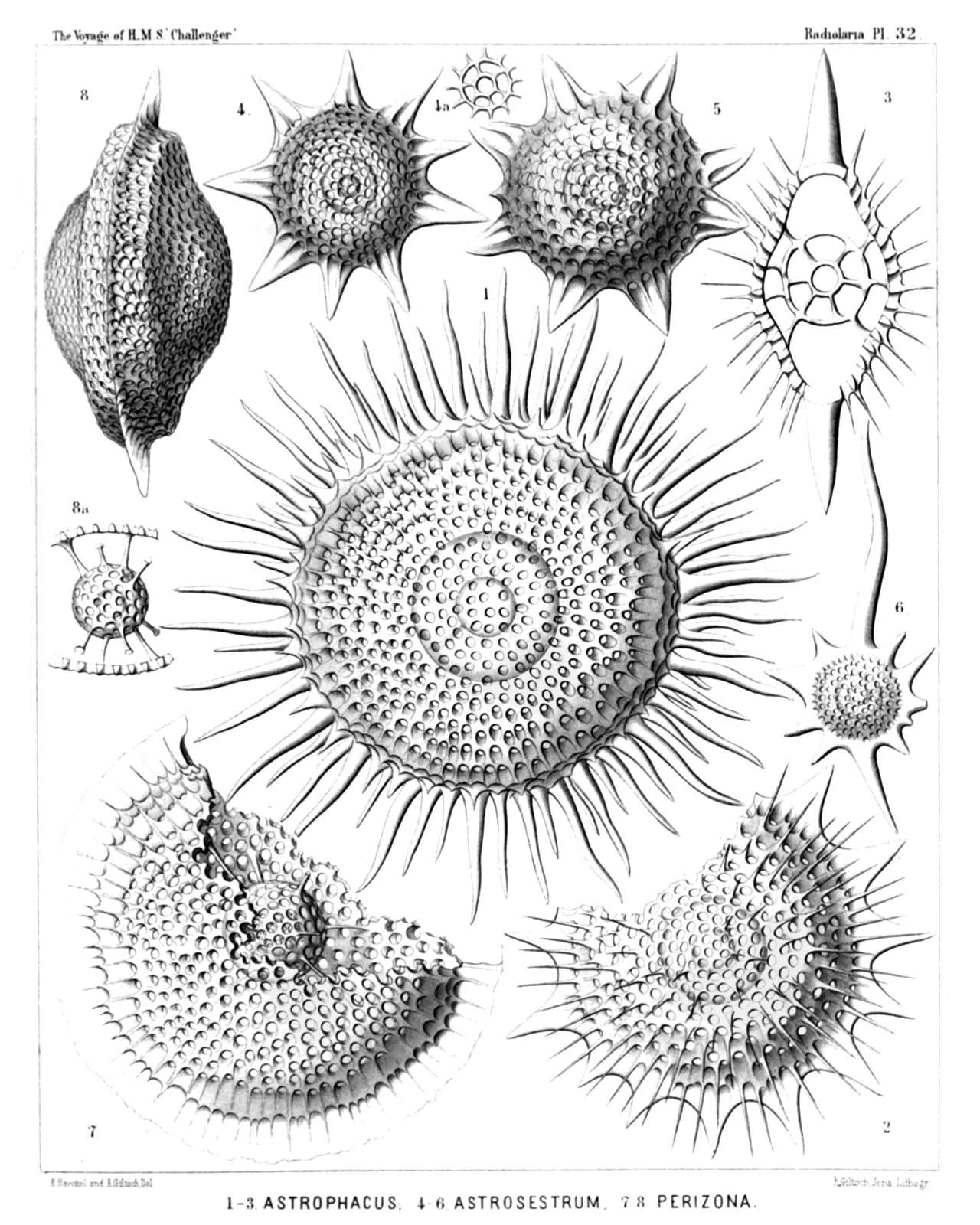
PLATE 32.
Phacodiscida.
Diam. Page.
Fig. 1. Astrophacus solaris, n. sp., × 300 453
Fig. 2. Astrophacus apollinis, n. sp., × 300 455
Fig. 3. Astrophacus phacodiscus, n. sp., × 300 454
Vertical section through the centrum.
Fig. 4. Astrosestrum ephyra, n. sp., × 300 442
Fig. 4a. Transverse section through the double medullary shell, × 300 442
Fig. 5. Astrosestrum nauphanta, n. sp., × 300 442
Fig. 6. Phacostylus caudatus, n. sp. (vel Astrosestrum caudatum), × 200 431
Fig. 7. Perizona scutella, n. sp., × 400 427
Fig. 8. Perizona pterygota, n. sp., × 400 427
Fig. 8a. Medullary shells and radial beams connecting them with the disk, × 300 427
PLATE 33.
Legion SPUMELLARIA.
Order DISCOIDEA.
Family Phacodiscida.
PLATE 33. Phacodiscida. Diam. Page. Fig. 1. Sethodiscus lenticula, n. sp., × 300 423 Fig. 2. Sethodiscus lenticula, n. sp., × 300 423 Vertical section. Fig. 3. Sethodiscus macrococcus, n. sp., × 300 423 Young shell, not yet closed, seen from the margin. Fig. 4. Periphæna cincta, n. sp., × 400 426 Fig. 5. Triactiscus tricuspis, n. sp., × 300 432 Marginal view. Fig. 6. Triactiscus tripyramis, n. sp., × 400 432 Fig. 7. Heliodiscus cingillum, n. sp., × 300 448 Fig. 8. Heliodiscus asteriscus, n. sp., × 300 445 Fig. 9. Heliodrymus dendrocyclus, n. sp.(vel Heliocladus dendrocyclus), × 300 451
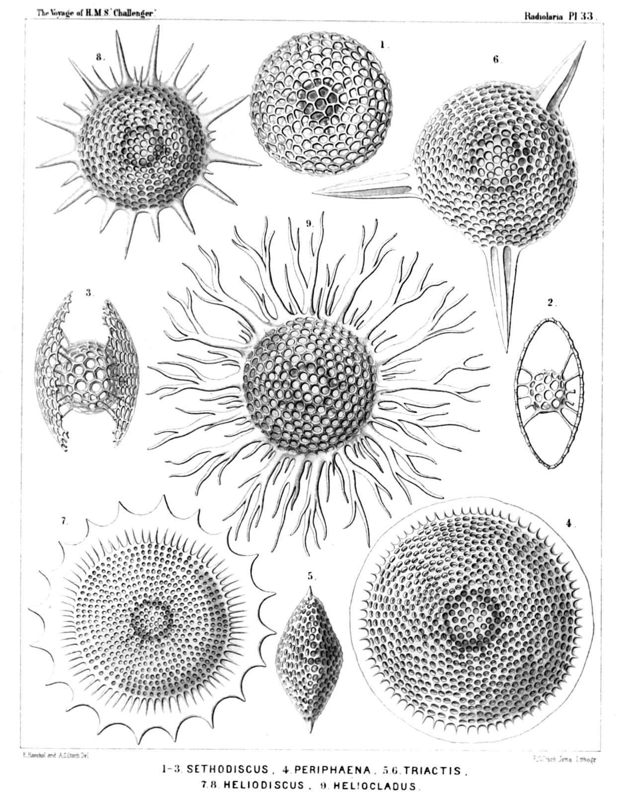
PLATE 33.
Phacodiscida.
Diam. Page.
Fig. 1. Sethodiscus lenticula, n. sp., × 300 423
Fig. 2. Sethodiscus lenticula, n. sp., × 300 423
Vertical section.
Fig. 3. Sethodiscus macrococcus, n. sp., × 300 423
Young shell, not yet closed, seen from the margin.
Fig. 4. Periphæna cincta, n. sp., × 400 426
Fig. 5. Triactiscus tricuspis, n. sp., × 300 432
Marginal view.
Fig. 6. Triactiscus tripyramis, n. sp., × 400 432
Fig. 7. Heliodiscus cingillum, n. sp., × 300 448
Fig. 8. Heliodiscus asteriscus, n. sp., × 300 445
Fig. 9. Heliodrymus dendrocyclus, n. sp.(vel Heliocladus dendrocyclus), × 300 451
PLATE 34.
Legion SPUMELLARIA.
Order DISCOIDEA.
Family Phacodiscida.
PLATE 34. Phacodiscida. Diam. Page. Fig. 1. Sethostylus dentatus, n. sp. (vel Heliostylus dentatus), × 300 429 Upper half of the disk. Fig. 2. Sethostylus serratus, n. sp. (vel Heliostylus serratus), × 300 429 Lower half of the disk. Fig. 3. Heliosestrum octonum, n. sp., × 300 440 Fig. 4. Heliodiscus solaster, n. sp., × 300 447 Fig. 5. Heliodiscus echiniscus, n. sp., × 400 448 Fig. 6. Heliosestrum medusinum, n. sp., × 300 438 Fig. 7. Sethostaurus conostaurus, n. sp., × 100 433 Normal form with four regular spines. Fig. 8. Sethostaurus conostaurus, n. sp., × 100 433 Abnormal form with five spines. Fig. 9. Heliodiscus marginatus, n. sp., × 100 449 Fig. 10. Heliodiscus trochiscus, n. sp., × 100 445 Fig. 11. Heliodiscus polymorphus, n. sp., × 100 447 Fig. 12. Heliodiscus polymorphus, n. sp., × 100 447 Fig. 13. Heliodiscus trochiscus, n. sp., × 100 445 Fig. 14. Astrophacus trochiscus, n. sp., × 100 453
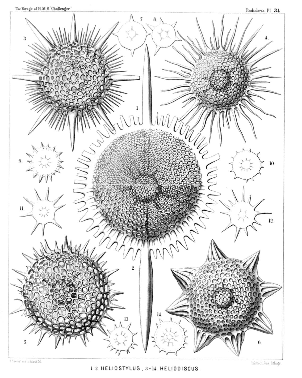
PLATE 34.
Phacodiscida.
Diam. Page.
Fig. 1. Sethostylus dentatus, n. sp. (vel Heliostylus dentatus), × 300 429
Upper half of the disk.
Fig. 2. Sethostylus serratus, n. sp. (vel Heliostylus serratus), × 300 429
Lower half of the disk.
Fig. 3. Heliosestrum octonum, n. sp., × 300 440
Fig. 4. Heliodiscus solaster, n. sp., × 300 447
Fig. 5. Heliodiscus echiniscus, n. sp., × 400 448
Fig. 6. Heliosestrum medusinum, n. sp., × 300 438
Fig. 7. Sethostaurus conostaurus, n. sp., × 100 433
Normal form with four regular spines.
Fig. 8. Sethostaurus conostaurus, n. sp., × 100 433
Abnormal form with five spines.
Fig. 9. Heliodiscus marginatus, n. sp., × 100 449
Fig. 10. Heliodiscus trochiscus, n. sp., × 100 445
Fig. 11. Heliodiscus polymorphus, n. sp., × 100 447
Fig. 12. Heliodiscus polymorphus, n. sp., × 100 447
Fig. 13. Heliodiscus trochiscus, n. sp., × 100 445
Fig. 14. Astrophacus trochiscus, n. sp., × 100 453
PLATE 35.
Legion SPUMELLARIA.
Order DISCOIDEA.
Family Phacodiscida.
PLATE 35. Phacodiscida. Diam. Page. Fig. 1. Heliodiscus pertusus, n. sp. (vel Heliosestrum pertusum), × 400 448 Irregular form with ten (instead of eight) larger latticed spines. Fig. 2. Heliodiscus glyphodon, n. sp. (vel Heliosestrum glyphodon), × 300 446 Fig. 3. Heliodrymus ramosus, n. sp., × 300 452 Fig. 4. Heliodrymus ramosus, n. sp., × 500 452 Medullary shell and a segment of the disk. Fig. 5. Heliodrymus viminalis, n. sp., × 400 452 Marginal view. Fig. 6. Phacodiscus clypeus, n. sp., × 400 425 Fig. 7. Phacodiscus rotula, n. sp., × 400 424 Marginal view. Fig. 8. Phacodiscus lentiformis, n. sp., × 400 425 Vertical section nearly through the centre. Fig. 9. Phacodiscus clypeus, n. sp., × 400 425 Vertical section nearly through the centre.
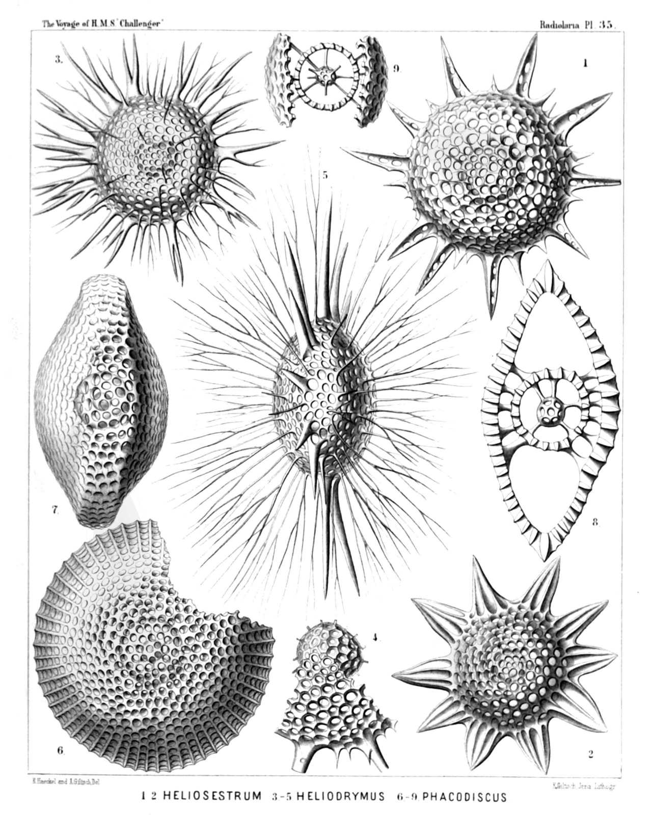
PLATE 35.
Phacodiscida.
Diam. Page.
Fig. 1. Heliodiscus pertusus, n. sp. (vel Heliosestrum pertusum), × 400 448
Irregular form with ten (instead of eight) larger latticed spines.
Fig. 2. Heliodiscus glyphodon, n. sp. (vel Heliosestrum glyphodon), × 300 446
Fig. 3. Heliodrymus ramosus, n. sp., × 300 452
Fig. 4. Heliodrymus ramosus, n. sp., × 500 452
Medullary shell and a segment of the disk.
Fig. 5. Heliodrymus viminalis, n. sp., × 400 452
Marginal view.
Fig. 6. Phacodiscus clypeus, n. sp., × 400 425
Fig. 7. Phacodiscus rotula, n. sp., × 400 424
Marginal view.
Fig. 8. Phacodiscus lentiformis, n. sp., × 400 425
Vertical section nearly through the centre.
Fig. 9. Phacodiscus clypeus, n. sp., × 400 425
Vertical section nearly through the centre.
PLATE 36.
Legion SPUMELLARIA.
Order DISCOIDEA.
Family Coccodiscida.
PLATE 36. Coccodiscida. Diam. Page. Fig. 1. Coccodiscus lamarckii, n. sp., × 500 459 The left half of the figure represents a horizontal section through the peripheral shell, the right half a view of the surface. Fig. 2. Coccodiscus gœthei, n. sp., × 500 461 Vertical section nearly through the centre. Fig. 3. Lithocyclia lenticula, n. sp., × 400 459 Fig. 4. Lithocyclia lenticula, n. sp., × 400 459 Vertical section through the centre. Fig. 5. Coccocyclia helianthus, n. sp., × 400 468 Fig. 6. Coccocyclia helianthus, n. sp., × 500 468 Vertical section through the outer medullary shell, showing the inner. Fig. 7. Astrocyclia solaster, n. sp., × 300 466 Fig. 8. Astrocyclia heterocycla, n. sp., × 400 468 Horizontal section through the equatorial plane.
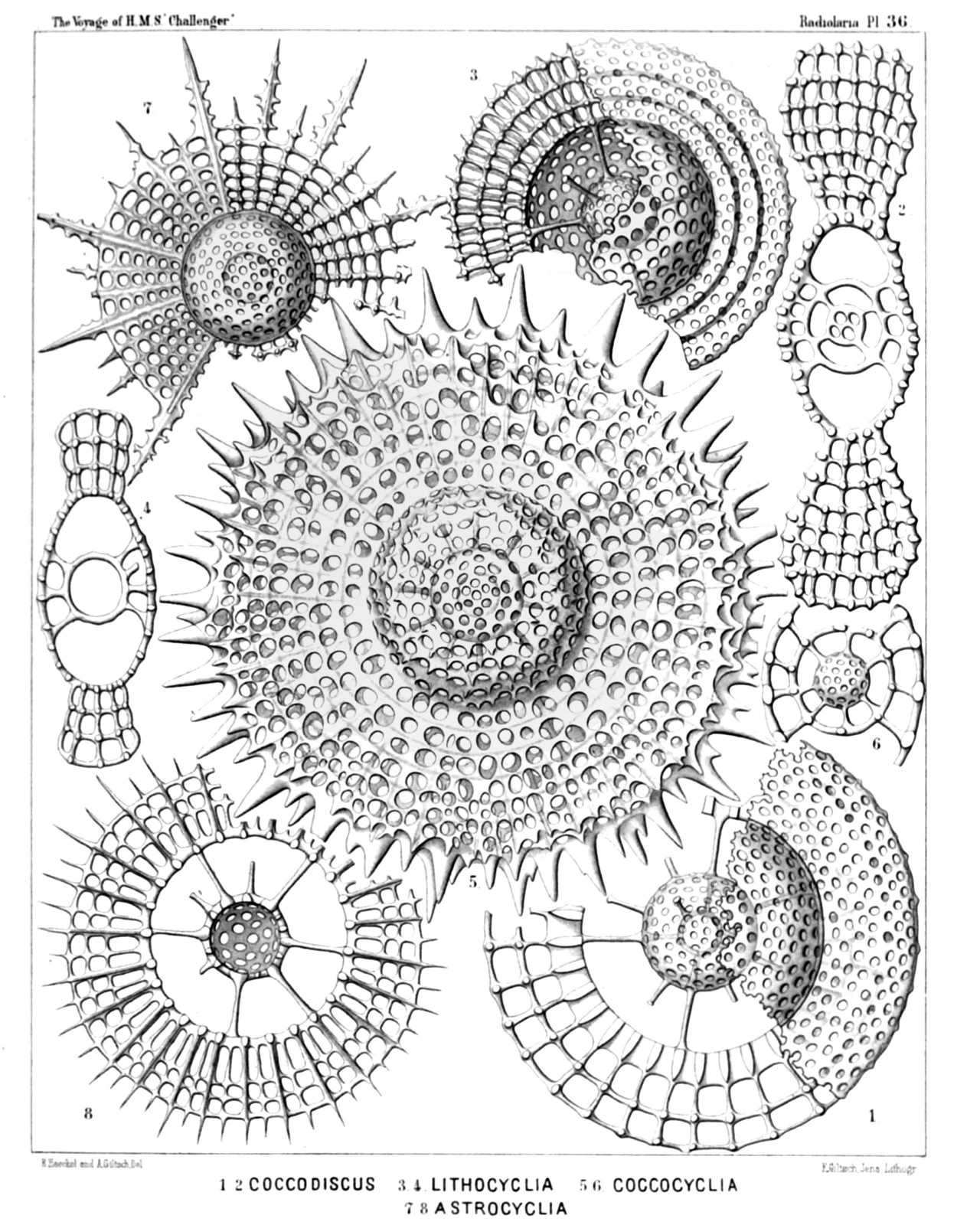
PLATE 36.
Coccodiscida.
Diam. Page.
Fig. 1. Coccodiscus lamarckii, n. sp., × 500 459
The left half of the figure represents a horizontal section through the peripheral shell, the right half a view of the surface.
Fig. 2. Coccodiscus gœthei, n. sp., × 500 461
Vertical section nearly through the centre.
Fig. 3. Lithocyclia lenticula, n. sp., × 400 459
Fig. 4. Lithocyclia lenticula, n. sp., × 400 459
Vertical section through the centre.
Fig. 5. Coccocyclia helianthus, n. sp., × 400 468
Fig. 6. Coccocyclia helianthus, n. sp., × 500 468
Vertical section through the outer medullary shell, showing the inner.
Fig. 7. Astrocyclia solaster, n. sp., × 300 466
Fig. 8. Astrocyclia heterocycla, n. sp., × 400 468
Horizontal section through the equatorial plane.
PLATE 37.
Legion SPUMELLARIA.
Order DISCOIDEA.
Family Coccodiscida.
PLATE 37. Coccodiscida. Diam. Page. Fig. 1. Staurocyclia cruciata, n. sp., × 400 465 Fig. 2. Staurocyclia phacostaurus, n. sp., × 300 465 Fig. 3. Staurocyclia phacostaurus, n. sp., × 300 465 Vertical section through the centre. Fig. 4. Staurocyclia magniducis, n. sp. (Coccostaurus magniducis), × 300 466 Fig. 5. Trigonocyclia triangularis, n. sp., × 400 464 Fig. 6. Stylocyclia prionacantha, n. sp., × 500 462 A great part of the peripheral shell is removed. Fig. 7. Amphicyclia amphistyla, n. sp., × 300 464 Vertical section through the centre. Fig. 8. Stylocyclia excavata, n. sp., × 200 463 Vertical section through the centre.
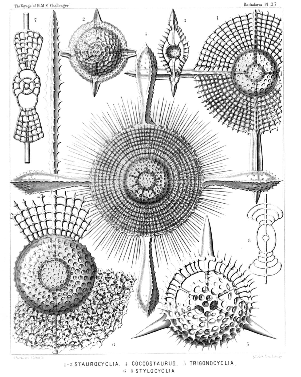
PLATE 37.
Coccodiscida.
Diam. Page.
Fig. 1. Staurocyclia cruciata, n. sp., × 400 465
Fig. 2. Staurocyclia phacostaurus, n. sp., × 300 465
Fig. 3. Staurocyclia phacostaurus, n. sp., × 300 465
Vertical section through the centre.
Fig. 4. Staurocyclia magniducis, n. sp. (Coccostaurus magniducis), × 300 466
Fig. 5. Trigonocyclia triangularis, n. sp., × 400 464
Fig. 6. Stylocyclia prionacantha, n. sp., × 500 462
A great part of the peripheral shell is removed.
Fig. 7. Amphicyclia amphistyla, n. sp., × 300 464
Vertical section through the centre.
Fig. 8. Stylocyclia excavata, n. sp., × 200 463
Vertical section through the centre.
PLATE 38.
Legion SPUMELLARIA.
Order DISCOIDEA.
Family Coccodiscida.
PLATE 38. Coccodiscida. Diam. Page. Fig. 1. Amphicyclia chronometra, n. sp., × 400 463 Fig. 2. Amphicyclia pachydiscus, n. sp., × 500 464 Vertical section through the centre. Fig. 3. Amphiactura amphibrachia, n. sp., × 300 470 Fig. 4. Amphiactura amphibrachia, n. sp., × 150 470 Vertical section through the centre. Fig. 5. Diplactura diploconus, n. sp., × 300 470 Fig. 6. Trigonactura triacantha, n. sp., × 200 472 Fig. 7. Trigonactura triacantha, n. sp., × 400 472 Vertical section nearly through the centre. Fig. 8. Hymenactura archimedis, n. sp., × 300 473 Fig. 9. Hymenactura copernici, n. sp., × 200 475
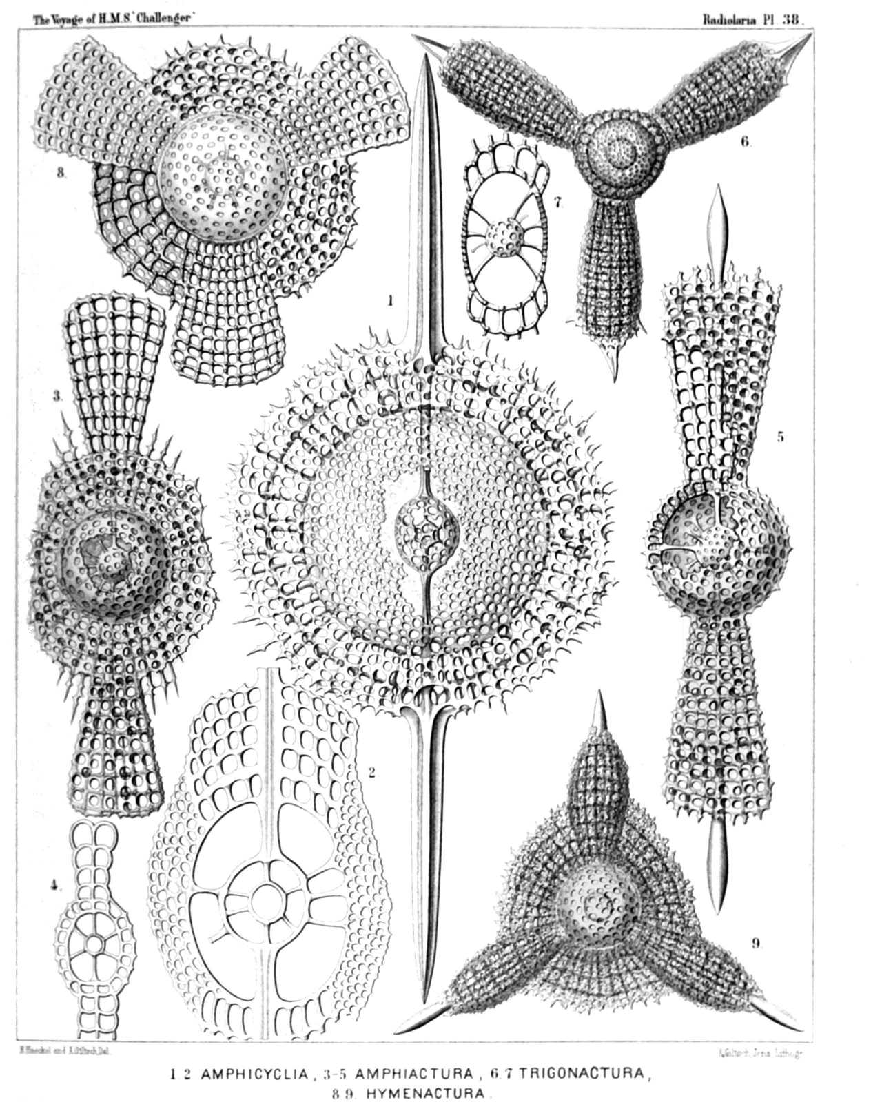
PLATE 38.
Coccodiscida.
Diam. Page.
Fig. 1. Amphicyclia chronometra, n. sp., × 400 463
Fig. 2. Amphicyclia pachydiscus, n. sp., × 500 464
Vertical section through the centre.
Fig. 3. Amphiactura amphibrachia, n. sp., × 300 470
Fig. 4. Amphiactura amphibrachia, n. sp., × 150 470
Vertical section through the centre.
Fig. 5. Diplactura diploconus, n. sp., × 300 470
Fig. 6. Trigonactura triacantha, n. sp., × 200 472
Fig. 7. Trigonactura triacantha, n. sp., × 400 472
Vertical section nearly through the centre.
Fig. 8. Hymenactura archimedis, n. sp., × 300 473
Fig. 9. Hymenactura copernici, n. sp., × 200 475
PLATE 39.
Legion SPUMELLARIA.
Order PRUNOIDEA.
Families Ellipsida, Druppulida, Artiscida et Cyphinida.
PLATE 39. Ellipsida, Druppulida, Artiscida et Cyphinida. Diam. Page. Fig. 1. Cenellipsis faceta, n. sp. (vel Ellipsis faceta), × 300 291 Fig. 2. Cenellipsis infundibulum, n. sp. (vel Ellipsis infundibulum), × 300 292 Fig. 3. Druppula pandanus, n. sp. (vel Coccymelium pandanus), × 300 308 Fig. 4. Prunulum coccymelium, n. sp. (vel Coccymelium prunulum), × 300 313 Fig. 5. Prunocarpus artocarpium, n. sp. (vel Artocarpium indicum), × 300 316 Fig. 6. Pipettella prismatica, n. sp., × 300 305 Fig. 7. Pipetta tuba, n. sp., × 300 337 Fig. 8. Pipetta fusus, n. sp., × 300 337 Fig. 8a. The enclosed medullary shell. Fig. 9. Artiscus nodosus, n. sp. (vel Artidium nodosum), × 400 356 Fig. 10. Cannartus violina, n. sp., × 300 358 Fig. 11. Cyphonium cribellum, n. sp., × 200 365 Fig. 12. Cyphonium virgineum, n. sp. (vel Ommatospyris virginea), × 400 363 Fig. 12a. Vertical section through the double medullary shell. Fig. 13. Cypassis puella, n. sp. (vel Didymospyris puella), × 400 367 The enclosed central capsule is visible. Fig. 14. Cyphinus amphilophus, N. sp., × 300 370 Fig. 15. Pipettaria tubaria, n. sp., × 300 339 Fig. 16. Cannartidium mammiferum, n. sp., × 300 375 Fig. 17. Cannartidium mastophorum, n. sp., × 150 375 Fig. 18. Cannartidium bicinctum, n. sp., × 300 374 Fig. 18a. Vertical section through the main axis. Fig. 19. Cannartiscus amphiconiscus, n. sp., × 300 372
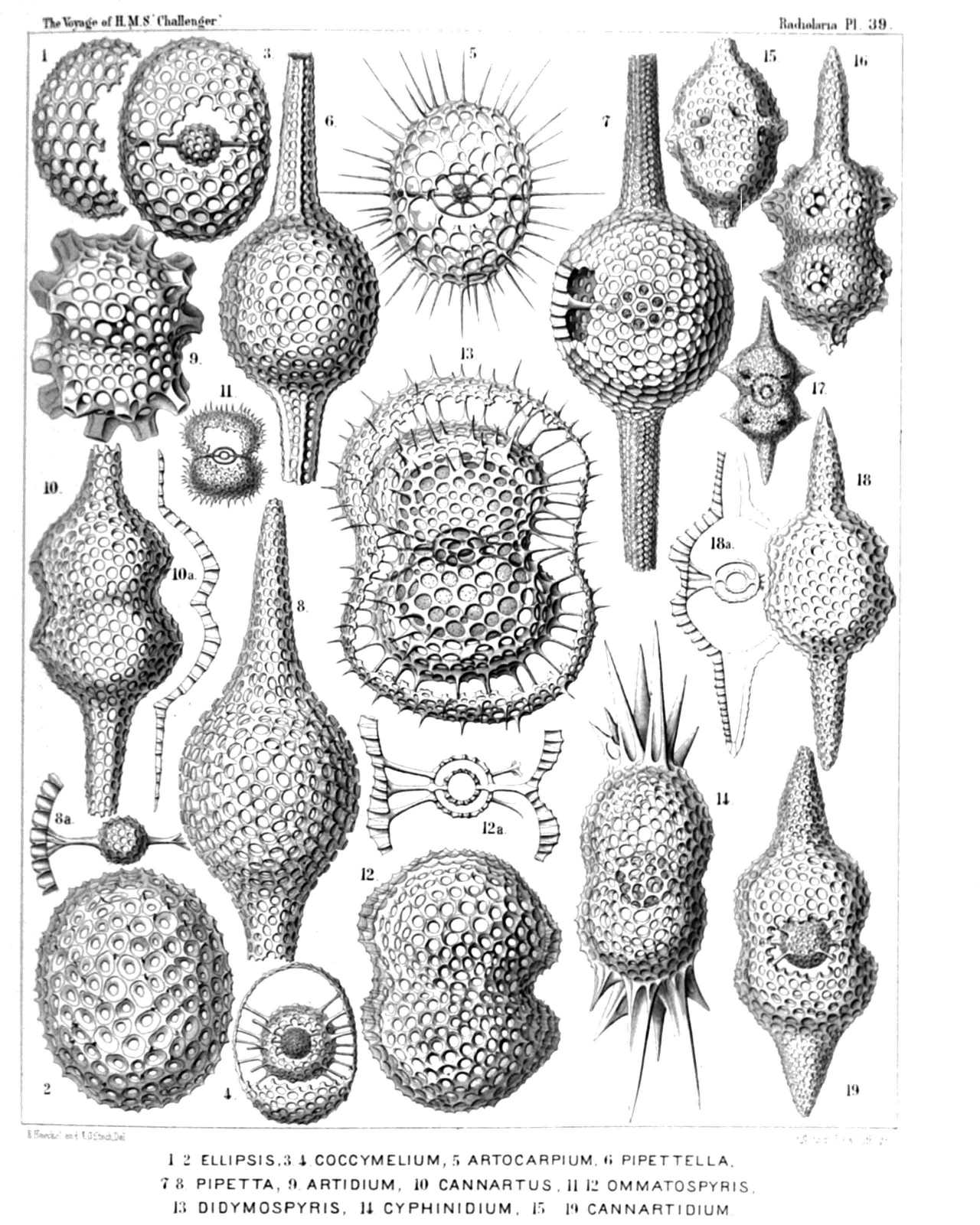
PLATE 39.
Ellipsida, Druppulida, Artiscida et Cyphinida.
Diam. Page.
Fig. 1. Cenellipsis faceta, n. sp. (vel Ellipsis faceta), × 300 291
Fig. 2. Cenellipsis infundibulum, n. sp. (vel Ellipsis infundibulum), × 300 292
Fig. 3. Druppula pandanus, n. sp. (vel Coccymelium pandanus), × 300 308
Fig. 4. Prunulum coccymelium, n. sp. (vel Coccymelium prunulum), × 300 313
Fig. 5. Prunocarpus artocarpium, n. sp. (vel Artocarpium indicum), × 300 316
Fig. 6. Pipettella prismatica, n. sp., × 300 305
Fig. 7. Pipetta tuba, n. sp., × 300 337
Fig. 8. Pipetta fusus, n. sp., × 300 337
Fig. 8a. The enclosed medullary shell.
Fig. 9. Artiscus nodosus, n. sp. (vel Artidium nodosum), × 400 356
Fig. 10. Cannartus violina, n. sp., × 300 358
Fig. 11. Cyphonium cribellum, n. sp., × 200 365
Fig. 12. Cyphonium virgineum, n. sp. (vel Ommatospyris virginea), × 400 363
Fig. 12a. Vertical section through the double medullary shell.
Fig. 13. Cypassis puella, n. sp. (vel Didymospyris puella), × 400 367
The enclosed central capsule is visible.
Fig. 14. Cyphinus amphilophus, N. sp., × 300 370
Fig. 15. Pipettaria tubaria, n. sp., × 300 339
Fig. 16. Cannartidium mammiferum, n. sp., × 300 375
Fig. 17. Cannartidium mastophorum, n. sp., × 150 375
Fig. 18. Cannartidium bicinctum, n. sp., × 300 374
Fig. 18a. Vertical section through the main axis.
Fig. 19. Cannartiscus amphiconiscus, n. sp., × 300 372
PLATE 40.
Legion SPUMELLARIA.
Order PRUNOIDEA.
Families Panartida et Zygartida.
PLATE 40. Panartida et Zygartida. Diam. Page. Fig. 1. Panartus diploconus, n. sp., × 300 379 Fig. 2. Panartus pluteus, n. sp., × 300 382 Fig. 3. Panartus tetrathalamus, n. sp., × 300 378 Fig. 4. Panicium coronatum, n. sp. (vel Panartidium coronatum), × 300 386 Fig. 5. Peripanartus amphiconus, n. sp., × 300 383 Fig. 6. Peripanartus cylindrus, n. sp., × 150 384 Fig. 7. Peripanartus atractus, n. sp., × 300 384 Fig. 8. Peripanicium amphicorona, n. sp., × 300 387 Fig. 9. Panarium tubularium, n. sp., × 300 390 Fig. 10. Ommatocampe nereides, n. sp., × 300 394 Fig. 11. Cyphocolpus virginis, n. sp. (vel Zygartus virginis) × 300 369 Fig. 12. Desmartus larvalis, n. sp. (vel Zygartus larvalis), × 300 398 Fig. 13. Zygartus chrysalis, n. sp. (vel Zygocampe chrysalis), × 400 401
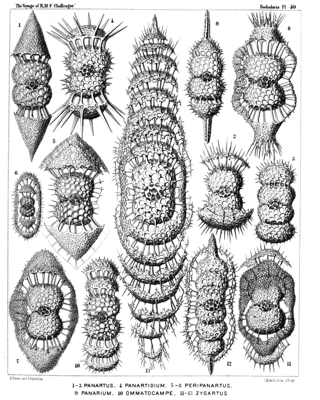
PLATE 40.
Panartida et Zygartida.
Diam. Page.
Fig. 1. Panartus diploconus, n. sp., × 300 379
Fig. 2. Panartus pluteus, n. sp., × 300 382
Fig. 3. Panartus tetrathalamus, n. sp., × 300 378
Fig. 4. Panicium coronatum, n. sp. (vel Panartidium coronatum), × 300 386
Fig. 5. Peripanartus amphiconus, n. sp., × 300 383
Fig. 6. Peripanartus cylindrus, n. sp., × 150 384
Fig. 7. Peripanartus atractus, n. sp., × 300 384
Fig. 8. Peripanicium amphicorona, n. sp., × 300 387
Fig. 9. Panarium tubularium, n. sp., × 300 390
Fig. 10. Ommatocampe nereides, n. sp., × 300 394
Fig. 11. Cyphocolpus virginis, n. sp. (vel Zygartus virginis) × 300 369
Fig. 12. Desmartus larvalis, n. sp. (vel Zygartus larvalis), × 300 398
Fig. 13. Zygartus chrysalis, n. sp. (vel Zygocampe chrysalis), × 400 401
PLATE 41.
Legion SPUMELLARIA.
Order DISCOIDEA.
Families Porodiscida et Spongodiscida.
PLATE 41. Porodiscida et Spongodiscida. Diam. Page. Fig. 1. Porodiscus flustrella, n. sp., × 300 493 Fig. 2. Porodiscus perispira, n. sp., × 200 495 The rings alone (equatorial section). Fig. 3. Porodiscus quadrigatus, n. sp., × 200 494 The rings alone (equatorial section). Fig. 4. Porodiscus semispiralis, n. sp., × 500 497 Fig. 5. Perichlamydium saturnus, n. sp., × 300 499 Fig. 6. Porodiscus centrospira, n. sp. (vel Perispongidium centrospira), × 200 495 The rings alone (equatorial section). Fig. 7. Porodiscus irregularis, n. sp. (vel Perispongidium irregulare), × 200 498 The rings alone (equatorial section). Fig. 8. Stylodictya heliospira, n. sp., × 400 512 Fig. 9. Stylodictya centrospira, n. sp., × 400 512 Fig. 10. Stylochlamydium asteriscus, n. sp., × 400 514 Fig. 11. Stylotrochus geddesii, n. sp., × 300 585
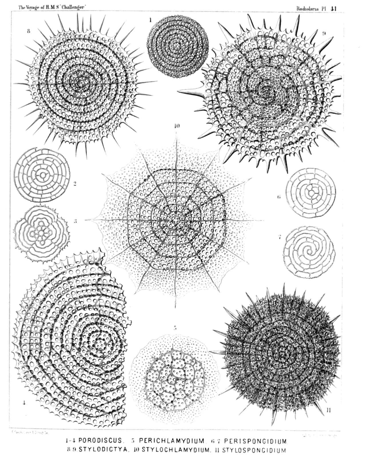
PLATE 41.
Porodiscida et Spongodiscida.
Diam. Page.
Fig. 1. Porodiscus flustrella, n. sp., × 300 493
Fig. 2. Porodiscus perispira, n. sp., × 200 495
The rings alone (equatorial section).
Fig. 3. Porodiscus quadrigatus, n. sp., × 200 494
The rings alone (equatorial section).
Fig. 4. Porodiscus semispiralis, n. sp., × 500 497
Fig. 5. Perichlamydium saturnus, n. sp., × 300 499
Fig. 6. Porodiscus centrospira, n. sp. (vel Perispongidium centrospira), × 200 495
The rings alone (equatorial section).
Fig. 7. Porodiscus irregularis, n. sp. (vel Perispongidium irregulare), × 200 498
The rings alone (equatorial section).
Fig. 8. Stylodictya heliospira, n. sp., × 400 512
Fig. 9. Stylodictya centrospira, n. sp., × 400 512
Fig. 10. Stylochlamydium asteriscus, n. sp., × 400 514
Fig. 11. Stylotrochus geddesii, n. sp., × 300 585
PLATE 42.
Legion SPUMELLARIA.
Order DISCOIDEA.
Family Porodiscida.
PLATE 42. Porodiscida. Diam. Page. Fig. 1. Staurodictya elegans, n. sp., × 500 507 Fig. 2. Staurodictya ciliata, n. sp., × 400 506 Fig. 3. Staurodictya medusa, n. sp., × 400 506 Fig. 4. Staurodictya cruciata, n. sp., × 300 507 Fig. 5. Staurodictya cruciata, n. sp., × 300 507 Vertical section through the disk. Fig. 6. Staurodictya grandis, n. sp., × 300 508 Vertical section through the disk. Fig. 7. Tripodictya triacantha, n. sp., × 400 505 Fig. 8. Tripodictya trigonaria, n. sp., × 400 505 Fig. 9. Tripodictya tribelonia, n. sp., × 400 505 Vertical section through the disk. Fig. 10. Xiphodictya amphibelonia, n. sp., × 300 503 Marginal view. Fig. 11. Xiphodictya amphirrhopalia, n. sp., × 400 504 Fig. 12. Xiphodictya staurospira, n. sp., × 500 504
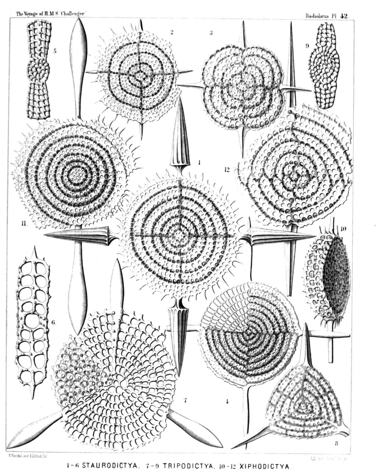
PLATE 42.
Porodiscida.
Diam. Page.
Fig. 1. Staurodictya elegans, n. sp., × 500 507
Fig. 2. Staurodictya ciliata, n. sp., × 400 506
Fig. 3. Staurodictya medusa, n. sp., × 400 506
Fig. 4. Staurodictya cruciata, n. sp., × 300 507
Fig. 5. Staurodictya cruciata, n. sp., × 300 507
Vertical section through the disk.
Fig. 6. Staurodictya grandis, n. sp., × 300 508
Vertical section through the disk.
Fig. 7. Tripodictya triacantha, n. sp., × 400 505
Fig. 8. Tripodictya trigonaria, n. sp., × 400 505
Fig. 9. Tripodictya tribelonia, n. sp., × 400 505
Vertical section through the disk.
Fig. 10. Xiphodictya amphibelonia, n. sp., × 300 503
Marginal view.
Fig. 11. Xiphodictya amphirrhopalia, n. sp., × 400 504
Fig. 12. Xiphodictya staurospira, n. sp., × 500 504
PLATE 43.
Legion SPUMELLARIA.
Order DISCOIDEA.
Family Porodiscida.
PLATE 43. Porodiscida. Diam. Page. Fig. 1. Rhopalastrum malleus, n. sp., × 100 527 Fig. 2. Rhopalastrum ypsilinum, n. sp., × 50 528 Fig. 3. Rhopalastrum hexaceros, n. sp., × 100 529 Fig. 4. Rhopalastrum triceros, n. sp., × 50 529 Fig. 5. Rhopalastrum trispinosum, n. sp. (vel Dictyastrum trispinosum), × 150 525 Fig. 6. Rhopalastrum arcticum, n. sp., × 300 539 Fig. 7. Rhopalastrum hexagonum, n. sp. (vel Dictyastrum hexagonum), × 100 525 Fig. 8. Rhopalastrum irregulare, n. sp., × 100 528 Fig. 9. Euchitonia lanceolata, n. sp., × 80 534 Fig. 10. Euchitonia carcinus, n. sp., × 300 535 Fig. 11. Euchitonia echinata, n. sp., × 120 536 Fig. 12. Euchitonia stohrii, n. sp., × 100 534 Fig. 13. Hymeniastrum euclidis, n. sp., × 200 531 Fig. 14. Chitonastrum jugatum, n. sp., × 200 537 Fig. 15. Chitonastrum lyra, n. sp., × 500 538 A living specimen observed. The entire shell is enveloped by the calymma and surrounded by radiating pseudopodia (drawn much too short). Between the two paired arms arises a large "sarcode-flagellum." The central chamber and the first enveloping ring are filled by the clear nucleus; the other rings and all the chambers of the arms contain numerous pink oil-globules. Fig. 16. Trigonastrum regulare, n. sp. (vel Chitonastrum regulare), × 200 539
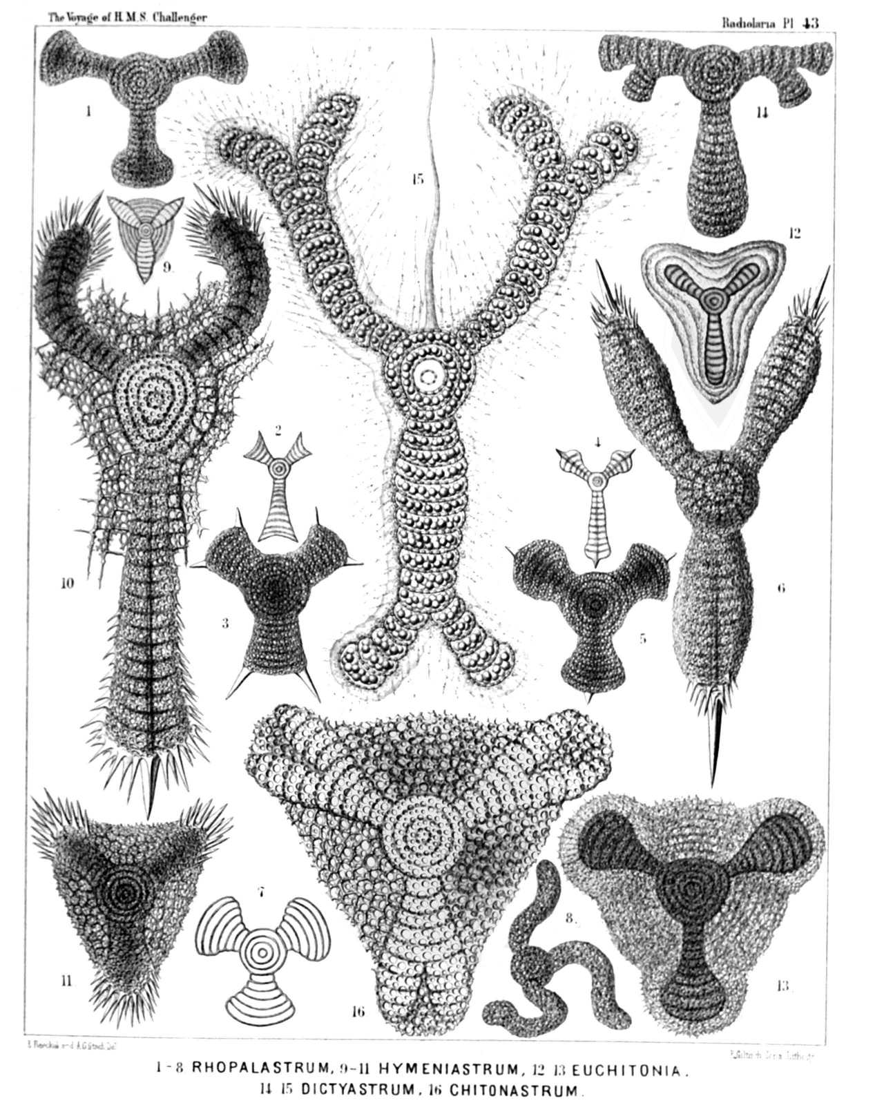
PLATE 43.
Porodiscida.
Diam. Page.
Fig. 1. Rhopalastrum malleus, n. sp., × 100 527
Fig. 2. Rhopalastrum ypsilinum, n. sp., × 50 528
Fig. 3. Rhopalastrum hexaceros, n. sp., × 100 529
Fig. 4. Rhopalastrum triceros, n. sp., × 50 529
Fig. 5. Rhopalastrum trispinosum, n. sp. (vel Dictyastrum trispinosum), × 150 525
Fig. 6. Rhopalastrum arcticum, n. sp., × 300 539
Fig. 7. Rhopalastrum hexagonum, n. sp. (vel Dictyastrum hexagonum), × 100 525
Fig. 8. Rhopalastrum irregulare, n. sp., × 100 528
Fig. 9. Euchitonia lanceolata, n. sp., × 80 534
Fig. 10. Euchitonia carcinus, n. sp., × 300 535
Fig. 11. Euchitonia echinata, n. sp., × 120 536
Fig. 12. Euchitonia stohrii, n. sp., × 100 534
Fig. 13. Hymeniastrum euclidis, n. sp., × 200 531
Fig. 14. Chitonastrum jugatum, n. sp., × 200 537
Fig. 15. Chitonastrum lyra, n. sp., × 500 538
A living specimen observed. The entire shell is enveloped by the calymma and surrounded by radiating pseudopodia (drawn much too short). Between the two paired arms arises a large "sarcode-flagellum." The central chamber and the first enveloping ring are filled by the clear nucleus; the other rings and all the chambers of the arms contain numerous pink oil-globules.
Fig. 16. Trigonastrum regulare, n. sp. (vel Chitonastrum regulare), × 200 539
PLATE 44.
Legion SPUMELLARIA.
Order DISCOIDEA.
Family Porodiscida.
PLATE 44. Porodiscida. Diam. Page. Fig. 1. Stephanastrum capitatum, n. sp., × 200 549 Fig. 2. Pentinastrum asteriscus, n. sp., × 300 557 Fig. 3. Pentalastrum ophidiaster, n. sp., × 100 557 Fig. 4. Hexinastrum geryonidum, n. sp., × 300 560 Fig. 5. Hexalastrum orchidaceum, n. sp., × 50 560 Fig. 6. Amphibrachium dilatatum, n. sp., × 50 517 Fig. 7. Amphymenium zygartus, n. sp., × 400 520 Fig. 8. Amphymenium pupula, n. sp., × 300 519 Fig. 9. Amphymenium amphistylium, n. sp., × 200 520 Fig. 10. Amphicraspedum murrayanum, n. sp., × 300 523 Fig. 11. Amphymenium monstrosum, n. sp., × 300 520
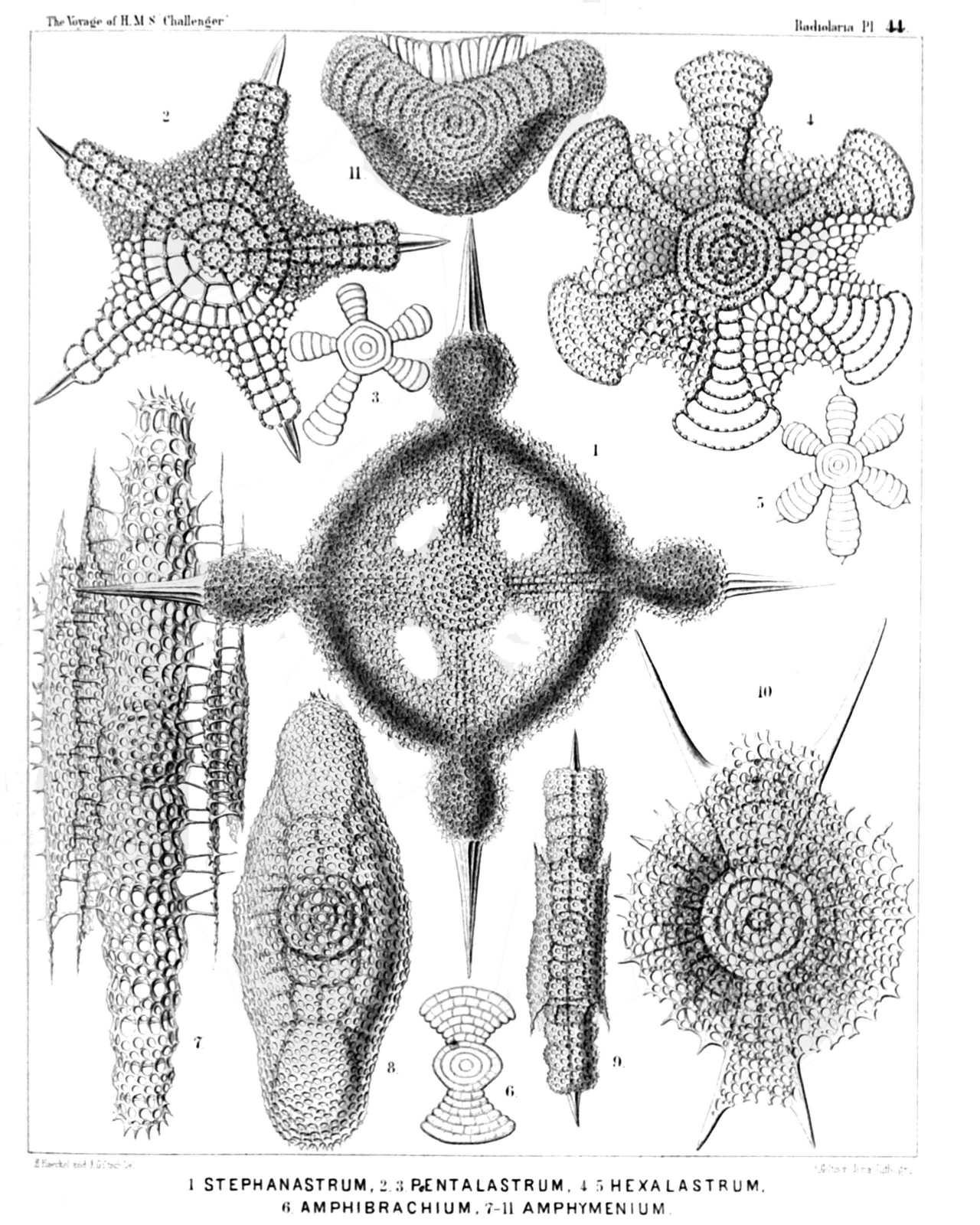
PLATE 44.
Porodiscida.
Diam. Page.
Fig. 1. Stephanastrum capitatum, n. sp., × 200 549
Fig. 2. Pentinastrum asteriscus, n. sp., × 300 557
Fig. 3. Pentalastrum ophidiaster, n. sp., × 100 557
Fig. 4. Hexinastrum geryonidum, n. sp., × 300 560
Fig. 5. Hexalastrum orchidaceum, n. sp., × 50 560
Fig. 6. Amphibrachium dilatatum, n. sp., × 50 517
Fig. 7. Amphymenium zygartus, n. sp., × 400 520
Fig. 8. Amphymenium pupula, n. sp., × 300 519
Fig. 9. Amphymenium amphistylium, n. sp., × 200 520
Fig. 10. Amphicraspedum murrayanum, n. sp., × 300 523
Fig. 11. Amphymenium monstrosum, n. sp., × 300 520
PLATE 45.
Legion SPUMELLARIA.
Order DISCOIDEA.
Family Porodiscida.
PLATE 45. Porodiscida. Diam. Page. Fig. 1. Stauralastrum rhopalophorum, n. sp., × 200 541 Fig. 2. Dicranastrum cornutum, n. sp., × 200 551 Fig. 3. Hagiastrum mosis, n. sp., × 100 543 Fig. 4. Hagiastrum mosis, n. sp., × 50 543 Lateral view, from the edge. Fig. 5. Hagiastrum buddhæ, n. sp., × 50 542 Fig. 6. Stauralastrum cruciforme, n. sp. (in glycerine), × 500 540 The central capsule contains a large central nucleus with nucleolus, and is surrounded by the jelly calymma and numerous small zooxanthellæ. The endoplasm is radially striped. Fig. 7. Tesserastrum democriti, n. sp., × 100 548 Fig. 8. Tesserastrum straussii, n. sp., × 500 547 Fig. 9. Tesserastrum brunonis, n. sp., × 200 548 Disk seen from the edge. Fig. 10. Amphirhopalum echinatum, n. sp., × 300 522 Fig. 11. Amphicraspedum maclaganium, n. sp., × 100 523 Fig. 12. Amphicraspedum wyvilleanum, n. sp., × 300 523
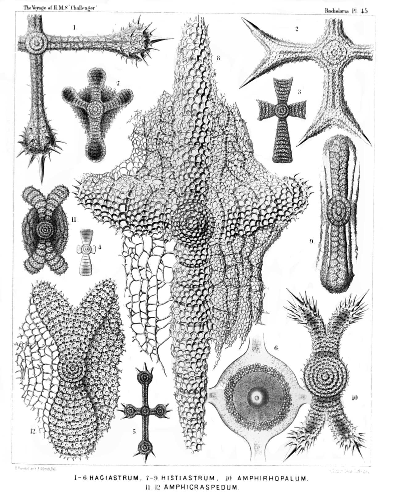
PLATE 45.
Porodiscida.
Diam. Page.
Fig. 1. Stauralastrum rhopalophorum, n. sp., × 200 541
Fig. 2. Dicranastrum cornutum, n. sp., × 200 551
Fig. 3. Hagiastrum mosis, n. sp., × 100 543
Fig. 4. Hagiastrum mosis, n. sp., × 50 543
Lateral view, from the edge.
Fig. 5. Hagiastrum buddhæ, n. sp., × 50 542
Fig. 6. Stauralastrum cruciforme, n. sp. (in glycerine), × 500 540
The central capsule contains a large central nucleus with nucleolus, and is surrounded by the jelly calymma and numerous small zooxanthellæ. The endoplasm is radially striped.
Fig. 7. Tesserastrum democriti, n. sp., × 100 548
Fig. 8. Tesserastrum straussii, n. sp., × 500 547
Fig. 9. Tesserastrum brunonis, n. sp., × 200 548
Disk seen from the edge.
Fig. 10. Amphirhopalum echinatum, n. sp., × 300 522
Fig. 11. Amphicraspedum maclaganium, n. sp., × 100 523
Fig. 12. Amphicraspedum wyvilleanum, n. sp., × 300 523
PLATE 46.
Legion SPUMELLARIA.
Order DISCOIDEA.
Family Porodiscida.
PLATE 46. Porodiscida. Diam. Page. Fig. 1. Histiastrum boseanum, n. sp., × 400 546 Fig. 2. Histiastrum pentadiscus, n. sp., × 200 546 Fig. 3. Histiastrum quadrigatum, n. sp., × 300 544 Fig. 4. Histiastrum velatum, n. sp., × 200 545 Fig. 5. Stephanastrum quadratum, n. sp., × 200 549
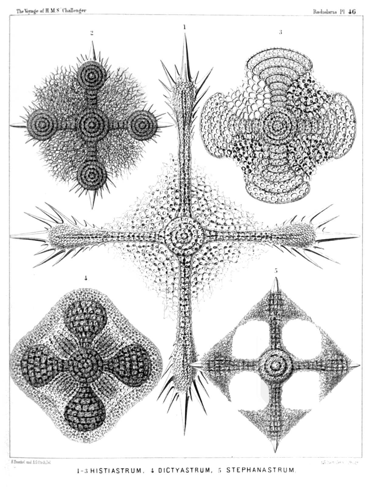
PLATE 46.
Porodiscida.
Diam. Page.
Fig. 1. Histiastrum boseanum, n. sp., × 400 546
Fig. 2. Histiastrum pentadiscus, n. sp., × 200 546
Fig. 3. Histiastrum quadrigatum, n. sp., × 300 544
Fig. 4. Histiastrum velatum, n. sp., × 200 545
Fig. 5. Stephanastrum quadratum, n. sp., × 200 549
PLATE 47.
Legion SPUMELLARIA.
Order DISCOIDEA.
Family Porodiscida.
PLATE 47. Porodiscida. Diam. Page. Fig. 1. Dicranastrum bifurcatum, n. sp., × 200 552 Fig. 1a. Central disc of the same, × 600 Fig. 2. Dicranastrum furcatum, n. sp., × 100 550 Fig. 3. Dicranastrum wyvillei, n. sp., × 100 551 Fig. 4. Pentophiastrum forcipatum, n. sp., × 50 559 Fig. 5. Pentophiastrum caudatum, n. sp., × 50 559 Fig. 6. Myelastrum papilio, n. sp., × 50 554 Fig. 7. Myelastrum decaceros, n. sp., × 20 554 Fig. 8. Myelastrum heteropterum, n. sp., × 20 553 Fig. 9. Myelastrum anomalum, n. sp., × 50 556 Fig. 10. Myelastrum farfalla, n. sp., × 50 554 Fig. 11. Myelastrum dodecaceros, n. sp., × 100 554 Fig. 12. Myelastrum octocorne, n. sp., × 90 553 Fig. 13. Myelastrum medullare, n. sp., × 50 553
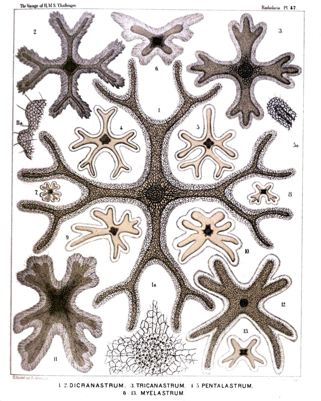
PLATE 47.
Porodiscida.
Diam. Page.
Fig. 1. Dicranastrum bifurcatum, n. sp., × 200 552
Fig. 1a. Central disc of the same, × 600
Fig. 2. Dicranastrum furcatum, n. sp., × 100 550
Fig. 3. Dicranastrum wyvillei, n. sp., × 100 551
Fig. 4. Pentophiastrum forcipatum, n. sp., × 50 559
Fig. 5. Pentophiastrum caudatum, n. sp., × 50 559
Fig. 6. Myelastrum papilio, n. sp., × 50 554
Fig. 7. Myelastrum decaceros, n. sp., × 20 554
Fig. 8. Myelastrum heteropterum, n. sp., × 20 553
Fig. 9. Myelastrum anomalum, n. sp., × 50 556
Fig. 10. Myelastrum farfalla, n. sp., × 50 554
Fig. 11. Myelastrum dodecaceros, n. sp., × 100 554
Fig. 12. Myelastrum octocorne, n. sp., × 90 553
Fig. 13. Myelastrum medullare, n. sp., × 50 553