Front matter (continued 3)
Reading edition
Fig. 5. Semantrum quadrifore, n. sp., × 400 958
Fig. 6. Semantidium hexastoma, n. sp., × 400 960
Fig. 7. Semantidium signatorium, n. sp., × 400 961
Fig. 8. Clathrocircus stapedius, n. sp., × 400 962
Fig. 9. Clathrocircus dictyospyris, n. sp., × 300 963
Fig. 10. Clathrocircus multiforis, n. sp., × 300 963
Fig. 11. Cortiniscus tripodiscus, n. sp., × 400 963
Fig. 12. Cortiniscus typicus, n. sp., × 300 964
Fig. 13. Cortiniscus dipylaris, n. sp., × 400 964
Fig. 14. Stephaniscus quadrifurcus, n. sp., × 300 965
Fig. 15. Stephaniscus quadrigatus, n. sp., × 400 965
Fig. 16. Semantiscus hexapodius, n. sp., × 400 966
Fig. 17. Semantiscus hexapylus, n. sp., × 400 967
Fig. 18. Semantiscus hexaspyris, n. sp., × 400 966
Fig. 19. Lithocircus tarandus, n. sp., × 400 944
Fig. 20. Stephanium quadrupes, n. sp., × 200 952
Fig. 21. Cortina cervina, n. sp., × 300 952
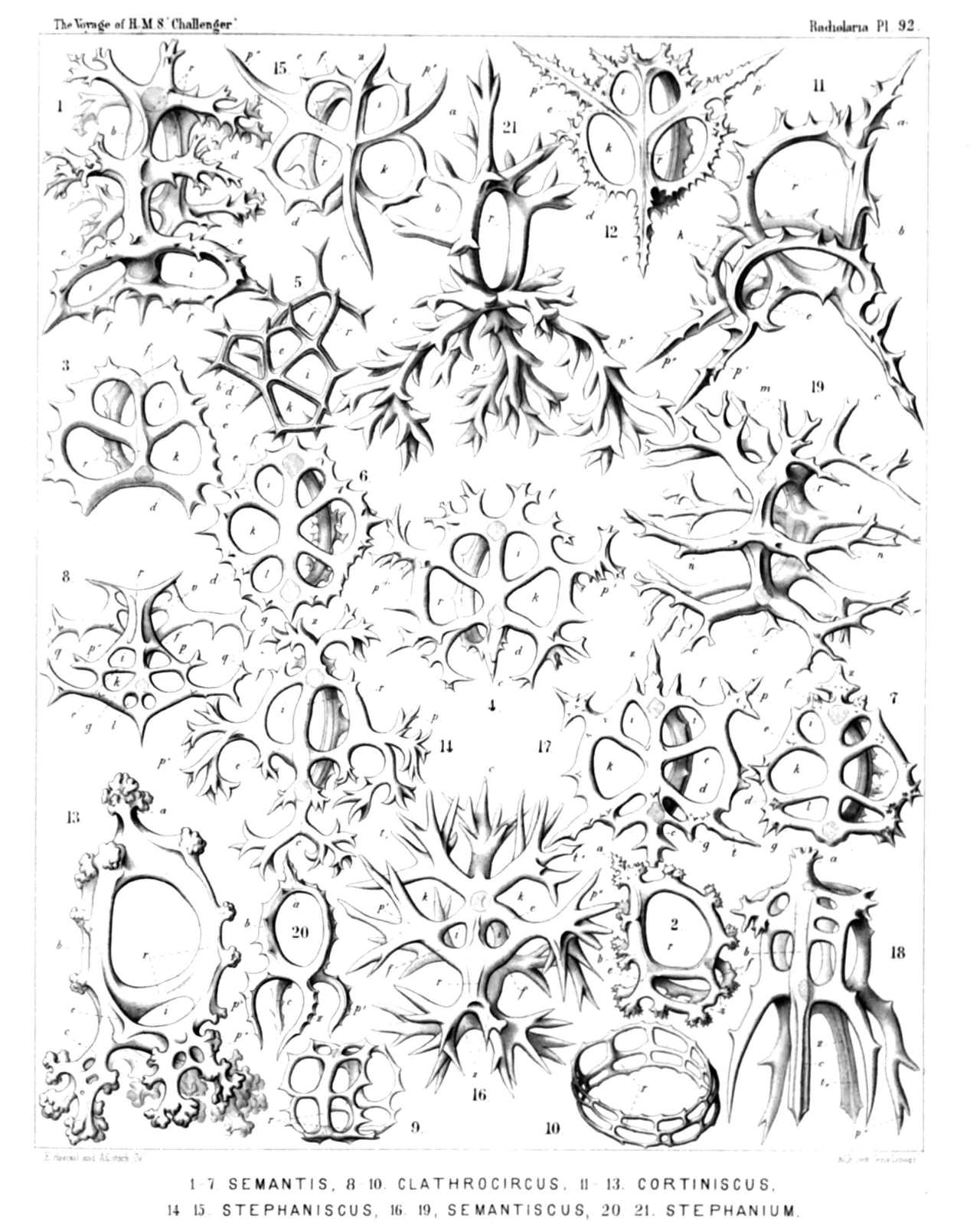
PLATE 93.
Legion NASSELLARIA.
Order STEPHOIDEA.
Families Coronida et Tympanida..
PLATE 93. Coronida et Tympanida.. Diam. Page. Fig. 1. Zygostephanus dissocircus, n. sp., × 300 971 Fig. 2. Zygostephanus bicornis, n. sp., × 300 972 Fig. 3. Zygostephanium dizonium, n. sp., × 300 973 Fig. 4. Zygostephanium paradictyum, n. sp., × 300 973 Fig. 5. Acanthodesmia corona, n. sp., × 400 976 Fig. 6. Plectocoronis pentacantha, n. sp., × 300 979 Fig. 7. Tristephanium quadricorne, n. sp., × 300 984 Fig. 8. Tristephanium octopyle, n. sp., × 300 983 Fig. 9. Tristephanium dimensivum, n. sp., × 400 983 Fig. 10. Trissocircus lentellipsis, n. sp., × 300 985 Fig. 11. Trissocircus octostoma, n. sp., × 300 986 Fig. 12. Trissocyclus sphæridium, n. sp., × 300 987 Fig. 13. Tricyclidium dictyospyris, n. sp., × 300 984 Fig. 14. Protympanium amphipodium, n. sp., × 300 992 Fig. 15. Acrocubus arcuatus, n. sp., × 300 993 Fig. 16. Acrocubus cortina, n. sp., × 300 994 Fig. 17. Acrocubus amphithectus, n. sp., × 300 995 Fig. 18. Toxarium thorax, n. sp., × 300 996 Fig. 19. Toxarium cordatum, n. sp., × 300 996 Fig. 20. Toxarium bifurcum, n. sp., × 300 997 Fig. 21. Parastephanus quadrispinus, n. sp., × 300 1008 Fig. 22. Prismatium tripodium, n. sp., × 300 1009
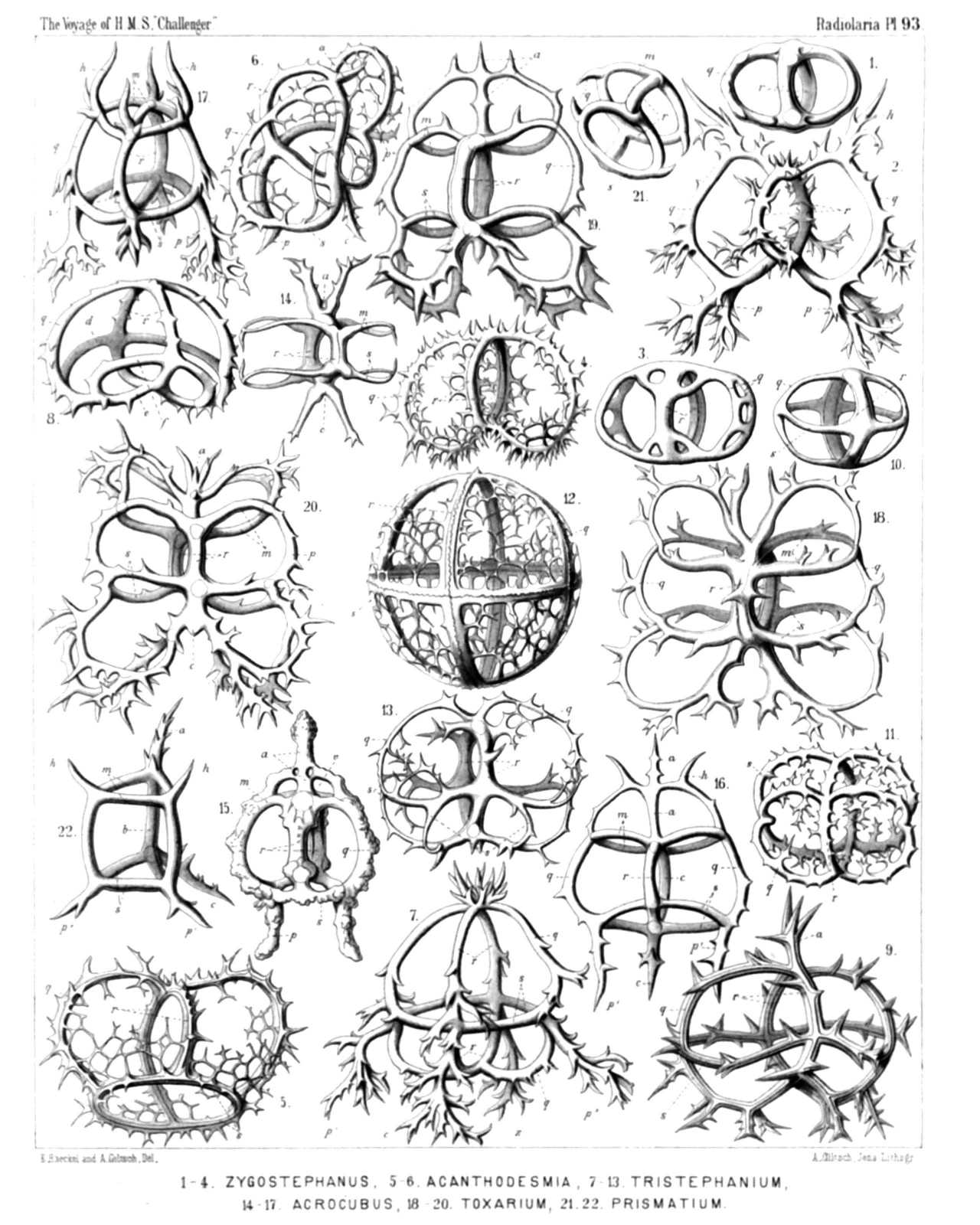
PLATE 93.
Coronida et Tympanida..
Diam. Page.
Fig. 1. Zygostephanus dissocircus, n. sp., × 300 971
Fig. 2. Zygostephanus bicornis, n. sp., × 300 972
Fig. 3. Zygostephanium dizonium, n. sp., × 300 973
Fig. 4. Zygostephanium paradictyum, n. sp., × 300 973
Fig. 5. Acanthodesmia corona, n. sp., × 400 976
Fig. 6. Plectocoronis pentacantha, n. sp., × 300 979
Fig. 7. Tristephanium quadricorne, n. sp., × 300 984
Fig. 8. Tristephanium octopyle, n. sp., × 300 983
Fig. 9. Tristephanium dimensivum, n. sp., × 400 983
Fig. 10. Trissocircus lentellipsis, n. sp., × 300 985
Fig. 11. Trissocircus octostoma, n. sp., × 300 986
Fig. 12. Trissocyclus sphæridium, n. sp., × 300 987
Fig. 13. Tricyclidium dictyospyris, n. sp., × 300 984
Fig. 14. Protympanium amphipodium, n. sp., × 300 992
Fig. 15. Acrocubus arcuatus, n. sp., × 300 993
Fig. 16. Acrocubus cortina, n. sp., × 300 994
Fig. 17. Acrocubus amphithectus, n. sp., × 300 995
Fig. 18. Toxarium thorax, n. sp., × 300 996
Fig. 19. Toxarium cordatum, n. sp., × 300 996
Fig. 20. Toxarium bifurcum, n. sp., × 300 997
Fig. 21. Parastephanus quadrispinus, n. sp., × 300 1008
Fig. 22. Prismatium tripodium, n. sp., × 300 1009
PLATE 94.
Legion NASSELLARIA.
Order STEPHOIDEA.
Family Tympanida.
PLATE 94. Tympanida. Diam. Page. Fig. 1. Tympanidium foliosum, n. sp., × 400 1003 Fig. 2. Octotympanum cervicorne, n. sp., × 400 1000 Fig. 3. Octotympanum octonarium, n. sp., × 400 1000 Fig. 4. Tympaniscus quadrupes, n. sp., × 400 1002 Fig. 5. Tympaniscus dipodiscus, n. sp., × 400 1001 Frontal view. Fig. 6. Tympaniscus dipodiscus, n. sp., × 400 1001 Lateral view. Fig. 7. Tympaniscus tripodiscus, n. sp., × 400 1002 Frontal view. Fig. 8. Microcubus zonarius, n. sp., × 300 998 Fig. 9. Microcubus dodecastoma, n. sp., × 300 998 Fig. 10. Microcubus amphispyris, n. sp., × 400 999 Fig. 11. Pseudocubus obeliscus, n. sp., × 400 1010 Fig. 12. Pseudocubus hexapylus, n. sp., × 300 1011 Fig. 13. Lithocubus geometricus, n. sp., × 200 1011 Fig. 14. Paratympanum octostylum, n. sp., × 400 1005 Fig. 15. Dystympanium dictyocha, n. sp., × 400 1007 Lateral view. Fig. 16. Dystympanium dictyocha, n. sp., × 400 1007 Apical view. Fig. 17. Circotympanum octogonium, n. sp., × 500 1013 Fig. 18. Tympanidium binoctonum, n. sp., × 400 1004
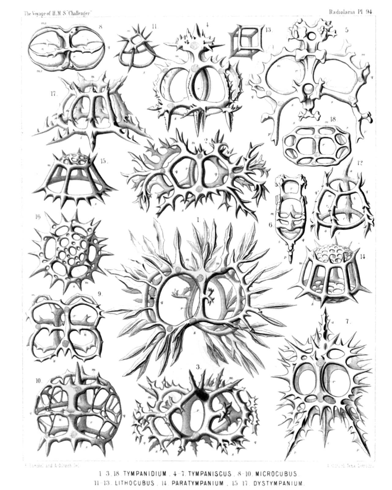
PLATE 94.
Tympanida.
Diam. Page.
Fig. 1. Tympanidium foliosum, n. sp., × 400 1003
Fig. 2. Octotympanum cervicorne, n. sp., × 400 1000
Fig. 3. Octotympanum octonarium, n. sp., × 400 1000
Fig. 4. Tympaniscus quadrupes, n. sp., × 400 1002
Fig. 5. Tympaniscus dipodiscus, n. sp., × 400 1001
Frontal view.
Fig. 6. Tympaniscus dipodiscus, n. sp., × 400 1001
Lateral view.
Fig. 7. Tympaniscus tripodiscus, n. sp., × 400 1002
Frontal view.
Fig. 8. Microcubus zonarius, n. sp., × 300 998
Fig. 9. Microcubus dodecastoma, n. sp., × 300 998
Fig. 10. Microcubus amphispyris, n. sp., × 400 999
Fig. 11. Pseudocubus obeliscus, n. sp., × 400 1010
Fig. 12. Pseudocubus hexapylus, n. sp., × 300 1011
Fig. 13. Lithocubus geometricus, n. sp., × 200 1011
Fig. 14. Paratympanum octostylum, n. sp., × 400 1005
Fig. 15. Dystympanium dictyocha, n. sp., × 400 1007
Lateral view.
Fig. 16. Dystympanium dictyocha, n. sp., × 400 1007
Apical view.
Fig. 17. Circotympanum octogonium, n. sp., × 500 1013
Fig. 18. Tympanidium binoctonum, n. sp., × 400 1004
PLATE 95.
Legion NASSELLARIA.
Order SPYROIDEA.
Families Zygospyrida, Tholospyrida, Phormospyrida et Androspyrida.
PLATE 95. Zygospyrida, Tholospyrida, Phormospyrida et Androspyrida. Diam. Page. Fig. 1. Tripospyris cortina, n. sp., × 300 1025 Basal view. Fig. 2. Tripospyris triplecta, n. sp., × 300 1027 Basal view. Fig. 3. Tripospyris semantrum, n. sp., × 400 1027 Basal view. Fig. 4. Tripospyris hexomma, n. sp., × 300 1028 Basal view. Fig. 5. Brachiospyris diacantha, n. sp., × 400 1038 Basal view. Fig. 6. Tetraspyris stephanium, n. sp., × 300 1044 Basal view. Fig. 7. Liriospyris amphithecta, n. sp., × 300 1050 Basal view. Fig. 8. Hexaspyris hexacorethra, n. sp., × 300 1048 Frontal view. Fig. 9. Clathrospyris pyramidalis, n. sp., × 500 1052 Frontal view. Fig. 10. Aegospyris aegoceras, n. sp., × 400 1054 Frontal view. Fig. 11. Pentaspyris pentacantha, n. sp., × 400 1054 Dorsal view. Fig. 12. Taurospyris cervina, n. sp., × 400 1058 Frontal view. Fig. 13. Circospyris nucula, n. sp., × 300 1072 Dorsal view. Fig. 14. Lophospyris dipodiscus, n. sp., × 400 1080 Frontal view. Fig. 15. Sepalospyris platyphylla, n. sp., × 400 1081 Dorsal view. Fig. 16. Pylospyris canariensis, n. sp., × 400 1084 Frontal view. Fig. 17. Acrospyris clathrocanium, n. sp., × 300 1085 Dorsal view. Fig. 18. Phormospyris tridentata, n. sp., × 400 1087 Frontal view. Fig. 19. Patagospyris anthocyrtis, n. sp., × 500 1088 Dorsal view. Fig. 20. Androspyris pithecus, n. sp., × 400 1093 Lateral view.
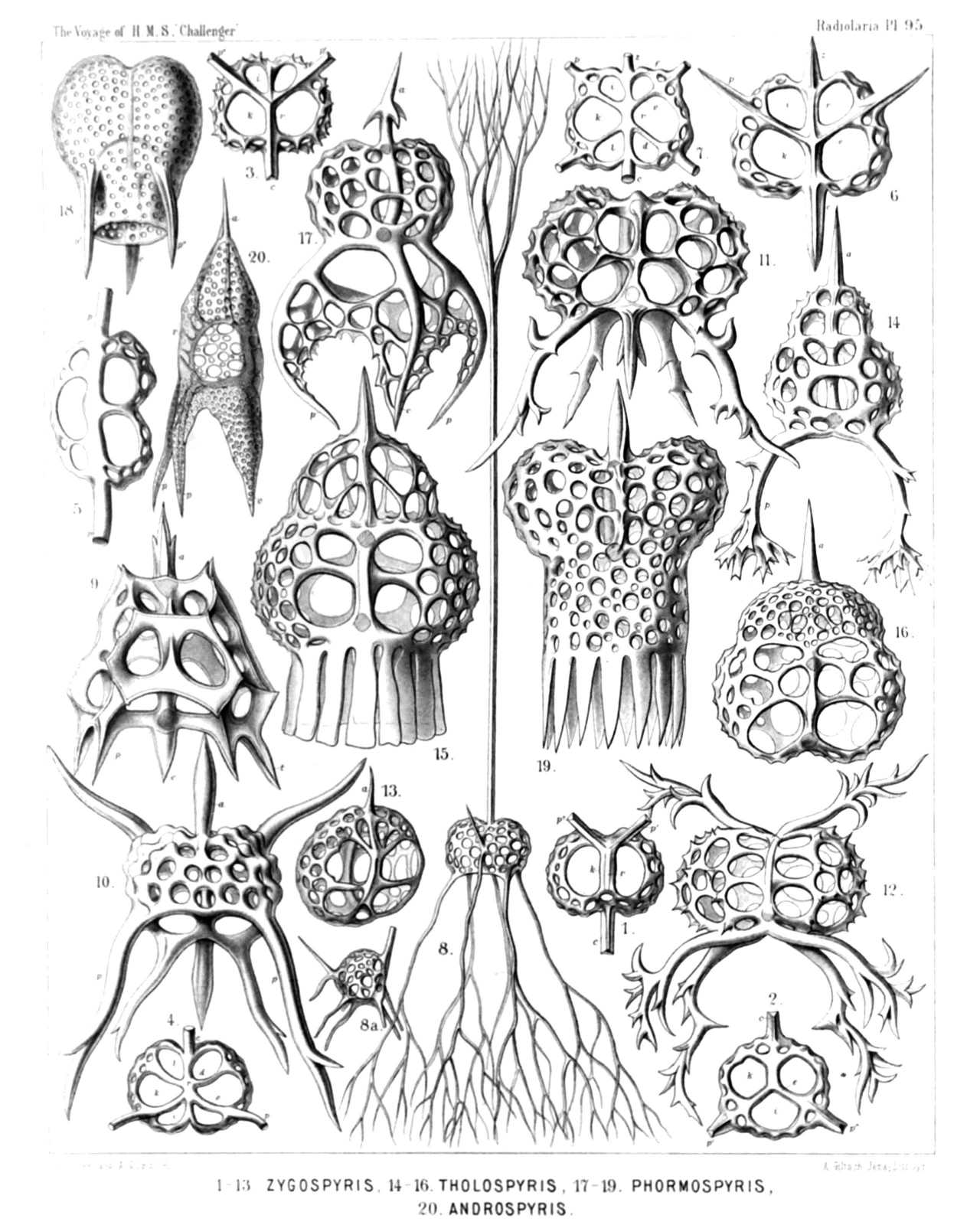
PLATE 95.
Zygospyrida, Tholospyrida, Phormospyrida et Androspyrida.
Diam. Page.
Fig. 1. Tripospyris cortina, n. sp., × 300 1025
Basal view.
Fig. 2. Tripospyris triplecta, n. sp., × 300 1027
Basal view.
Fig. 3. Tripospyris semantrum, n. sp., × 400 1027
Basal view.
Fig. 4. Tripospyris hexomma, n. sp., × 300 1028
Basal view.
Fig. 5. Brachiospyris diacantha, n. sp., × 400 1038
Basal view.
Fig. 6. Tetraspyris stephanium, n. sp., × 300 1044
Basal view.
Fig. 7. Liriospyris amphithecta, n. sp., × 300 1050
Basal view.
Fig. 8. Hexaspyris hexacorethra, n. sp., × 300 1048
Frontal view.
Fig. 9. Clathrospyris pyramidalis, n. sp., × 500 1052
Frontal view.
Fig. 10. Aegospyris aegoceras, n. sp., × 400 1054
Frontal view.
Fig. 11. Pentaspyris pentacantha, n. sp., × 400 1054
Dorsal view.
Fig. 12. Taurospyris cervina, n. sp., × 400 1058
Frontal view.
Fig. 13. Circospyris nucula, n. sp., × 300 1072
Dorsal view.
Fig. 14. Lophospyris dipodiscus, n. sp., × 400 1080
Frontal view.
Fig. 15. Sepalospyris platyphylla, n. sp., × 400 1081
Dorsal view.
Fig. 16. Pylospyris canariensis, n. sp., × 400 1084
Frontal view.
Fig. 17. Acrospyris clathrocanium, n. sp., × 300 1085
Dorsal view.
Fig. 18. Phormospyris tridentata, n. sp., × 400 1087
Frontal view.
Fig. 19. Patagospyris anthocyrtis, n. sp., × 500 1088
Dorsal view.
Fig. 20. Androspyris pithecus, n. sp., × 400 1093
Lateral view.
PLATE 96.
Legion NASSELLARIA.
Order BOTRYODEA.
Families Cannobotryida, Lithobotryida et Pylobotryida.
PLATE 96. Cannobotryida, Lithobotryida et Pylobotryida Diam. Page. Fig. 1. Botryopera cyrtoloba, n. sp., × 500 1108 Apical view. Fig. 2. Botryopera quinqueloba, n. sp., × 500 1109 Half lateral, half frontal view. Fig. 3. Cannobotrys tricanna, n. sp., × 400 1110 View half from the frontal, half from the left side. Fig. 4. Cannobotrys cortina, n. sp., × 400 1110 Basal view. Fig. 5. Botryopyle inclusa, n. sp., × 500 1113 Frontal view. Fig. 6. Botryopyle dictyocephalus, n. sp., × 500 1113 Lateral view (right side). Fig. 7. Botryopyle sethocorys, n. sp., × 400 1112 Frontal view. Fig. 8. Acrobotrys trisolenia, n. sp., × 400 1115 Lateral view (right side). Fig. 9. Acrobotrys acuminata, n. sp., × 400 1115 Lateral view (right side). Fig. 10. Acrobotrys disolenia, n. sp., × 400 1114 Lateral view (left side). Fig. 11. Acrobotrys auriculata, n. sp., × 500 1115 Lateral view (right side). Fig. 12. Botryocella multicellaris, n. sp., × 500 1117 Lateral view (left side). Fig. 13. Botryocella quadricellaris, n. sp., × 400 1117 Lateral view (left side). Fig. 14. Botryocella quadrigemina, n. sp., × 400 1117 Collar septum, between cephalis and thorax. Fig. 15. Lithobotrys sphærothorax, n. sp., × 500 1119 Lateral view (right side). Fig. 16. Lithobotrys mascula, n. sp., × 500 1119 Frontal view. Fig. 17. Lithobotrys orchidea, n. sp., × 500 1119 Frontal view. Fig. 18. Botryocyrtis cerebellum, n. sp., × 400 1121 Apical view. Fig. 19. Botryocyrtis theocampe, n. sp., × 500 1121 Lateral view (left side). Fig. 20. Pylobotrys fontinalis, n. sp., × 400 1122 Apical view. Fig. 21. Pylobotrys putealis, n. sp., × 500 1121 Lateral view (right side). Fig. 22. Pylobotrys cerebralis, n. sp., × 500 1122 Dorsal view. Fig. 23. Botryocampe rotalia, n. sp., × 400 1123 Collar septum. Fig. 24. Botryocampe camerata, n. sp., × 500 1124 Lateral view (left side). Fig. 25. Phormobotrys cannothalamia, n. sp., × 400 1125 Lateral view (right side). Fig. 26. Phormobotrys trithalamia, n. sp., × 500 1124 Frontal section. The dorsal wall is visible, in the cephalis the cruciform frontal septum. Fig. 27. Phormobotrys pentathalamia, n. sp., × 400 1124 Lateral view (left side). Fig. 28. Cephalospyris triangulata, n. sp., × 400 1035 The central capsule encloses numerous spherical concrements.
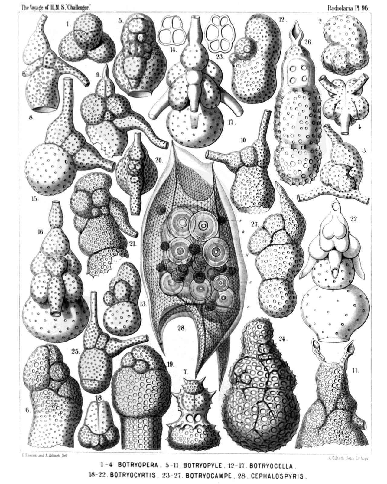
PLATE 96.
Cannobotryida, Lithobotryida et Pylobotryida
Diam. Page.
Fig. 1. Botryopera cyrtoloba, n. sp., × 500 1108
Apical view.
Fig. 2. Botryopera quinqueloba, n. sp., × 500 1109
Half lateral, half frontal view.
Fig. 3. Cannobotrys tricanna, n. sp., × 400 1110
View half from the frontal, half from the left side.
Fig. 4. Cannobotrys cortina, n. sp., × 400 1110
Basal view.
Fig. 5. Botryopyle inclusa, n. sp., × 500 1113
Frontal view.
Fig. 6. Botryopyle dictyocephalus, n. sp., × 500 1113
Lateral view (right side).
Fig. 7. Botryopyle sethocorys, n. sp., × 400 1112
Frontal view.
Fig. 8. Acrobotrys trisolenia, n. sp., × 400 1115
Lateral view (right side).
Fig. 9. Acrobotrys acuminata, n. sp., × 400 1115
Lateral view (right side).
Fig. 10. Acrobotrys disolenia, n. sp., × 400 1114
Lateral view (left side).
Fig. 11. Acrobotrys auriculata, n. sp., × 500 1115
Lateral view (right side).
Fig. 12. Botryocella multicellaris, n. sp., × 500 1117
Lateral view (left side).
Fig. 13. Botryocella quadricellaris, n. sp., × 400 1117
Lateral view (left side).
Fig. 14. Botryocella quadrigemina, n. sp., × 400 1117
Collar septum, between cephalis and thorax.
Fig. 15. Lithobotrys sphærothorax, n. sp., × 500 1119
Lateral view (right side).
Fig. 16. Lithobotrys mascula, n. sp., × 500 1119
Frontal view.
Fig. 17. Lithobotrys orchidea, n. sp., × 500 1119
Frontal view.
Fig. 18. Botryocyrtis cerebellum, n. sp., × 400 1121
Apical view.
Fig. 19. Botryocyrtis theocampe, n. sp., × 500 1121
Lateral view (left side).
Fig. 20. Pylobotrys fontinalis, n. sp., × 400 1122
Apical view.
Fig. 21. Pylobotrys putealis, n. sp., × 500 1121
Lateral view (right side).
Fig. 22. Pylobotrys cerebralis, n. sp., × 500 1122
Dorsal view.
Fig. 23. Botryocampe rotalia, n. sp., × 400 1123
Collar septum.
Fig. 24. Botryocampe camerata, n. sp., × 500 1124
Lateral view (left side).
Fig. 25. Phormobotrys cannothalamia, n. sp., × 400 1125
Lateral view (right side).
Fig. 26. Phormobotrys trithalamia, n. sp., × 500 1124
Frontal section. The dorsal wall is visible, in the cephalis the cruciform frontal septum.
Fig. 27. Phormobotrys pentathalamia, n. sp., × 400 1124
Lateral view (left side).
Fig. 28. Cephalospyris triangulata, n. sp., × 400 1035
The central capsule encloses numerous spherical concrements.
PLATE 97.
Legion NASSELLARIA.
Orders STEPHOIDEA et CYRTOIDEA.
Families Stephanida, Coronida, Tripocalpida, Phænocalpida, Tripocyrtida, Podocyrtida et Podocampida.
PLATE 97. Stephanida, Coronida, Tripocalpida, Phænocalpida, Tripocyrtida, Podocyrtida et Podocampida. Diam. Page. Fig. 1. Cortina typus, n. sp., × 300 951 View from the right side. The upper part of the central capsule includes the nucleus, the lower part the podoconus, besides some oil-globules. The two pectoral feet are partly broken off. Fig. 2. Podocoronis cortiniscus, n. sp., × 400 981 View from the right anterior side. Fig. 3. Tripocalpis cortinaris, n. sp., × 400 1137 Fig. 4. Phænocalpis petalospyris, n. sp., × 400 1173 Lateral view (inverted). Fig. 5. Haliphormis lagena, n. sp., × 200 1167 Fig. 6. Halicapsa lithapium, n. sp., × 300 1190 Basal view. Fig. 7. Peridium alatum, n. sp., × 300 1155 Basal view. Fig. 8. Sethopilium orthopus, n. sp., × 300 1202 Basal view. Fig. 9. Sethopilium macropus, n. sp., × 400 1203 Fig. 10. Amphiplecta acrostoma, n. sp., × 400 1223 Fig. 11. Sethopera tricostata, n. sp., × 400 1232 Fig. 12. Acanthocorys macroceras, n. sp., × 200 1264 Fig. 13. Sethophæna hexaptera, n. sp., × 400 1286 Fig. 14. Theopodium tricostatum, n. sp., × 400 1328 Fig. 15. Podocampe trictenota, n. sp., × 500 1446
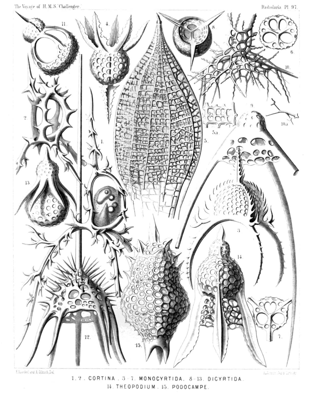
PLATE 97.
Stephanida, Coronida, Tripocalpida, Phænocalpida, Tripocyrtida, Podocyrtida et Podocampida.
Diam. Page.
Fig. 1. Cortina typus, n. sp., × 300 951
View from the right side. The upper part of the central capsule includes the nucleus, the lower part the podoconus, besides some oil-globules. The two pectoral feet are partly broken off.
Fig. 2. Podocoronis cortiniscus, n. sp., × 400 981
View from the right anterior side.
Fig. 3. Tripocalpis cortinaris, n. sp., × 400 1137
Fig. 4. Phænocalpis petalospyris, n. sp., × 400 1173
Lateral view (inverted).
Fig. 5. Haliphormis lagena, n. sp., × 200 1167
Fig. 6. Halicapsa lithapium, n. sp., × 300 1190
Basal view.
Fig. 7. Peridium alatum, n. sp., × 300 1155
Basal view.
Fig. 8. Sethopilium orthopus, n. sp., × 300 1202
Basal view.
Fig. 9. Sethopilium macropus, n. sp., × 400 1203
Fig. 10. Amphiplecta acrostoma, n. sp., × 400 1223
Fig. 11. Sethopera tricostata, n. sp., × 400 1232
Fig. 12. Acanthocorys macroceras, n. sp., × 200 1264
Fig. 13. Sethophæna hexaptera, n. sp., × 400 1286
Fig. 14. Theopodium tricostatum, n. sp., × 400 1328
Fig. 15. Podocampe trictenota, n. sp., × 500 1446
PLATE 98.
Legion NASSELLARIA.
Order CYRTOIDEA.
Families Tripocalpida et Phænocalpida.
PLATE 98. Tripocalpida et Phænocalpida. Diam. Page. Fig. 1. Euscenium plectaniscus, n. sp., × 300 1146 Half frontal, half basal view. Fig. 2. Cladoscenium pectinatum, n. sp., × 400 1150 Shell opened by a vertical section. Fig. 3. Archiscenium cyclopterum, n. sp., × 400 1151 View from the dorsal side. Fig. 4. Pteroscenium arcuatum, n. sp., × 400 1152 The central capsule contains a large spherical nucleus with a nucleolus. Fig. 5. Archipera cortiniscus, n. sp., × 400 1155 Fig. 6. Archibursa tripodiscus, n. sp., × 400 1157 Basal view. Fig. 7. Archipilium orthopterum, n. sp., × 400 1139 Fig. 8. Tripilidium costatum, n. sp., × 300 1141 Fig. 8a. Central capsule in the upper part of the shell, Fig. 8b. Cortinar septum, Fig. 9. Phænoscenium hexapodium, n. sp., × 300 1175 Fig. 10. Archiphæna gorgospyris, n. sp., × 300 1178 Fig. 10a. Cortinar septum with four collar pores, × 300 Fig. 11. Archiphormis urceolata, n. sp., × 300 1168 Fig. 12. Halicalyptra petalospyris, n. sp., × 400 1169 Fig. 13. Arachnocalpis ellipsoides, n. sp., × 300 1172 The central capsule is filled up by clear vacuoles and exhibits in the upper half the ellipsoidal nucleus and four oil-globules, in the lower half the slender striated podoconus. Fig. 13a. A piece of the network, more enlarged, × 900
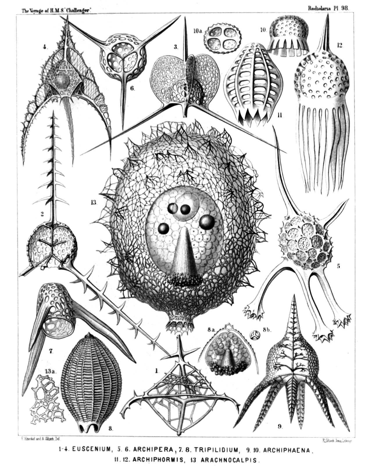
PLATE 98.
Tripocalpida et Phænocalpida.
Diam. Page.
Fig. 1. Euscenium plectaniscus, n. sp., × 300 1146
Half frontal, half basal view.
Fig. 2. Cladoscenium pectinatum, n. sp., × 400 1150
Shell opened by a vertical section.
Fig. 3. Archiscenium cyclopterum, n. sp., × 400 1151
View from the dorsal side.
Fig. 4. Pteroscenium arcuatum, n. sp., × 400 1152
The central capsule contains a large spherical nucleus with a nucleolus.
Fig. 5. Archipera cortiniscus, n. sp., × 400 1155
Fig. 6. Archibursa tripodiscus, n. sp., × 400 1157
Basal view.
Fig. 7. Archipilium orthopterum, n. sp., × 400 1139
Fig. 8. Tripilidium costatum, n. sp., × 300 1141
Fig. 8a. Central capsule in the upper part of the shell,
Fig. 8b. Cortinar septum,
Fig. 9. Phænoscenium hexapodium, n. sp., × 300 1175
Fig. 10. Archiphæna gorgospyris, n. sp., × 300 1178
Fig. 10a. Cortinar septum with four collar pores, × 300
Fig. 11. Archiphormis urceolata, n. sp., × 300 1168
Fig. 12. Halicalyptra petalospyris, n. sp., × 400 1169
Fig. 13. Arachnocalpis ellipsoides, n. sp., × 300 1172
The central capsule is filled up by clear vacuoles and exhibits in the upper half the ellipsoidal nucleus and four oil-globules, in the lower half the slender striated podoconus.
Fig. 13a. A piece of the network, more enlarged, × 900
PLATE 99.
Legion PHÆODARIA.
Order PHÆOGROMIA.
Family Challengerida.
PLATE 99. Challengerida. Diam. Page. (The central capsule is coloured red and the phæodium green in Figs. 1, 6, 10, 14-17, 20). Fig. 1. Challengeria murrayi, n. sp., × 50 1653 From the dorsal side. Numerous streams of sarcode arise from the central capsule and pierce the calymma inside the shell. Fig. 2. Challengeria wildi, n. sp., × 400 1653 The peristome from the left side. Fig. 3. Challengeria bromleyi, n. sp., × 400 1652 From the dorsal side. Fig. 4. Challengeria sloggettii, John Murray × 150 1649 The ventral corner broken off. From the left side. Fig. 4a. Vertical section through the shell-wall. Fig. 5. Challengeria tritonis, n. sp., × 150 1649 Fig. 6. Challengeron diodon, n. sp., × 400 1654 From the dorsal side. The shell contains two central capsules. Fig. 7. Challengeron pearceyi, n. sp., × 300 1654 From the dorsal side. Fig. 8. Challengeron richardsii, n. sp., × 100 1655 From the oral margin Fig. 9. Challengeron fergusoni, n. sp., × 100 1656 From the right side. Fig. 10. Challengeron triangulum, n. sp., × 200 1656 From the right side. Fig. 11. Challengeron crosbiei, n. sp., × 300 1657 From the ventral side. Fig. 12. Challengeron buchanani, n. sp., × 300 1657 From the right side. Fig. 13. Challengeron willemoesii, n. sp., × 400 1659 From the ventral side. Fig. 14. Challengeron moseleyi, n. sp., × 300 1658 From the right side. Fig. 15. Challengeron wyvillei, n. sp., × 300 1660 From the left side. Fig. 16. Porcupinia cordiformis, n. sp., × 200 1663 From the right side. Fig. 17. Pharyngella gastræa, n. sp., × 150 1662 Fig. 18. Pharyngella gastrula, n. sp., × 150 1662 Fig. 19. Entocannula infundibulum, n. sp., × 100 1661 Fig. 20. Entocannula hirsuta, n. sp., × 150 1661 Fig. 21. Lithogromia diatomacea, n. sp., × 400 1647 A piece of the shell with diatomaceous structure. Fig. 21a. Vertical section through the shell-wall. Fig. 22. Lithogromia silicea, n. sp., × 150 1647
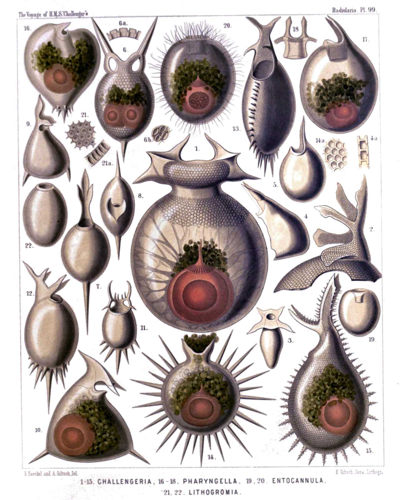
PLATE 99.
Challengerida.
Diam. Page.
(The central capsule is coloured red and the phæodium green in Figs. 1, 6, 10, 14-17, 20).
Fig. 1. Challengeria murrayi, n. sp., × 50 1653
From the dorsal side. Numerous streams of sarcode arise from the central capsule and pierce the calymma inside the shell.
Fig. 2. Challengeria wildi, n. sp., × 400 1653
The peristome from the left side.
Fig. 3. Challengeria bromleyi, n. sp., × 400 1652
From the dorsal side.
Fig. 4. Challengeria sloggettii, John Murray × 150 1649
The ventral corner broken off. From the left side.
Fig. 4a. Vertical section through the shell-wall.
Fig. 5. Challengeria tritonis, n. sp., × 150 1649
Fig. 6. Challengeron diodon, n. sp., × 400 1654
From the dorsal side. The shell contains two central capsules.
Fig. 7. Challengeron pearceyi, n. sp., × 300 1654
From the dorsal side.
Fig. 8. Challengeron richardsii, n. sp., × 100 1655
From the oral margin
Fig. 9. Challengeron fergusoni, n. sp., × 100 1656
From the right side.
Fig. 10. Challengeron triangulum, n. sp., × 200 1656
From the right side.
Fig. 11. Challengeron crosbiei, n. sp., × 300 1657
From the ventral side.
Fig. 12. Challengeron buchanani, n. sp., × 300 1657
From the right side.
Fig. 13. Challengeron willemoesii, n. sp., × 400 1659
From the ventral side.
Fig. 14. Challengeron moseleyi, n. sp., × 300 1658
From the right side.
Fig. 15. Challengeron wyvillei, n. sp., × 300 1660
From the left side.
Fig. 16. Porcupinia cordiformis, n. sp., × 200 1663
From the right side.
Fig. 17. Pharyngella gastræa, n. sp., × 150 1662
Fig. 18. Pharyngella gastrula, n. sp., × 150 1662
Fig. 19. Entocannula infundibulum, n. sp., × 100 1661
Fig. 20. Entocannula hirsuta, n. sp., × 150 1661
Fig. 21. Lithogromia diatomacea, n. sp., × 400 1647
A piece of the shell with diatomaceous structure.
Fig. 21a. Vertical section through the shell-wall.
Fig. 22. Lithogromia silicea, n. sp., × 150 1647
PLATE 100.
Legion PHÆODARIA.
Order PHÆOGROMIA.
Family Tuscarorida.
PLATE 100. Tuscarorida. Diam. Page. Fig. 1. Tuscarora bisternaria, John Murray, × 30 1706 View from the dorsal side. Fig. 1a. View from the mouth pole × 25 Fig. 2. Tuscarora murrayi, n. sp., × 30 1706 View from the dorsal side. The central capsule (in the aboral half), and the phæodium (in the middle of the shell-cavity) are visible. A fine network of pseudopodia pierces the calymma, which fills up the shell-cavity. Fig. 3. Tuscarora wyvillei, n. sp., × 30 1707 View from the dorsal side. Fig. 3a. Base of a tooth, × 100 Fig. 3b. Transverse section through the base of a tooth. Fig. 3c. Base of a foot. Fig. 4. Tuscarora tetrahedra, John Murray, × 15 1707 View from the dorsal side. Fig. 4a. Mouth with the three teeth, × 50 Fig. 5. Tuscarora tubulosa, John Murray, × 40 1707 View from the ventral side. Fig. 5a. Mouth with the two teeth, × 100 Fig. 5b. Basal part of a single tooth, × 150 Fig. 6. Tuscarora porcellana, John Murray, × 600 1708 Fig. 6a. A piece of the shell, with five pores. Fig. 6b. A piece of a tooth, with the internal axial rod and its transverse branches. Fig. 7. Tuscarusa medusa, n. sp., × 25 1709 View from the side. Fig. 7a. View from the mouth, × 50 Fig. 8. Tuscaridium lithornithium, n. sp., × 20 1710 View from the ventral side. Central capsule and calymma as in fig. 2. Fig. 8a. Peristome from the ventral side. Fig. 8b. Peristome from the right side.
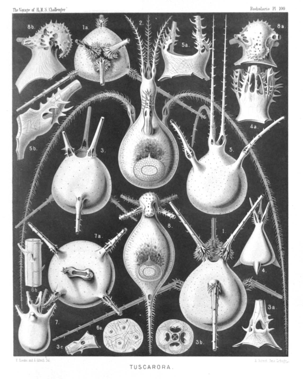
PLATE 100.
Tuscarorida.
Diam. Page.
Fig. 1. Tuscarora bisternaria, John Murray, × 30 1706
View from the dorsal side.
Fig. 1a. View from the mouth pole × 25
Fig. 2. Tuscarora murrayi, n. sp., × 30 1706
View from the dorsal side. The central capsule (in the aboral half), and the phæodium (in the middle of the shell-cavity) are visible. A fine network of pseudopodia pierces the calymma, which fills up the shell-cavity.
Fig. 3. Tuscarora wyvillei, n. sp., × 30 1707
View from the dorsal side.
Fig. 3a. Base of a tooth, × 100
Fig. 3b. Transverse section through the base of a tooth.
Fig. 3c. Base of a foot.
Fig. 4. Tuscarora tetrahedra, John Murray, × 15 1707
View from the dorsal side.
Fig. 4a. Mouth with the three teeth, × 50
Fig. 5. Tuscarora tubulosa, John Murray, × 40 1707
View from the ventral side.
Fig. 5a. Mouth with the two teeth, × 100
Fig. 5b. Basal part of a single tooth, × 150
Fig. 6. Tuscarora porcellana, John Murray, × 600 1708
Fig. 6a. A piece of the shell, with five pores.
Fig. 6b. A piece of a tooth, with the internal axial rod and its transverse branches.
Fig. 7. Tuscarusa medusa, n. sp., × 25 1709
View from the side.
Fig. 7a. View from the mouth, × 50
Fig. 8. Tuscaridium lithornithium, n. sp., × 20 1710
View from the ventral side. Central capsule and calymma as in fig. 2.
Fig. 8a. Peristome from the ventral side.
Fig. 8b. Peristome from the right side.
PLATE 101.
Legion PHÆODARIA.
Order PHÆOCYSTINA.
Families Phæodinida, Cannorrhaphida et Aulacanthida.
PLATE 101. Phæodinida, Cannorrhaphida et Aulacanthida. Diam. Page. Fig. 1. Phæocolla primordialis, n. sp., × 300 1544 Central capsule, isolated. The double contoured outer membrane exhibits only one opening, with a radiate operculum and long proboscis. The granular protoplasm encloses clear spherical vacuoles. The sphæroidal nucleus contains irregular amœboid nucleoli. Fig. 2. Phæodina tripylea, n. sp., × 300 1545 A central capsule in self-division, with two elliptical nuclei. The astropyle is already bisected and has two proboscides. Fig. 3. Cannorrhaphis spinulosa, n. sp., × 300 1552 A complete specimen with two central capsules, each of which contains two nuclei. The alveolate calymma contains a dark phæodium and is surrounded by tangential tubular needles. Fig. 4. Cannorrhaphis spinulosa, n. sp., × 300 1552 A single tangential tube. Fig. 5. Cannorrhaphis spathillata, n. sp., × 300 1552 A single tangential tube. Fig. 6. Aulactinium actinastrum, n. sp., × 100 1574 A complete specimen, seen in optical meridional section. In the centre the spheroidal central capsule, with its double membrane and three openings (above two lateral parapylæ, below the large astropyle with its radiate operculum). The capsule encloses numerous spherical vacuoles and two hemispherical nuclei, each with numerous nucleoli. The anterior half of the capsule is surrounded by the blackish phæodium. The spherical calymma contains numerous globular alveoles and is pierced by the radial tubes, the proximal ends of which are in contact with the surface of the central capsule (compare Pl. 103, fig. 1). Fig. 7. Aulactinium actinastrum, n. sp., × 300 1574 A single radial tube. Fig. 8. Aulactinium actinelium, n. sp., × 200 1574 A single radial tube. Fig. 9. Mesocena stellata, n. sp., × 600 1557 A single annular piece of the skeleton Fig. 10. Dictyocha stapedia, n. sp., × 300 1561 A complete specimen, observed living at Ceylon. In the centre is visible the large, spheroidal, tripylean central capsule, with its three openings, containing a large nucleus with numerous nucleoli. Its oral half is covered with the dark phæodium. The voluminous spherical calymma contains numerous globular alveoles and its surface is covered with scattered, stirrup-shaped pieces of the skeleton. Numerous free pseudopodia arise from the surface. Fig. 11. Dictyocha stapedia, n. sp., × 800 1561 A single piece of the skeleton, from above. Fig. 12. Dictyocha stapedia, n. sp., × 800 1561 A twin piece of the skeleton. Fig. 13. Dictyocha medusa, n. sp., × 800 1560 A single piece of the skeleton, from the side. Fig. 14. Dictyocha medusa, n. sp., × 800 1560 A single piece of the skeleton, from above.
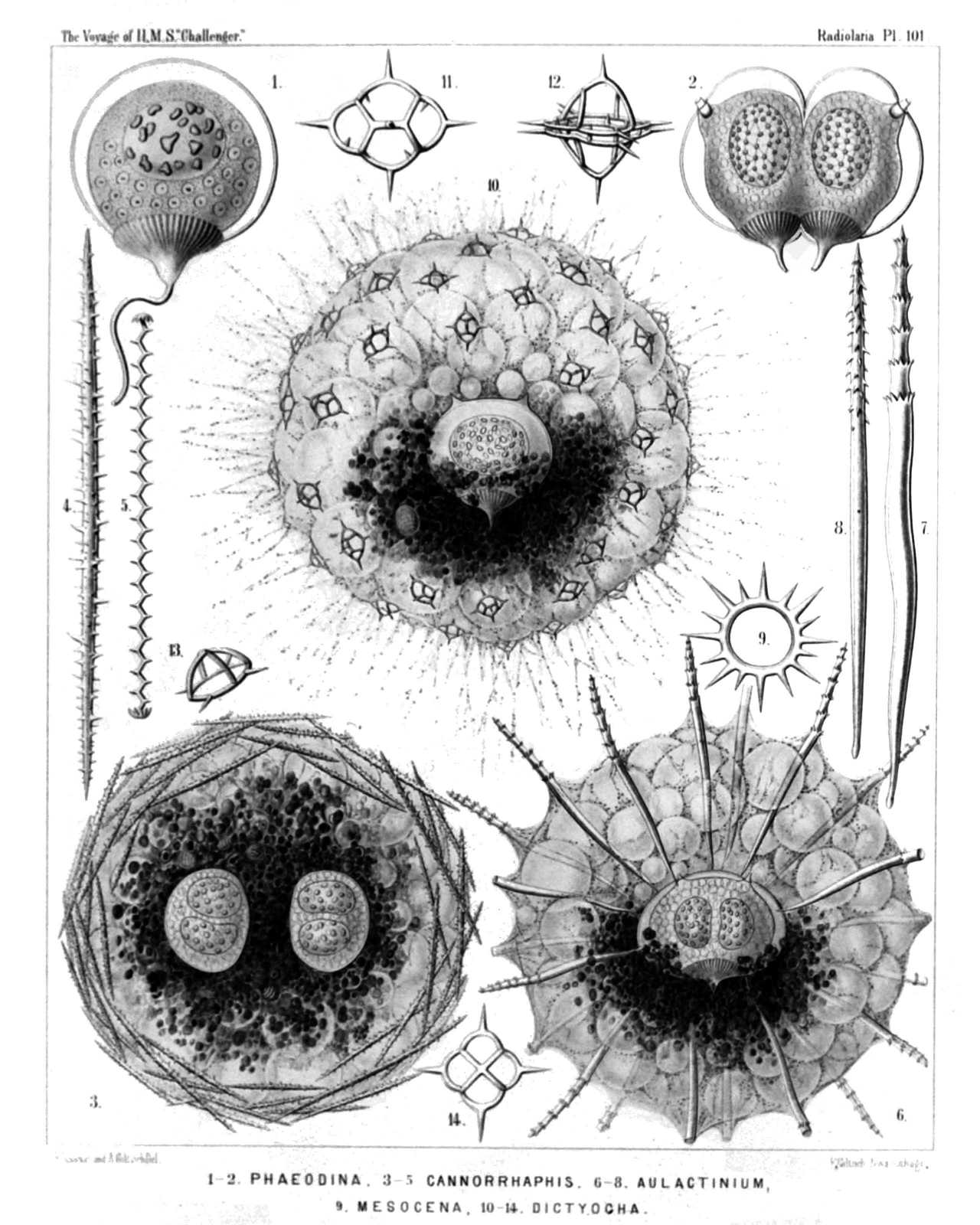
PLATE 101.
Phæodinida, Cannorrhaphida et Aulacanthida.
Diam. Page.
Fig. 1. Phæocolla primordialis, n. sp., × 300 1544
Central capsule, isolated. The double contoured outer membrane exhibits only one opening, with a radiate operculum and long proboscis. The granular protoplasm encloses clear spherical vacuoles. The sphæroidal nucleus contains irregular amœboid nucleoli.
Fig. 2. Phæodina tripylea, n. sp., × 300 1545
A central capsule in self-division, with two elliptical nuclei. The astropyle is already bisected and has two proboscides.
Fig. 3. Cannorrhaphis spinulosa, n. sp., × 300 1552
A complete specimen with two central capsules, each of which contains two nuclei. The alveolate calymma contains a dark phæodium and is surrounded by tangential tubular needles.
Fig. 4. Cannorrhaphis spinulosa, n. sp., × 300 1552
A single tangential tube.
Fig. 5. Cannorrhaphis spathillata, n. sp., × 300 1552
A single tangential tube.
Fig. 6. Aulactinium actinastrum, n. sp., × 100 1574
A complete specimen, seen in optical meridional section. In the centre the spheroidal central capsule, with its double membrane and three openings (above two lateral parapylæ, below the large astropyle with its radiate operculum). The capsule encloses numerous spherical vacuoles and two hemispherical nuclei, each with numerous nucleoli. The anterior half of the capsule is surrounded by the blackish phæodium. The spherical calymma contains numerous globular alveoles and is pierced by the radial tubes, the proximal ends of which are in contact with the surface of the central capsule (compare Pl. 103, fig. 1).
Fig. 7. Aulactinium actinastrum, n. sp., × 300 1574
A single radial tube.
Fig. 8. Aulactinium actinelium, n. sp., × 200 1574
A single radial tube.
Fig. 9. Mesocena stellata, n. sp., × 600 1557
A single annular piece of the skeleton
Fig. 10. Dictyocha stapedia, n. sp., × 300 1561
A complete specimen, observed living at Ceylon. In the centre is visible the large, spheroidal, tripylean central capsule, with its three openings, containing a large nucleus with numerous nucleoli. Its oral half is covered with the dark phæodium. The voluminous spherical calymma contains numerous globular alveoles and its surface is covered with scattered, stirrup-shaped pieces of the skeleton. Numerous free pseudopodia arise from the surface.
Fig. 11. Dictyocha stapedia, n. sp., × 800 1561
A single piece of the skeleton, from above.
Fig. 12. Dictyocha stapedia, n. sp., × 800 1561
A twin piece of the skeleton.
Fig. 13. Dictyocha medusa, n. sp., × 800 1560
A single piece of the skeleton, from the side.
Fig. 14. Dictyocha medusa, n. sp., × 800 1560
A single piece of the skeleton, from above.
PLATE 102.
Legion PHÆODARIA.
Order PHÆOCYSTINA.
Family Aulacanthida.
PLATE 102. Aulacanthida. Diam. Page. Fig. 1. Auloceros elegans, n. sp., × 80 1584 A complete specimen, observed living at Ceylon. In the centre is visible the red central capsule with its three openings, containing a large nucleus of half the size, with numerous nucleoli. The alveolate calymma encloses a green excentric phæodium, is surrounded by a veil of interwoven tangential needles, and forms conical elevations, which enclose the piercing radial tubes. Between these radiate numerous pseudopodia (compare for the single parts, Pl. 103, fig. 1 and Pl. 104, figs. 1-3, and their explanation). Figs. 2-6. Auloceros furcosus, n. sp., × 100 1583 Distal ends of different radial tubes, exhibiting the great variability of this species. Fig. 7. Auloceros trigeminus, n. sp., × 300 1584 Distal end of a single tube. Fig. 8. Auloceros capreolus, n. sp., × 200 1584 Distal End of a Single Tube. Figs. 9, 10. Auloceros cervinus, n. sp., × 300 1584 Distal ends of two single tubes. Fig. 12. Auloceros spathillaster, n. sp., × 300 1585 Distal end of a single tube. Figs. 11, 13. Auloceros arborescens, n. sp., × 300 1585 Distal ends of two single tubes.
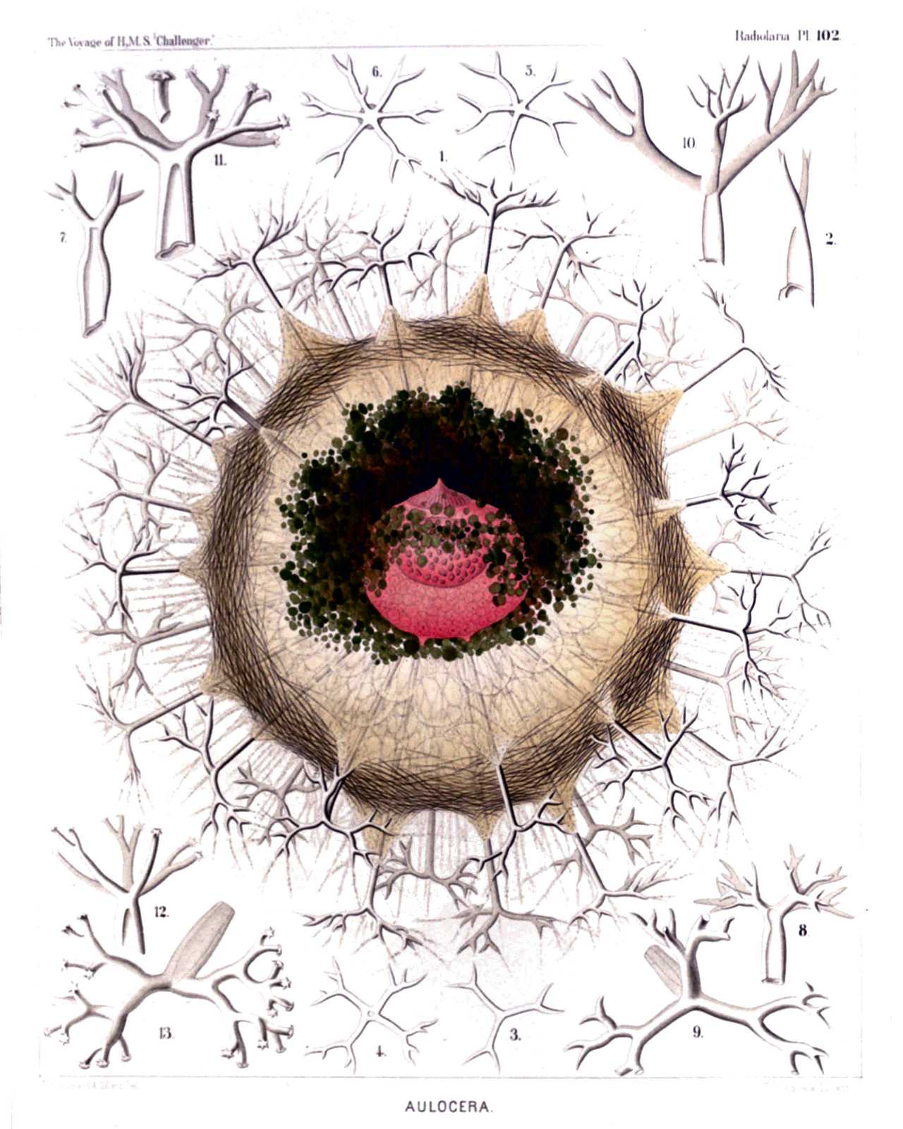
PLATE 102.
Aulacanthida.
Diam. Page.
Fig. 1. Auloceros elegans, n. sp., × 80 1584
A complete specimen, observed living at Ceylon. In the centre is visible the red central capsule with its three openings, containing a large nucleus of half the size, with numerous nucleoli. The alveolate calymma encloses a green excentric phæodium, is surrounded by a veil of interwoven tangential needles, and forms conical elevations, which enclose the piercing radial tubes. Between these radiate numerous pseudopodia (compare for the single parts, Pl. 103, fig. 1 and Pl. 104, figs. 1-3, and their explanation).
Figs. 2-6. Auloceros furcosus, n. sp., × 100 1583
Distal ends of different radial tubes, exhibiting the great variability of this species.
Fig. 7. Auloceros trigeminus, n. sp., × 300 1584
Distal end of a single tube.
Fig. 8. Auloceros capreolus, n. sp., × 200 1584
Distal End of a Single Tube.
Figs. 9, 10. Auloceros cervinus, n. sp., × 300 1584
Distal ends of two single tubes.
Fig. 12. Auloceros spathillaster, n. sp., × 300 1585
Distal end of a single tube.
Figs. 11, 13. Auloceros arborescens, n. sp., × 300 1585
Distal ends of two single tubes.
PLATE 103.
Legion PHÆODARIA.
Order PHÆOCYSTINA.
Family Aulacanthida.
PLATE 103. Aulacanthida. Diam. Page. Fig. 1. Aulographis candelabrum, n. sp., × 100 1583 p, The dark phæodium surrounding the central capsule on its oral part; a, a part of the surrounding alveolate calymma, also surrounding the central capsule; s, the veil of tangential needles covering the surface of the alveolate calymma; r, the big radial tubes, seven of which are visible, with an elegant verticil of terminal branches; f, the numerous pseudopodia radiating between the branches. The central capsule exhibits the following parts:—o, Astropyle; u, parapylæ; e, outer membrane; i, inner membrane; v, vacuoles; n, nucleus; l, nucleoli. Figs. 2-9. Aulographis pandor, n. sp., × 100 1577 Distal ends of various radial tubes of a single specimen, exhibiting the extraordinary variability of this species. Fig. 10. Aulographis furcula, n. sp., × 400 1580 A two-branched tube. Fig. 11. Aulographis furcula, n. sp., × 400 1580 A three-branched tube. Figs. 12, 13. Aulographis bovicornis, n. sp., × 200 1577 Two tubes with two branches. Fig. 14. Aulographis bovicornis, n. sp., × 200 1577 A tube with three branches. Fig. 15. Aulographis triangulum, n. sp., × 200 1580 A single tube. Fig. 16. Aulographis taumorpha, n. sp., × 300 1577 Two tubes, each with two branches. Fig. 17. Aulographis triglochin, n. sp., × 300 1578 A tube with three branches. Figs. 18, 19. Aulographis hexancistra, n. sp., × 300 1581 Distal end of two tubes (one with four, the other with five terminal branches). Fig. 20. Aulographis dentata, n. sp., × 200 1582 Distal end of a single tube. Fig. 21. Aulographis ancorata, n. sp., × 300 1578 Two tubes, each with four recurved branches. Fig. 22. Aulographis tetrancistra, n. sp., × 300 1581 A single tube. Fig. 23. Aulographis stellata, n. sp., × 300 1578 a and b, Two rudimentary or incompletely developed tubes; c, a well-developed tube of the usual form. Fig. 24. Aulographis asteriscus, n. sp., × 300 1581 Terminal verticil of a single tube. Fig. 25. Aulographis cruciata, n. sp., × 300 1578 Distal end of a single tube. Fig. 26. Aulographis pulvinata, n. sp., × 400 1582 Distal end of a single tube. Fig. 27. Aulographis serrulata, n. sp., × 400 1582 Distal end of a single tube.
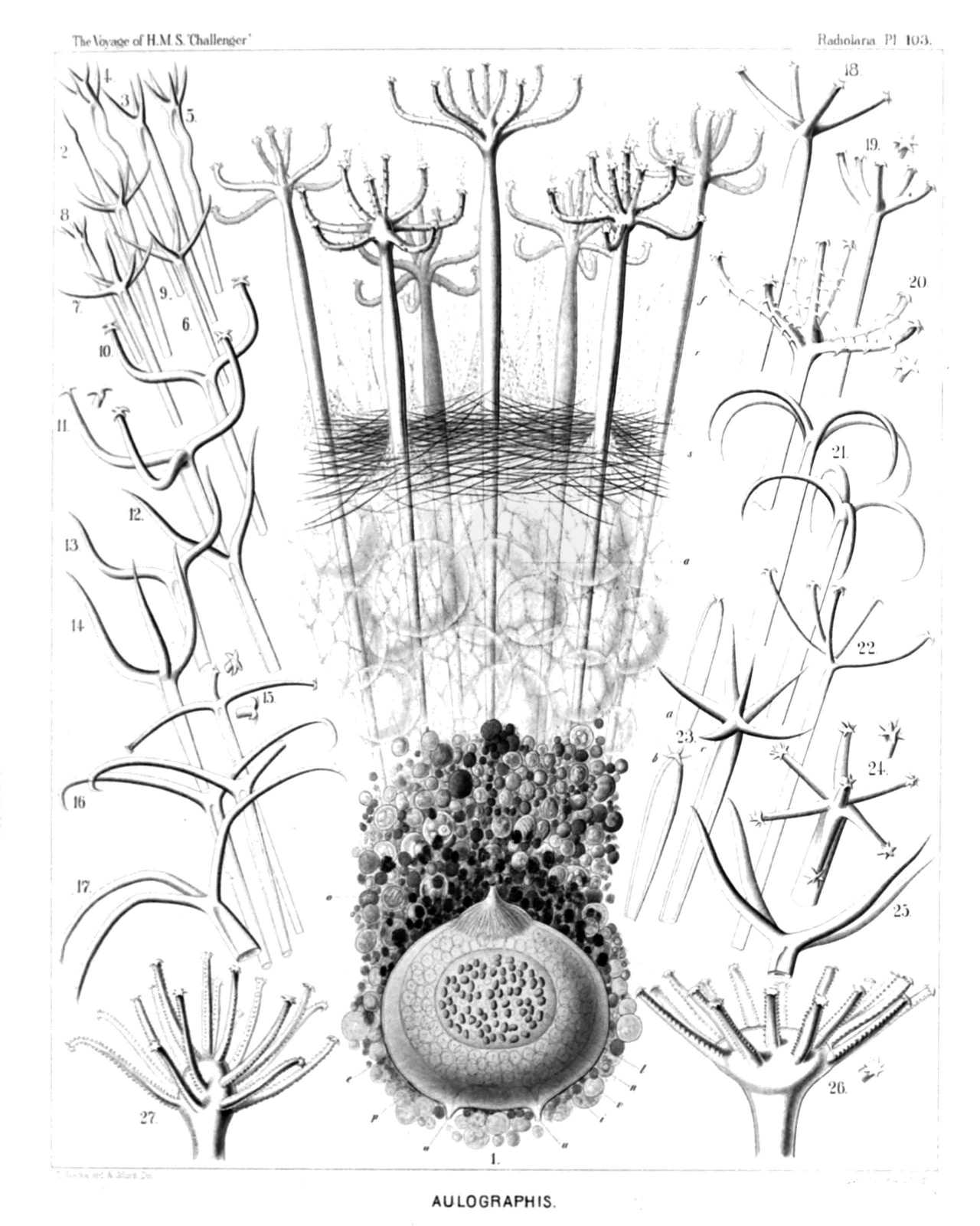
PLATE 103.
Aulacanthida.
Diam. Page.
Fig. 1. Aulographis candelabrum, n. sp., × 100 1583
p, The dark phæodium surrounding the central capsule on its oral part; a, a part of the surrounding alveolate calymma, also surrounding the central capsule; s, the veil of tangential needles covering the surface of the alveolate calymma; r, the big radial tubes, seven of which are visible, with an elegant verticil of terminal branches; f, the numerous pseudopodia radiating between the branches. The central capsule exhibits the following parts:—o, Astropyle; u, parapylæ; e, outer membrane; i, inner membrane; v, vacuoles; n, nucleus; l, nucleoli.
Figs. 2-9. Aulographis pandor, n. sp., × 100 1577
Distal ends of various radial tubes of a single specimen, exhibiting the extraordinary variability of this species.
Fig. 10. Aulographis furcula, n. sp., × 400 1580
A two-branched tube.
Fig. 11. Aulographis furcula, n. sp., × 400 1580
A three-branched tube.
Figs. 12, 13. Aulographis bovicornis, n. sp., × 200 1577
Two tubes with two branches.
Fig. 14. Aulographis bovicornis, n. sp., × 200 1577
A tube with three branches.
Fig. 15. Aulographis triangulum, n. sp., × 200 1580
A single tube.
Fig. 16. Aulographis taumorpha, n. sp., × 300 1577
Two tubes, each with two branches.
Fig. 17. Aulographis triglochin, n. sp., × 300 1578
A tube with three branches.
Figs. 18, 19. Aulographis hexancistra, n. sp., × 300 1581
Distal end of two tubes (one with four, the other with five terminal branches).
Fig. 20. Aulographis dentata, n. sp., × 200 1582
Distal end of a single tube.
Fig. 21. Aulographis ancorata, n. sp., × 300 1578
Two tubes, each with four recurved branches.
Fig. 22. Aulographis tetrancistra, n. sp., × 300 1581
A single tube.
Fig. 23. Aulographis stellata, n. sp., × 300 1578
a and b, Two rudimentary or incompletely developed tubes; c, a well-developed tube of the usual form.
Fig. 24. Aulographis asteriscus, n. sp., × 300 1581
Terminal verticil of a single tube.
Fig. 25. Aulographis cruciata, n. sp., × 300 1578
Distal end of a single tube.
Fig. 26. Aulographis pulvinata, n. sp., × 400 1582
Distal end of a single tube.
Fig. 27. Aulographis serrulata, n. sp., × 400 1582
Distal end of a single tube.
PLATE 104.
Legion PHÆODARIA.
Order PHÆOCYSTINA.
Family Aulacanthida.
PLATE 104. Aulacanthida. Diam. Page. Aulacanthida. Fig. 1. Aulospathis bifurca, n. sp., × 50 1586 A complete specimen, excellently preserved, with an ovate alveolate calymma and two central capsules. The surface of the calymma is covered with tangential needles. Fig. 2. Aulospathis bifurca, n. sp., × 100 1586 An isolated central capsule of another specimen, surrounded by granules of the phæodium. o, Radiate operculum of the astropyle; u, the two lateral parapylæ; e, external membrane of the capsule; i, internal membrane; c, vacuoles in the protoplasm; n, nucleus; l, numerous nucleoli. Fig. 3. Aulospathis bifurca, n. sp., × 80 1586 Two central capsules of another specimen, surrounded by the phæodium (Self-division). Characters as in fig. 2. Fig. 4. Aulospathis bifurca, n. sp., × 100 1586 A single radial tube. Fig. 5. Aulospathis bifurca, n. sp., × 200 1586 Distal part of another radial tube, partly filled up by air-bubbles. Fig. 6. Aulospathis trifurca, n. sp., × 200 1586 Distal part of a single radial tube. Fig. 7. Aulospathis trifurca, n. sp., × 200 1586 Distal part of another radial tube. Fig. 8. Aulospathis triodon, n. sp., × 100 1587 A single radial tube. Fig. 9. Aulospathis tetrodon, n. sp., × 200 1588 Distal end of single tube. Figs. 10-13. Aulospathis polymorpha, n. sp., × 400 1587 Four single terminal branches with very different forms of spathillæ. Figs. 14-17. Aulospathis variabilis, n. sp., × 400 1588 Four single terminal branches with very different forms of spathillæ.
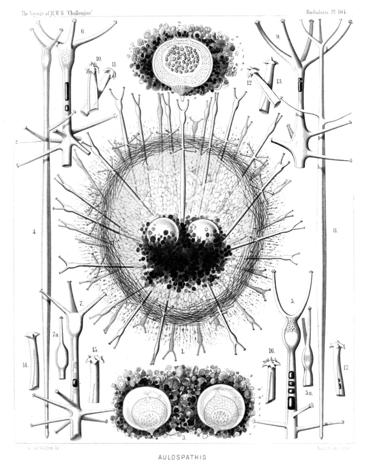
PLATE 104.
Aulacanthida.
Diam. Page.
Aulacanthida.
Fig. 1. Aulospathis bifurca, n. sp., × 50 1586
A complete specimen, excellently preserved, with an ovate alveolate calymma and two central capsules. The surface of the calymma is covered with tangential needles.
Fig. 2. Aulospathis bifurca, n. sp., × 100 1586
An isolated central capsule of another specimen, surrounded by granules of the phæodium. o, Radiate operculum of the astropyle; u, the two lateral parapylæ; e, external membrane of the capsule; i, internal membrane; c, vacuoles in the protoplasm; n, nucleus; l, numerous nucleoli.
Fig. 3. Aulospathis bifurca, n. sp., × 80 1586
Two central capsules of another specimen, surrounded by the phæodium (Self-division). Characters as in fig. 2.
Fig. 4. Aulospathis bifurca, n. sp., × 100 1586
A single radial tube.
Fig. 5. Aulospathis bifurca, n. sp., × 200 1586
Distal part of another radial tube, partly filled up by air-bubbles.
Fig. 6. Aulospathis trifurca, n. sp., × 200 1586
Distal part of a single radial tube.
Fig. 7. Aulospathis trifurca, n. sp., × 200 1586
Distal part of another radial tube.
Fig. 8. Aulospathis triodon, n. sp., × 100 1587
A single radial tube.
Fig. 9. Aulospathis tetrodon, n. sp., × 200 1588
Distal end of single tube.
Figs. 10-13. Aulospathis polymorpha, n. sp., × 400 1587
Four single terminal branches with very different forms of spathillæ.
Figs. 14-17. Aulospathis variabilis, n. sp., × 400 1588
Four single terminal branches with very different forms of spathillæ.
PLATE 105.
Legion PHÆODARIA.
Order PHÆOCYSTINA.
Family Aulacanthida.
PLATE 105. Aulacanthida. Diam. Page. AULACANTHIDA. Fig. 1. Aulodendron indicum, n. sp., × 200 1590 A single tube. Fig. 2. Aulodendron pacificum, n. sp., × 400 1589 Distal half of a tube. Fig. 3. Aulodendron australe, n. sp., × 300 1589 A single tube. Fig. 4. Aulacantha spinosa, n. sp., × 300 1575 Distal half of a tube. Fig. 5. Aulodendron antarcticum, n. sp., × 300 1589 A single tube. Fig. 6. Aulographis pistillum, n. sp., × 300 1579 A single tube. Fig. 7. Aulographis martagon, n. sp., × 300 1579 Distal end of a single tube. Fig. 8. Aulographis triæna, n. sp., × 80 1579 A single tube. Fig. 9. Aulographis flammabunda, n. sp., × 100 1579 Distal end of a tube. Fig. 10. Aulographis flosculus, n. sp., × 300 1580 Distal end of a tube. Fig. 11. Aulographis gemmascens, n. sp., × 100 1580 Distal end of a tube. Fig. 12. Aulographis verticillata, n. sp., × 400 1582 Distal end of a tube. Fig. 12a. Apical view, with four verticils of five branches. Fig. 13. Aulographis tripentas, n. sp., × 300 1582 Distal end of a tube. Fig. 13a. Apical view, with three verticils of five branches. Fig. 14. Auloceros dicranaster, n. sp., × 400 1585 Distal end of a tube, seen from the side. Fig. 15. Auloceros dicranaster, n. sp., × 200 1585 Distal end of a tube, seen from the terminal face. Fig. 16. Aulacantha cannulata, n. sp., × 300 1576 Distal end of a tube.
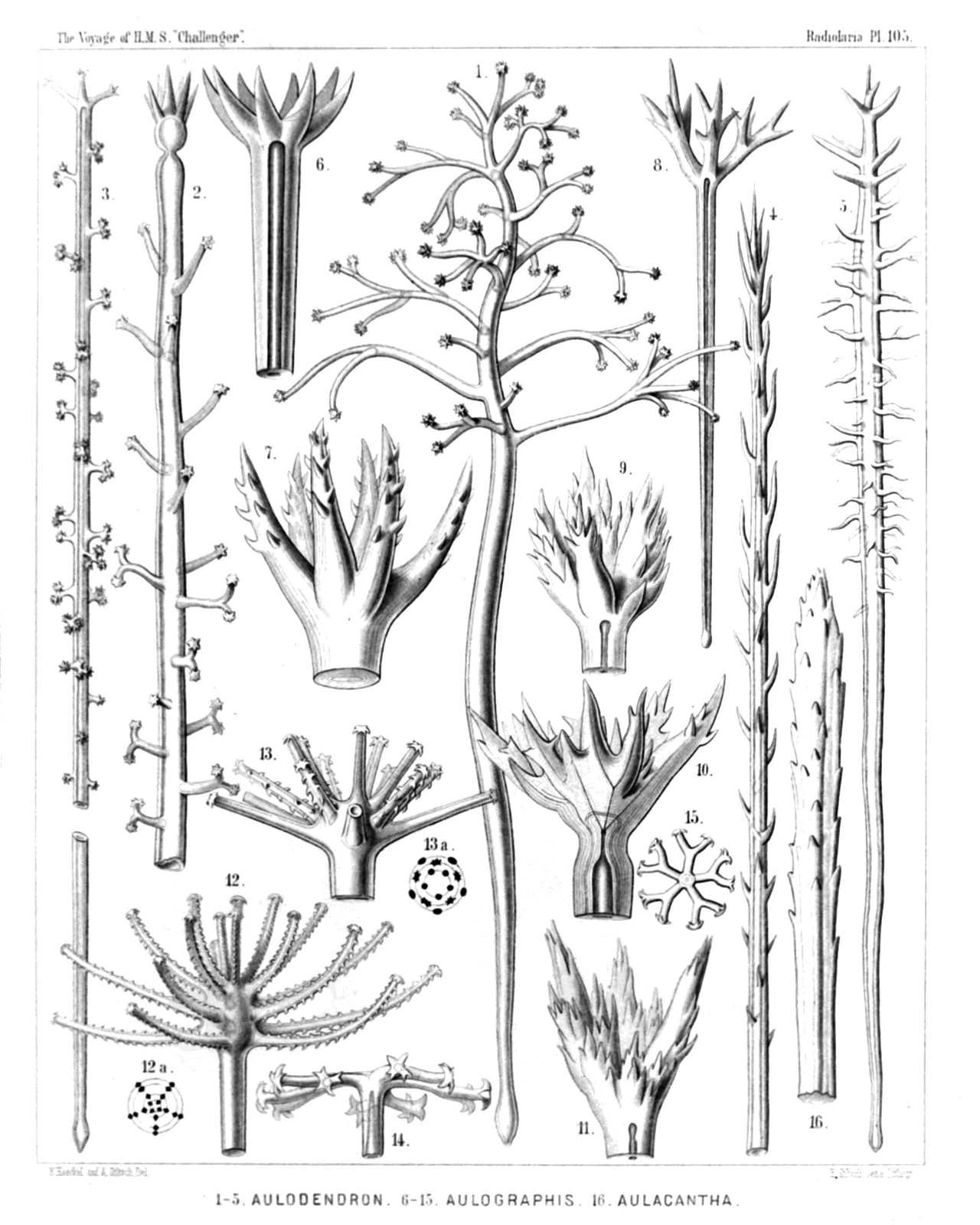
PLATE 105.
Aulacanthida.
Diam. Page.
AULACANTHIDA.
Fig. 1. Aulodendron indicum, n. sp., × 200 1590
A single tube.
Fig. 2. Aulodendron pacificum, n. sp., × 400 1589
Distal half of a tube.
Fig. 3. Aulodendron australe, n. sp., × 300 1589
A single tube.
Fig. 4. Aulacantha spinosa, n. sp., × 300 1575
Distal half of a tube.
Fig. 5. Aulodendron antarcticum, n. sp., × 300 1589
A single tube.
Fig. 6. Aulographis pistillum, n. sp., × 300 1579
A single tube.
Fig. 7. Aulographis martagon, n. sp., × 300 1579
Distal end of a single tube.
Fig. 8. Aulographis triæna, n. sp., × 80 1579
A single tube.
Fig. 9. Aulographis flammabunda, n. sp., × 100 1579
Distal end of a tube.
Fig. 10. Aulographis flosculus, n. sp., × 300 1580
Distal end of a tube.
Fig. 11. Aulographis gemmascens, n. sp., × 100 1580
Distal end of a tube.
Fig. 12. Aulographis verticillata, n. sp., × 400 1582
Distal end of a tube.
Fig. 12a. Apical view, with four verticils of five branches.
Fig. 13. Aulographis tripentas, n. sp., × 300 1582
Distal end of a tube.
Fig. 13a. Apical view, with three verticils of five branches.
Fig. 14. Auloceros dicranaster, n. sp., × 400 1585
Distal end of a tube, seen from the side.
Fig. 15. Auloceros dicranaster, n. sp., × 200 1585
Distal end of a tube, seen from the terminal face.
Fig. 16. Aulacantha cannulata, n. sp., × 300 1576
Distal end of a tube.
PLATE 106.
Legion PHÆODARIA.
Orders PHÆOSPHÆRIA.
Family Orosphærida.
PLATE 106. Orosphærida. Diam. Page. Orosphærida. Fig. 1. Orosphæra serpentina, n. sp., × 50 1595 The entire shell. Fig. 2. Orosphæra horrida, n. sp., × 50 1596 The entire shell. Fig. 3. Orosphæra arborescens, n. sp. (vel Orothamnus arborescens), × 50 1597 The entire shell. Fig. 4. Oroscena gegenbauri, n. sp., × 50 1597 The entire shell. (Compare Pl. 12, fig. 1.)
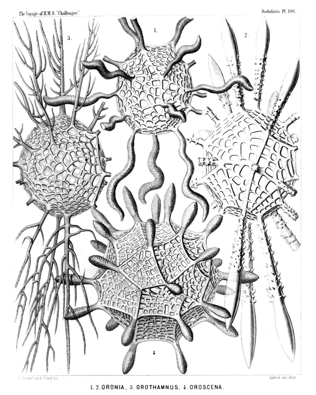
PLATE 106.
Orosphærida.
Diam. Page.
Orosphærida.
Fig. 1. Orosphæra serpentina, n. sp., × 50 1595
The entire shell.
Fig. 2. Orosphæra horrida, n. sp., × 50 1596
The entire shell.
Fig. 3. Orosphæra arborescens, n. sp. (vel Orothamnus arborescens), × 50 1597
The entire shell.
Fig. 4. Oroscena gegenbauri, n. sp., × 50 1597
The entire shell.
(Compare Pl. 12, fig. 1.)
PLATE 107.
Legion PHÆODARIA.
Order PHÆOSPHÆRIA.
Family Orosphærida.
PLATE 107. Orosphærida. Diam. Page. (Fig. 8 of this Plate has no number, by mistake; it is at the top in the middle.) Fig. 1. Oroplegma diplosphæra, n. sp., × 50 1600 The entire shell, enveloped by an outer mantle of spongy framework. Fig. 2. Oroplegma giganteum, n. sp., × 200 1601 A small piece of the spongy framework. Fig. 3. Oroplegma spongiosum, n. sp., × 50 1601 A pyramidal elevation of the inner shell, with its spongy framework, and a radial spine on the top. Fig. 4. Oroscena bærii, n. sp., × 100 1598 A pyramidal elevation of the shell, with a radial spine on its top. Fig. 5. Orona maxima, n. sp., × 300 1594 A small piece of the network; the central canals of the bars are partly filled by air. Fig. 6. Oroscena cuvieri, n. sp., × 50 1598 A single radial spine. Fig. 7. Orona crassissima, n. sp., × 300 1594 A single bar of the coarse network, with dimpled surface. Fig. 8. Oroscena mülleri, n. sp., × 50 1598 A single radial spine.
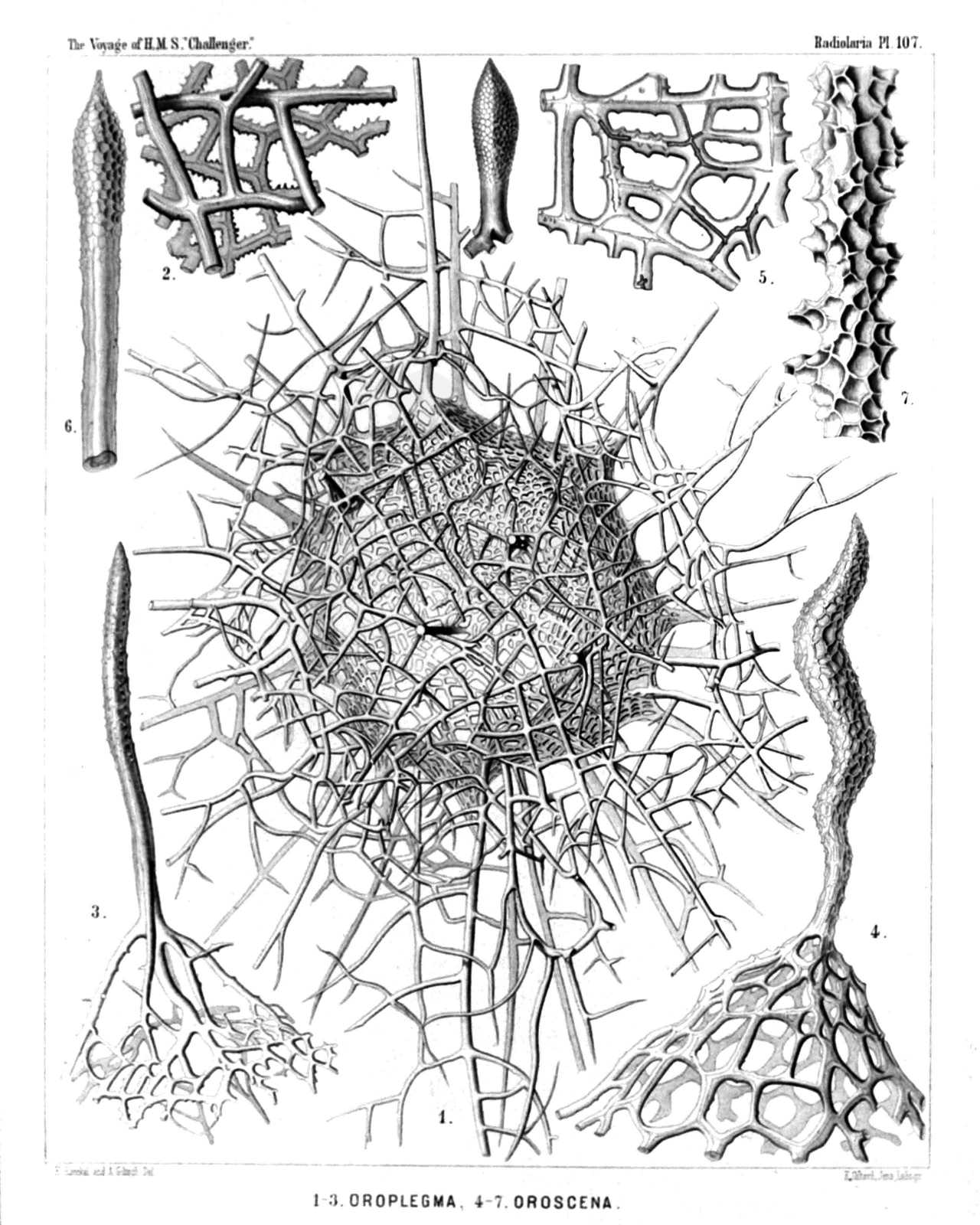
PLATE 107.
Orosphærida.
Diam. Page.
(Fig. 8 of this Plate has no number, by mistake; it is at the top in the middle.)
Fig. 1. Oroplegma diplosphæra, n. sp., × 50 1600
The entire shell, enveloped by an outer mantle of spongy framework.
Fig. 2. Oroplegma giganteum, n. sp., × 200 1601
A small piece of the spongy framework.
Fig. 3. Oroplegma spongiosum, n. sp., × 50 1601
A pyramidal elevation of the inner shell, with its spongy framework, and a radial spine on the top.
Fig. 4. Oroscena bærii, n. sp., × 100 1598
A pyramidal elevation of the shell, with a radial spine on its top.
Fig. 5. Orona maxima, n. sp., × 300 1594
A small piece of the network; the central canals of the bars are partly filled by air.
Fig. 6. Oroscena cuvieri, n. sp., × 50 1598
A single radial spine.
Fig. 7. Orona crassissima, n. sp., × 300 1594
A single bar of the coarse network, with dimpled surface.
Fig. 8. Oroscena mülleri, n. sp., × 50 1598
A single radial spine.
PLATE 108.
Legion PHÆODARIA.
Order PHÆOSPHÆRIA.
Family Sagosphærida.
PLATE 108. Sagosphærida. Diam. Page. Sagosphærida. Fig. 1. Sagoscena castra, n. sp., × 50 1608 Half the shell, with the enclosed central capsule and the phæodium, stained by carmine. (The central nucleus dark.) Fig. 2. Sagmarium spongodictyum, n. sp., × 50 1612 Half the shell, with its delicate spongy framework. Fig. 3. Sagenoscena stellata, n. sp., × 300 1610 Top and axial rod of a pyramid, prolonged into a crowned radial spine. Fig. 4. Sagenoscena ornata, n. sp., × 300 1610 A single pyramid with its axial rod, prolonged into a crowned radial spine. Fig. 5. Sagoscena pellorium, n. sp., × 300 1609 A single pyramid of the shell-surface. Fig. 6. Sagoscena tentorium, n. sp., × 100 1608 A piece of the shell with eight pyramids. Fig. 7. Sagoscena prætorium, n. sp., × 400 1609 Top of a pyramid. Fig. 8. Sagena ternaria, n. sp., × 400 1606 A single triangular mesh of the lattice sphere. Fig. 9. Sagmidium crucicorne, n. sp., × 400 1613 A single nodal point with three radial spines. Fig. 9a. A portion of a spine, more highly magnified. Fig. 10. Sagosphæra penicilla, n. sp., × 400 1607 One nodal point and its radial spine. Fig. 11. Sagosphæra furcilla, n. sp., × 300 1607 Two nodal points of the network. Fig. 11a. Extremity of a spine. Fig. 12. Sagmidium quadricorne, n. sp., × 400 1614 A nodal point of the shell surface, with four divergent spines. Fig. 13. Sagoplegma scenophora, n. sp., × 300 1615 Tops of two pyramids. Fig. 14. Sagmarium plegmosphærium, n. sp., × 300 1612 A nodal point of the spongy framework.
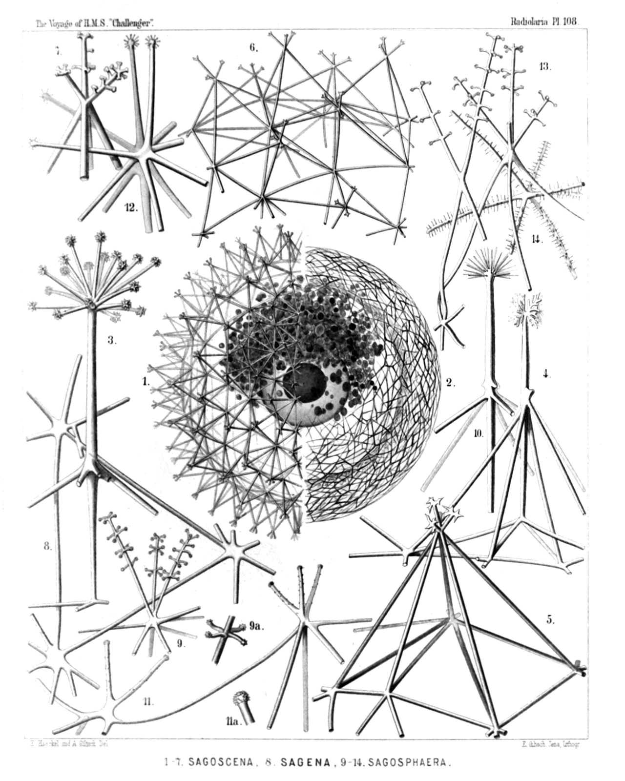
PLATE 108.
Sagosphærida.
Diam. Page.
Sagosphærida.
Fig. 1. Sagoscena castra, n. sp., × 50 1608
Half the shell, with the enclosed central capsule and the phæodium, stained by carmine. (The central nucleus dark.)
Fig. 2. Sagmarium spongodictyum, n. sp., × 50 1612
Half the shell, with its delicate spongy framework.
Fig. 3. Sagenoscena stellata, n. sp., × 300 1610
Top and axial rod of a pyramid, prolonged into a crowned radial spine.
Fig. 4. Sagenoscena ornata, n. sp., × 300 1610
A single pyramid with its axial rod, prolonged into a crowned radial spine.
Fig. 5. Sagoscena pellorium, n. sp., × 300 1609
A single pyramid of the shell-surface.
Fig. 6. Sagoscena tentorium, n. sp., × 100 1608
A piece of the shell with eight pyramids.
Fig. 7. Sagoscena prætorium, n. sp., × 400 1609
Top of a pyramid.
Fig. 8. Sagena ternaria, n. sp., × 400 1606
A single triangular mesh of the lattice sphere.
Fig. 9. Sagmidium crucicorne, n. sp., × 400 1613
A single nodal point with three radial spines.
Fig. 9a. A portion of a spine, more highly magnified.
Fig. 10. Sagosphæra penicilla, n. sp., × 400 1607
One nodal point and its radial spine.
Fig. 11. Sagosphæra furcilla, n. sp., × 300 1607
Two nodal points of the network.
Fig. 11a. Extremity of a spine.
Fig. 12. Sagmidium quadricorne, n. sp., × 400 1614
A nodal point of the shell surface, with four divergent spines.
Fig. 13. Sagoplegma scenophora, n. sp., × 300 1615
Tops of two pyramids.
Fig. 14. Sagmarium plegmosphærium, n. sp., × 300 1612
A nodal point of the spongy framework.
PLATE 109.
Legion PHÆODARIA.
Order PHÆOSPHÆRIA.
Family Aulosphærida.
PLATE 109. Aulosphærida. Diam. Page. Fig. 1. Aulosphæra dendrophora, n. sp., × 50 1625 The entire shell, with the central capsule and its nucleus, enveloped by the dark granular phæodium. Fig. 2. Aulosphæra dendrophora, n. sp., × 300 1625 A single radial tube. Fig. 3. Aulosphæra sceptrophora, n. sp., × 300 1625 A hexagonal group of six triangular meshes. Fig. 4. Aulosphæra sceptrophora, n. sp., × 300 1625 A similar group, seen from the side, with three radial tubes. Fig. 5. Aulosphæra spinosa, n. sp., × 300 1627 A hexagonal group of six triangular meshes. Fig. 6. Aulosphæra undulata, n. sp., × 400 1627 A single radial tube. Fig. 7. Aulosphæra spathillata, n. sp., × 400 1624 A single radial tube. Fig. 7a. An abnormal variety, × 400 Fig. 8. Aulosphæra triodon, n. sp., × 400 1623 A single radial tube. Fig. 9. Aulosphæra trifurca, n. sp., × 400 1626 A single radial tube. Fig. 10. Aulosphæra cruciata, n. sp., × 300 1624 A single radial tube. Fig. 11. Aulosphæra bisternaria, n. sp., × 300 1624 A single radial tube. Fig. 12. Aulosphæra bisternaria, n. sp., × 600 1624 Distal end of a single radial tube.
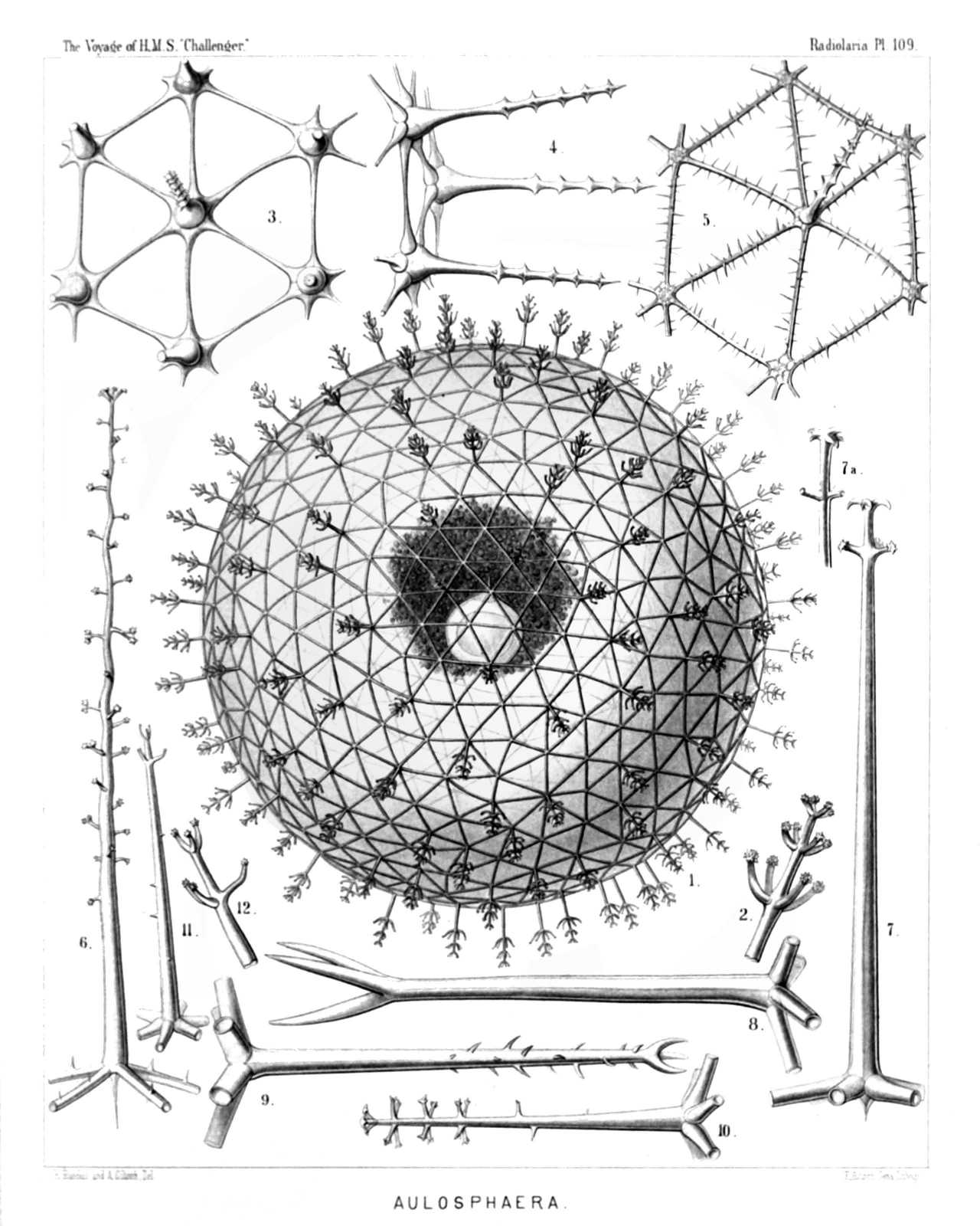
PLATE 109.
Aulosphærida.
Diam. Page.
Fig. 1. Aulosphæra dendrophora, n. sp., × 50 1625
The entire shell, with the central capsule and its nucleus, enveloped by the dark granular phæodium.
Fig. 2. Aulosphæra dendrophora, n. sp., × 300 1625
A single radial tube.
Fig. 3. Aulosphæra sceptrophora, n. sp., × 300 1625
A hexagonal group of six triangular meshes.
Fig. 4. Aulosphæra sceptrophora, n. sp., × 300 1625
A similar group, seen from the side, with three radial tubes.
Fig. 5. Aulosphæra spinosa, n. sp., × 300 1627
A hexagonal group of six triangular meshes.
Fig. 6. Aulosphæra undulata, n. sp., × 400 1627
A single radial tube.
Fig. 7. Aulosphæra spathillata, n. sp., × 400 1624
A single radial tube.
Fig. 7a. An abnormal variety, × 400
Fig. 8. Aulosphæra triodon, n. sp., × 400 1623
A single radial tube.
Fig. 9. Aulosphæra trifurca, n. sp., × 400 1626
A single radial tube.
Fig. 10. Aulosphæra cruciata, n. sp., × 300 1624
A single radial tube.
Fig. 11. Aulosphæra bisternaria, n. sp., × 300 1624
A single radial tube.
Fig. 12. Aulosphæra bisternaria, n. sp., × 600 1624
Distal end of a single radial tube.
PLATE 110.
Legion PHÆODARIA.
Order PHÆOSPHÆRIA.
Family Aulosphærida.
PLATE 110. Aulosphærida. Diam. Page. Fig. 1. Auloscena mirabilis, n. sp., × 50 1628 The complete shell, representing a regular latticed sphere, which is composed of equal hexagonal pyramids; the top of each pyramid bears a radial tube with a terminal corona. Fig. 2. Auloscena mirabilis, n. sp., × 600 1628 Terminal corona of a single radial tube. Fig. 3. Auloscena penicillus, n. sp., × 200 1629 A single tent-shaped elevation or six-sided pyramid, bearing on the top a brush-shaped radial tube. Fig. 4. Auloscena flammabunda, n. sp., × 400 1629 A single radial tube, with a centripetal free prolongation at the base and a verticil of undulate terminal branches at the distal end. Fig. 5. Auloscena serrata, n. sp., × 600 1630 Terminal corona of a single radial tube. Fig. 6. Auloscena tentorium, n. sp., × 400 1628 A single radial tube, with a centripetal prolongation at the base and a terminal corona at the distal end. Fig. 7. Auloscena gigantea, n. sp., × 400 1629 Basal part of a radial tube, exhibiting the internal axial thread and its connection with the six tubes, which form the edges of a flat six-sided pyramid (usually more elevated than the figure exhibits). Fig. 8. Auloscena spectabilis, n. sp., × 400 1628 Apex of an abnormal pyramid (sometimes occurring), in which seven radial tubes are united, instead of six. Fig. 9. Auloscena spectabilis, n. sp., × 800 1628 Basal part of a radial tube, in the top of a flat six-sided pyramid; above it the distal part of the same tube with its terminal corona (middle part of the tube wanting). Fig. 10. Auloscena verticillus, n. sp., × 300 1629 Apex of a six-sided pyramid, seen from the inside. Fig. 11. Auloscena verticillus, n. sp., × 400 1629 Distal part of a single radial tube, with the terminal corona.
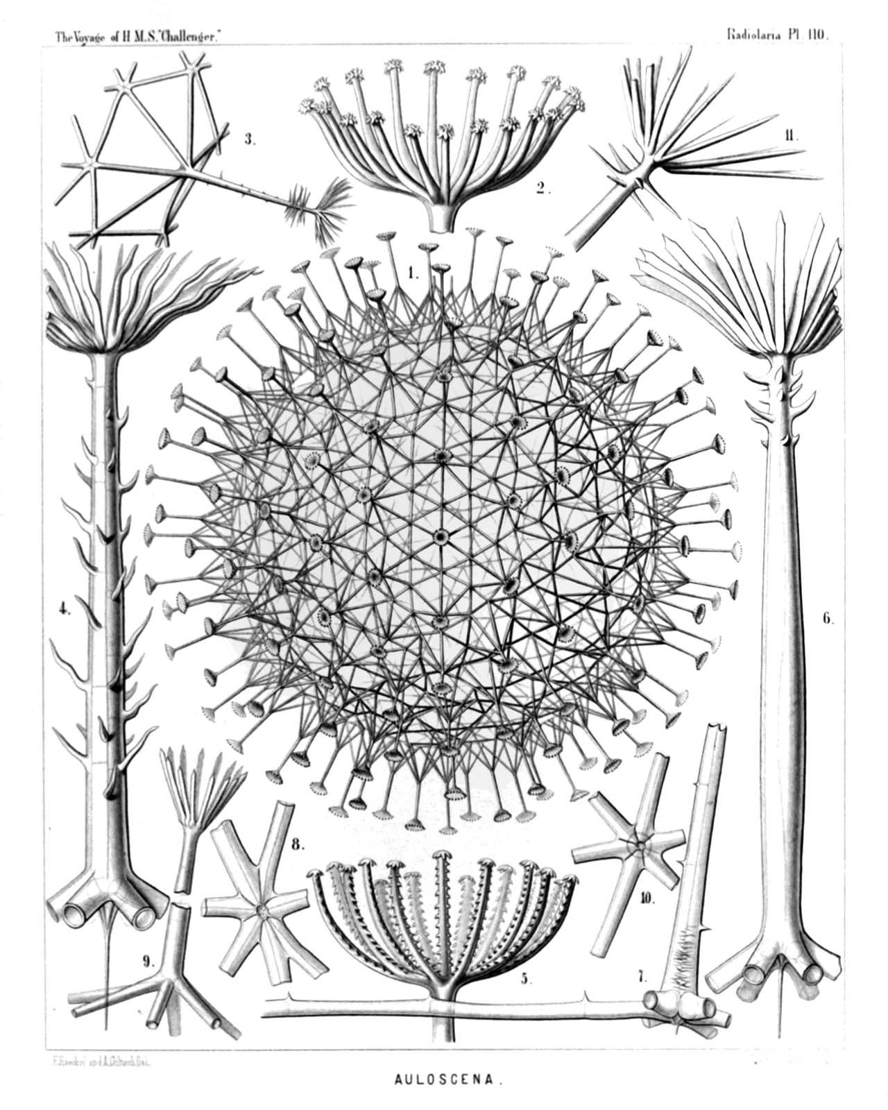
PLATE 110.
Aulosphærida.
Diam. Page.
Fig. 1. Auloscena mirabilis, n. sp., × 50 1628
The complete shell, representing a regular latticed sphere, which is composed of equal hexagonal pyramids; the top of each pyramid bears a radial tube with a terminal corona.
Fig. 2. Auloscena mirabilis, n. sp., × 600 1628
Terminal corona of a single radial tube.
Fig. 3. Auloscena penicillus, n. sp., × 200 1629
A single tent-shaped elevation or six-sided pyramid, bearing on the top a brush-shaped radial tube.
Fig. 4. Auloscena flammabunda, n. sp., × 400 1629
A single radial tube, with a centripetal free prolongation at the base and a verticil of undulate terminal branches at the distal end.
Fig. 5. Auloscena serrata, n. sp., × 600 1630
Terminal corona of a single radial tube.
Fig. 6. Auloscena tentorium, n. sp., × 400 1628
A single radial tube, with a centripetal prolongation at the base and a terminal corona at the distal end.
Fig. 7. Auloscena gigantea, n. sp., × 400 1629
Basal part of a radial tube, exhibiting the internal axial thread and its connection with the six tubes, which form the edges of a flat six-sided pyramid (usually more elevated than the figure exhibits).
Fig. 8. Auloscena spectabilis, n. sp., × 400 1628
Apex of an abnormal pyramid (sometimes occurring), in which seven radial tubes are united, instead of six.
Fig. 9. Auloscena spectabilis, n. sp., × 800 1628
Basal part of a radial tube, in the top of a flat six-sided pyramid; above it the distal part of the same tube with its terminal corona (middle part of the tube wanting).
Fig. 10. Auloscena verticillus, n. sp., × 300 1629
Apex of a six-sided pyramid, seen from the inside.
Fig. 11. Auloscena verticillus, n. sp., × 400 1629
Distal part of a single radial tube, with the terminal corona.
PLATE 111.
Legion PHÆODARIA.
Order PHÆOSPHÆRIA.
Family Aulosphærida.
PLATE 111. Aulosphærida. Diam. Page. Fig. 1. Aulonia hexagonia, n. sp., × 30 1634 The complete spherical shell. Fig. 2. Aularia ternaria, n. sp., × 300 1621 A group of six triangular meshes, with seven nodal points of radial tubes. Behind the central capsule, with its double membrane (e, outer; i, inner) and radiate operculum (o); u, the two outer parapylæ; v, vacuoles in the protoplasm. The ellipsoidal nucleus (n) contains numerous nucleoli (l). Fig. 3. Aulastrum triceros, n. sp., × 50 1635 The complete shell. Fig. 3a. Aulastrum triceros, n. sp., × 300 1635 A single radial tube. Figs. 4a, 4b, 4c. Aulastrum dendroceros, n. sp., × 400 1635 Three single radial spines (taken from three different specimens). Fig. 5a. Aulophacus lenticularis, n. sp., × 300 1631 A single radial spine. Fig. 5b. Aulophacus amphidiscus, n. sp., × 300 1631 A single radial spine. Fig. 6. Aulatractus fusiformis, n. sp., × 5 1632 The complete shell, five times enlarged. Fig. 6a. Aulatractus fusiformis, n. sp., × 20 1632 Apical part of the shell. Fig. 6b. Aulatractus fusiformis, n. sp., × 400 1632 A single radial tube. Fig. 7. Aulatractus diploconus, n. sp., × 20 1632 Apical part of the shell. Fig. 7a. Aulatractus diploconus, n. sp., × 400 1632 A single radial tube. Fig. 8. Auloplegma perplexum, n. sp., × 50 1630 Half the shell. Fig. 8a. Auloplegma perplexum, n. sp., × 400 1630 A single radial tube. Fig. 9. Auloplegma spongiosum, n. sp., × 300 1631 A single radial tube.
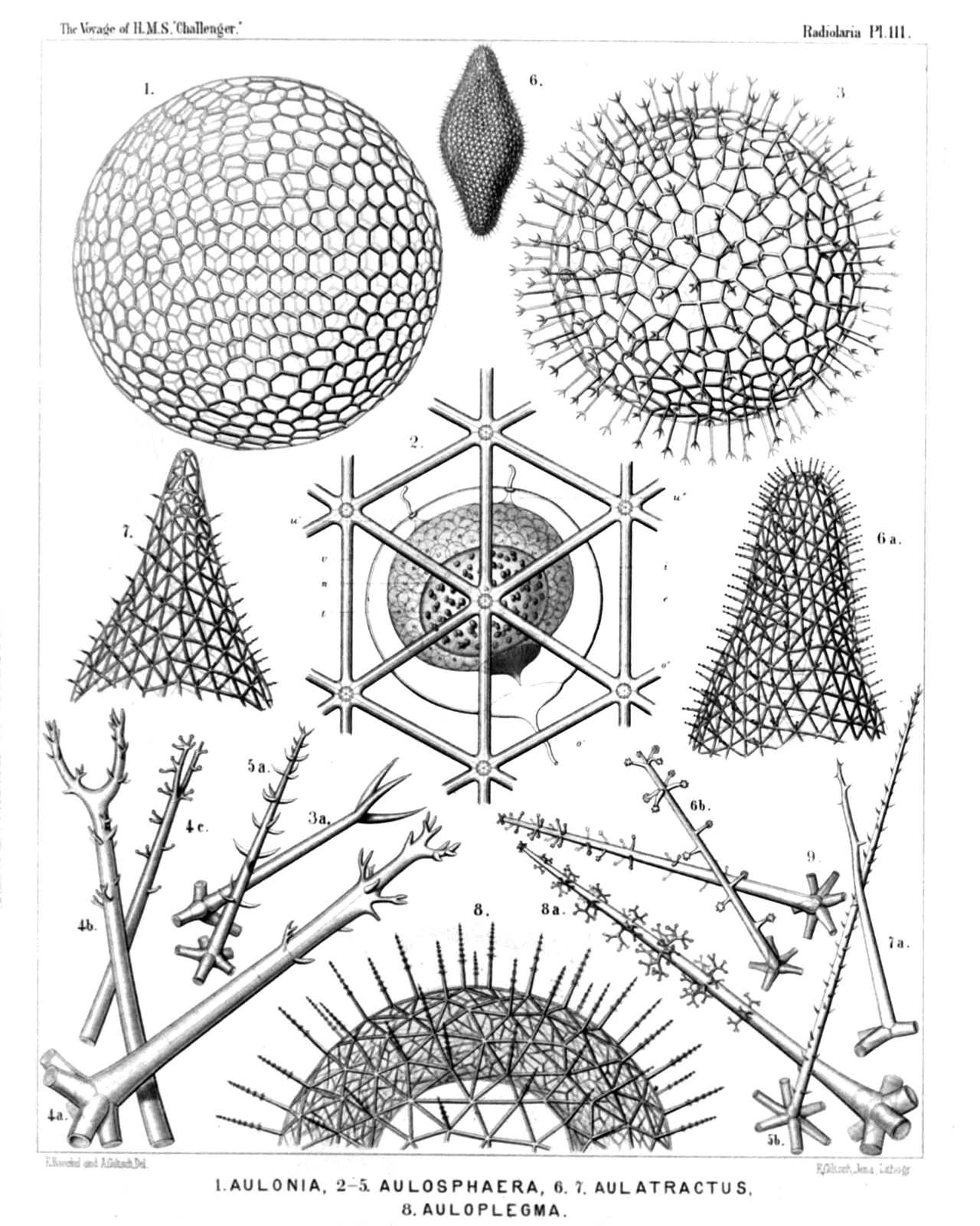
PLATE 111.
Aulosphærida.
Diam. Page.
Fig. 1. Aulonia hexagonia, n. sp., × 30 1634
The complete spherical shell.
Fig. 2. Aularia ternaria, n. sp., × 300 1621
A group of six triangular meshes, with seven nodal points of radial tubes. Behind the central capsule, with its double membrane (e, outer; i, inner) and radiate operculum (o); u, the two outer parapylæ; v, vacuoles in the protoplasm. The ellipsoidal nucleus (n) contains numerous nucleoli (l).
Fig. 3. Aulastrum triceros, n. sp., × 50 1635
The complete shell.
Fig. 3a. Aulastrum triceros, n. sp., × 300 1635
A single radial tube.
Figs. 4a, 4b, 4c. Aulastrum dendroceros, n. sp., × 400 1635
Three single radial spines (taken from three different specimens).
Fig. 5a. Aulophacus lenticularis, n. sp., × 300 1631
A single radial spine.
Fig. 5b. Aulophacus amphidiscus, n. sp., × 300 1631
A single radial spine.
Fig. 6. Aulatractus fusiformis, n. sp., × 5 1632
The complete shell, five times enlarged.
Fig. 6a. Aulatractus fusiformis, n. sp., × 20 1632
Apical part of the shell.
Fig. 6b. Aulatractus fusiformis, n. sp., × 400 1632
A single radial tube.
Fig. 7. Aulatractus diploconus, n. sp., × 20 1632
Apical part of the shell.
Fig. 7a. Aulatractus diploconus, n. sp., × 400 1632
A single radial tube.
Fig. 8. Auloplegma perplexum, n. sp., × 50 1630
Half the shell.
Fig. 8a. Auloplegma perplexum, n. sp., × 400 1630
A single radial tube.
Fig. 9. Auloplegma spongiosum, n. sp., × 300 1631
A single radial tube.
PLATE 112.
Legion PHÆODARIA.
Orders PHÆOSPHÆRIA.
Family Cannosphærida.
PLATE 112. Cannosphærida. Diam. Page. Cannosphærida. Fig. 1. Cannosphæra antarctica, n. sp., × 50 1640 The entire shell. The inner mammillate shell, from the month of which is prominent the phæodium, in connected by numerous radial beams with the outer shell. Fig. 2. Cannosphæra antarctica, n. sp., × 200 1640 The inner shell, from the mouth of which is prominent the phæodium, and a single hexagonal mesh of the outer shell, connected with the former by thin radial threads. Fig. 3. Cannosphæra antarctica, n. sp., × 200 1640 A single radial spine, with four terminal branches. Fig. 4. Cannosphæra pacifica, n. sp., × 200 1641 The inner shell, exhibiting on its base the widely open mouth, and in its upper half the transparent spherical central capsule with its nucleus. Of the outer shell (which is connected with the inner by thin radial threads), only a few polygonal meshes are visible. Fig. 5. Cannosphæra atlantica, n. sp., × 200 1640 The inner shell, connected by spiny radial beams with the outer shell, a quadrant only of which is visible. Fig. 6. Cannosphæra atlantica, n. sp., × 200 1640 A single radial spine, with five terminal branches.
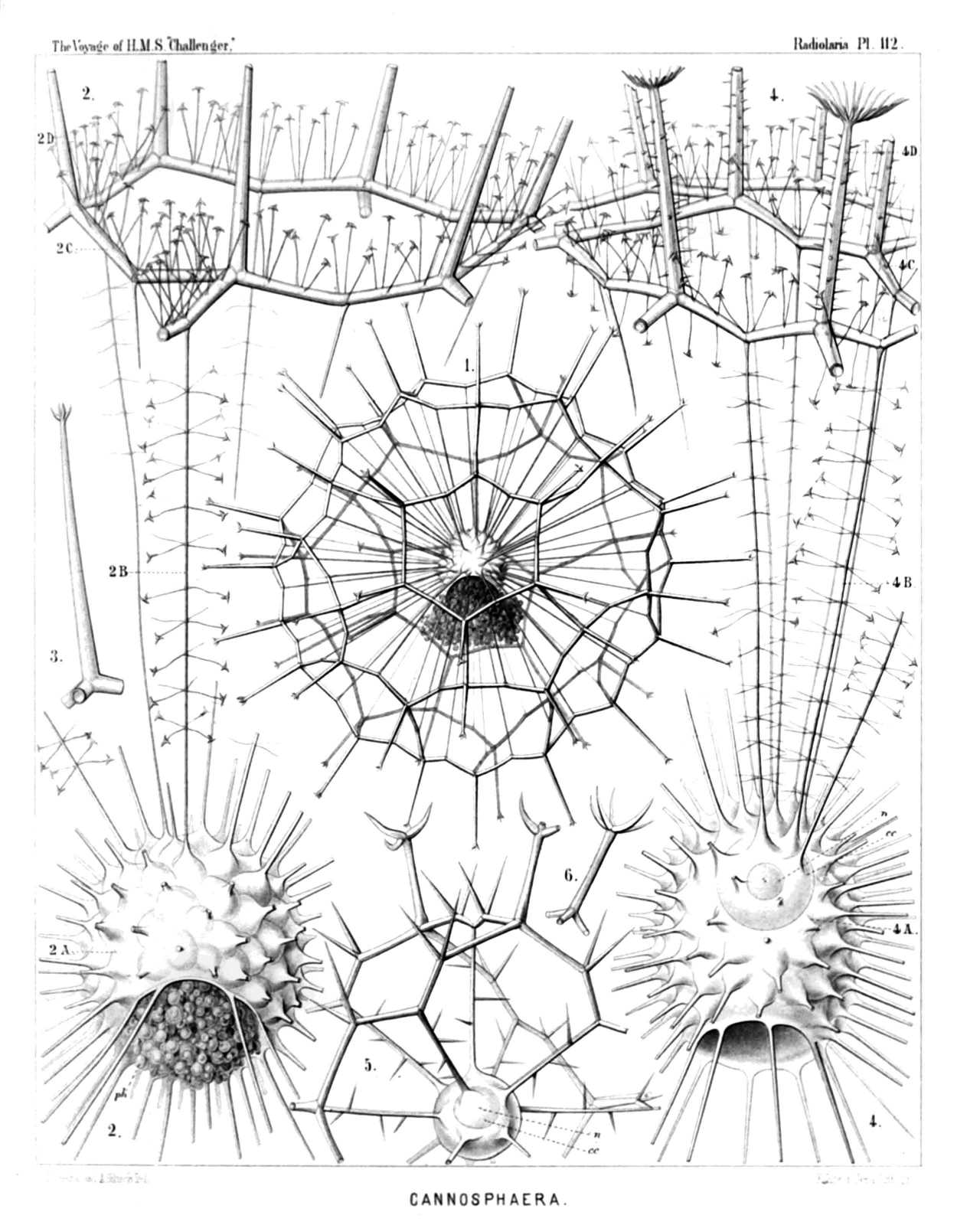
PLATE 112.
Cannosphærida.
Diam. Page.
Cannosphærida.
Fig. 1. Cannosphæra antarctica, n. sp., × 50 1640
The entire shell. The inner mammillate shell, from the month of which is prominent the phæodium, in connected by numerous radial beams with the outer shell.
Fig. 2. Cannosphæra antarctica, n. sp., × 200 1640
The inner shell, from the mouth of which is prominent the phæodium, and a single hexagonal mesh of the outer shell, connected with the former by thin radial threads.
Fig. 3. Cannosphæra antarctica, n. sp., × 200 1640
A single radial spine, with four terminal branches.
Fig. 4. Cannosphæra pacifica, n. sp., × 200 1641
The inner shell, exhibiting on its base the widely open mouth, and in its upper half the transparent spherical central capsule with its nucleus. Of the outer shell (which is connected with the inner by thin radial threads), only a few polygonal meshes are visible.
Fig. 5. Cannosphæra atlantica, n. sp., × 200 1640
The inner shell, connected by spiny radial beams with the outer shell, a quadrant only of which is visible.
Fig. 6. Cannosphæra atlantica, n. sp., × 200 1640
A single radial spine, with five terminal branches.
PLATE 113.
Legion PHÆODARIA.
Order PHÆOGROMIA.
Family Castanellida.
PLATE 113. Castanellida. Diam. Page. Fig. 1. Castanissa challengeri, n. sp., × 100 1686 In the lower part of the figure is visible the large corona of teeth around the mouth (a). Fig. 2. Castanidium moseleyi, n. sp., × 80 1686 In the upper part of the figure, at left, is visible the irregular polygonal mouth (a). Fig. 3. Castanopsis naresi, n. sp., × 80 1688 In the upper part of the figure is visible the smooth circular mouth (a). Fig. 4. Castanura tizardi, n. sp., × 80 1689 Fig. 4a. A single main-spine of the same, × 400 Fig. 5. Castanidium murrayi, n. sp., × 100 1685 With a large phæodium, partly protruded through the circular mouth. Fig. 5a. A single main-spine of the same, hexagonally dimpled, × 400 Fig. 6. Castanella wyvillei, n. sp., × 100 1683 A piece of the shell with the mouth, armed with six large teeth (a). Fig. 7. Castanidium buchanani, n. sp., × 100 1685 A piece of the shell with the smooth roundish mouth (a).
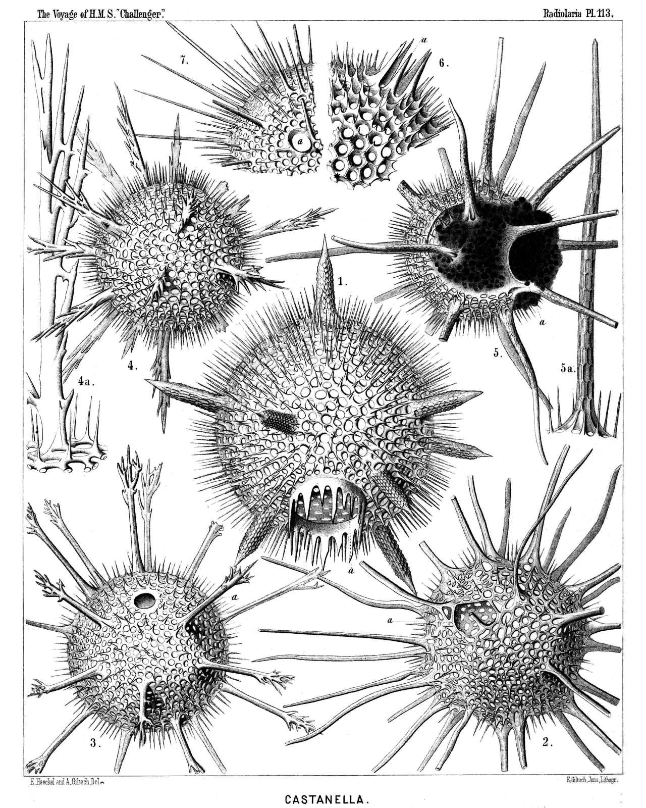
PLATE 113.
Castanellida.
Diam. Page.
Fig. 1. Castanissa challengeri, n. sp., × 100 1686
In the lower part of the figure is visible the large corona of teeth around the mouth (a).
Fig. 2. Castanidium moseleyi, n. sp., × 80 1686
In the upper part of the figure, at left, is visible the irregular polygonal mouth (a).
Fig. 3. Castanopsis naresi, n. sp., × 80 1688
In the upper part of the figure is visible the smooth circular mouth (a).
Fig. 4. Castanura tizardi, n. sp., × 80 1689
Fig. 4a. A single main-spine of the same, × 400
Fig. 5. Castanidium murrayi, n. sp., × 100 1685
With a large phæodium, partly protruded through the circular mouth.
Fig. 5a. A single main-spine of the same, hexagonally dimpled, × 400
Fig. 6. Castanella wyvillei, n. sp., × 100 1683
A piece of the shell with the mouth, armed with six large teeth (a).
Fig. 7. Castanidium buchanani, n. sp., × 100 1685
A piece of the shell with the smooth roundish mouth (a).
PLATE 114.
Legion PHÆODARIA.
Orders PHÆOCYSTINA et PHÆOGROMIA.
Families Cannorrhaphida et Circoporida.
PLATE 114. Cannorrhaphida et Circoporida. Diam. Page. Fig. 1. Haeckeliana darwiniana, n. sp., × 200 1702 A complete shell. Fig. 2. Haeckeliana darwiniana, n. sp., × 400 1702 A single coronet of pores. Fig. 3. Haeckeliana gœtheana, n. sp., × 300 1702 The oral part of the shell with the mouth. Fig. 4. Haeckeliana lamarckiana, n. sp., × 400 1701 A single coronet of pores. Fig. 5. Haeckeliana maxima, n. sp., × 300 1701 A single coronet of pores. Fig. 6. Haeckeliana porcellana, John Murray, × 200 1701 A complete shell. Fig. 7. Distephanus corona, n. sp., × 800 1566 A single pileated piece (half from the side, half from below). Fig. 8. Distephanus corona, n. sp., × 800 1566 Two coupled pileated pieces caught into one another (twin-piece). Fig. 9. Distephanus corona, n. sp., × 800 1566 A single pileated piece, seen from above. Fig. 10. Cannopilus diplostaurus, n. sp., × 800 1568 A single pileated piece, seen from above. Fig. 11. Cannopilus cyrtoides, n. sp., × 800 1569 A single pileated piece, seen obliquely from the side. Fig. 12. Cannopilus cyrtoides, n. sp., × 800 1569 A single pileated piece, seen from below. Fig. 13. Haeckeliana porcellana, John Murray, × 600 1526 The radiate operculum of the central capsule.
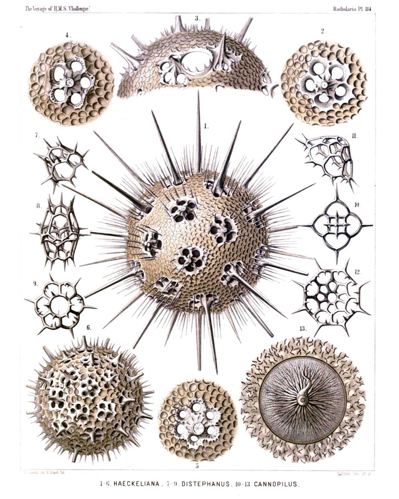
PLATE 114.
Cannorrhaphida et Circoporida.
Diam. Page.
Fig. 1. Haeckeliana darwiniana, n. sp., × 200 1702
A complete shell.
Fig. 2. Haeckeliana darwiniana, n. sp., × 400 1702
A single coronet of pores.
Fig. 3. Haeckeliana gœtheana, n. sp., × 300 1702
The oral part of the shell with the mouth.
Fig. 4. Haeckeliana lamarckiana, n. sp., × 400 1701
A single coronet of pores.
Fig. 5. Haeckeliana maxima, n. sp., × 300 1701
A single coronet of pores.
Fig. 6. Haeckeliana porcellana, John Murray, × 200 1701
A complete shell.
Fig. 7. Distephanus corona, n. sp., × 800 1566
A single pileated piece (half from the side, half from below).
Fig. 8. Distephanus corona, n. sp., × 800 1566
Two coupled pileated pieces caught into one another (twin-piece).
Fig. 9. Distephanus corona, n. sp., × 800 1566
A single pileated piece, seen from above.
Fig. 10. Cannopilus diplostaurus, n. sp., × 800 1568
A single pileated piece, seen from above.
Fig. 11. Cannopilus cyrtoides, n. sp., × 800 1569
A single pileated piece, seen obliquely from the side.
Fig. 12. Cannopilus cyrtoides, n. sp., × 800 1569
A single pileated piece, seen from below.
Fig. 13. Haeckeliana porcellana, John Murray, × 600 1526
The radiate operculum of the central capsule.
PLATE 115.
Legion PHÆODARIA.
Order PHÆOGROMIA.
Family Circoporida.
PLATE 115. Circoporida. Diam. Page. Fig. 1. Circoporus sexfuscinus, n. sp., × 100 1695 The cruciform mouth is visible in the upper part of the figure, to the right. Fig. 2. Circoporus sexfuscinus, n. sp., × 200 1695 A single radial spine, with four cruciate pores at the base. Fig. 3. Circoporus sexfuscinus, n. sp., × 600 1695 The radiate operculum of the central capsule, with the proboscis. Fig. 4. Circospathis furcata, n. sp., × 100 1696 Five of the nine spines are visible, two others (on the upper face) broken off. Between the latter the pentagonal mouth (with five teeth). Fig. 5. Circospathis furcata, n. sp., × 300 1696 The mouth with its five teeth. Fig. 6. Circospathis furcata, n. sp., × 400 1696 A piece of the shell with a radial spine. Fig. 7. Circospathis furcata, n. sp., × 400 1696 Vertical section through the base of a radial spine, to show the central funicle. Fig. 8. Circogonia dodecacantha, n. sp., × 100 1698 The central capsule with the elliptical nucleus (to the right) and the dark phæodium (to the left) are visible, in the upper part (to the left) the mouth of the shell, with six teeth. Fig. 9. Circogonia dodecacantha, n. sp., × 400 1698 A fragment of the shell, exhibiting its peculiar structure (needles tangentially scattered in the cement of the porcellanous substance), and a circle of nine pores around the base of a broken spine. Fig. 10. Circospathis tetrodonta, n. sp., × 400 1697 The mouth with four teeth, in profile view.
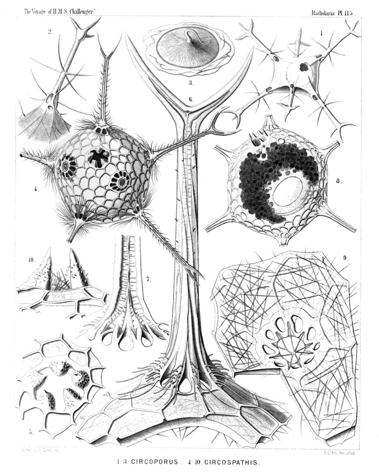
PLATE 115.
Circoporida.
Diam. Page.
Fig. 1. Circoporus sexfuscinus, n. sp., × 100 1695
The cruciform mouth is visible in the upper part of the figure, to the right.
Fig. 2. Circoporus sexfuscinus, n. sp., × 200 1695
A single radial spine, with four cruciate pores at the base.
Fig. 3. Circoporus sexfuscinus, n. sp., × 600 1695
The radiate operculum of the central capsule, with the proboscis.
Fig. 4. Circospathis furcata, n. sp., × 100 1696
Five of the nine spines are visible, two others (on the upper face) broken off. Between the latter the pentagonal mouth (with five teeth).
Fig. 5. Circospathis furcata, n. sp., × 300 1696
The mouth with its five teeth.
Fig. 6. Circospathis furcata, n. sp., × 400 1696
A piece of the shell with a radial spine.
Fig. 7. Circospathis furcata, n. sp., × 400 1696
Vertical section through the base of a radial spine, to show the central funicle.
Fig. 8. Circogonia dodecacantha, n. sp., × 100 1698
The central capsule with the elliptical nucleus (to the right) and the dark phæodium (to the left) are visible, in the upper part (to the left) the mouth of the shell, with six teeth.
Fig. 9. Circogonia dodecacantha, n. sp., × 400 1698
A fragment of the shell, exhibiting its peculiar structure (needles tangentially scattered in the cement of the porcellanous substance), and a circle of nine pores around the base of a broken spine.
Fig. 10. Circospathis tetrodonta, n. sp., × 400 1697
The mouth with four teeth, in profile view.
PLATE 116.
Legion PHÆODARIA.
Order PHÆOGROMIA.
Families Medusettida et Circoporida.
PLATE 116. Medusettida et Circoporida. Diam. Page. Fig. 1. Polypetta mammillata, n. sp., × 500 1677 In the upper part of the figure the dentate proboscis. Fig. 1a. Vertical section through the shell-wall, showing two of the hollow alveoles, opening on its inside, × 1000 Fig. 2. Polypetta tabulata, n. sp., × 500 1677 In the upper part of the figure the dentate proboscis. Fig. 2a. A piece of the shell, seen from the surface, with the triangular plates, × 1000 Fig. 2b. Vertical section through the shell-wall, with an alveole, × 1000 Fig. 3. Circostephanus coronarius, n. sp., × 150 1699 The polyhedral shell exhibits in its wall the small tangential needles. The radial spines are partly broken off. The mouth of the shell, surrounded by eight short conical teeth, is visible on the left side of the figure. Fig. 3a. The mouth of the shell, seen in profile, with eight conical spinulate teeth, × 400 Fig. 3b. The base of a radial spine broken off, to show the corona of (five or six) basal pores, × 400
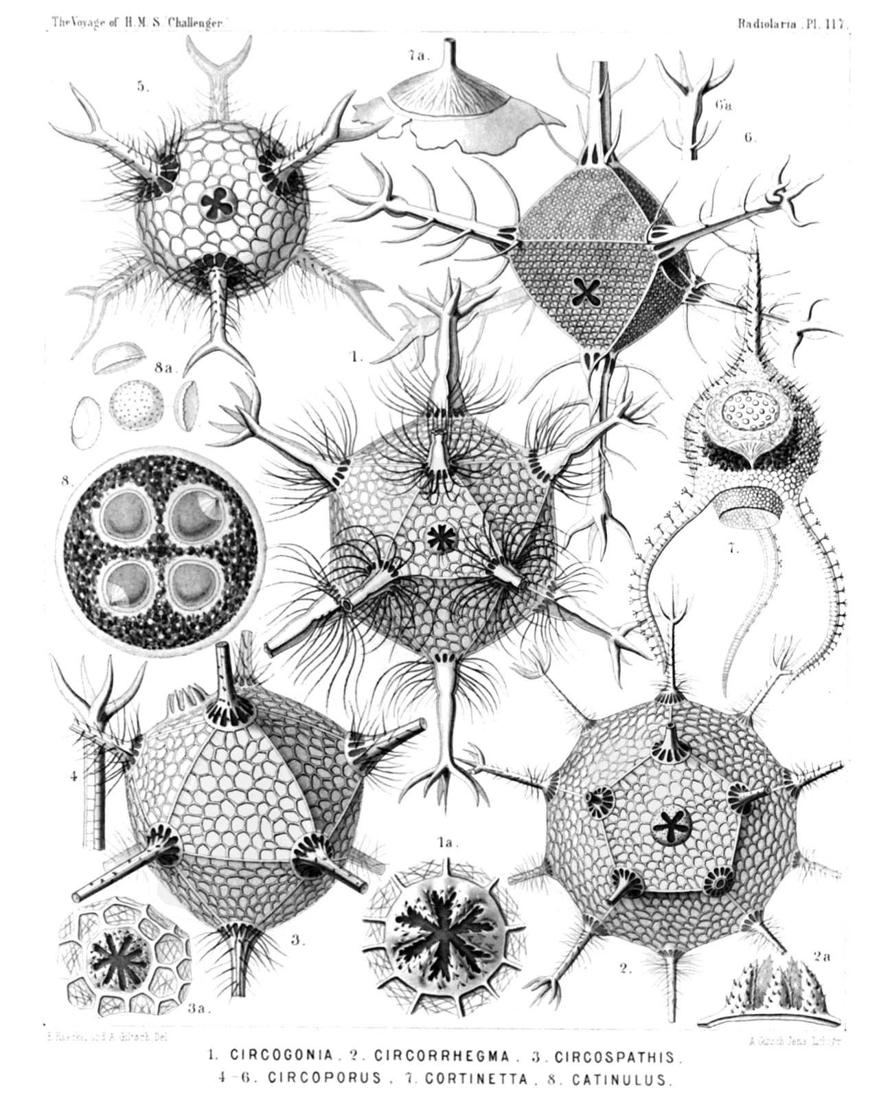
PLATE 116.
Medusettida et Circoporida.
Diam. Page.
Fig. 1. Polypetta mammillata, n. sp., × 500 1677
In the upper part of the figure the dentate proboscis.
Fig. 1a. Vertical section through the shell-wall, showing two of the hollow alveoles, opening on its inside, × 1000
Fig. 2. Polypetta tabulata, n. sp., × 500 1677
In the upper part of the figure the dentate proboscis.
Fig. 2a. A piece of the shell, seen from the surface, with the triangular plates, × 1000
Fig. 2b. Vertical section through the shell-wall, with an alveole, × 1000
Fig. 3. Circostephanus coronarius, n. sp., × 150 1699
The polyhedral shell exhibits in its wall the small tangential needles. The radial spines are partly broken off. The mouth of the shell, surrounded by eight short conical teeth, is visible on the left side of the figure.
Fig. 3a. The mouth of the shell, seen in profile, with eight conical spinulate teeth, × 400
Fig. 3b. The base of a radial spine broken off, to show the corona of (five or six) basal pores, × 400
PLATE 117.
Legion PHÆODARIA.
Orders PHÆOCYSTINA ET PHÆOGROMIA.
Families Cannorrhaphida, Medusettida et Circoporida.
PLATE 117. Cannorrhaphida, Medusettida et Circoporida. Diam. Page. Fig. 1. Circogonia icosahedra, n. sp., × 80 1698 The entire shell, with twelve radial tubes and twenty triangular faces. In the centre of one face is the mouth, with six teeth. Fig. 1a. The mouth alone, with its six spinulate teeth, × 400 Fig. 2. Circorrhegma dodecahedra, n. sp., × 80 1699 The entire shell, with twenty radial tubes and twelve pentagonal faces. In the centre of one face is the mouth, with five teeth. Fig. 2a. The mouth alone, with its five spinulate teeth, seen in profile, × 200 Fig. 3. Circospathis novena, n. sp., × 100 1696 The entire shell, with nine radial tubes and fourteen triangular faces. In one face (to the left above) is the mouth with nine teeth. Fig. 3a. The mouth alone, with its nine spinulate teeth, × 150 Fig. 4. Circoporus hexastylus, n. sp., × 80 1695 A single radial spine. Fig. 5. Circoporus sexfurcus, n. sp., × 80 1694 The entire spherical shell with six forked and ciliated radial tubes. In the centre the cruciform mouth with four teeth. Fig. 6. Circoporus octahedrus, n. sp., × 300 1695 The entire shell, with six verticillate radial tubes and eight triangular faces. In the centre of one face is the mouth, with four teeth. Fig. 7. Cortinetta tripodiscus, n. sp., × 300 1667 The entire shell with the enclosed central capsule, and the phæodium around the astropyle. Fig. 7a. The astropyle, partly detached from the wall of the central capsule, seen in profile, × 800 Fig. 8. Catinulus quadrifidus, n. sp., × 80 1553 A complete specimen, with four equal central capsules, united in a single spherical calymma. Fig. 8a. Some single pieces of the skeleton, × 400
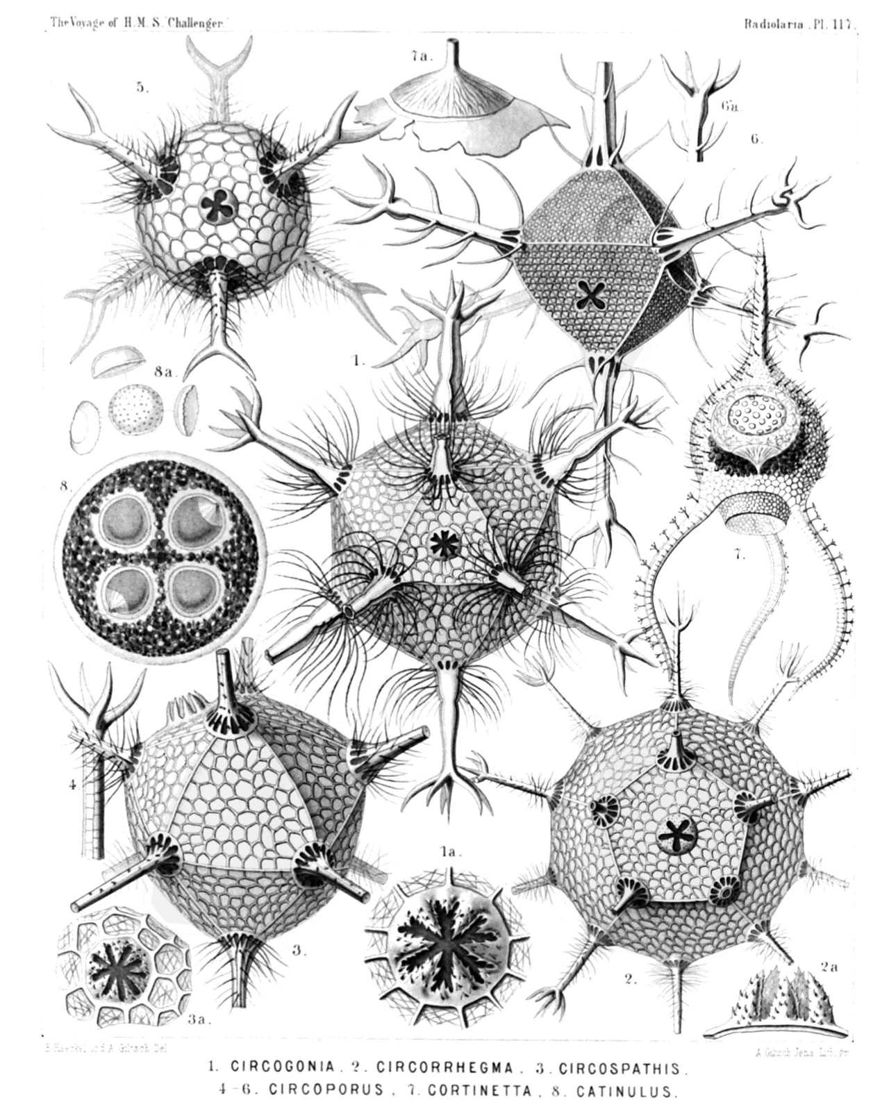
PLATE 117.
Cannorrhaphida, Medusettida et Circoporida.
Diam. Page.
Fig. 1. Circogonia icosahedra, n. sp., × 80 1698
The entire shell, with twelve radial tubes and twenty triangular faces. In the centre of one face is the mouth, with six teeth.
Fig. 1a. The mouth alone, with its six spinulate teeth, × 400
Fig. 2. Circorrhegma dodecahedra, n. sp., × 80 1699
The entire shell, with twenty radial tubes and twelve pentagonal faces. In the centre of one face is the mouth, with five teeth.
Fig. 2a. The mouth alone, with its five spinulate teeth, seen in profile, × 200
Fig. 3. Circospathis novena, n. sp., × 100 1696
The entire shell, with nine radial tubes and fourteen triangular faces. In one face (to the left above) is the mouth with nine teeth.
Fig. 3a. The mouth alone, with its nine spinulate teeth, × 150
Fig. 4. Circoporus hexastylus, n. sp., × 80 1695
A single radial spine.
Fig. 5. Circoporus sexfurcus, n. sp., × 80 1694
The entire spherical shell with six forked and ciliated radial tubes. In the centre the cruciform mouth with four teeth.
Fig. 6. Circoporus octahedrus, n. sp., × 300 1695
The entire shell, with six verticillate radial tubes and eight triangular faces. In the centre of one face is the mouth, with four teeth.
Fig. 7. Cortinetta tripodiscus, n. sp., × 300 1667
The entire shell with the enclosed central capsule, and the phæodium around the astropyle.
Fig. 7a. The astropyle, partly detached from the wall of the central capsule, seen in profile, × 800
Fig. 8. Catinulus quadrifidus, n. sp., × 80 1553
A complete specimen, with four equal central capsules, united in a single spherical calymma.
Fig. 8a. Some single pieces of the skeleton, × 400
PLATE 118.
Legion PHÆODARIA.
Order PHÆOGROMIA.
Family Medusettida.
PLATE 118. Medusettida. Diam. Page. Fig. 1. Gazelletta melusina, n. sp., × 300 1674 From the peristome of the thorny campanulate shell arise six large descending feet, which are studded with arborescent fragile lateral branches, and armed at the distal end with stouter dichotomous terminal branches. Fig. 2. Euphysetta staurocodon, n. sp., × 300 1670 The peristome of the ovate shell bears an odd large foot with three terminal branches and three cruciate rudimentary feet. In the upper part of the shell-cavity is visible the sphæroidal central capsule (containing a nucleus of half the size, with numerous nucleoli); in the lower half the dark pigment-masses of the green phæodium. Fig. 3. Euphysetta amphicodon, n. sp., × 300 1670 The shell-wall exhibits the regular alveolate structure. From the mouth are prominent large masses of the phæodium, which is more voluminous than the shell-cavity, and seems to contain nucleated cells.
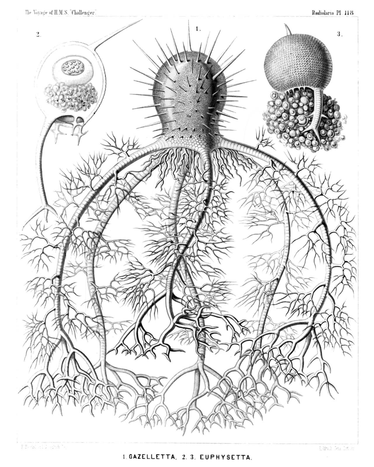
PLATE 118.
Medusettida.
Diam. Page.
Fig. 1. Gazelletta melusina, n. sp., × 300 1674
From the peristome of the thorny campanulate shell arise six large descending feet, which are studded with arborescent fragile lateral branches, and armed at the distal end with stouter dichotomous terminal branches.
Fig. 2. Euphysetta staurocodon, n. sp., × 300 1670
The peristome of the ovate shell bears an odd large foot with three terminal branches and three cruciate rudimentary feet. In the upper part of the shell-cavity is visible the sphæroidal central capsule (containing a nucleus of half the size, with numerous nucleoli); in the lower half the dark pigment-masses of the green phæodium.
Fig. 3. Euphysetta amphicodon, n. sp., × 300 1670
The shell-wall exhibits the regular alveolate structure. From the mouth are prominent large masses of the phæodium, which is more voluminous than the shell-cavity, and seems to contain nucleated cells.
PLATE 119.
Legion PHÆODARIA.
Order PHÆOGROMIA.
Family Medusettida.
PLATE 119. Medusettida. Diam. Page. Fig. 1. Gorgonetta mirabilis, n. sp., × 100 1674 The entire body. From the margin of the cap-shaped shell arise six ascending arborescent feet and six alternating descending feet, which are covered with anchor-pencils and branched at the distal end. From the mouth of the delicately alveolate shell depend prominent parts of the dark voluminous phæodium. Fig. 2. Gorgonetta mirabilis, n. sp., × 300 1674 The distal end of an ascending foot; the branches bear a terminal spathilla with small recurved teeth. Fig. 3. Gorgonetta mirabilis, n. sp., × 300 1674 The distal end of a descending foot, with three lateral anchor-pencils and three terminal branches (broken off). Odd alveole contains an air-bubble. Fig. 4. Gorgonetta mirabilis, n. sp., × 600 1674 A single thread of an anchor-pencil with two quadridentate spathillæ, a larger proximal and a smaller distal (terminal).
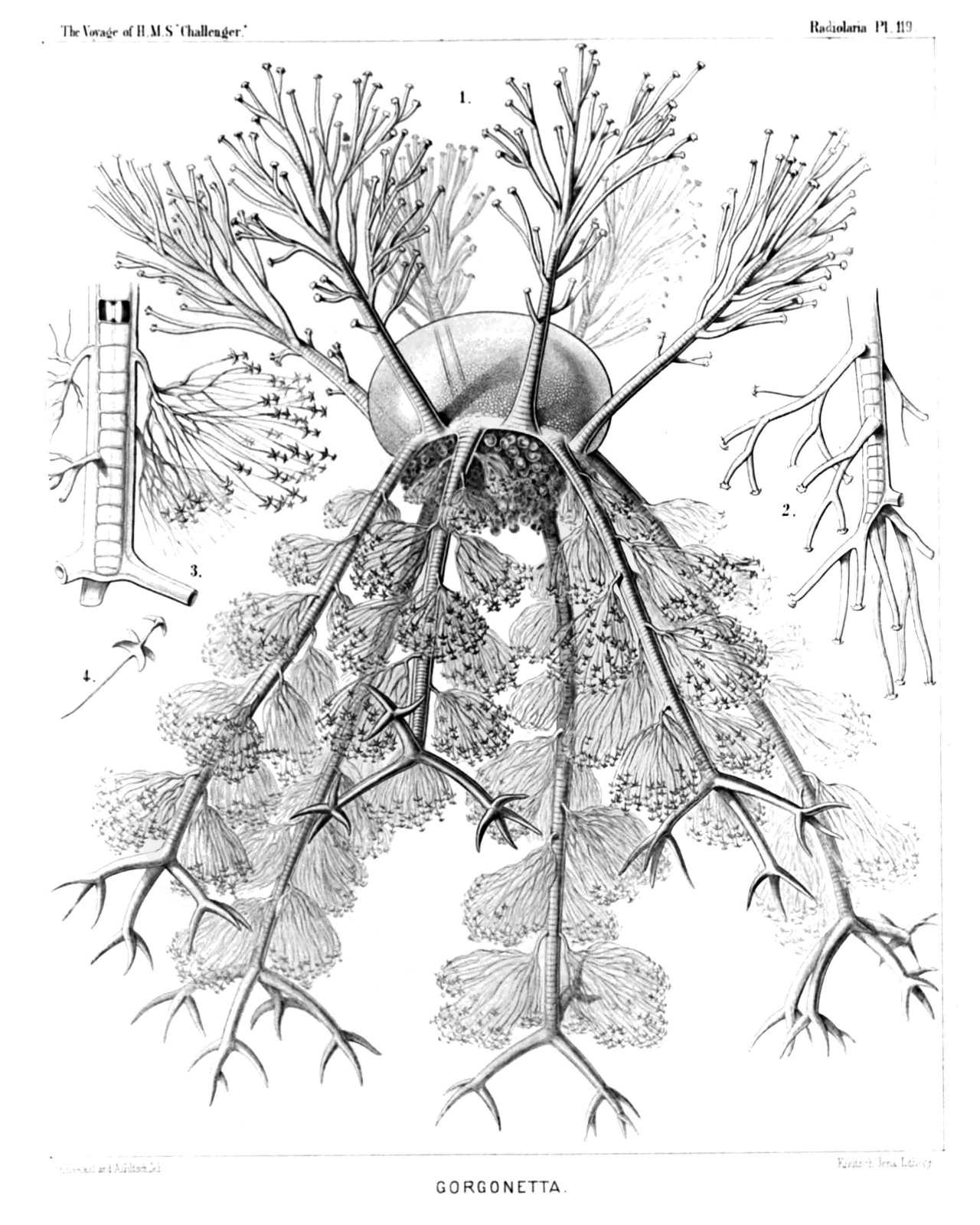
PLATE 119.
Medusettida.
Diam. Page.
Fig. 1. Gorgonetta mirabilis, n. sp., × 100 1674
The entire body. From the margin of the cap-shaped shell arise six ascending arborescent feet and six alternating descending feet, which are covered with anchor-pencils and branched at the distal end. From the mouth of the delicately alveolate shell depend prominent parts of the dark voluminous phæodium.
Fig. 2. Gorgonetta mirabilis, n. sp., × 300 1674
The distal end of an ascending foot; the branches bear a terminal spathilla with small recurved teeth.
Fig. 3. Gorgonetta mirabilis, n. sp., × 300 1674
The distal end of a descending foot, with three lateral anchor-pencils and three terminal branches (broken off). Odd alveole contains an air-bubble.
Fig. 4. Gorgonetta mirabilis, n. sp., × 600 1674
A single thread of an anchor-pencil with two quadridentate spathillæ, a larger proximal and a smaller distal (terminal).
PLATE 120.
Legion PHÆODARIA.
Order PHÆOGROMIA.
Family Medusettida.
PLATE 120. Medusettida. Diam. Page. Fig. 1. Medusetta codonium, n. sp., × 400 1668 Fig. 2. Medusetta quadrigata, n. sp., × 400 1668 The central capsule is visible in the upper half, the phæodium in the lower half of the shell-cavity. Fig. 3. Medusetta tetranema, n. sp., × 400 1669 Fig. 4. Medusetta craspedota, n. sp., × 400 1669 Fig. 5. Gazelletta hexanema, n. sp., × 300 1671 Fig. 6. Gazelletta bifurca, n. sp., × 300 1672 A single alveolate foot. Fig. 7. Gazelletta macronema, n. sp., × 200 1671 Oral view of the shell. Fig. 8. Gazelletta macronema, n. sp., × 800 1671 Three joints of an alveolate foot. Fig. 9. Gazelletta cyrtonema, n. sp., × 300 1671 The upper part of the shell encloses the central capsule with its nucleus. The voluminous phæodium is prominent over the mouth. Fig. 10. Gazelletta orthonema, n. sp., × 200 1671 The central capsule and its nucleus are visible in the shell-cavity. Fig. 11. Gazelletta schleinitzii, n. sp., × 400 1673 Oblique apical view, with the enclosed central capsule, the nucleus of which contains numerous nucleoli. Fig. 12. Gazelletta schleinitzii, n. sp., × 300 1673 A single alveolate foot. Fig. 13. Gazelletta trispathilla, n. sp., × 400 1673 The middle part of a foot. Fig. 14. Gazellatta robusta, n. sp., × 300 1673 The base of a foot, exhibiting the pores of the alveoli. Fig. 15. Gazelletta studeri, n. sp., × 400 1673 The distal end of a foot; four alveoli filled up by air-bubbles. Fig. 16. Gazelletta dendronema, n. sp., × 300 1674 A part of the velum, seen from the inside. The alveoles are partly filled by air.
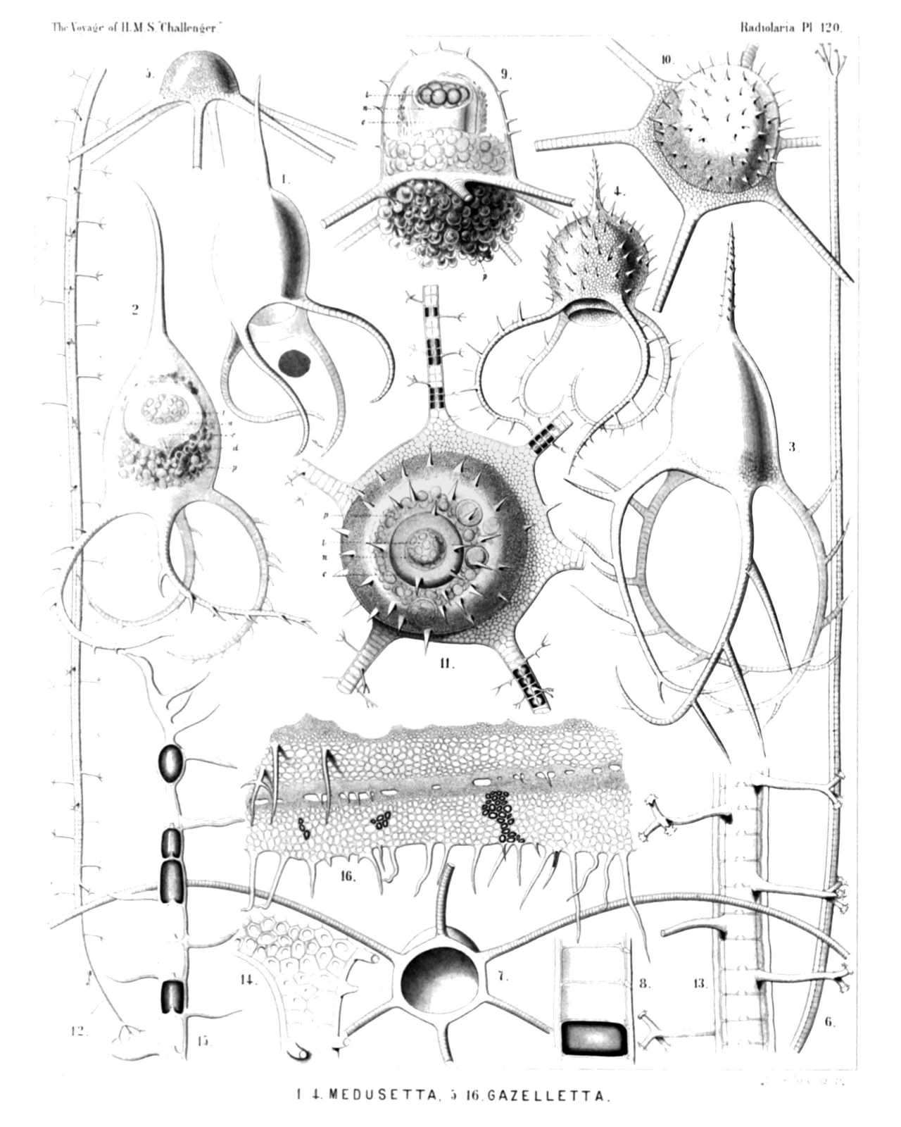
PLATE 120.
Medusettida.
Diam. Page.
Fig. 1. Medusetta codonium, n. sp., × 400 1668
Fig. 2. Medusetta quadrigata, n. sp., × 400 1668
The central capsule is visible in the upper half, the phæodium in the lower half of the shell-cavity.
Fig. 3. Medusetta tetranema, n. sp., × 400 1669
Fig. 4. Medusetta craspedota, n. sp., × 400 1669
Fig. 5. Gazelletta hexanema, n. sp., × 300 1671
Fig. 6. Gazelletta bifurca, n. sp., × 300 1672
A single alveolate foot.
Fig. 7. Gazelletta macronema, n. sp., × 200 1671
Oral view of the shell.
Fig. 8. Gazelletta macronema, n. sp., × 800 1671
Three joints of an alveolate foot.
Fig. 9. Gazelletta cyrtonema, n. sp., × 300 1671
The upper part of the shell encloses the central capsule with its nucleus. The voluminous phæodium is prominent over the mouth.
Fig. 10. Gazelletta orthonema, n. sp., × 200 1671
The central capsule and its nucleus are visible in the shell-cavity.
Fig. 11. Gazelletta schleinitzii, n. sp., × 400 1673
Oblique apical view, with the enclosed central capsule, the nucleus of which contains numerous nucleoli.
Fig. 12. Gazelletta schleinitzii, n. sp., × 300 1673
A single alveolate foot.
Fig. 13. Gazelletta trispathilla, n. sp., × 400 1673
The middle part of a foot.
Fig. 14. Gazellatta robusta, n. sp., × 300 1673
The base of a foot, exhibiting the pores of the alveoli.
Fig. 15. Gazelletta studeri, n. sp., × 400 1673
The distal end of a foot; four alveoli filled up by air-bubbles.
Fig. 16. Gazelletta dendronema, n. sp., × 300 1674
A part of the velum, seen from the inside. The alveoles are partly filled by air.
PLATE 121.
Legion PHÆODARIA.
Order PHÆOCONCHIA.
Family Cœlodendrida.
PLATE 121. Cœlodendrida. Diam. Page. Fig. 1. Cœlodendrum furcatissimum, n. sp., × 50 1735 A complete specimen with the central capsule and the big phæodium. The spherical calymma envelops almost the entire skeleton. Fig. 2. Cœlodendrum furcatissimum, n. sp., × 300 1735 A distal branch with its terminal ramification. Fig. 3. Cœlodendrum furcatissimum, n. sp., × 100 1735 One valve of the shell, with its galea and the four hollow forked tubes arising from it. Fig. 4. Cœlodendrum furcatissimum, n. sp., × 100 1735 The central capsule with its nucleus; on the left side one valve of the closely enveloping shell (seen in vertical section), and its galea with the origin of the four tubes. Fig. 5. Cœlodendrum serratum, n. sp., × 400 1737 A flabellate terminal branch. Fig. 6. Cœlodendrum flabellatum, n. sp., × 150 1737 A flabellate terminal branch. Fig. 7. Cœlodendrum spinosissimum, n. sp., × 300 1735 Forked distal end of a terminal branch. Fig. 8. Cœlodendrum cervicorne, n. sp., × 150 1736 One valve of the shell, with its galea and the four tubes arising from it. A network of protoplasm connects the distal branches. Fig. 9. Cœlodrymus ancoratus, n. sp., × 50 1738 A complete specimen, with the central capsule and the enveloping phæodium. The surface of the spherical calymma is covered by a dense network, from which arise numerous, anchor-bearing, radial tubules. Fig. 10. Cœlodrymus ancoratus, n. sp., × 150 1738 A small piece of the superficial network of the skeleton, with the zigzag radial tubules arising from it, each of which bears an anchor with two recurved denticulate teeth on the distal end.
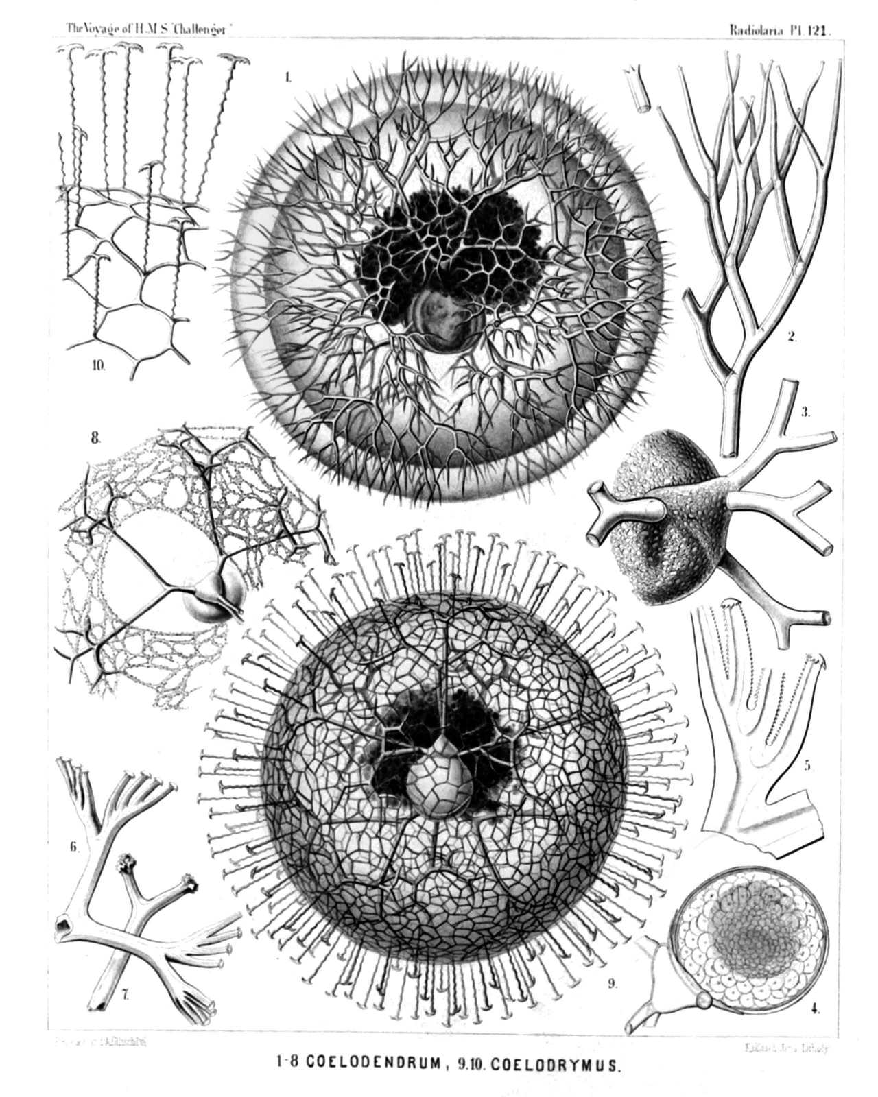
PLATE 121.
Cœlodendrida.
Diam. Page.
Fig. 1. Cœlodendrum furcatissimum, n. sp., × 50 1735
A complete specimen with the central capsule and the big phæodium. The spherical calymma envelops almost the entire skeleton.
Fig. 2. Cœlodendrum furcatissimum, n. sp., × 300 1735
A distal branch with its terminal ramification.
Fig. 3. Cœlodendrum furcatissimum, n. sp., × 100 1735
One valve of the shell, with its galea and the four hollow forked tubes arising from it.
Fig. 4. Cœlodendrum furcatissimum, n. sp., × 100 1735
The central capsule with its nucleus; on the left side one valve of the closely enveloping shell (seen in vertical section), and its galea with the origin of the four tubes.
Fig. 5. Cœlodendrum serratum, n. sp., × 400 1737
A flabellate terminal branch.
Fig. 6. Cœlodendrum flabellatum, n. sp., × 150 1737
A flabellate terminal branch.
Fig. 7. Cœlodendrum spinosissimum, n. sp., × 300 1735
Forked distal end of a terminal branch.
Fig. 8. Cœlodendrum cervicorne, n. sp., × 150 1736
One valve of the shell, with its galea and the four tubes arising from it. A network of protoplasm connects the distal branches.
Fig. 9. Cœlodrymus ancoratus, n. sp., × 50 1738
A complete specimen, with the central capsule and the enveloping phæodium. The surface of the spherical calymma is covered by a dense network, from which arise numerous, anchor-bearing, radial tubules.
Fig. 10. Cœlodrymus ancoratus, n. sp., × 150 1738
A small piece of the superficial network of the skeleton, with the zigzag radial tubules arising from it, each of which bears an anchor with two recurved denticulate teeth on the distal end.
PLATE 122.
Legion PHÆODARIA.
Order PHÆOCONCHIA.
Family Cœlographida.
PLATE 122. Cœlographida. Diam. Page. Fig. 1. Cœlotholus octonus, n. sp., × 30 1749 The entire bivalved shell, seen obliquely from the dorsal and somewhat from the right side, enveloped by the yellowish calymma. Fig. 2. Cœlotholus octonus, n. sp., × 100 1749 One valve of the shell (h) with its large galea and the origin of the three styles. The base of the two lateral styles (g1, g2) is connected by two latticed lateral frenula (b1, b2) with the mouth (m) of the rhinocanna (t). The odd style (g3) is free. Fig. 3. Cœlothauma duodenum, n. sp., × 20 1750 The entire shell, seen from the dorsal side. The long styles are enveloped by the yellowish calymma. Fig. 4. Cœlothauma duodenum, n. sp., × 80 1750 One valve of the shell (h), seen from the apical side; t, rhinocanna; m, its mouth; b1, b2, the two lateral frenula; g1, g2, the two paired styles; g3, the odd style. Fig. 5. Cœlothauma duodenum, n. sp., × 80 1750 One valve of the shell, seen in profile. Characters as in fig. 4. Fig. 6. Cœlothamnus bivalvis, n. sp., × 30 1751 The entire shell, enveloped by the yellowish calymma, seen from the left side; between the two valves is the central capsule, with nucleus and astropyle. Fig. 7. Cœlothamnus bivalvis, n. sp., × 100 1751 A single lateral anchor-pencil. Fig. 8. Cœlothamnus bivalvis, n. sp., × 200 1751 Distal end of a style, with its anchor-pencils. Fig. 9. Cœlothamnus bivalvis, n. sp., × 400 1751 A single anchor-thread, with its quadridentate terminal spathilla.
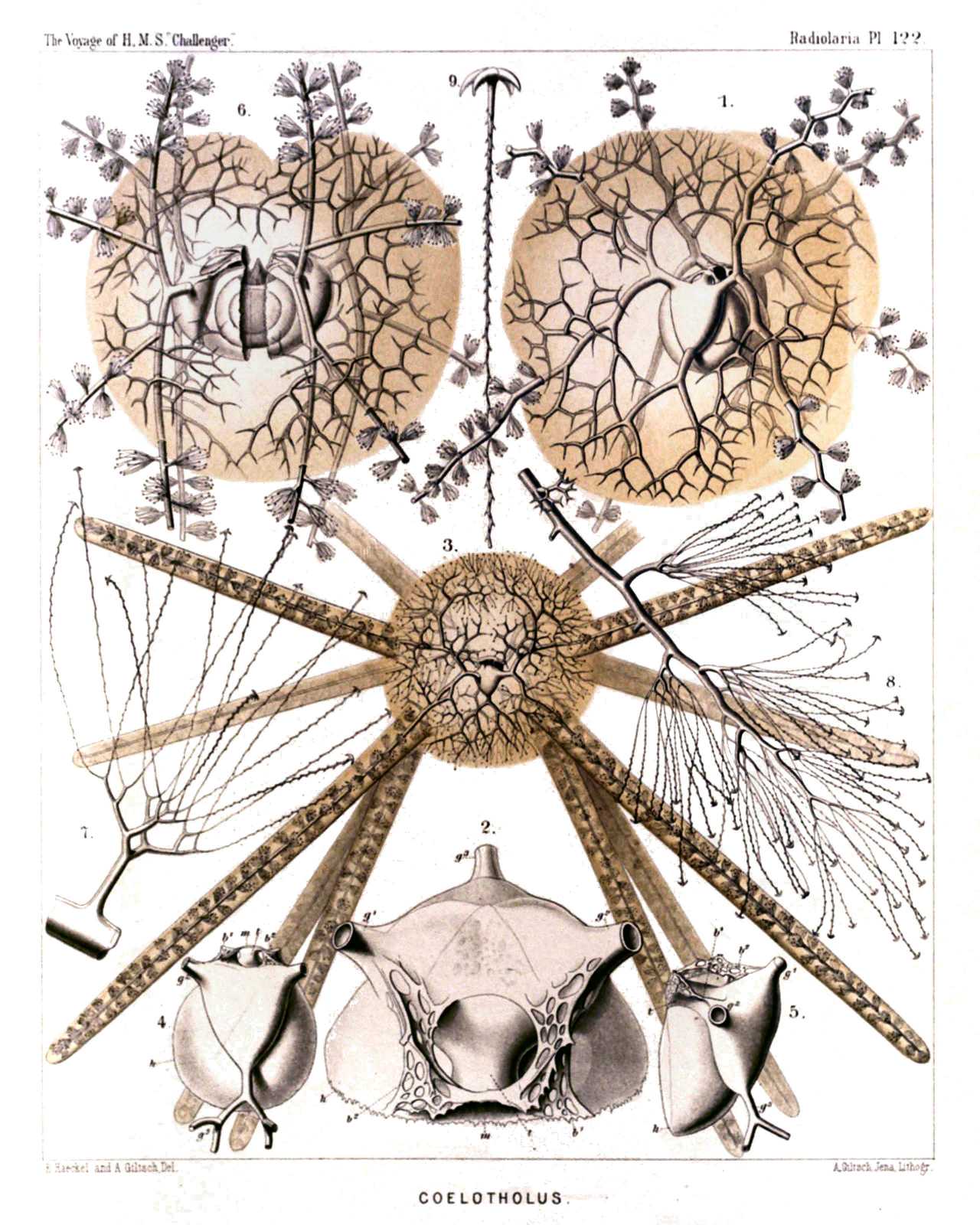
PLATE 122.
Cœlographida.
Diam. Page.
Fig. 1. Cœlotholus octonus, n. sp., × 30 1749
The entire bivalved shell, seen obliquely from the dorsal and somewhat from the right side, enveloped by the yellowish calymma.
Fig. 2. Cœlotholus octonus, n. sp., × 100 1749
One valve of the shell (h) with its large galea and the origin of the three styles. The base of the two lateral styles (g1, g2) is connected by two latticed lateral frenula (b1, b2) with the mouth (m) of the rhinocanna (t). The odd style (g3) is free.
Fig. 3. Cœlothauma duodenum, n. sp., × 20 1750
The entire shell, seen from the dorsal side. The long styles are enveloped by the yellowish calymma.
Fig. 4. Cœlothauma duodenum, n. sp., × 80 1750
One valve of the shell (h), seen from the apical side; t, rhinocanna; m, its mouth; b1, b2, the two lateral frenula; g1, g2, the two paired styles; g3, the odd style.
Fig. 5. Cœlothauma duodenum, n. sp., × 80 1750
One valve of the shell, seen in profile. Characters as in fig. 4.
Fig. 6. Cœlothamnus bivalvis, n. sp., × 30 1751
The entire shell, enveloped by the yellowish calymma, seen from the left side; between the two valves is the central capsule, with nucleus and astropyle.
Fig. 7. Cœlothamnus bivalvis, n. sp., × 100 1751
A single lateral anchor-pencil.
Fig. 8. Cœlothamnus bivalvis, n. sp., × 200 1751
Distal end of a style, with its anchor-pencils.
Fig. 9. Cœlothamnus bivalvis, n. sp., × 400 1751
A single anchor-thread, with its quadridentate terminal spathilla.
PLATE 123.
Legion PHÆODARIA.
Order PHÆOCONCHIA.
Family Concharida.
PLATE 123. Concharida. Diam. Page. (The central capsule is coloured red in the figures of this plate, the phæodium green). Fig. 1. Concharium diatomeum, n. sp., 1717 Dorsal view. The central capsule (red) exhibits above the anterior tubular main-opening (astropyle), and below the two small posterior lateral openings (right and left parapylæ). Fig. 2. Concharium bivalvum, n. sp., × 150 1717 Dorsal view. The central capsule is visible in the lower part, the margin of the two valves in the upper part of the figure. Fig. 2a exhibits the two smooth lateral margins of the valves, catching into one another. (Lateral view). Fig. 3. Concharium nucula, n. sp., 1717 The dorsal valve alone, seen from the outside. Fig. 4. Concharium bacillarium, n. sp., 1718 Lateral view from the smooth margin, by which the two valves are united. Fig. 5. Conchasma radiolites, n. sp., × 300 1719 Lateral view. In the aboral half of the shell-cavity lies the red central capsule, in the oral half the green phæodium. Fig. 6. Conchasma sphærulites, n. sp., × 300 1719 Lateral view. On the aboral pole the two horns of the hinge. Fig. 7. Conchellium tridacna, n. sp., × 200 1720 Oblique lateral view (from the right and ventral side). Fig. 7a. Three pores of the same, with their hexagonal frames and six internal denticles, × 400 Fig. 8. Conchopsis carinata, n. sp., × 150 1725 Lateral view, from the left side. Fig. 9. Conchopsis lenticula, n. sp., × 150 1726 Lateral view, from the right side. The two membranes of the central capsule are separated by a wide interval in this and the preceding figure. The nucleus contains numerous nucleoli. Fig. 9a. Two of the peculiar cells, which are contained in the green phæodium in large numbers, × 400
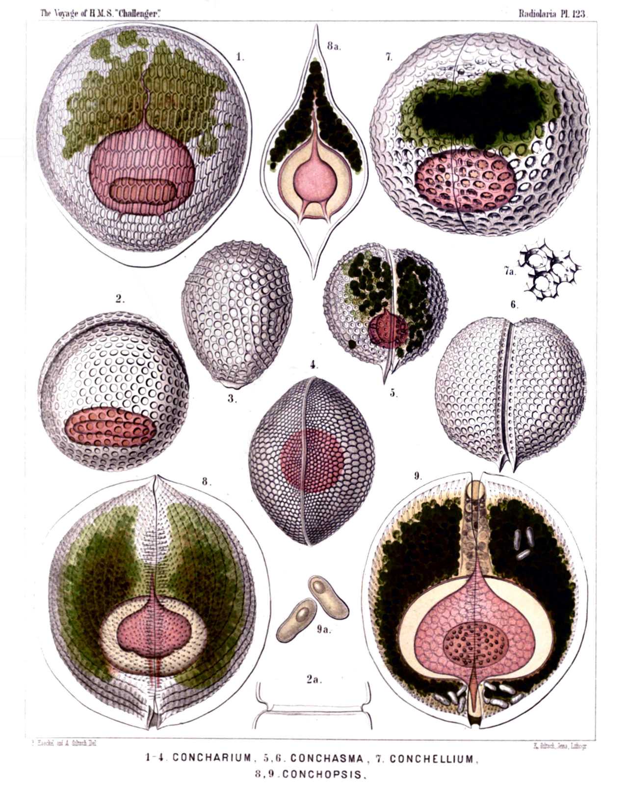
PLATE 123.
Concharida.
Diam. Page.
(The central capsule is coloured red in the figures of this plate, the phæodium green).
Fig. 1. Concharium diatomeum, n. sp., 1717
Dorsal view. The central capsule (red) exhibits above the anterior tubular main-opening (astropyle), and below the two small posterior lateral openings (right and left parapylæ).
Fig. 2. Concharium bivalvum, n. sp., × 150 1717
Dorsal view. The central capsule is visible in the lower part, the margin of the two valves in the upper part of the figure.
Fig. 2a exhibits the two smooth lateral margins of the valves, catching into one another. (Lateral view).
Fig. 3. Concharium nucula, n. sp., 1717
The dorsal valve alone, seen from the outside.
Fig. 4. Concharium bacillarium, n. sp., 1718
Lateral view from the smooth margin, by which the two valves are united.
Fig. 5. Conchasma radiolites, n. sp., × 300 1719
Lateral view. In the aboral half of the shell-cavity lies the red central capsule, in the oral half the green phæodium.
Fig. 6. Conchasma sphærulites, n. sp., × 300 1719
Lateral view. On the aboral pole the two horns of the hinge.
Fig. 7. Conchellium tridacna, n. sp., × 200 1720
Oblique lateral view (from the right and ventral side).
Fig. 7a. Three pores of the same, with their hexagonal frames and six internal denticles, × 400
Fig. 8. Conchopsis carinata, n. sp., × 150 1725
Lateral view, from the left side.
Fig. 9. Conchopsis lenticula, n. sp., × 150 1726
Lateral view, from the right side. The two membranes of the central capsule are separated by a wide interval in this and the preceding figure. The nucleus contains numerous nucleoli.
Fig. 9a. Two of the peculiar cells, which are contained in the green phæodium in large numbers, × 400
PLATE 124.
Legion PHÆODARIA.
Order PHÆOCONCHIA.
Family Concharida.
PLATE 124. Concharida. Diam. Page. Fig. 1. Conchidium terebratula, n. sp., × 400 1721 Lateral view, from the left side. Fig. 2. Conchidium terebratula, n. sp., × 800 1721 A piece of the frontal girdle-fissure, with the teeth of both valves catching into one another. Fig. 3. Conchidium rhynchonella, n. sp., × 200 1722 Lateral view, from the left side. Fig. 4. Conchidium leptæna, n. sp., × 300 1722 Girdle-fissure with the teeth, seen from the left side. Fig. 5. Conchidium leptæna, n. sp., × 800 1722 A single tooth with its base. Fig. 6. Conchidium thecidium, n. sp., × 300 1721 Lateral view, from the left side. In the oral part of the shell-cavity the dark phæodium, in the aboral part the central capsule with two nuclei (a dorsal and a ventral). Fig. 7. Conchidium argiope, n. sp., × 300 1722 Oblique oral view (half from the anterior, half from the left side). Fig. 8. Conchidium argiope, n. sp., × 300 1722 Dorsal valve, from below. Fig. 9. Conchidium argiope, n. sp., × 600 1722 A piece of the valve margin, with four teeth. Fig. 10. Conchonia diodon, n. sp., × 200 1723 Lateral view, from the left side. In the anterior part of the shell-cavity the dark phæodium, in the posterior part the central capsule with the nucleus. The two valves are connected at the posterior hinge by a ligament (to the right in the figure). Fig. 11. Conchonia diodon, n. sp., × 400 1723 Mouth of the shell, with its two lips, seen from the oral pole. Fig. 12. Conchonia diodon, n. sp., × 400 1723 A piece of the valve-margin, with four teeth. Fig. 13. Conchonia triodon, n. sp., × 300 1724 Ventral valve, seen from the lower face. Fig. 14. Conchonia triodon, n. sp., × 300 1724 Dorsal valve, seen from the left side. Fig. 15. Conchoceras caudatum, n. sp., × 300 1727 Lateral view, from the left side. Fig. 16. Conchoceras cornutum, n. sp., × 200 1728 Lateral view, from the left side.
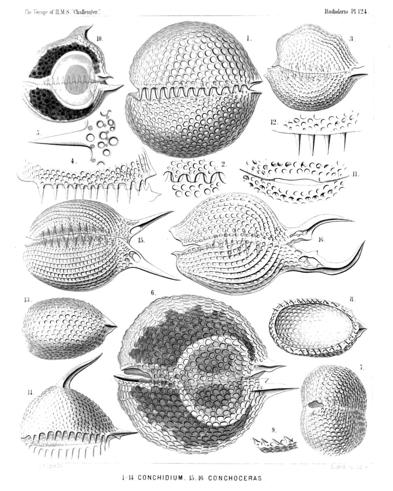
PLATE 124.
Concharida.
Diam. Page.
Fig. 1. Conchidium terebratula, n. sp., × 400 1721
Lateral view, from the left side.
Fig. 2. Conchidium terebratula, n. sp., × 800 1721
A piece of the frontal girdle-fissure, with the teeth of both valves catching into one another.
Fig. 3. Conchidium rhynchonella, n. sp., × 200 1722
Lateral view, from the left side.
Fig. 4. Conchidium leptæna, n. sp., × 300 1722
Girdle-fissure with the teeth, seen from the left side.
Fig. 5. Conchidium leptæna, n. sp., × 800 1722
A single tooth with its base.
Fig. 6. Conchidium thecidium, n. sp., × 300 1721
Lateral view, from the left side. In the oral part of the shell-cavity the dark phæodium, in the aboral part the central capsule with two nuclei (a dorsal and a ventral).
Fig. 7. Conchidium argiope, n. sp., × 300 1722
Oblique oral view (half from the anterior, half from the left side).
Fig. 8. Conchidium argiope, n. sp., × 300 1722
Dorsal valve, from below.
Fig. 9. Conchidium argiope, n. sp., × 600 1722
A piece of the valve margin, with four teeth.
Fig. 10. Conchonia diodon, n. sp., × 200 1723
Lateral view, from the left side. In the anterior part of the shell-cavity the dark phæodium, in the posterior part the central capsule with the nucleus. The two valves are connected at the posterior hinge by a ligament (to the right in the figure).