RNA SYNTHESIS: HOW TO TRANSLATE ONE LANGUAGE INTO ANOTHER
Reading edition
Mathematicians are like Frenchmen: whatever you say to them they translate into their own language, and forthwith it is something entirely different.
Wolfgang von Goethe
We have mentioned previously that there are two main types of nucleic acids: DNA, the genetic material itself, and RNA, the molecule that translates the genetic message from DNA into terms the cell can use as “instructions” for making protein. Cells differ from each other on the basis of kinds of proteins they contain, and, since differences among cells determine differences among organisms, it follows that differences in the composition of DNA serve to explain the variety in living organisms populating the world. However, if differences between two organisms can be explained by differences in the chemical composition of their respective DNA’s, how can we explain differences between cells of the same organism? How can we explain that cells of the human pancreas secrete insulin, whereas other cells in man produce no insulin? Or how can we explain that certain cells make bone and others make fat? If indeed all cells in the same organism contain the same amount and kind of DNA (since all DNA in an organism derives from the duplication of the DNA of the fertilized egg cell and its descendants), it would seem, at first glance, that DNA is not the molecule responsible for differences among the cells. The clarification of this apparent contradiction is found in the remarkable properties of the other nucleic acid, the translator molecule, RNA.
Three Kinds of RNA
In the first place, there are at least three different kinds of RNA. The largest quantity is a special kind called ribosomal RNA, or r-RNA. It is found in close conjunction with proteins and makes up the structural frame upon which the protein-synthesizing machinery is built. The r-RNA and the proteins to which it is firmly bound form 26 the ribosomes, the RNA-rich microsomes that are attached to the endoplasmic reticulum. Proteins are synthesized on ribosomes. We shall see later what determines the differences among proteins and how these differences are dictated directly by RNA and indirectly by DNA.
Besides r-RNA, there is a kind of RNA called soluble RNA, or transfer RNA, or s-RNA. It combines with r-RNA to complete the sequence of events that synthesizes the proteins. A bond between r-RNA and s-RNA is established by a third RNA molecule called messenger RNA, or template RNA, or m-RNA. This m-RNA molecule is truly the messenger that carries the genetic message from DNA to the protein-synthesizing apparatus.
Dr. Michael Shimkin, a Temple University scientist, in his analogy has compared the DNA → RNA → protein sequence to the activities of a newspaper staff. DNA is the editor; m-RNA molecules are copyboys who carry the editorials to the typesetters, the r-RNA and s-RNA, who then take the “letters” of nucleic acid and set them into slots in accordance with the editor’s directions. There are also workers who melt down outworn letters and still other workers who make new letters for further use; these are the enzymes, special kinds of proteins. If we wish to continue the analogy, we may say that each kind of cell in the organism has a different subeditor, who writes that cell’s own editorial. Actually we might say that all cells have the same board of editors in common, but only one editor functions in any given type of cell. In biological terms this means that only a portion of all the cellular DNA is active in each cell.
The active DNA is the DNA that makes m-RNA that will carry instructions to the protein-synthesizing machinery of that type of cell. Cells of the same organism therefore differ from each other on the basis of the segment of DNA that is active in making m-RNA. Let us now see how we can use radioactive isotopes to investigate the synthesis of RNA.
Labeling RNA with a Radioactive Isotope
RNA synthesis is investigated with radioactive tracers in the same way as DNA synthesis. If we can mark, with a 27 radioactive atom, a small molecule that is incorporated into newly formed RNA, we can then trace the course of the labeled RNA molecule with a radiation-detection device. DNA had one advantage in this regard—the fact that one compound, thymidine, was a precursor of DNA, a specific material that could be incorporated only into DNA. We do not know similar specific precursors of RNA. But we know several precursors that are predominantly incorporated into RNA; the most common of these are the nucleosides adenine, cytidine, and uridine, and the smaller molecule, orotic acid. All these precursors can be labeled with either ³H or ¹⁴C, and their incorporation into RNA can be measured.
Detecting RNA with Autoradiography
As in DNA synthesis, we can use autoradiography to follow the incorporation of precursors into RNA. By proper treatment of the tissues, we can make sure that all the radioactivity visible by autoradiography is due to labeled RNA, even though some of the precursor also enters DNA molecules. Even so, the kind of information obtained from autoradiographs of tissues exposed to RNA precursors is different from that obtained with DNA precursors. The advantage of high-resolution autoradiography in DNA studies is the possibility of identifying particular cells that are synthesizing nucleic acid. This advantage is apparently lost in the case of RNA. The reason is that, at any given time, only a few cells are making DNA, whereas practically all cells are synthesizing RNA constantly. The only exceptions are cells in the midpoint of mitosis. At the beginning (prophase) and at the end of cell division (telophase), RNA is synthesized. If we want a quantitative measurement of RNA synthesis, other methods, to be examined presently, are considerably more precise. But autoradiography can still give us valuable information.
Other Methods of Detecting RNA
If we look at cells soon after they have been exposed to an RNA precursor, we find that the radioactivity detectable by autoradiography is only in the nuclei of the cells. No 28 radioactivity can be detected in the cytoplasm, although we know that the cytoplasm of living cells contains large amounts of r-RNA and s-RNA. One or two hours later, however, radioactive RNA appears in the cytoplasm as well as in the nucleus. What autoradiography is telling us is that RNA is made in the nucleus and then is slowly transferred to the cytoplasm.
Autoradiography cannot tell us whether the RNA that has been newly synthesized in the nuclei of cells is m-RNA, s-RNA, or r-RNA. The methods necessary to make this distinction are based on the chemical fractionation of the tissue, isolation of RNA, determination of its amount by quantitative analysis, and determination of the amount of radioactivity by physical methods. Let us examine these steps separately.
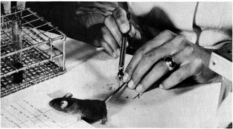
Figure 17 Injecting a mouse with a radioactive solution.
Chemical Fractionation of Tissue After an animal has been injected with a radioactive precursor of RNA, some of it will be incorporated into DNA as well as into RNA (remember that the precursors of RNA lack specificity), and part of the precursor will be broken down into smaller molecules. The injected animal can be sacrificed, and an organ or another tissue, for instance, the liver, can be removed. Then the liver is homogenized, that is, ground to a pulp with a modern version of the mortar and pestle. The homogenate (pulp) is treated with cold (weak) acid. Proteins and nucleic acids are insoluble in cold acids and 29 therefore precipitate to the bottom of the test tube. All molecules that are soluble in a cold acid are left in the supernatant (the remaining liquid); among these are small molecules, like those of the RNA precursor. The precipitate (the solid material that settles to the bottom), now containing proteins and nucleic acids, is then treated with a strong alkali, for instance, sodium hydroxide. Alkali will digest RNA into smaller molecules but does not affect DNA. If we now add acid to the solution, DNA, being insoluble in acid, will precipitate again; RNA, having been broken down into small molecules, will remain in the supernatant. DNA can then be extracted from the precipitate by boiling in strong acid. Proteins from the tissue remain in the final residue.
We have now fractionated the tissue into four portions: the acid-soluble fraction (containing small molecules), RNA, DNA, and proteins. (The cell’s lipids and sugars come out during alcohol rinses between the weak acid and the alkali steps.) Chemical analysis allows us to measure precisely the amount of RNA or DNA in its respective fraction and therefore in the tissue or organ. The amount of radioactivity in the RNA fraction can then be determined by a technique known as liquid scintillation counting.
Liquid Scintillation Counting Liquid scintillation counting is the preferred method for the measurement of low-energy beta-emitting radioisotopes commonly used in cell-fractionation studies (see Figure 18). It is convenient, sensitive, and rapid for routine measurement of radiation in hydrocarbons, other organic compounds, and aqueous solutions containing such isotopes as ³H, ¹⁴C, and ³²P.
Liquid scintillation solutions share with other scintillating materials the property of converting into visible light the energy deposited in them by ionizing radiation. In theory, if a sample of a beta emitter is dissolved in a liquid scintillator solution, every beta particle emitted will be absorbed completely because the range of penetration of beta particles in liquids is quite short (ranging from 0.008 millimicron for ³H to 7.9 millimicrons for ³²P in a medium of unit density). The kinetic energy of the beta particles is largely used up in the ionization and excitation of the most abundant molecular species present, the 30 solvent in which the scintillating material was dissolved. A fraction of the energy thus expended by each beta particle is transferred from excited solvent molecules to scintillator molecules; thus the electrons in the atoms of the scintillator molecules are raised to an excited (higher energy) state. When these electrons return to the ground, or unexcited, state, a fraction of them emit a photon of light. Thus each beta particle produces a burst of photons.
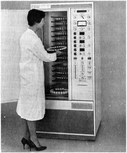
Figure 18 Technician placing a tray of samples in a liquid scintillation counter. The radioactivity of each sample is recorded as the trays revolve.
If a vessel containing the liquid scintillator and the radioactive sample is placed near a suitably sensitive instrument known as a photomultiplier tube, each burst of scintillator photons activates this device and causes it to release a burst of photoelectrons. Each burst of photoelectrons is multiplied successively in a series of electronic steps; as a result, there is a suitably large electrical-output pulse to be recorded.
One of the principal advantages of the liquid scintillation method is the ease of sample preparation. We need only transfer a known volume of a liquid sample or weigh a given mass of a solid sample into a sample bottle, add a known amount of the liquid scintillator solution, and stir until there is a homogeneous solution. Samples thus prepared are placed in a refrigerated counting apparatus. 31 After a short waiting period to allow time for the samples to cool and for a natural, short-lived phosphorescence (due to exposure to room light) to subside, the samples are ready to be measured.
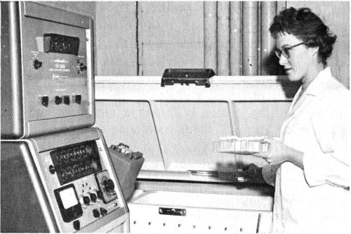
Figure 19 Placing radioactive samples in a refrigerated unit for liquid scintillation counting.
One disadvantage of liquid scintillation counting is that different compounds show different degrees of quenching (loss of emitted photons), and the effect must be checked for each class of compounds in each concentration range. This checking is usually done with an internal standard technique, the sample being counted before and after a standard, or known, emitter is added.
Another difficulty is that the best scintillating solvents are not the best chemical solvents for most biological materials. The solubility problem is also aggravated by the low temperatures at which liquid scintillation counters are usually operated for more effective instrument performance.
With the method we have described, we can obtain a fairly accurate idea of the rate of RNA synthesis in a given tissue. There are other things we would like to know about RNA. The first of these is the kind of RNA being synthesized. During alkaline digestion all kinds of RNA are broken down into their component nucleotides; we must therefore use other methods if we wish to know the kind of 32 RNA in which the radioactivity of the precursor has been incorporated.
Isolation of RNA Native RNA, that is, RNA not broken down into its smaller constituents, can be obtained in a variety of ways, but the most popular one makes use of phenol extraction, which removes DNA and proteins and leaves RNA in solution. If this phenol-purified RNA is dissolved in a concentrated sugar solution and spun in a centrifuge at a very high velocity, it will separate into three major components. These components separate because they have different molecular weights, and the larger the molecule, the faster it forms a sediment in the centrifugal field. Two of these components are s-RNA, the lightest of all, and r-RNA, which is divided into two subfractions. We can also identify a third component, m-RNA, with the centrifuge system but only with some difficulty and only after labeling it with a radioactive precursor, because the amount of m-RNA in a cell is very small.
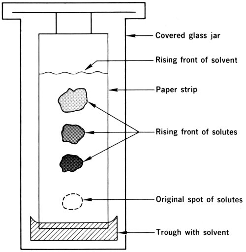
Figure 20 Diagram of ascending paper chromatography.
Quantitative Analysis Another important feature of RNA (or DNA, for that matter) is its base composition, that is, the percentage of each of the nucleotides that make it up. 33 The four bases that, with ribose and phosphoric acid, comprise the RNA molecule are guanine, adenine, cytosine, and uracil. It will be noted that three of the four—guanine, adenine, and cytosine—are the same as those in DNA, but thymidine in DNA has been replaced by another base, uracil. To determine the percentage of each base in a given RNA molecule, we must digest RNA with alkali to produce mononucleotides, which are smaller molecules, each consisting of a base, ribose, and phosphoric acid. We can now separate the four nucleotides by using paper chromatography (see Figure 20).
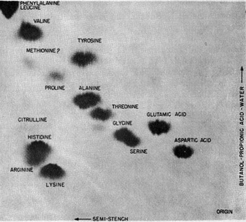
Figure 21 A paper chromatography showing separation of amino acids in two directions. Radioactivity in samples then produced this record by radioautography.
In this technique a mixture of compounds is deposited on the edge of a special type of paper. This edge is then immersed in a solvent that slowly permeates the paper (at a constant speed) by capillary action. As the solvent moves from the immersed edge toward the other edge, which is hanging freely, it carries the mixture of nucleotides with 34 it. Each of the compounds in the mixture travels at a different speed, however; thus, as the solvent front moves along the paper, the dissolved compounds are separated from each other and appear as distinct spots on the paper. To locate the nucleotides on the paper and to determine the percentage composition, we can use a chromatogram scanner, a device that scans the paper chromatograms, measures the radiation from them, and thus locates the labeled substances (see Figure 22).
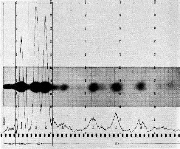
Figure 22 Recording of radioactivity in a sample by radioautography and paper chromatography. The peaks of the trace prepared by a chromatogram scanner coincide with the areas of separated components on the same chromatogram, as revealed by radioautography. The radioautograph is superimposed on the chromatogram recording.
Another technique used to separate the nucleotides of RNA is column chromatography. In this method mixtures of nucleotides are separated as they pass down a column of chemicals (see Figure 23).
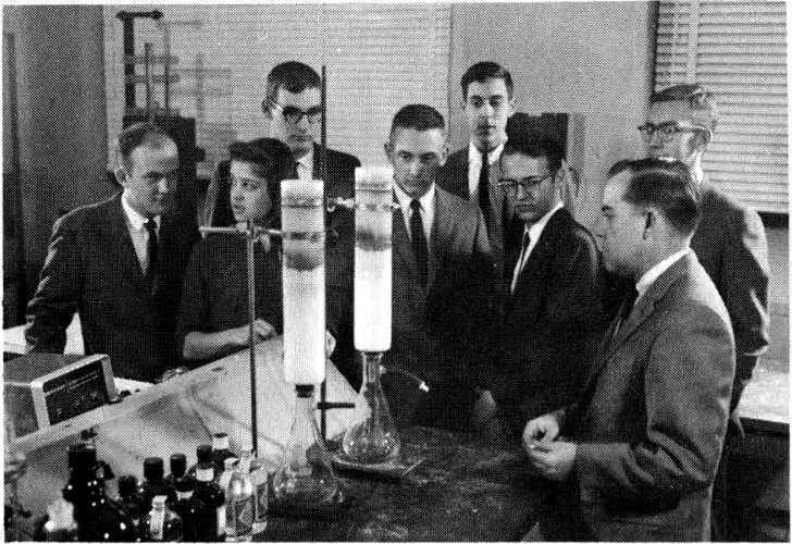
Figure 23 Students visiting Argonne National Laboratory listen to a scientist explain the column chromatography process, in which mixtures of nucleotides are separated as they pass down a column of chemicals.
35
We have now learned how to use radioisotopes to investigate the synthesis of RNA, the molecule that translates the DNA message into the language of proteins. Let us now see what we can learn about the synthesis and function of proteins.