CELL THEORY: DNA IS THE SECRET OF LIFE
Reading edition
We have seen that all organisms are composed of essentially like parts, namely cells; that these cells are formed and grow in accordance with essentially the same laws; hence that these processes must everywhere result from the operation of the same forces.
Theodor Schwann
Unit of Life
The cell theory, based on the concept that higher organisms consist of smaller units called cells, was formulated in 1838 by two German biologists, Mathias-Jacob Schleiden, a botanist, and Theodor Schwann, an anatomist. The theory had far-reaching effect upon the study of biological phenomena. It suggested that living things had a common basis of organization. Appreciation of its full significance, however, had to await more precise knowledge of the structure and activities of cells.
Some organisms,[1] for instance, amoebae, consist of a single cell each and are therefore called unicellular organisms. Higher animals are multicellular, containing aggregations of cells grouped into tissues and organs. A 3 man, for instance, consists of millions of many different cells performing a variety of different functions. Cells of higher animals differ vastly from one another in size, shape, and function; they are specialized cells.
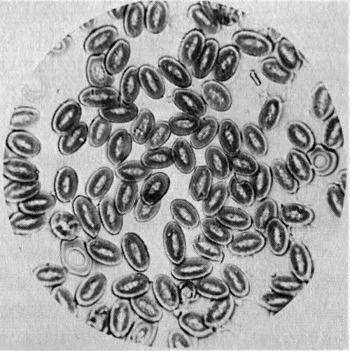
Figure 1 One of the earliest photographs of cells taken with a microscope. This photomicrograph shows cells in the blood of a pigeon. It was made by J. J. Woodward, U. S. Army surgeon, in 1871. Woodward had made the first cell micrograph (a graphic reproduction of the image of an object formed by a microscope) in 1866.
There is a remarkable similarity, moreover, in the molecular composition and metabolism[2] of all living things. This similarity has been taken to mean that life could have originated only once in the past and had a specific chemical composition on which its metabolic processes depended. This structure and metabolism were handed down to subsequent living things by reproduction, and all variations thereafter resulted from occasional mutation, or changes in the nature of the heredity-transmitting units. One of the most extraordinary of all the attributes of life is its ordered complexity, both in function and structure.
It is agreed among biologists that the functional manifestations of life include movement, respiration, growth, irritability (reaction to environmental changes), and reproduction and that these phenomena are therefore possessed by all cells. The first four of these can be grouped under a single word: metabolism. We can therefore say that living things have two common properties: metabolism and reproduction. Therefore, when we say we are studying life processes, we actually are studying the metabolism and reproduction of cells. Since metabolism is the sum of 4 the biochemical reactions taking place in a living organism, it properly belongs to the field of investigation of biochemists. Cell reproduction is the concern of both biochemists and morphologists[3] since it can be studied by either biochemical or morphological techniques.
Cell Structure
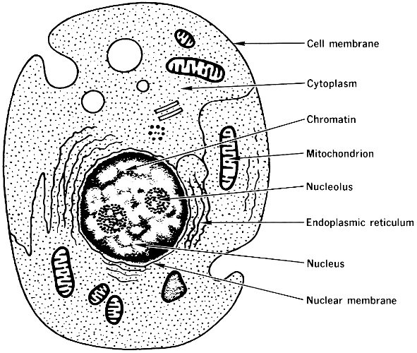
Figure 2 Generalized diagram of a cell, showing the organelles, or “little organs”, of its internal structure. The organelles that are labeled are important for this booklet.
The basic structure of a cell is shown in Figure 2. Each cell consists of a dense inner structure called the nucleus, which is surrounded by a less dense mass of cytoplasm. The nucleus is separated from the cytoplasm by a double envelope, called the nuclear membrane, which is peppered with perforations. The cytoplasm contains a network of membranes, which form the boundaries of countless canals 5 and vesicles (or pouches), and is laden with small bodies called ribosomes. This membranous network is called the endoplasmic reticulum and is distinct from the mitochondria, which are membranous organelles (little organs) structurally independent of other components of the cytoplasm. The outer coat of the cell is called the cell membrane, or plasma membrane, and forms the cell boundary.
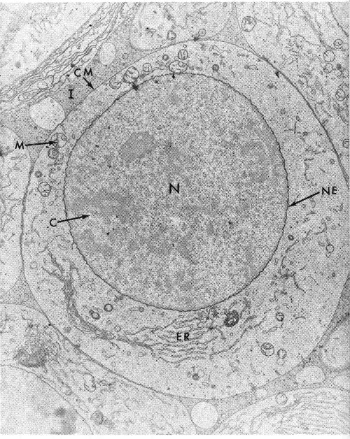
Figure 3 Electron micrograph of a primary spermatocyte cell of a grasshopper, showing the nucleus (N), endoplasmic reticulum (ER), mitochondrion (M), chromatin (C), nuclear membrane or nuclear envelope (NE), cell membrane (CM), and intercellular space (I). The magnification is about 25,000 times the actual size.
The nucleus, which in many cells is the largest and most central body, is of special importance. It contains a number of threadlike bodies, or chromosomes, that are the carriers of the cell’s heredity-controlling system. These contain granules of a material called chromatin, which is rich in a nucleic acid, DNA (deoxyribonucleic acid). The chromosomes usually are not readily seen in the nucleus except when the cell, along with its nucleus, is dividing. When the nucleus is not dividing, a spherical body, the nucleolus, can be seen. (In some nuclei there may be more than one.) When the nucleus is dividing, the nucleolus disappears.
6
Not all cells possess all these structures. For instance, the red cells of the blood do not have a nucleus, and in other cells the endoplasmic reticulum is at a minimum. The diagram (Figure 2) is valid for a great majority of the cells of higher organisms.
The cell structures shown in Figure 3 are visible with an electron microscope. They contain the chemical components of the cell. The chief classes of these constituents are the carbohydrates (sugars), the lipids (fats), the proteins, and the nucleic acids. However, a cell also contains water (about 70% of the cell weight is due to water) and several other organic and inorganic compounds, such as vitamins and minerals.
Carbohydrates serve mostly as foodstuff within the cell. They can be stored in several related forms. Further, they may serve a number of functions outside the cell, especially as structural units. In this way structure and function are correlated.
Lipids in the cell occur in a great variety of types: alcohols, fats, steroids, phospholipids, and aldehydes. They are found in all fractions of the cell. Their most important functions seem to be to form membranes and to give these membranes specific permeability. They are also important as stores of chemical energy, mostly in the form of neutral fats.
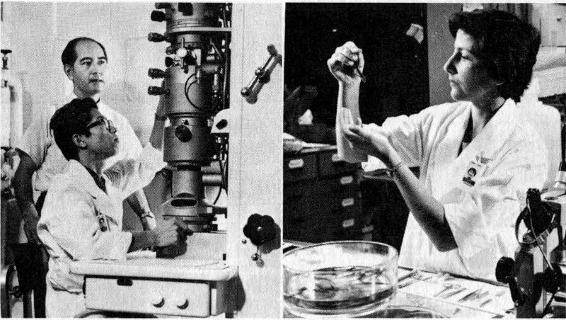
Figure 4 Scientists using an electron microscope (left) and an optical microscope (right) in fundamental biochemical research. Both instruments are important tools in studies of life processes.
7
The proteins occur in many cell structures and are of many kinds: Enzymes, the catalysts for the cell’s metabolic processes, are proteins, for instance. The nucleic acids are DNA and RNA (ribonucleic acid), which function together to manufacture the cell’s proteins. Since a large share of the remaining pages will be devoted to a discussion of proteins and nucleic acids, at this point we need only emphasize that these two types of materials are interrelated in their function and that both are essential.
The Two Nucleic Acids
It is not very fruitful to discuss whether proteins or nucleic acids are more important. That question is something like the one about the chicken and the egg. We cannot think of one without thinking of the other. Although our insight into the mutual dependence of these two materials has greatly increased in recent years and although we know the relation between them is a fundamental factor in such events as reproduction, mutation, and differentiation (or specialization) of cells, our understanding of their interplay is far from complete. Real understanding of the relation between them would give us insight into the essence of growth—both normal and abnormal—or, indeed, one could almost say, into the complexity of life itself.
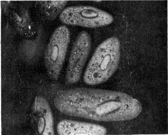
Figure 5 Photomicrograph of Paramecia, one-celled animals, magnified 1100 times. Many of the same structures that appear in Figure 3 can be seen here. This photo was taken with an “interference” microscope designed to permit continuous variation of contrast in the subject under study.
Practically all the DNA of most cells is concentrated in the nucleus. RNA, on the other hand, is distributed throughout the cell. Some RNA is present in the nucleus, but most of it is associated with minute particles in the cytoplasm known as microsomes, some of which are especially rich in RNA and are accordingly named ribosomes. These are much smaller particles than the mitochondria.
8
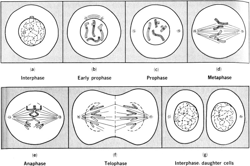
Figure 6 Stages of the mitotic cycle in a hypothetical cell with four chromosomes.
Mitosis
One of the most remarkable characteristics of cells is their ability to grow and divide. New cells come from preexisting cells. When a cell reaches a certain stage in its life, it divides into two parts. These parts, after another period of growth, can in turn divide. In this way plants and animals grow to their normal size and injured tissues are repaired. Cell division occurs when some of the contents of the cell have been doubled by replication, or copying (to be discussed later). The division of a cell results in two roughly equal new parts, the daughter cells. The process of cell division is known as mitosis and is diagrammed in Figure 6.
Mitosis is a continuous process; the following stages of the process are designated only for convenience. During interphase the cell is busy metabolizing, synthesizing new cellular materials, and preparing for self-duplication by synthesizing new chromosomes. In prophase the chromosomes, each now composed of two identical strands called chromatids, shorten by coiling, and the nucleolus and nuclear membrane disappear. During metaphase the chromosomes line up in one plane near the cell equator. At anaphase the sister chromatids of each chromosome separate, and each part moves toward the ends, or poles, of the cell. During telophase the chromosomes uncoil and return to invisibility; a new nucleus, nucleolus, and nuclear membrane are reconstituted at each end, and division of the cell body occurs between the new nuclei, forming the two new cells. Each daughter cell thereby receives a full 9 set of chromosomes, and, since the genes are in the chromosomes, each daughter cell has the same genetic complement.
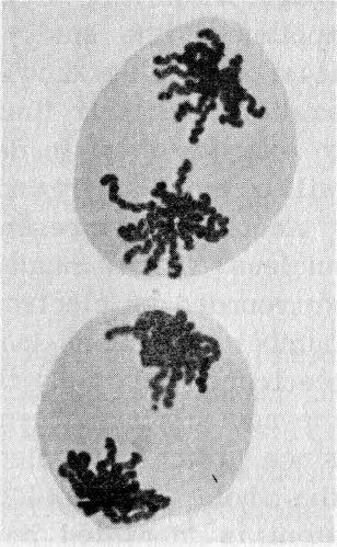
Figure 7 Photomicrograph of cells of the Trillium plant, which has five chromosomes, in anaphase. Note the duplicate sets of chromosomes moving to opposite poles of the cell.
All life processes use up energy and therefore require fuel. The mitochondria have a central role in the reactions by which the energy of sugars is supplied for cellular activity. The importance of this vital activity is obvious. In this booklet, however, we are concerned with the processes, involving nucleic acids and proteins, that can be described as making up “the gene-action system”. The gene-action system is the series of biochemical events that regulate and direct all life processes by “transcription” of the genetic “information” contained in molecules of DNA.
10