Chapter III · ITS CONTROL AND SUPPRESSION (continued 2)
Reading edition
Where the occupants resist this action sanitary orders should be issued in the usual manner and interference should be stopped until the order is served and complied with. There are a number of most insanitary and unsuitable shelters of bamboo, tin, etc., used for houses by a number of families in this square and it is desirable to tear down these huts if permission can be secured. If permission is refused orders should be issued on the usual form.
[Signed] T. W. Jackson,
In Charge of Plague Suppression.
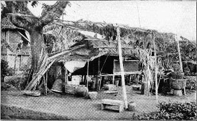
VIEW OF HOUSE AT 447 CALLE CONSERVADOR, TONDO, WHERE INFECTED RATS WERE FOUND (MANILA PLAGUE CAMPAIGN)
Specimen order issued by the Medical Inspector in charge of Plague Suppression.
Station "C," Tondo, May 21, 1913.
Redistribution of rat catchers and laborers engaged in antiplague work. Effective May 2, 1913.
Sanitary Inspector Kennard and 20 rat catchers will move into Tondo District and trap and poison rats in the district bounded on the west by Manila Bay and on the east by Estero Reina. The work will be begun at the extreme north water boundary of this district and will proceed toward the south.
Sanitary Inspector Brantigan with a similar number of rat catchers (20) will work within the same east and west boundaries and will begin trapping and poisoning at Calle Moriones, proceeding north. The poisoning and trapping is to be done in the most thorough manner possible, as this is a dangerously infected district and rat-plague must be controlled and terminated here.
The laborers, 60 men, divided into 4 parties of 15 men each under Assistant Sanitary Inspectors Jesús, De la Rosa, Laxamana and Parás, will continue the cleaning operations now under way on both sides of C. Juan Luna south of C. Moriones (plague localities in the same neighborhood), and thoroughly disinfect.
One party of 15 men will work in the vicinity of C. Perla, vacate basements as habitations, search for dead rats in yards, houses, bamboos, under broken concrete, etc., and will close up openings in structural bamboo by means of tin and cement. Emphasis is placed upon the necessity for permanently vacating basements and men will be sent back over the ground daily to see that the persons moved out do not return. Reports are desired so that prosecutions for violations of the law may be instituted if necessary.
[Signed] T. W. Jackson,
Medical Inspector in Charge of Plague Suppression.
Specimen order issued to Assistants.
May 4, 1913. Station "C," Bureau of Health:
Please place work parties in (interior) 1627–1629 Sande and 525 C. Azcarraga, to clean, disinfect and thoroughly investigate these premises and the houses, stables and other buildings in the vicinity. Search for rats, living and dead, rat nests and rats in bamboos and wood piles, stone piles, stables, under planks and elsewhere. Cement the openings in bamboos in houses or close with tin. Make notes on needed structural work. Do the work as thoroughly as possible.
[Signed] T. W. Jackson,
Medical Inspector in Charge of Plague Suppression.
Method of Procedure in Collecting and Forwarding Rats Suspected of Plague Infection to the Laboratory in Manila, P. I.—Rat catching,—trapping and poisoning,—is conducted in accordance with instructions contained in the Sanitary Inspector's Handbook (pp. 36, 37, 38) issued by the Bureau of Health.
Rats are collected in Manila and forwarded to the Bureau of Science for autopsy and for biologic examination for the presence of plague bacilli in the following manner:
The various groups of rat catchers are provided with receptacles (iron pails) and a supply of a mixture of kerosene, cresol and water (kerosene 10 parts, cresol 2 parts; water 88 parts).
In these vessels, filled with the pulicidal mixture, the rats are immersed, with a minimum amount of handling, as soon as they are found (whether in traps or dead from poison).
If captured alive they are killed and then promptly immersed. The mixture must be well shaken or stirred when used, as it separates upon standing. The immersion is, of course, for the purpose of destroying any fleas which may be present upon the captured rat.
A paper tag showing the date and the exact location of the place of capture, with the name or group number of the rat catcher, is next affixed to a foot or to the tail of the rat and firmly tied upon the same, where it remains until the rat cadaver is finally disposed of. This tag is a card of strong Manila paper and the record upon it is made with an ordinary lead-pencil, as both ink and indelible pencil marks are apt to become illegible from wetting, whereas lead-pencil marks are little affected thereby.
If desired, the disinfected tag in any given case of rat plague may be returned to the Bureau of Health, for identification, where an accurate record of every rat captured is kept.
After dipping and tagging, the rats are taken to a central point, again dipped, and placed in large, tightly-covered, galvanized iron cans, in which containers they are delivered to the laboratory by cart, once or twice daily.
The Case of Mr. C.—The following are the facts concerning the case of Mr. W. C., a prominent American resident of Manila who suffered and died from plague in 1914.
Mr. C., an editor, was taken ill with plague on the night of September 18, sought medical advice and entered St. Paul's Hospital September 19, and was transferred to San Lazaro Hospital, September 20, with an established clinical and bacteriologic diagnosis of bubonic plague. He survived till September 22.
Upon September 21, in the course of disinfecting the business office of Mr. C., located in a district which had furnished a number of cases of both rat and human plague, a dead rat, mummified, was found in the right hand drawer of his desk and fleas were seen to hop from the drawer upon opening it.
A flea killed by the disinfecting mixture at this desk was identified at the Bureau of Science as a rat flea (Xenopsylla cheopis).
The rat cadaver was sent to the Bureau of Science and the following facts were reported from there some days later:
The mummified rat and skeleton were pulverized in a sterile mortar and an emulsion was made and injected into guinea-pigs. The animals died from plague in a few days and plague bacilli were recovered from the tissues, as well as from the rat cadaver, by culture.
A second rat cadaver, found at the same time in the same building, during cleaning operations, was similarly treated with identical results.
There could scarcely be a stronger chain of convincing evidence against the rat and the flea, nor a more complete and convincing explanation of Mr. C.'s death than that afforded by these established facts and official documents. So far as I know there is no more striking case on record in the modern history of plague.
Letter of Warning and Appeal.—The following letter of warning and appeal for coöperation was suggested and framed by me February 10, 1914, at the time that extensive rat plague was discovered in the heart of the business district of Manila. I presented it to the Director of Health with a strong recommendation for approval and publication and after consideration he approved and authorized publication upon February 10. No change was made in the wording of the proclamation, but it was issued over the signature of the Director of Health to give added force and authority to the appeal. The results were, as I had hoped they might be, highly beneficial. The taking of the public into the confidence of the health authorities brought about a coöperation, without which our efforts in this difficult situation would have been sadly handicapped. It is my belief that this method should often be used by health authorities, particularly where an intelligent community is threatened.
To Whom It May Concern:
You are hereby informed that the district bounded by Calles Rosario, Juan Luna, Dasmariñas and Plaza Calderon (and possibly the neighborhood bordering upon this congested district) is a dangerous one for all persons living or conducting business therein, on account of the presence there of extensive rat plague. Six human cases (with five deaths) have recently developed there and many dead rats have been found. All human cases have been directly traced to rats dead from plague.
The Bureau of Health is now doing everything within its power to make this district safe, but the attention of all citizens, property owners and tenants is called to the fact that they are required by law to keep their premises free from rats and to abolish all structural conditions of the buildings which favor the harboring of rats. This means rat-proofing, and owners are earnestly urged to perform this necessary work now, under the direction of the Bureau of Health.
As a temporary expedient and safeguard all interiors, walls, floors and ceilings should be sprayed with kerosene daily, or at intervals of two days, to kill the fleas which carry plague from rats to human beings. All dark insanitary places used for living rooms should be vacated at once; all merchandise should be piled upon trusses at least a foot above the floor; all straw, shavings and other material attractive to rats for nesting, should be removed and burned and all food materials upon which rats may feed and live should be placed in covered boxes, bins or cans.
All rat-holes should be permanently closed and all broken cement or masonry should be repaired.
Observance of these instructions may save the lives of yourselves, your families and your tenants. It is your duty to do your part in this matter, a part which neither the Bureau of Health nor the Government can do for you.
Through very great effort the Bureau of Health has controlled plague in Manila and the Philippine Islands during the last two years.
Residents must now do their part, and owners of property must permanently make their buildings safe for tenants, both for business and residential purposes.
Bacteriologic Observations Made by Dr. Otto Schöbl.—The following observations upon the bacteriologic aspect of the Manila epidemic which we are considering were made by Dr. Otto Schöbl of the Biological Laboratory of the Bureau of Science, Manila, and pertain to the cases of the first year of the epidemic. They were printed in the December number of the Philippine Journal of Science in 1913, but as they belong so definitely to the epidemic I am describing and as Dr. Schöbl has expressed his willingness for me to quote them in full, I gladly accept his permission. Dr. Schöbl advanced the possibilities of blood-culture diagnosis to such a point of reliability that it became practically possible for us to expect positive culture in nearly every case of true plague and the whole matter of bacteriologic diagnosis was perfected to a high degree of efficiency under his administration of the laboratory work.
He relates his observations as follows:
During the recent outbreak of plague in Manila, I had the opportunity to make certain observations which are of interest. These observations were made in the examination of: (1) Specimens taken from patients and from dead bodies at autopsies, (2) samples of blood-sucking insects collected in houses where plague patients had lived, (3) rodents caught by trap or poisoned in the parts of the city where plague cases occurred from time to time, and (4) domestic animals suspected of plague infection.
I. Bacteriological Examination of Plague Patients
In order to secure as early diagnosis as possible, the following procedure of investigation was adopted:
The bubo was aspirated by means of a sterile hypodermic [129]syringe. The material thus obtained was placed in the water of condensation of an agar-slant culture tube.
At least 7 centimetres of blood were withdrawn from the cubital vein by means of another sterile syringe, and 5 centimetres of it were placed in an Erlenmeyer's flask, containing 200 centimetres of neutral meat broth. The rest of the blood was emptied into a sterile tube, and used for agglutination tests.
Cultures obtained by this method were examined microscopically, and the growths on various culture media were studied. Gram stain, Löffler's methylene blue, and hanging-drop method were used. Polar-staining and chain formation in liquid media and the characteristic type of colony on the surface of agar were looked for. Animal inoculation was performed in every case, and the culture isolated from each case was identified by agglutination test, rabbit's immune serum being used.
The results of the bacteriological examination of a series of 24 patients are tabulated in the two following tables. Table I includes the fatal cases and Table II those cases which recovered.
The diagnosis of plague could be safely made from the microscopical examination of the liquid aspirated from the bubo in the majority of the cases. However, in certain instances the amount of the aspirated fluid being small and the bacilli very few, it was impossible to diagnose the case, especially when the cultures from the bubo were negative. Repeated examination of the patient was necessary under those conditions, but it happened in cases 22 and 23 that the patients died of plague before a second examination could be made. The smears and cultures from case 22 remained sterile, while the smears and cultures
[130]Table I.—Examination of Fatal Cases of Plague
Patient Race Sex Age Date of examination Duration of illness Hours before death Bubo Blood Skin Sputum
Smear Culture Animal inoculation Culture Agglutination Smear Culture Animal inoculation Smear Culture Animal inoculation
Years 1912 Days
1. Sing Nu Chinese Male (?) July 11 5 48 + + + 0 0 + + + - - -
3. Aluncion Raymundo Filipino Male 15 Sept. 29 3 + + + 0 0 0 0 0 0 0 0
4. Filo Almalas Filipino Male 39 Oct. 10 4 22 + + + + - + + + 0 0 0
6. Polycarpio Guzman Filipino Male 34 Oct. 22 2 + + + 0 0 0 0 0 0 0 0
7. José Sarmiento Filipino Male 37 Oct. 22 3 + + + 0 0 0 0 0 0 0 0
8. Julian Gonzales Filipino Male 41 Oct. 22 3 23½ 0 0 0 + - 0 0 0 + + +
9. Valeriano Buencamino Filipino Male 31 Oct. 22 3 10 + + + + - 0 0 0 0 0 0
10. Pedro Nicomedes Filipino Male 30 Oct. 22 2 5¾ + + + + - 0 0 0 0 0 0
12. Regino Gulano Filipino Male 34 {Oct. 22 2 106 0 0 0 + - 0 0 0 0 0 0
{Oct. 24 4 82 0 0 0 0 0 0 0 0 + + +
13. Martin Dimalanta Filipino Male 35 Oct. 23 3 25½ + + + + - 0 0 0 0 0 0
14. Roberto Obiso Filipino Male 25 Oct. 23 1 53 + + + + - 0 0 0 0 0 0
15. Juan Barceta Filipino Male 23 Oct. 24 3 37 + + + + - 0 0 0 0 0 0
16. Yu Tum Chinese Male 14 Oct. 24 2 + + + 0 0 0 0 0 0 0 0
17. Augustin Monterey Filipino Male 29 Nov. 1 1 27 + + + + - 0 0 0 0 0 0
18. Demetrio Pabraw Filipino Male 27 Nov. 23 4 15 0 0 0 + - + + + 0 0 0
21. Ambrosio Sobremonte Filipino Male 20 Dec. 7 6 1 + + + + - 0 0 0 0 0 0
22. Mateo Marcelo Filipino Male 8 Aug. 20 (?) - - - 0 0 0 0 0 0 0 0
23. Alejandro Gita Filipino Male [A]17 Nov. 24 3 - - - 0 0 0 0 0 0 0 0
[131]Table II.—Examination of Plague Patients Who Recovered
Patient Race Sex Age Date of examination Duration of disease Bubo Blood
Smear Culture Animal inoculation Culture Agglutination
Years 1912 Days
{Sept. 29 2 - - - 0 0
{Oct. 2 5 + + + 0 0
2. Dionisio Capate Filipino Male 18 {Oct. 3 6 0 0 0 - +1:16
{Oct. 7 10 - - - 0 0
{Oct. 15 18 - - - - +1:64
5. Alejandra Fisher European Female 6 Oct. 20 7 + + + 0 0
{Oct. 22 2 + + + + -
{Oct. 24 4 + + + 0 0
11. Gabriel Sevilla Filipino Male 21 {Oct. 26 6 0 0 0 - +1:16
{Nov. 8 18 - - - 0 0
{Nov. 15 25 - - - - +1:64
{Nov. 26 3 + + + + -
{Dec. 6 13 0 0 0 - +1:32
19. Esteban Roa Filipino Male 15 {Dec. 16 23 - - - - +1:60
1913
{Jan. 11 48 - - - - +1:120
{Dec. 2 (?) + + + 0 0
20. Sia Su Chinese Male 35 {Dec. 5 - 0 0 0 + -
{Dec. 16 - - - - - +1:80
24. Purificacion del Val Filipino Female 19 {Dec. 11 3 + + + 0 0
{Feb. 11 33 - - - 0 0
Note.—The bubo in Nos. 2, 5, and 24 never opened spontaneously. The pus was aspirated at the time of the second, eventually third, examination. Nos. 11 and 19 opened spontaneously. A fistula formed along the canal which was caused by the puncture, and healed up in several weeks. Hard inguinal buboes of secondary order persisted in patient 19 at the time of second examination. No plague bacilli were found either in the bubo of the first or second order. Patient 20 had a considerable amount of pus in the inguinal primary bubo, but it was not opened until after the last examination. made from the swelling on the neck of patient 23 revealed the presence of pneumococci. Both patients died of plague, as was ascertained by examination of the organs after death.
Two of the patients, cases 8 and 12, had numerous plague bacilli in the sputum at the time when the expectoration showed the presence of blood (twenty-three and one-half and eighty-two hours, respectively, before death). In 3 cases I was able to prove the presence of Bacillus pestis in the skin lesions, intra vitam, fifteen, twenty-two, and forty-eight hours, respectively, before death. In case 18 there was no doubt that the skin lesions, which covered the whole body and the face, were of secondary nature, as the patient died shortly afterward. It was undoubtedly a case similar to those reported by Gotschlich and Zabolotny.[5] In the other two patients there was only 1 maculopapulous efflorescence on the foot in case 1 (with a corresponding femoral bubo) and 2 lesions of the same type on the arm and forearm in case 4 (with a corresponding axillary bubo). It is possible that these lesions were the original port of entry of infection. Numerous plague bacilli were found in the skin lesions of these cases, both microscopically and in culture.
The plague patients tabulated in Table II recovered. They were all treated with antiplague serum. While cases 5, 11, 19, and 24 appeared clinically to be rather severe, cases 2 and 20 were mild.
It can be seen from the table that the plague bacilli may not be detected in the enlarged gland at first (case 2) and that their presence may be revealed only after repeated examination of the bubo. It is also evident from the results of repeated examinations that the plague bacilli disappear from the infected gland in a comparatively short time, as a rule at the time when pus starts to form. Contrary to the findings in patients who died, distinct phagocytosis was noticed in the smears made from the aspirated liquid in those patients who recovered and who had been treated with serum soon after the onset of the disease. It is undoubtedly this process that clears the gland of the infectious agents.
The general opinion in regard to the presence of Bacillus pestis in the circulating blood seems to have been, as Thompson remarks, that "the bacillus is rarely to be found in the peripheral blood stream before the agonal stage."[6]
The Austrian Commission, using few drops of blood, found positive blood culture in 40 per cent; Calvert in Manila in 100 per cent when examined twenty-four hours before death; Choksy, Berestneff, and Mayr in 45 per cent; and Greig in 60 per cent. The Indian Commission examined 28 patients, and obtained positive blood cultures in 16 out of 23 fatal cases. Not a single positive blood culture was obtained from the patients who survived. The time of blood examination in positive cases was three and one-half to seventy-five and one-half hours before death. The amount of blood used was 1 cubic centimetre. Only 6 out of the 30 samples, which gave positive blood culture, were found positive by microscopical examination of blood smears. The following conclusions are based on these observations in regard to the septicæmic stage of bubonic plague: (1) "A severe septicæmia may be present at a comparatively early stage of the disease and for a considerable number of hours before death, and (2) the septicæmia may be of an irregular and fluctuating type."[7]
From the tables it will be seen that out of 15 patients examined by me, 14 gave positive blood culture; and of these 3 recovered. One blood culture revealed the presence of streptococcus in addition to Bacillus pestis. The results of the examinations tabulated in Tables I and II show, in agreement with the findings of the Indian Commission, the occasional early occurrence of plague bacilli in the blood stream, as the time of examination in the positive cases varied from one hour to one hundred six hours before death. In consideration of the ephemeral character of the septicæmic stage of plague, as evidenced by repeated blood cultures in the three patients who recovered, one can hardly avoid the impression that there is a certain degree of septicæmia in every case of plague. The possibility of detecting the bacillus in the circulating blood increases in proportion with the quantity of blood used for culture. The best chance to recover plague bacilli from the circulating blood seems to be in the stage of high fever and general prostration.
The phenomenon of agglutination of plague bacilli by the serum of patients was first observed by Wissokowitsch and Zabolotny in 1897[8] and later confirmed by the German Plague Commission. Vagedes, Klein, and others[8] pointed out the defects of the reaction as a diagnostic means. Aside from the technical difficulties, the reaction was found inconstant, and its occurrence was not noticed until the second week of the disease and even then only in low dilutions of the serum.
Although the recent work of Strong[9] and of Strong and Teague[10] has reduced the technical difficulties, the fact remains that positive agglutination of plague bacilli by the patient's serum cannot be obtained in the first week of the disease, and, therefore, the isolation of plague bacilli from the body of the patient is still the only quick and safe method of plague diagnosis.
Having utilized the technic devised by Teague, I have had no difficulty in performing the agglutination test in plague. The emulsion of plague bacilli, to be used for the test, was prepared by suspending young cultures of virulent plague bacilli, grown at 30° C., in salt solution and filtering the suspension through filter paper. No antiseptic was added nor heat applied. Serial dilutions of unheated patient's serum were mixed with equal amounts of bacterial suspension in small test tubes. Incubation at 35° C. followed. Controls, consisting of serial dilutions of normal human serum as well as bacterial suspensions without serum, excluded any possible error which might have been caused by spontaneous sedimentation of the bacterial suspension; while a parallel test with highly agglutinant serum facilitated the reading of positive results.
Altogether, 22 tests were performed on 15 patients, 11 of whom were fatal cases and 4 of whom recovered. In the negative reactions, the duration of the disease at the time of examination ranges from two to six days. The non-fatal cases showed slight agglutination from the sixth day on. From that day, the agglutination titer of the serum was found to rise, and the agglutinins persisted in the blood of convalescents up to the seventh week of the disease.[11]
It must be borne in mind that the patients, who showed positive agglutination, had been vigorously treated with antiplague serum. Nevertheless, in consideration of the low titer of the curative serum (dilution 1:32, agglutination positive; dilution 1:64, agglutination negative), the rise of the agglutinant power of the patient's serum in dilutions higher than 1:16 cannot be explained as wholly due to passive immunity, but rather to active immunity arrived at on the principle of simultaneous immunization.
From the preceding observations the following conclusions are drawn:
The importance of blood cultures as a diagnostic means is evident from the fact that positive blood culture was obtained in practically every case that was examined in the febrile stage of the disease, even when buboes or signs of pulmonary involvement had not been detected clinically.
It is also evident that Bacillus pestis may be found in the circulating blood of the patients even in cases which subsequently recover.
The period of time during which Bacillus pestis circulates in the blood is evidently short and irregular.
[137]Mixed infection may be encountered in plague septicæmia (Streptococcus, Pneumococcus).
The agglutination test is of no value for the diagnosis of plague, as it was found positive only in convalescents.
Phagocytosis of plague bacilli in the bubo was noticed only in patients who recovered after being vigorously treated with curative serum.
The presence of numerous plague bacilli in comparatively insignificant skin lesions during the life of the patient points to the possibility of direct transmission, while the fact that a patient without any apparent bubo, who is not so sick as to be detained from his daily occupation, may expectorate large numbers of plague bacilli, are facts of great importance with regard to the communication of the disease. It is obvious that the last-mentioned condition might, and very likely does, give rise to an epidemic of pneumonic plague if the atmospheric and sanitary conditions are favorable.
[138]Table III.—Insects Found to Contain Bacillus Pestis
Author Insect Source of infection Experimental transmission
Yersin Flies Laboratory infection
Nuttal Flies Experimental infection
Nuttal Bedbugs Experimental infection Negative by bite.
Nuttal Flea Experimental infection Negative.
Hankin Ant's fæces Fed on plague material
Hankin Bedbugs Plague hospital
Ogata Flea Plague rats
Simond Flea Plague rats, experimental Positive.
Tindswell, 1900 Flea Plague rats Negative.
Tindswell, 1903 Flea Plague rats Negative.
Kolle Flea Experimental infection Negative.
Gauthier and Raybaud Flea Experimental infection Positive.
Liston Flea Epidemic among pigs; harbored fleas; dead rats found Positive.
Zirolia Flea Retained Bacillus pestis, 7–8 days
British Commission Flea Repeated experiments Positive.
Verbijtski Flea and bedbug Experimental infection Positive.
La Bonadière and Xanthopulides Fly
Herzog Pediculus capitis Dead body of a plague case
II. Observations on the Transmission of Plague By Blood-sucking Insects
Judging from the data which have been collected from the literature[12] on the transmission of plague (Table III), Simond seems to have been the first to call attention to the important part which blood-sucking insects, particularly fleas, play in the transmission of plague. Although many investigators have been successful in demonstrating the presence of Bacillus pestis in the digestive system of blood-sucking insects, it was not until the experiments of Gauthier and Raybaud that the actual transmission of plague infection by fleas was convincingly proved. Ever since the exhaustive and conclusive experiments, which were carried out both under natural and artificial conditions by the British Plague Commission, and the work of Verbijtski, which antedates the British Commission, were presented, there has been no doubt that the transmission of plague by blood-sucking insects, particularly by the fleas, is one, although not the only, mode of spreading this disease. It is obvious, as Herzog correctly remarks, that the factors which are responsible for the spreading of plague must be considered individually in each epidemic and in various parts of the world as well. There is no doubt that the importance of any insect in the transmission of plague depends on its habits as well as on those of the host, be it either animal or man.
During the recent outbreak of plague in Manila, several samples of bed-bugs from the beds of the plague patients and dog fleas from a plague-infected house were collected and examined, but with negative result.
In spite of the fact that it adds nothing new to the question of whether or not plague can be transmitted by fleas, since the question has been conclusively answered by the work of the Indian Commission, nevertheless the following observations of a small outbreak of plague among animals, the spreading of which was due solely to fleas, are of interest.
One wild rat was inoculated with strain Iloilo 3 of Bacillus pestis. The skin adjoining the root of the right ear was scarified, and a loopful of the culture was smeared on the scarified skin. The rat was found dead three days after the inoculation.
The cage containing the dead rat was immersed in kreolin solution. At autopsy the cervical glands were found slightly swollen, somewhat reddened, but no hæmorrhagic œdema of the surrounding tissue was noticeable. There was slight necrosis at the place of inoculation, showing superficial, purulent discharge. Clear effusion in both pleural cavities and one hemorrhage in the pleura were found. The lungs were hyperæmic, but otherwise normal. The spleen was of somewhat darker color, but otherwise normal in size and appearance. The liver showed a slight degree of parenchymatous degeneration, the congestion making prominent the structure of the organ. The typical, although not constant, changes of the organ, which are characteristic of natural plague infection in rats, were absent. The kidneys were without macroscopic change. The lymph glands, with exception of the cervical nodes, were normal.
Examination of the rat's fur revealed ectoparasites on the neck, under the chin, and back of the ears; these at the time of the examination apparently were dead. About 6 common rat fleas were found and identified as Lœmopsylla cheopis Rothsch. The parasites were immersed in sterile salt solution for three hours. When removed in a dry test tube, they began to move about sluggishly. The intestinal tract of these fleas contained blood.
Five of the fleas were crushed by means of sterile forceps, and inserted in a pocket under the shaved skin of a guinea-pig. The animal died of plague within three days, showing considerable hemorrhagic œdema around the place of inoculation, typical bilateral inguinal buboes, and characteristic changes in the spleen. Smears and cultures made from the bubo and spleen were positive for Bacillus pestis.
Another wild rat, which was in a separate cage in the same room where rat 1 had been kept, died twenty-four hours after rat 1. The two cages were at least 10 centimetres apart. Rat 2 harbored fleas of the same species as were found on rat 1.
Numerous severe bites were detected back of the ears and on the neck of the dead animal. The post-mortem findings were identical with those described in rat 1; that is, cervical buboes, pleural effusion, and slightly enlarged spleen.
It is well to remark that both rats had been kept in the same room for about six months. Fleas had never been noticed on our guinea-pigs. During the time the rats had been kept in the plague house no irregular results were noticed in plague-inoculated animals. At the time the first rat was inoculated no other plague-infected animals were in the plague house, and since that time another building has been used for plague-infected animals.
Two days after the death of rat 2 three guinea-pigs, which were kept in separate cages in the same room, were found dead of plague (smears and cultures were both positive). Several fleas (Lœmopsylla cheopis) were found on the necks of these animals. They were collected and inoculated in the same way as the fleas from the first rat. The experimental animal, which was inoculated with the fleas, was killed and found to be infected with plague. The findings were local reaction, inguinal buboes, and typical spleen. Smears and cultures were positive for Bacillus pestis.
Although numerous healthy guinea-pigs were examined in the same plague house, no fleas could be found at that time, only the 2 rats and the first 3 guinea-pigs are positively known to have harbored fleas, the latter after the death of the rats and not before.
The gross lesions in these naturally infected guinea-pigs were somewhat unlike those found in guinea-pigs infected either by vaccination or by intraperitoneal or subcutaneous inoculation. All except one showed primary buboes on the neck with more or less extensive hemorrhagic œdema extending in some cases over the thorax. There was little pleural effusion present; the spleen always showed typical changes of necrotic foci varying in size and number. In one instance similar foci were found also in the liver, large enough to be visible macroscopically. This was in a case where like changes were found in the lungs.
Only one of the guinea-pigs showed an exception, in that the primary buboes were located in the inguinal region, with pelvic and axillary glands secondarily involved. These are the findings usually met within guinea-pigs artificially infected with plague by the vaccination method, if the lower part of the abdomen be chosen for inoculation. The reason for such a deviation from the findings in the rest of the guinea-pigs may lie in the fact that this animal was almost completely deprived of hair by a skin disease.
It is of importance to mention the skin lesions which were found on the necks of the guinea-pigs, particularly under the chin. Besides small red spots which appeared to be fresh flea bites, small, elevated, and fairly deep infiltrations partly covered with moist scab were found in the skin under the chin. Other animals showed changes usually found in the scarified skin of guinea-pigs after artificial inoculation with plague material. The base of each cutaneous efflorescence was hemorrhagic and œdematous.
A histological study of the tissues of these guinea-pigs known to be naturally infected by plague fleas showed the following changes:
The Cervical Bubo.—The enlarged lymphatic gland was surrounded with a thickened capsule. Necrosis existed in the subcapsular part of the gland, where it formed an almost continuous circular zone, leaving the central part less changed. Smaller irregular necrotic foci were scattered throughout the section. Polymorphonuclears in various stages of disintegration were found throughout the section.
The Lungs.—Very few blood extravasations were present in the alveoli; otherwise normal.
The Spleen.—The capsule was thin. There were subcapsular hemorrhages. The Malpighian bodies were somewhat enlarged, but of normal structure. Throughout the parenchyma irregular multiple necrotic foci were found, leaving but little of spleen tissue intact. Numerous polymorphonuclears which were present showed varying degrees of karyorrhexis.
The Kidneys.—The outline of the cells was indefinite; a few miliary hemorrhages existed in the cortical part of the organ.
The Liver.—There was excessive congestion, fatty degeneration, and pigmentation of the cells. The capsule was slightly thickened.
The Skin.—The epithelium was missing in one place in the section, and cellular infiltration extended from that place into the subepithelial layer of the surrounding skin. The same kind of infiltration reached deep into the skin, stripes of cellular infiltration penetrating into the tissue along the muscle fibres. There was no direct connection between the cellular infiltration and the follicles of the hair.
It may be well to describe in detail the time of death from plague among these and the other animals in this outbreak, as well as the time when the plague house was disinfected.
The first animal (rat 1) having been inoculated on August 27, in the afternoon, died of plague within three days (August 30). The second animal (rat 2) died twenty-four hours later. Guinea-pigs 3, 4, and 5 (see plan) were found dead on the morning of September 2; that is, two days after the death of rat 2 and three days after the death of rat 1.
The same day that the three guinea-pigs were found dead of plague, rooms I, III, IV, and VI were thoroughly disinfected. The floor, the ceiling, and the walls were sprayed with kerosene and lysol solution. The remaining animals in room VI were destroyed, and the cages disinfected. No animals were kept in rooms I, III, and IV at that time.
Three days after the death of animal 5, guinea-pigs 6 and 7 were found dead of plague, while the next day guinea-pigs 8 and 9 died. No death occurred on September 7, but the next two days each recorded two plague guinea-pigs (10, 11, 12, and 13). On September 11, the last guinea-pig died of plague in this outbreak. The whole building was then thoroughly disinfected. No plague-inoculated animals were kept in the rooms after the first sign of the epidemic. After September 11, no more cases of spontaneous plague infection were observed.
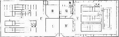
ANIMAL HOUSE
It will be noticed that the epidemic lasted eleven days after the first animal died and fourteen days after animal 1 was inoculated. Altogether, 14 animals out of at least 200 animals exposed died of plague.
No death occurred among rabbits, although these animals were distributed among the guinea-pigs. In fact, 2 rabbits were surrounded by plague guinea-pigs 8, 9, and 10, but did not contract plague.
From the epidemiological standpoint it is interesting to know the dimensions and location of the cages in which the animals were kept.
Aside from the 2 rats which were confined in ordinary traps that stood on a table 80 centimetres high, the rest of the animals were kept in regular metal animal cages. The dimensions of the cages are: Fifty centimetres long, 36 centimetres broad, and 30 centimetres high. The cage stands on four legs each 10 centimetres long; the centre of the bottom of the cage holds a drain opening 8 centimetres above the floor.
The majority of the cages in room II were located on the floor; some on the second shelf of a wooden rack. This last-mentioned arrangement, judging from the construction of the wooden frame, allowed a continuous passageway for the fleas to the second shelf of the racks. On the other hand, the deaths among the guinea-pigs in room V were restricted to the cages standing on the floor, the majority of cages in that room being placed on tables 80 centimetres high.
Only a theoretical explanation can be given of the short duration and sudden cessation of the outbreak. One can assume with great probability that the first partial disinfection drove the fleas away from the primary source of infection, and that they traveled as far as possible. They finally settled in those guinea-pig cages which had not been molested by the first disinfection. Having no new supply of plague blood (all of the plague-infected guinea-pigs having been removed, most of them before death), the fleas soon cleared themselves of plague bacilli. The peculiar feature of the outbreak, namely, the failure to find fleas on the animals in rooms II and V, finds its explanation in the observation of the Indian Commission who found that the fleas "died or disappeared very rapidly."
The following conclusions can be drawn from these observations:
The common rat flea (Lœmopsylla cheopis) prefers the rat to the guinea-pig.
In the absence of rats it will attack guinea-pigs rather than rabbits.
The fleas which have sucked blood from rats or guinea-pigs afflicted with plague septicæmia were found to harbor virulent plague bacilli inside of their bodies.
The transmission of plague infection by direct or indirect contact being excluded in our case, the fact that fleas of the same species and harboring plague bacilli were found on the rat and on the guinea-pigs, the presence of flea bites on the rats and on the guinea-pigs with positive findings of skin lesions on that part of the body where the fleas and flea bites were located, together with the anatomical picture of the findings in the guinea-pigs, lead to but one explanation; namely, that the plague infection was transmitted by fleas.
III. Observations on Animals Suspected of Plague
Out of the several tens of thousands of rodents examined during the antirat campaign, we have found only two plague rats which showed the typical picture of natural plague infection in rat; that is, cervical buboes with surrounding œdema, subcutaneous injection, pleural effusion, enlarged spleen, and such changes of the liver as are characteristic of natural plague infection in rats. Microscopically, large numbers of plague bacilli were found in these cases, and pure cultures of Bacillus pestis were recovered from the spleen. Histological examination of internal organs, particularly that of the liver, confirmed the bacteriological findings. The remainder of the plague rats exhibited only two of the signs of plague infection, namely, bubo and œdema of the surrounding tissue, and eventually hemorrhages.
Besides plague infection, a great number of rats showed purulent conditions from causes other than plague. Abscesses of the lungs were frequently met with, and cervical or axillary buboes are not uncommon in Manila rats. Various pyogenic bacteria were found in the pus of such abscesses. Of the less common was Bacillus pyocyaneus and the pneumobacillus of Friedländer. Chronic plague was excluded in these cases since the animal inoculation failed to produce plague infection.
More than half of the rats examined harbored parasites in their organs. Echinococcus taeniæformis was found in the liver of practically every gray rat, while a small Ascaris and Tænia diminuta were not uncommon in the intestines. Two rats were found to have sarcosporidiosis, 2.6 per cent. showed rat leprosy, and 7.4 per cent. trypanosomiasis. One tumor of the mammary gland and one tumor in the axillary region were encountered, while one tumor of the large curvature of the stomach proved to be a chronic inflammatory tumor due to parasites. One peritoneal tumor in a rat (Mus decumanus) gave the impression of a malignant tumor on account of the miliary dissemination of the peritoneum. It was found to consist of muscle and spindle-cell sarcomatous tissue. Ectoparasites were very seldom noticed, on account of the method of collecting the rats. When present, they were mites and fleas.
In the naturally infected plague rats the rigidity of the fresh cadaver was pronounced. The primary bubo was in every case cervical. Cervical glands were enlarged and hemorrhagic with slight œdema of the surrounding tissue. The subcutaneous injection extended over the neck and chest. The inguinal glands were small and pigmented. The lungs were collapsed, and showed hemorrhagic foci. The spleen was slightly enlarged, firm, and dark red. The liver was rather large, firm, pale red, with shade of yellow, which was caused by minute yellowish foci thickly scattered throughout the tissue and visible through the capsule. The kidneys were hyperæmic. The intestines were without change. The serous membranes were pale with no hemorrhages.
Histological examination of the tissue of naturally infected plague rats showed the following changes:
Liver.—The structure of the organ was well marked; the veins dilated, trabeculæ slightly compressed, nuclei well stained, and few of the liver cells showed vacuoles. Small foci, most numerous under Glisson's capsule, were scattered throughout the organ; they varied in size, but were not larger than a miliary tubercle. The small necrotic foci were found to consist of few necrotic liver cells. The centre of the larger foci was formed by degenerated and necrotic liver tissue, surrounded by round-cell infiltration. Polymorphonuclears were also found in the zone of cellular infiltration. There was a slight degree of hemorrhage in each focus. Epithelioid cells and large vesicular cells with several nuclei were to be found. The foci, mentioned above, were sharply demarcated from the surrounding liver tissue, which appeared to be intact.
Spleen.—The structure was well preserved, the capsule thin. The Malpighian bodies were normal as to the elements of which they consist. Cells with pycnotic nuclei were scattered throughout the organ, and vesicular cells with small, deeply stained, excentrically located nuclei were present. Polymorphonuclears were found in the tissue in considerable numbers. No localized necrotic foci could be found in sections through the spleen.
Cervical Glands.—The blood-vessels were considerably distended. A few hemorrhages and polymorphonuclears were present. Œdema of the capsules and surrounding tissue existed. Part of the gland was necrotic.
Lungs.—The blood-vessels were distended. The alveoli contained homogeneous masses and blood. There were numerous subpleural hemorrhages. The bronchi were collapsed, and contained mucus.
Kidneys.—The cortical part showed subdued structure; the epithelial cells had an indefinite outline and occasionally showed vacuolization. The medullar part was better preserved. There were miliary subcapsular hemorrhages. A few small foci were scattered throughout both medullar and cortical parts. They consisted of round-cell infiltration.
Natural Plague Infection in a Cat
The experiments of the German Plague Commission proved that cats showed considerable resistance to plague infection as cutaneous and subcutaneous inoculations failed to infect them. According to the Austrian Commission, cats develop submaxillary buboes if fed on plague material. They are said by Albrecht and Gohn[13] sometimes to recover. Out of four cats fed on plague material two died of plague, one showing submaxillary, the other mesenterial buboes. Virulent plague bacilli were found in the discharge from the nose and also in the fæces of cats which apparently did not become infected after having been fed on plague material.
One case of spontaneous plague infection of a cat was recorded by Thompson[14] in Sydney.
W. Hunter,[15] in Hongkong made observations on cats suffering from plague infection. The author also undertook a few experiments, and arrived at the following conclusions:
Cats suffer from plague.
The disease may be acute or chronic.
The type of the disease is septicæmic.
The animals may occasionally play a part in the dissemination of plague.
In plague-infected areas cats probably become infected through rats, which they devour as food.
In plague-infected districts possible plague infection in cats is of great importance from a domestic point of view.
On November 27, 1912, a sick cat was brought to the laboratory for examination. It was reported that the animal was found in a warehouse in which dead rats had been found some time previously. The rats were not examined. In the morning of the 30th, the cat was found dead in the cage where it had been kept under observation. The following are the post-mortem findings:
The animal was a fairly well-nourished female.[16] The subcutaneous tissue, pericardium, mediastinum, and mesenterium contained considerable amounts of fat.
The subcutaneous tissue of the neck showed œdema and small hemorrhages. The submaxillary tissues were swollen on both sides. When the fasciæ and superficial muscles of the neck were removed, enlarged glands were found on both sides. These were closely attached to the submaxillary salivary glands. The surrounding tissue was œdematous, but no hemorrhages were noticed in the vicinity of the enlarged glands. Upon section the glands were found to be necrotic, and upon pressure a thin purulent liquid escaped. There were no hemorrhages within the glands. Several enlarged lymph-nodes, smaller in size, could be followed down the neck on the left side. The lymph-nodes in the axillæ as well as in the groins and peribronchial nodes were normal. The mesenteric glands were slightly enlarged and reddened.
The lungs were slightly collapsed. A clear, sanguineous, slightly coagulated effusion was observed in both pleural cavities. The tissue of the lungs showed considerable œdema and hypostasis. The bronchi and pharynx showed no changes, the mucous membrane being pale and thin.
The heart was normal.
The spleen was enlarged, of light red color, with follicles slightly prominent.
The stomach contents was blackish in color; there were no hemorrhages or ulcers in the mucosa.
The liver was somewhat enlarged. The organ showed prominent structure, the centres of the acini being red, the periphery lighter in color.
The kidneys were slightly enlarged and pale. The capsule peeled off easily, the venæ stellatæ were prominent, the surface smooth; there were no hemorrhages. The cortex was increased in breadth and was of the same color as the surface; the pyramids were darker in color. The organ was of fragile consistence.
Suprarenals were normal, as were also intestine and bladder.
The histological findings were as follows:
Bubo.—The capsule of the gland was œdematous. The whole gland as seen in cross section had undergone necrosis, except a few foci which still showed cellular structure.
Lungs.—The alveoli were filled with homogeneous masses, containing but few degenerated epithelial cells and leucocytes. The blood-vessels were dilated, particularly in the subpleural part of the organ. In some places capillary mycotic emboli with subsequent hemorrhage were encountered. The large blood-vessels and bronchi were normal.
Salivary Glands.—Those glands attached to the primary bubo showed the normal structure of a combined mucous and serous gland.
Liver.—There was considerable congestion. The centres of the acini showed parenchymatous and fatty degeneration. The cells on the periphery of the acini exhibited typical fatty infiltration. The large blood-vessels and small ducts were without change.
Kidney.—The cells of the kidney showed various degrees of degeneration, ranging from parenchymatous to fatty infiltration. There were a few capillary hemorrhages and hyaline casts present.
Suprarenals.—These showed slight degeneration.
Spleen.—This organ showed congestion, a few hemorrhages, and bacterial emboli; otherwise normal.
The bacteriological examination of the material from this cat gave the following results:
Smears: From the buboes showed degenerated leucocytes, many lymphocytes, and numerous bacteria, some of which resembled Bacillus pestis in their polar staining. From the spleen showed numerous plague-like, polar-stained bacilli. Round involution forms were present.
Cultures: From the buboes were badly contaminated with Bacillus coli and Bacillus pyocyaneus colonies. From the spleen: A few scattered colonies of Bacillus pyocyaneus developed on the surface of the agar. Between the large colonies a scanty growth of dewy appearance was noticed. Smears made from this growth revealed plague-like bacilli of the cultural type, showing a few club-shaped involution forms. Subcultures were made in order to secure pure culture. They showed a pure growth of Bacillus pestis as indicated by the morphology of bacilli and shape of the colonies. Agglutination with plague-immune serum was positive.
[154]Inoculation experiments (vaccination method): One guinea-pig was inoculated with the material from the left bubo, another one with material from the right bubo. They died of plague on the third and fifth days, respectively. One guinea-pig was inoculated with the material from the spleen. It died of plague on the third day. One guinea-pig was inoculated with material from the nostrils obtained by swab. The animal survived, showing no indication of plague. One guinea-pig was inoculated with material from the rectum obtained by swab. It died of plague on the fifth day.
Although plague infection among cats is apparently a rare occurrence, the fact that cats may contract the disease in spite of the high degree of resistance to plague infection has to be considered from the hygienic standpoint.
To appreciate the important rôle which cats may play in the spreading of the disease one need only consider the close contact of these animals with rats on one side and human beings on the other. It is also a well-established fact that not only plague-infected cats, but also those which have devoured plague-infected material and remained apparently normal, may excrete plague bacilli which have retained their full virulence.
Notes on Plague in Hong Kong by Dr. Roberg.—During the Hong Kong epidemic of plague which preceded and was coincident with that of Manila, I visited that city twice (December, 1913, and July, 1914), but I did not closely investigate the methods adopted and carried out by the authorities there, for the reason that the Manila plan was so much more productive of results, as shown by the apparent inability of the Hong Kong officials to gain control of the disease. However, I received from Dr. David Roberg, of the Oregon State Board of Health, a copy of a report made by him to the Secretary of his State Board of Health, following an investigation of the Hong Kong epidemic and the methods there followed. I have Dr. Roberg's permission to use his report and it is herewith presented. It is dated Manila, April 16, 1914, and is as follows:
I have the following notes to present on the epidemic of bubonic plague in Hongkong.
On April 5th when I arrived in Hongkong the epidemic was rapidly approaching its height. With its onset in January there were 47 cases, in February 42, and in March 223. During the week previous to April 5th, there were 91 cases; during the six days I was in Hongkong they averaged 15 a day.
Judging from previous epidemics the present one will be exceptionally severe. The season for the occurrence of human plague is from the months of February to July. The onset is gradual; in May it reaches its maximum and then declines. In the epidemic of 1912, for the city of Victoria the monthly rate showed the following, January 9, February 22, March 61, April 265, May 513, June 346, July 105, August 11, and September 1. Comparing these rates with those of the present year it will be seen that the number for March far exceeds that of two years previous.
Illustrating the season for human plague, with its onset, maximum and decline, are the monthly rates for the city of Kowloon during 1912, when the following cases occurred: February 2, March 12, April 52, May 246, June 152, July 39, August 8, and September 3.
The season for human cases is determined by the condition of the rats. At the close of the season in July the rats die off from plague in great numbers as it is then the hottest time of the year. During the months from September to February the rats increase in number and in susceptibility to the extent of being sufficient to again infect human beings. Moreover every other year shows a marked severity in the epidemics of human bubonic plague. This is explained by the fact that it requires two years' time for the rat population to become of sufficient greatness and susceptibility to cause a severe human outbreak. This is shown by the yearly number of cases since the year 1911. During the years 1911, 1912 and 1913 respectively, there were 253, 1847, and 408 cases. During the present year the monthly rate is exceeding that of the heavy year of 1912.
The severe epidemic in 1912 was a result of the influx of 50,000 Chinese refugees into Hongkong during the revolution in 1911. The number of rats in the native district depends upon the available food supply, and as a result of this human overcrowding the amount of waste food so increased in the houses, yards and streets, that the over accumulation of garbage could not be kept pace with. This influx also brought in great numbers of susceptible rats.
The number of rats killed off during the epidemic in 1912 were so great that in 1913 they had not recovered sufficiently to cause a severe outbreak during that year, and as a result of the lightness of epidemic in 1913, they are so increased in number and susceptibility now that they are causing a very severe epidemic in human beings.
Of rats in Hongkong they have the Mus decumanus or drain rat and the Mus rattus or house rat. It is noteworthy that the drain rat is found plague-infected throughout the year, while the house rat is found infected only during the period in which the human epidemics occur, namely from February to July. The number of infected rats a year run parallel to the number of monthly cases.
The bulk of human infection is due to the spread of house rats. Man also becomes infected by the drain rat when the drains are flooded by rain storms and the rats are driven into the houses.
What has made plague permanent in Hongkong is the overcrowding of the native districts. Besides there is a floating population entering and leaving the native quarters, numbering about 4000 a day. The native houses have been built with double floors and walls which harbor the rats. Where the construction is of wood it is possible to remove the rat spaces. It has been found since the introduction of plague into Hongkong in 1894, that those districts containing the greatest number of soft brick houses with hollow walls, have shown the greatest incidence of plague. This can not be remedied as it would involve the destruction of buildings on too large a scale.
The Work of the Sanitary Board
The area under the control of the Board comprises the Island of Hongkong containing 32 square miles, with a sea frontage of 13 miles in length. Included also is the old city of Kowloon which is situated one mile and a third across the harbor and contains two and three-fourths square miles. The city of Victoria on the northern shore of the Island of Hongkong has a sea frontage of 5 miles, contains about ten thousand domestic buildings, of which about one thousand are non-Chinese.
The population of Hongkong is difficult to estimate, as the floating population is so great. In the 1912 census there were 446,614 Chinese and 21,163 non-Chinese.
The city of Victoria is divided into 10 Urban Health Districts and old Kowloon into 2. There is an inspector in charge of each. These districts are built over an area averaging from 31 to 140 acres. The houses in these districts average one thousand and the population from 8000 to 33,000. There are four inspectors in charge of the scavenging work, one for the disinfection stations in Victoria and old Kowloon, one for the cemeteries and two for general duty.
The measures employed by the Sanitary Board are summarized as follows:
The exclusion of rats from all dwellings by means of concreted ground surfaces, the protection of all drain [159]openings and ventilating openings by iron gratings, and the prohibition of ceilings and of hollow walls in new buildings and in those existing buildings from which they have been removed by order.
The collection and bacteriological examination of all dead rats. Facilities for the collection of rats in the quarters are provided in the shape of small covered bins attached to lamp posts, telephone posts, electric light poles, etc. These bins contain a carbolic acid disinfectant, and the inhabitants are invited to at once put into them all rats found or killed by them. There are 650 of these bins distributed throughout the city and its suburbs, and each of them is visited twice daily by rat collectors who take all rats found by them to the City Bacteriologist. Each rat is at once labelled with the number of the bin from which it is taken, and if subsequently found to be plague infected, a special survey is immediately made of the block of houses in that vicinity. All rat-holes and rat runs are filled up with broken glass and cement, defective gratings and drains dealt with, and rat poison distributed free to the occupants. If several plague-infected rats are found in one locality, a special house-to-house survey and cleansing of that district is made.
The destruction of rats by poison, traps and birdlime boards; special efforts in this direction being made just before the onset of the regular plague season which is in the months of from March to July.
The encouraging of the community to keep cats.
The systematic cleaning and washing out of all native dwellings at least once in three months with a flea killing mixture made by emulsifying kerosene in water.
An efficient daily scavenging of all streets and lanes [160]and the daily removal of refuse from the houses, coupled with the provision of covered metal dust-bins, to reduce as far as possible the amount of food available for rats.
The disinfection of plague-infected premises by stripping them and washing them out thoroughly with a kerosene emulsion. The bedding, clothing, carpets, rugs, etc., are conveyed in a huge covered basket to the disinfecting plant and sterilized with superheated steam. No objection is made to the treatment of plague cases in native hospitals, and no restrictions are imposed in regard to the burial of those dead with plague except the provision of a substantial coffin.
Every effort is made by means of lectures, addresses and explanations to induce the native population to participate in the above preventive measures.
Upon my last visit to Hong Kong, in July last, plague was abating. The South China Morning Post of July 15, 1914, contained the following statement:
Plague is gradually disappearing from Hongkong. Last week's return shows that there were 26 cases, of which 19 were fatal. All were Chinese. The total number of cases for the current year to date is 2093, with 1939 deaths resulting.
I regret that circumstances do not permit me to relate in detail the work done and the observations made during the closing six months of the Manila epidemic.
Up to the day of my departure from the Philippines, in July, 1914, I remained in charge of plague suppression, but the added duties of administration at San Lazaro Hospital and the coincident occurrence of a cholera epidemic prevented me from keeping a detailed record in such form as to permit reproduction here. It will therefore suffice to say that the first six months of 1914 witnessed the passing of the most threatening situation that has confronted the city of Manila in years. The record of plague rats found does not convey an accurate idea of the prevalence of rat plague by any means, for the simple reason that, when found, the rat cadavers were in such condition as to forbid bacteriologic examination; and inasmuch as the bacteriologic test of plague had been used exclusively in determining rat plague up to this time, it seemed desirable to adhere to the original method.
In February we found in one of the districts, in which we undertook systematic work in consequence of a few cases of human plague, a very large number of dead rats, in and adjacent to houses which furnished human plague cases. In one building alone more than 150 rat cadavers were found during our cleaning and rat-proofing operations. It is this district concerning which the letter to the public (already quoted) was written.
The methods followed in treating this new and dangerous focus of infection did not differ from those practised during the previous year, except in the matter of intensity. Forces of the cleaning and rat-catching gangs were increased and the utmost thoroughness of treatment was insisted upon. The results fully justified our policy and demonstrated again how feasible it is to fight plague successfully if adequate authority be given.
During the last year of the epidemic in Manila it became the rule for us to expect our plague workers to locate and find the identical rat cadaver from which the infected fleas bore the disease to the human victim, provided the spot upon the floor where the patient's sleeping mat had been placed was known. In the better class of houses the rat (sometimes more than one) was found dead beneath the floor, behind some post casing, or in other space caused by double construction. Time and again I have directed the removal of some panel of woodwork, some post casing, or a board of the floor with the full expectation (seldom unrealized) of finding a dead rat or a rat nest. These experiences were positively uncanny at times. In the houses of the poorer class, usually of bamboo and thatch construction, the finding of the rat was less easy and more uncertain, although the nest was repeatedly found, and as related elsewhere the dead rat itself might be found in a hollow bamboo timber, or in the thatch construction of the wall. In a house on Calle Echague, from which a Filipino and his wife were removed, dead, within a few hours of each other, several dead rats were found in the floor (the only piece of double construction in the whole house) within four feet from the spot where the sleeping mats were placed. A rat hole led to the nest and through this hole the fleas from the dead rats found their way to the human victims sleeping on the floor above the encased nest.
These instances could be multiplied many times, but there is no longer any special reason to do so, as the rat and the rat-flea are so completely incriminated as to render these repetitions quite unnecessary, however interesting they may be to the plague worker. The danger of pursuing these investigations, to the persons so engaged, must not be lost sight of, and exposure of such nests and rat cadavers should invariably be preceded by thorough spraying of the place, and particularly of the spot where tearing out of double construction is to be done. I know of no more dangerous employment than this, both for laborer and bystander.
[165]