III. PUZZLES AND ERRORS IN THE DIAGNOSIS OF NEUROSYPHILIS
Reading edition
This part of the case collection, dealing with puzzles and errors, is ushered in by six cases (39–44) drawn from a group of errors in diagnosis made some years since at the Danvers Hospital. These six are autopsied cases. Attention is called to the fact that modern methods of diagnosis might have prevented the errors.
DIFFUSE NEUROSYPHILIS (“cerebrospinal syphilis”) versus PARETIC NEUROSYPHILIS (“general paresis”). Autopsy.
Case 39. Caroline Davis, dead at 49 years, was a case of error in the diagnosis of general paresis. Like Cases 40 to 44, Case 39 was diagnosticated by the full Danvers staff as a case of general paresis; however, it must be added, before the days of the W. R. and the modern methods of systematic diagnosis. As will transpire in the sequel, there is a large question whether Case 39 is not after all really a case of neurosyphilis, possibly not of the paretic group. The details are as follows:
Caroline Davis was a normal school girl till 15, apt in studies, mill worker till marriage at 18; one child, dead (cause unknown). Habits good. Moderate deafness set in in the forties and in 1901 patient became completely deaf in three months’ time. In 1905 she became unable to take care of her house and had a shock in which the right leg was affected.
On commitment patient showed good development and nutrition with slight enlargement of capillaries of cheeks, redness and roughening of skin of right ankle. Teeth 166absent. Slight radial and brachial arteriosclerosis. Urine negative. Sluggish pupil reactions to light both directly and consensually. Deafness absolute, bone conduction defective. Arm reflexes brisk, knee-jerks equal, brisk. Bilateral Babinski reaction more marked on the right side, tremor of tongue, Romberg’s sign, gait defective. Speech stumbling, writing clear, without tremor.
Communicated by writing only. Consciousness normal, disorientation for day of month, for place (misnames hospital) and for persons (recognizing nurses, not patients).
Patient wrote many letters complaining of pain, headaches and especially of pain in the abdomen and side. The patient was thought to show a slight defect of memory, but her deafness rendered diagnosis difficult. The patient died suddenly on May 23, 1908, shortly after supper, falling backwards, and dying in five minutes with marked respiratory distress.
Post Mortem Findings. The cause of death was not clear. The heart’s blood and cerebrospinal fluid were sterile. There was a small hemorrhage in the anterior part of the right ventricle derived from a small artery of the caudate nucleus. There was about 400 cc. of blood between the dura mater and the pia mater. There was a slight sclerosis of the basal and Sylvian arteries. The brain substance was uniformly softer than normal.
It is possible that the hemorrhage had taken place some time before the patient’s fall and that the brain substance had swollen in consequence. Just before the fall she had a weeping spell.
The anatomical diagnoses were as follows:
Obesity, unequal pupils, fresh wound near left ear, edema of legs, slight focal adhesive pleuritis, hypostatic congestion of lungs, chronic endocarditis, chronic myocarditis, congestion of kidneys, congestion of pancreas, subacute splenitis, chronic adhesive pelvic peritonitis, hematoma and cystic condition of Fallopian tubes, calvarium dense and thick, subdural hemorrhage, slight chronic leptomeningitis, general cerebral atrophy, marked in tips of frontal lobes, old cyst of softening between left corpora albicantia and optic chiasm, small 167punctures of left ear drum, drums opaque, chronic spinal leptomeningitis; brain weight, 1190 grams.
There were marked firm interadhesions between dura and pia throughout. A lumbar puncture soon after admission in 1907 had shown:
Per cent
Endothelial cells 10
Lymphocytes 30
Plasma cells 0
Phagocytes 0
Polymorphonuclear cells 51
Unclassified 9
Fibroblasts 0
Cells in 100 fields 125
It will be noted that the lumbar puncture yielded no plasma cells and yet showed 30% of lymphocytes. Alzheimer, in 1904, attempted to distinguish the histology of the cerebral syphilitic from that of the general paretic, maintaining that lymphocytosis was the characteristic feature of the ordinary neurosyphilitic, whereas plasma cells were associated with the lymphocytes in the paretic. This case showed lymphocytic deposits. To be sure, they were decidedly subordinate in the cerebral cortex, cerebellum, and basal ganglia, to the marked evidences of nerve cell destruction, although there were perivascular infiltrations about a few of the larger vessels in the white matter of the cerebral cortex.
The spinal cord, however, showed a most severe infiltration, especially in the gray matter, where the infiltration accompanied severe nerve cell changes and arterial changes. The pia mater of the spinal cord was also packed with mononuclear elements, among which, however, no plasma cells could be found.
But although the inflammatory changes in the shape of lymphocytosis were relatively more prominent in the spinal cord than in the cortex, yet the cortex yielded evidence of an exceedingly marked destructive process. Perhaps no layer of any of the areas of the cortex examined failed to show some atrophic alteration. The upper layers of the cortex were everywhere more severely diseased than the lower layers. Here we are dealing with an instance of an active meningomyelitis 168and subcortical encephalitis. It is, of course, probable that the W. R., had it been performed, would have been positive in this case. On the basis of the histology, we are inclined to regard the clinical picture in this case as belonging among cases of Non-paretic Diffuse Neurosyphilis.
This case, as also the next several, is especially instructive in teaching the difficulty in differentiating paretic and non-paretic neurosyphilis. Not only is this difficulty met in clinical diagnosis, but in pathological diagnosis as well.
The histological diagnosis depends in large part on the work of the Nissl-Alzheimer school, which has received great recognition. At the present time, however, there is beginning to be considerable doubt as to the entire validity of this teaching. At any rate there are many borderline cases in which the differentiation is well nigh impossible. In this case note chronic meningoencephalitis, with cortical degeneration, in the absence of plasmocytosis.
From the clinical standpoint the intensity of the W. R., the character of the gold sol reaction, and the result of therapy have added new points in differentiation. Much more work controlled by autopsies is still needed, however, to put us on sure ground in borderline cases.
169
VASCULAR NEUROSYPHILIS(?) versus PARETIC NEUROSYPHILIS (“general paresis”). Autopsy.
Case 40. Case 40 like Case 41 was an error in the diagnosis of general paresis which might be regarded as academic rather than practical. Both were cases of arteriosclerotic brain disease with severe cerebellar involvement. Case 40 had a spinal cord that was not quite normal. There was a tabetiform lesion in the cervical spinal cord (not elsewhere), together with a unilateral degeneration suggesting in some respects a radicular origin. The most striking feature, however, of Case 40 as in Case 41, was a lesion of the cerebellum. In Case 40 the dentate nuclei were in large part destroyed by cysts of softening, although the cerebellar cortex was fairly well preserved on both sides. The details of Case 40 are as follows:
H. F., male, gear maker, born 1850.
Heredity. Maternal grandmother insane. Mother insane at 52, became demented and lost use of limbs, died at 71. Aunt insane.
Personal History. Common school education. Capable workman till within a few months. Early in life alcoholic. Drunk almost every week until 1899 or 1900. Irritable, nervous, selfish, loose in relations with women. Venereal disease denied by wife. Married in 1883. Three frail children. No miscarriages. Neuralgia in 1901 or 1902.
January, 1904, patient left carriage shop on account of mistakes in work, became more pleasant, childish, fearful, talkative, did funny things, later became vagrant, stole from fruit stores, smoked cigarettes picked up in the street, and became restless and irritable.
Committed to Danvers, June 24, 1904, with slightly enlarged heart, somewhat heightened blood pressure, and a slight sediment of epithelial cells in urine.
Romberg’s sign was present, but there was little or no demonstrable incoördination otherwise. Very slight tremor 170of fingers. Left knee-jerk absent, right obtained on reinforcement. Achilles jerk absent. Triceps, wrist and normal plantar reflexes present. Pupils react to accommodation, but very slightly, if at all, to light. Sensations normal except in legs. The legs show preservation of tactile and temperature senses, but abolition of pain sense except over dorsum of foot.
Speech showed slurring of syllables and “brigrade” for “brigade.” Disorientation for time, place and in part for persons. Admitted that his work had been deficient but regarded himself as well. Emotionally variable, crying at times and suddenly becoming jocular. Eloped July 3 and somehow reached his wife’s house in a neighboring city.
Euphoria persisted. The pupils continued Argyll-Robertson, and the knee-jerks remained absent. Became oriented for place and partially as to time (month and day of week correct).
During 1905 failure became rapid, with ataxia of legs, persistent euphoria, and loss of weight.
Convulsions, regarded as general paretic, developed in 1906. Death sudden, December 7, 1906.
Post Mortem Findings. The cause of death was streptococcus septicemia, probably derived from a gangrenous bronchopneumonia or related with a small thrombus of the right auricular appendix. There was also an acute purulent otitis media, mastoiditis and sphenoidal sinusitis, as well as extensive decubitus. From this decubitus or from the intestinal tract may have been derived the numerous colonies of bacillus coli communis which developed on plates from the cerebrospinal fluid.
Arteriosclerosis was little in evidence, being confined to the coronary, right vertebral and carotid arteries (slight in all). Cysts of softening existed in the posterior part of each dentate nucleus and may probably be interpreted as indicating vascular disease.
Chronic disease outside the nervous system was prominent and in part suggestive of senile findings; milky patches of pericardium, adhesions about liver and gall-bladder, adhesions about spleen, adhesions and fibrous thickening of parietal 171peritoneum, adhesions in both pleural cavities, chronic diffuse nephritis, hypertrophy of bladder wall, dense calvarium, dural adhesions.
The nervous system showed several unexpected features. The absence of chronic leptomeningitis was striking: the pia mater was everywhere delicate and transparent except that the walls of the cerebellar and chiasmal cisternæ were thickened and that there were slight opacities along the sulcal veins of the convexity. Brain weight 1090 grams. There was a generalized sclerosis and pigmentation of the cerebral cortex. The sclerosis varied in degree and was most marked in the prefrontal regions, the anterior halves of the superior frontal gyri, the middle third of the right precentral gyrus, the region of the splenium on the left side, and the sagittal rami. If the bacillus coli communis found in the cerebrospinal fluid had any effect upon the consistence of the brain, obviously hard to prove in a brain of leathery consistence at the outset, it was shown only in the right Rolandic area in the vicinity of the sclerotic part of the precentral gyrus. Granular ependymitis of all ventricles. Weight of cerebellum, pons and bulb, 135 grams.
Perhaps the most remarkable feature of all in the case was the occurrence of cysts of softening in the posterior part of each dentate nucleus. For discussion, see Case 41.
172
VASCULAR NEUROSYPHILIS (?) versus PARETIC NEUROSYPHILIS (“general paresis”). Autopsy.
Case 41, like Case 40, was one of arteriosclerotic brain disease with severe cerebellar involvement. Here is another case in which the Danvers staff made a diagnosis of general paresis without dissenting voice. There were some tabetic symptoms, and the spinal cord at autopsy did show a moderate lymphocytic infiltration of the meninges, entirely consistent with the picture in the spinal fluid. In this case, the dentate nuclei of the cerebellum were not destroyed as in Case 40, but were affected by cell atrophies of variable degree in different parts of the nuclei. There was also a severe gliosis of the cerebellar cortex. The left hemisphere of the cerebellum was more severely diseased than the right. The cortex showed far more marked and generalized cell atrophies throughout the layers than did Case 40. The details of this case, which was that of a colored coachman, Samuel North, are as follows:
He was born in 1871. Learned to read and write at school. Stableman and coachman. Alcoholic till 1902. Took much quinine, possibly impairing hearing thereby. Memory impaired and growing worse since 1902. Gait unsteady for a longer but unknown period. August 13, 1907, wandered about, instead of attending boot-black stand, muttered, talked incoherently. In the next few days talked about religion and apparently had hallucinations of hearing. Committed August 16, 1907.
On commitment stoop-shouldered, flat-chested. Gait staggering. Unsteadiness in Romberg’s position. Incoördination of arms and fingers. Coarse tremor of tongue. Tremor of lower jaw. Exaggeration of left knee-jerk and diminution of right. Exaggerated Achilles jerks. Spurious left ankle clonus. Questionable Babinski reaction of left side. Abdominal and epigastric reflexes present but cremasteric 173absent. Left pupil smaller than right and fails to react to light. Reaction of right pupil sluggish. Moderate defect of hearing of both sides.
During the first week the patient developed hallucinations of sight and hearing, but of no other senses. Disorientation for time, place, and persons. Answers to arithmetical problems given with assurance but as a rule incorrectly (as 17 and 32 are 90; 18 divided by 3 is 88). Handwriting scarcely legible. Memory poor, especially for recent events (recalled a lumbar puncture as an exercise in baptism). Impressibility and attention poor. Euphoria.
Death after gradual failure July 29, 1908.
Lumbar puncture showed: Per Cent.
Endothelial cells 9
Lymphocytes 81
Plasma cells 6
Phagocytes 0
Polymorphonuclear cells 4
Unclassified 0
Fibroblasts 0
Cells in 100 fields 700
Post Mortem Findings. The cerebrospinal fluid showed a pure culture of Bacillus coli communis, and the heart’s blood showed many colonies of an unidentified bacillus. Culture from mesenteric lymph nodes sterile.
The cause of death is somewhat in doubt. There was an early pneumonic process with fibrinous pleurisy, and there was an early acute hemorrhagic ileitis with a very slight overlying peritonitis and slight corresponding enlargement of mesenteric lymph nodes. There was an infection of the meninges with Bacillus coli communis.
Evidences of chronic disease outside the nervous system were: coronary and pulmonary arteriosclerosis, chronic fibrous endocarditis, mitral sclerosis, aortic sclerosis with calcification, chronic splenitis, chronic interstitial nephritis, hepatic atrophy (wt., 900 grams), thickening of cartilaginous portion of right auricle (old trauma), scars of apices of lungs.
The calvarium was dense and the dura mater everywhere adherent. The arachnoidal villi were but slightly developed, 174but there was one small focus of cortical herniation through the dura mater of the left middle cranial fossa. The pia mater was delicate except for slight opacities along sulci. There was some pial thickening over the region of the interparietal sulci on both sides. There was pial pigmentation anteriorly and superiorly.
There is no gross evidence of intracranial arteriosclerosis, except (1) that afforded by the lesions of the dentate nuclei of the cerebellum mentioned below and (2) the swerving to the right of the basilar artery, possibly due not to arteriosclerotic lengthening of the artery but to an unusual shape of the pons (see below).
The brain weighed 1245 grams (cerebellum and pons 165 grams). The anatomical diagnoses of central nervous system were:
Slight general encephalomalacia (post mortem imbibition of fluid, 31 hours). Slight gliosis of right prefrontal and frontal gyri. Slight gliosis of right optic thalamus. Generalized granular ependymitis, especially near fornix and about foramina of Monro. Anomaly of pons (not gliotic, but possessing far more white matter on the left side than the right). Severe arteriosclerosis confined to the dentate nuclei of the cerebellum.
As we now look over the data in Cases 40 and 41 we are inclined to ask the question, whether modern systematic diagnosis would not have shown these cases to be Neurosyphilitic? One is inclined to answer this question in the affirmative, on the basis that Case 40 showed somewhat questionable Argyll-Robertson pupils, and Case 41 showed unilateral Argyll-Robertson effect. Both cases showed Romberg sign, but the dentate nucleus and other cerebellar disease in each case may in some way have contributed to or imitated this phenomenon. Whether Case 40 was a tabetic must remain a question, but Case 41 must be regarded as a case with spinal and meningeal changes highly characteristic of syphilis.
175
VASCULAR NEUROSYPHILIS plus TABETIC NEUROSYPHILIS (“tabes dorsalis”) simulating paretic neurosyphilis (“general paresis”). Autopsy.
Case 42. The case of Elizabeth Brown was at one time carefully studied by Dr. A. M. Barrett in his work on mental diseases associated with cerebral arteriosclerosis and, like Case 43, was one in which tabes dorsalis was a factor. Elizabeth Brown’s maternal grandfather and mother were insane; there had also been insanity in a sister. Mrs. Brown was struck on the head at 44, and was unconscious for an hour, but there were no sequelae to this accident. At 48, there was a shock, or apoplectiform attack, followed by unconsciousness for two hours and by left hemiplegia, right ptosis, and thick speech. Mrs. Brown began to walk again after two weeks, but was found to be forgetful and fabulatory. She seemed at times to be hearing music, and somewhat repeatedly became helpless and unable to walk. She could not remember from day to day, showed incontinence of urine and feces, and was brought to the Danvers Hospital. The physical and mental deterioration was progressive. There were some signs of organic brain disease. The musculature was especially flabby on the left side. The left angle of the mouth drooped, and the left nasolabial fold was smoothed out. The arm movements were ataxic, the tongue protruded to the left, the right pupil reacted but slightly to light (eye blind from cataract), the knee-jerks, Achilles, wrist, and elbow reflexes, were absent. The patient was unable to stand, and there was a marked tremor of the hand, tongue, and lips. There was a zone of anesthesia for pain and tactile stimulation extending round the body, from the 3d to the 6th rib, and there were symmetrical areas of anesthesia on the inner surface of the forearms and the legs.
The autopsy showed a general arteriosclerosis with chronic and acute meningitis. The brain weighed 1110 grams; 176the pia mater was moderately thickened; the basal vessels were highly arteriosclerotic. The brain itself, however, normal externally, upon dissection, showed a number of small cysts irregularly scattered in the white substance. The basal ganglia were porous, and there were several small cysts in the pons. Microscopically, there was evidence of severe vascular disease, involving not only the arteries but also the veins. It was the superficial rather than the deep arteries that were more often attacked. There was a marked perivascular gliosis. Extensive search yielded no evidence of lymphocyte infiltrations, either in the brain or in the spinal cord.
The spinal cord showed degenerations in both the lateral and posterior columns, of which the explanation may possibly be like that in our paradigm, Case 1.
Is the case of Elizabeth Brown one of neurosyphilis? We cannot definitely say on account of the non-availability of the modern systematic tests, but it may well be that the case, although certainly not one of paretic neurosyphilis, was one of Tabes with Vascular Complications.
177
TABETIC NEUROSYPHILIS (“tabes dorsalis”) with symptoms of cerebral origin producing a picture resembling taboparetic neurosyphilis (“taboparesis”). Autopsy.
Case 43. Robert Allen was the fifth case of error in the diagnosis of general paresis analyzed some years since from the staff meeting records of the Danvers Hospital. The Allen case resembles the case of Elizabeth Brown in that there was a combination of tabetic phenomena with cerebral lesions of a non-paretic character at autopsy. But although there seemed to be an utter absence of inflammatory cells (lymphocytosis) in the case of Elizabeth Brown (42), there were some slight perivascular cell accumulations in the Allen case, with a few mononuclear cells suggestive of lymphocytes. The cerebrum, however, failed to show plasmocytosis. It was seriously diseased, showing a marked neuroglia proliferation about the atrophic nerve cells.
Robert Allen was a printer coming from a long-lived race. The following are the main facts:
Married in 1875 (two children, healthy); again married in 1893 (one child, healthy). Compositor from 1890. In 1898 and 1899 girdle and lancinating pains. Thereafter for several years gait was unsteady. During 1904 and 1905 freedom from pains and improvement in gait but gradually increasing irritability and nervousness. Stopped work on last of March, 1905, owing to sudden increase of irritability, emotionality, boastfulness, expansive schemes, and ataxia.
Habits: no tobacco, very little alcohol at long intervals. No drug habits, no sexual irregularity known.
Committed to Danvers April 3, 1905, with slight muscular development, poor nutrition, acne, irregular, poorly preserved teeth, gingivitis, flat-foot, slight radial arteriosclerosis, slight arcus senilis, a few hyaline casts, leucocytes, epithelial cells, and trace of albumin in the urine, scar in sulcus, and enlarged inguinal lymph nodes.
178Ataxic gait, Romberg’s sign, fibrillary twitching of chest, abdominal and facial muscles when standing; right pupil slightly larger than left, pupillary margins irregular, light reactions (electric bulb test) both consensual and direct absent, slight pupillary reaction in accommodation; biceps, triceps and wrist reflexes lively and equal; abdominal, cremasteric and plantar reflexes normal, knee-jerks, Achilles and front taps negative even on reinforcement.
The patient himself stated that his ataxia began in 1904, that he had been under treatment for swelling of legs and feet and pain in limbs since 1903, and that there had been some trouble with limbs since 1895. He had been told that his disease was lead-poisoning. About three weeks before commitment patient said he had had an attack of unconsciousness.
The patient’s speech showed considerable defect. Words were pronounced slowly with slurring and tripping especially of the labials. Orientation perfect. School knowledge well retained. The easier arithmetical problems were accurately performed. Memory imperfect for minor recent events. Estimations of space and time often very imperfect. Variability of mood, sometimes euphoric, sometimes tearful and irritable. Occasional expansive estimates of personal powers (“Can lift three five-hundred pound weights with one finger”). Indistinct expansive financial ideas.
The patient continued oriented, euphoric, expansive, untidy, till October, 1905, but on October 12 developed an infection at the site of a callus on the sole of the foot and died with pyemic symptoms, October 17.
Post Mortem Findings. The cause of death was streptococcus septicemia with acute ulcerative colitis, acute splenitis, bilateral purulent pleuritis, multiple infarctions of lungs.
There were no signs of chronic disease outside the nervous system except a moderate thickening of the mitral valves, and slight dural adhesions.
The brain weighed 1450 grams. The vessels at the base showed a slight degree of sclerosis. There was a slight opacity of the frontal, parietal, and temporal pia overlying slightly atrophied convolutions, whose surfaces showed in a 179few places slight cuppings. The ependyma over the thalami and the floor of the fourth ventricle was finely roughened. The spinal cord showed a typical Tabes Dorsalis.
Although we probably cannot regard either Case 42 or Case 43 as a case of paretic neurosyphilis, and although it must remain doubtful whether they are cases of any form whatever of neurosyphilis (in the absence of the modern tests), yet it seems clear that both these cases may very well have been cases of neurosyphilis on account of the existence of a definite tabetic process in each. The symptoms of these cases, like those of Cases 38 to 41, suggest how difficult it must be to make a clinical diagnosis of general paresis safely without employing available laboratory tests. Yet how frequently in the past have neurologists brought data concerning various phenomena in long series of so-called paretics in which the error of diagnosis was certainly between 5 and 15% and frequently still greater. The entire question of the symptomatology of paretic and non-paretic neurosyphilis, therefore, needs re-opening and revision.
180
CEREBRAL GLIOSIS (probably non-syphilitic) producing the clinical picture of paretic neurosyphilis (“general paresis”). Autopsy.
Case 44. John Hughes was a hostler, and later assistant with a wholesale drug company, with which he remained for 32 years. He had been moderately but constantly alcoholic all his adult life up to 50 years of age, and at 45 had had an attack of so-called nervous prostration, in which his head had troubled him and he had been seclusive. At 49, he had a serious attack of otitis media, associated with delirium, swelling of the feet, and what was called rheumatism. After this attack of otitis media, Hughes appears to have been not altogether right.
At 53, after a quarrel with his employer, Hughes quit work, began to trade a little in hens and pigs, became forgetful, especially of recent events, and did “a variety of peculiar things.” He was a married man but he had no children. There had been miscarriages but of unknown origin; venereal disease was denied. At 55, a week before admission, Hughes had a spell of unconsciousness for several hours, after which his speech was thick, and restlessness, insomnia, and a wandering tendency set in. Visual hallucinations, fabulation, tremors, “excited-looking” eyes, are described. He would sweep things from the dining-room table, pulled a hot stove into the middle of the floor, attempted to sweep paint off the floor, and cut up a carpet with a knife.
The patient on commitment November 5, 1904, was well developed and nourished. The mucous membranes were rather pale. Bruises and excoriations of limbs. Harsh breathing at the base of each lung. Enlargement of heart; sounds irregular. Accentuation of aortic second sound; tension fair, rate 80. Slight brachial arteriosclerosis. Abdomen slightly distended. The urine contained a faint trace of albumin and many hyaline casts.
181Moderate tremor of extended hands. Slight tongue tremor. Romberg’s sign absent (slight swaying). Considerable ataxia of extremities (inability to stand with foot on opposite knee). Vision poor. Hearing could not be tested accurately. Prompt pupil reactions with direct light. Slight consensual reaction in left pupil, absent in right. Deep reflexes equal and lively.
Quiet and orderly at first. Later restless and noisy. Questions were answered at times relevantly, more often irrelevantly. Patient irritable, intractable. Required repeated urging to take nourishment. Consciousness clouded. Orientation imperfect. Attendants are possibly “officers.” Date September, 1995. Slight errors in repeating alphabet. Mistakes in Lord’s Prayer with rhyming tendency. Simple arithmetical tests answered automatically with many mistakes. More complex combinations incorrect. Handwriting tremulous (noted as “typical of general paresis”). Auditory hallucinations (answering invisible persons), “All right, I’m coming.” Amnesia and confabulation. Q. “Have you had breakfast?” A. “No,” (later) “Yes, I had a very light breakfast.” Q. “What did you have?” A. “Anything that came along. A few green peas and beans that were left, bread and butter and pie. I had a good breakfast. Guess feed is very high.” Q. “Give names of your sisters and brothers.” A. “There are three or four I never see. I will have to think them up.” (Later)—“Lillie, Abbie, Julia, George.” On repetition of question, “Elizabeth, Julia, Annie and Lizzie.”
Delusions somewhat doubtful. At no time euphoria.
The patient remained only nine days in the hospital, developing diarrhea a week after admission.
Post Mortem Findings. The cause of death was bilateral bronchopneumonia of hypostatic distribution, accompanied by bronchitis and acute splenitis. The intestinal tract was normal (despite the diarrhea). No cultures. The heart showed acute myocarditis.
The vessels in general showed no sclerosis, except that the aorta showed a few patches with calcification near bifurcation. There was a moderate degree of mitral sclerosis. The kidneys 182showed a moderate degree of chronic interstitial nephritis. The heart weighed 530 grams and there was moderate dilatation of all the valves.
There were some evidences of chronic disease outside the nervous system, namely, an obliterative pleuritis on the right side, chronic perisplenitis, and chronic external adhesive pachymeningitis.
The nervous system showed a pia mater thin and transparent, with a moderate congestion of larger and smaller vessels. No noteworthy change of the brain substance or of the ventricles was found, except that the cerebral substance was of unusual firmness (autopsy twelve hours after death).
It is clear that the brain was not wholly normal, exhibiting a general induration due in part to subpial gliosis and in part doubtless to perivascular gliosis. Microscopically the tissues showed features of great interest, especially multiple focal neuroglia cell proliferations of a perivascular distribution, considerable subpial fibrillar gliosis of an unusually focal type, and a rather general subpial cellular gliosis. Histologically, it seemed that this chronic progressive process had started, not so much in relation with dying nerve cells, as in relation with blood vessels. The perivascular deposits of neuroglia cells were confined almost exclusively to the infragranular cortex layers. It seems plain that the diagnosis of general paresis was not justified. It is probable that the diagnosis of neurosyphilis is not justified. The explanation may be that now and then cases of cerebral sclerosis may clinically imitate the neurosyphilitic process. It must be borne in mind that the diagnosis in this case was made, like the other cases at head of Part III, without the advantage of modern systematic methods. Clinically speaking, of course, there was no definite Argyll-Robertson pupil, although the consensual reaction, slight on the left side, was absent in the right pupil. The general picture appeared to be one of the so-called demented form of paretic neurosyphilis.
183
Differential diagnosis between NEUROSYPHILIS and NEURASTHENIA.
Case 45. Albert Robinson, a man of 28 years, was shipwrecked on one of the Great Lakes. The ship was on the rocks for eight days, and Robinson was under a great strain. Ever after the wreck, Robinson had felt severe pain in the head, neck, and back, and a feeling of great weakness whenever he exerted himself physically or mentally, and seven months after the wreck, he had several attacks of fainting.
For a number of weeks he had worried a good deal about his inability to make money, especially as money was badly needed on account of his wife’s approaching confinement. A few days before entrance, Robinson had become very forgetful, and was unable to recall, the night before entrance, where he had been during the day. On the whole, however, on mental examination no actual evidence of memory defect could be shown to exist.
Physically, Robinson was entirely negative, except for some hard glands in each groin. Mentally, there was little to show except depression, worry over his financial condition, and his inability to work. The serum W. R. proved negative.
Diagnosis: On the whole, the diagnosis of psychoneurosis (see case Harrison (9)) due to the shock at the time of the shipwreck seemed to be proper. To be sure, the patient gave a history of a chancre at 25, treated for two years, after which he was declared cured.
However, following up the clue of admitted syphilis, rigorous questioning elicited the fact that a few months before there had been diplopia, lasting part of a day.
Lumbar puncture seemed desirable. The fluid was clear but contained 125 cells per cmm. with appropriately increased amounts of albumin and globulin. The spinal fluid W. R. was positive. The diagnosis of Cerebrospinal Syphilis seemed established.
184The lesson of this case appears to be that perhaps we should never exclude syphilis until we have made an examination of the cerebrospinal fluid. The W. R. of the blood in meningovascular (non-paretic syphilis) is negative in many cases (the figure is sometimes set as high as 40%).
Treatment: After a half dozen injections of salvarsan, all symptoms disappeared and Robinson went back to work, claiming to be in a better condition than for some time past.
1. How shall we explain such a symptom as the transient diplopia? This diplopia is probably an example of a neurorecidive, but it will be observed that it occurred without salvarsan therapy. See discussion above under the case of Bennett (34), where the general result of the neurorecidive inquiry launched by Ehrlich early in the history of salvarsan therapy showed that precisely similar phenomena had always occurred in neurosyphilis, whether under treatment or not. The anatomical and histopathological explanation of such phenomena is, of course, doubtful, but a review of the findings in the case of Alice Morton (1) will show how many apparently serious symptoms in neurosyphilitics are actually irritative or at least due to lesions which are entirely recoverable. We may suppose, first, a local proliferation of spirochetes; second, a local over-formation of toxic substances, directly or indirectly the product of spirochetosis; thirdly, a local exudation; fourthly, a local proliferation; fifthly, a combination of these phenomena, any or all of which may be regarded as but transient. We have sometimes found at autopsy very little exudate except in small areas; sometimes not more than a few mm. or cm. in superficial extent. Note, for example, the small areas of lymphocytosis demonstrable in but two foci in the case of Alice Morton, the paradigm placed at the beginning of this book.
185
NEUROSYPHILIS(?) in the SECONDARY STAGE of syphilis. HYSTERICAL symptoms. Diagnosis?
Case 46. Alice Caperson was a colored girl of 18 years. She had acquired syphilis five months before admission to the hospital, and the secondary symptoms of this syphilis had just disappeared before admission.
Very shortly after acquiring syphilis, the young negress began to act peculiarly. She describes herself as having a sort of nightmare, both when asleep and also when awake. For instance, she saw her dead grandmother. It appeared at first like a seraph; then it came nearer to her and seemed to fill out; and then was dressed precisely as her grandmother had been. This seraph appeared as though trying to tell her something, but she could not make out what the something was. The vision had appeared on two or three occasions.
Our examination detected little beyond instability and irritability of mood with some depression. The patient readily fell to weeping. She soon made friends in the wards, however, and got on well. Physical examination was entirely negative but the W. R. of the blood serum was positive. The W. R. of the spinal fluid was negative, as was the gold sol reaction; there was an excess of albumin and a positive globulin test; there were seven cells per cmm.
The psychiatric diagnosis of a case like that of Alice Caperson would waver between hysteria and dementia praecox. However, as for dementia praecox there are hardly any typical symptoms. There is insight into the hallucinations, which are hypnagogic. There are, however, no hysterical stigmata.
The spinal fluid reaction is typical of the secondary stage of syphilis. It is commonly said that in every case of syphilis the nervous system is involved at some period, if only to the degree shown in the present case. However, such involvement tends to disappear both with and without antisyphilitic treatment, just as do the secondary skin symptoms. 186So far as syphilis is concerned, the prognosis under radical treatment is as good as usual. We are inclined to regard the case as one of the Hysterical or Psychopathic group and inasmuch as cases occurring in the developmental stage of a patient’s life are of fairly good general prognosis, we are inclined to regard the prognosis in this particular case as good under proper therapy and hygiene.
1. What is the relation of neuroses to syphilis? Neurasthenia, chorea, hysteria, and epilepsy are often grouped (for example, by Nonne) as neuroses bearing at times important relations to neurosyphilis. (For the relations of neurasthenia, chorea, and epilepsy, see cases of Greeley Harrison (9), Margaret Green (72), and David Borofski (49), respectively.) As for the hysteria shown in Caperson, Charcot enumerated syphilis among agents provocateurs of hysteria along with alcohol, lead, arsenic, and the like. Fournier has also considered the problem. It is clearly necessary to show that before infection there were no hysterical symptoms, and that the hysteria developed during the operation of the syphilitic process, and it is probably necessary to show that the symptoms will clear up under antisyphilitic treatment, if we are to concede the existence of a syphilitic hysteria.
2. What are the evidences of neurosyphilis in the secondary and primary stages of syphilis? As above stated, the findings in Caperson are typical enough. Wile and Stokes at first stated that 60 to 70% of the secondary syphilitics show changes in the spinal fluid; in a further article they maintain that probably every case shows such changes and that clinical symptoms of neurosyphilis of the secondary period can probably be determined. They claim that it is probable also that the same holds for primary syphilis itself. The importance of these claims lodges partly in the relation of these early signs of neurosyphilis to the whole question of latency and to the question of paresis sine paresi. For a discussion of paresis sine paresi see cases Lawlor (25), Vogel (52).
187
Differential diagnosis between NEUROSYPHILIS and MANIC-DEPRESSIVE PSYCHOSIS.[9]
Case 47. As in other instances (compare Martha Bartlett (21) and Annie Monks (85)) so also in the case of Ethel Hunter, a woman 61 years of age, there was no initial suspicion of neurosyphilis. Mrs. Hunter was brought to the hospital stuporous as a result of an overdose of paraldehyd. The paraldehyd had been administered by a physician to combat insomnia and agitation. As soon as Mrs. H. had recovered from the drug stupor, this agitation appeared once more, and it was clear that she was suffering from marked depression. There was tremendous worry over the sickness of a woman with whom the patient lived. The patient was very self-accusatory, blaming herself for many things that had happened in the household. Besides her agitation, depression, self-accusations, and insomnia, the patient showed a good deal of the symptom frequently termed “retardation”—a kind of lagging of all mental processes found, according to Kraepelin, in manic-depressive psychosis.
Accordingly, the diagnosis of manic-depressive psychosis might well have been rendered. The fact that the psychosis so far as known began in the involution period was not against the diagnosis since the so-called involution-melancholia of this period is at least in a certain fraction of cases nothing more or less than a form of manic-depressive psychosis. However, the physical examination made the diagnosis of manic-depressive psychosis a little doubtful. There was a superficial thickening of the arteries (blood pressure: systolic, 170; diastolic, 104), which thickening would not in itself be against the diagnosis of manic-depressive psychosis. (In point of fact, arteriosclerosis is rather common late in this 188disease and previous attacks could not be excluded on the basis of available history.) The contracted pupils were irregular and both reacted sluggishly to light, although better to accommodation; the right pupil was larger than the left. The arm reflexes were pretty active. The left knee-jerk could not be obtained, nor was the right knee-jerk more than very sluggish. The Achilles reflexes could not be obtained. Although there was not a positive Romberg sign, there was a considerable swaying in Romberg position. There was no speech defect. The other reflexes showed nothing abnormal. On the whole, we had to conclude that, although Mrs. Hunter might be an instance of manic-depressive psychosis, still there was much of neurological interest in the case.
This conclusion was emphasized when the W. R. of the blood serum was found to be positive. The spinal fluid W. R. was also positive, and the gold sol index was of the “paretic” type. There were 74 cells to the cmm. Globulin stood at ++++, and albumin at ++++.
This case, therefore, again illustrates, as well the protean nature of General Paresis (the diagnosis rendered), as the doubtful value of making a psychiatric diagnosis without due consideration of the physical examination and laboratory findings. How easy might it have been, at least some years ago, to consider that this patient of 61 years had suffered a slight shock at some previous time (left knee-jerk absent), but was as a matter of fact a case of manic-depressive psychosis with a vascular complication!
Note: We must again duly insist that the merely sluggish light reactions of the pupils in such a case as this do not especially point to general paresis. The literature seems to establish that sluggishness of light reaction precedes the classical Argyll-Robertson pupil. Yet it does not do to say that, if the Argyll-Robertson pupil pretty conclusively points to neurosyphilis (for exceptions see cases Falvey (55), Murphy (60)), then a sluggish pupillary reaction to light looks in the same direction. Sluggishness may precede stiffness in many, or perhaps all, cases, but sluggishness of pupils is a frequent phenomenon outside the syphilitic group of cases.
1891. What part is played by emotional shock and psychic causes in the starting up of general paresis? The answer to this question cannot be definite. That a paretic process can be started up after trauma is admitted on all sides; but we here suppose actual physical or chemical brain disturbance permitting increased spirochetosis or inflammatory reaction. In the case of psychic shock, or what might be called psychogenic general paresis, our best resort will be to the indirect effects of hormone action, or of vasomotor and other autonomic disturbances produced directly or indirectly by emotion. We are clearly here dealing with material too speculative to be of practical service at this time.
2. Was the depressive drug therapy in the case of Hunter justifiable? The paraldehyd had been administered by a physician apparently on purely symptomatic grounds to combat the insomnia and agitation of this woman of 61 years. With all due acknowledgment of the difficulties of private practice, we must insist that when ordinary measures in the relief of insomnia and agitation are insufficient to curb these conditions, then a positive danger ensues with the larger doses. As a rule, with these larger doses and with the withdrawal of sensory stimulation, the patients relapse into a stupor of grave moment. We need only recall the situation in delirium tremens where adequately depressive drugs often tend to kill the patient.
190
Case for diagnosis. Errors in the diagnosis of NEUROSYPHILIS are possible even when abundant clinical and laboratory data are available.
Case 48. The first error chosen for demonstration is that in the case of the machinist, Milton Safsky.
Safsky, about 8 months before his entrance to the hospital in the 42d year of his life, had begun to lose strength, to grow thin and pale, and to suffer from an extreme and continuous thirst. He was said to have drunk as much as 6½ gal. in a day, and passed appropriately large quantities of urine. After a time, his management at a general hospital became difficult, as Safsky became confused, cried “hysterically,” and was at times very noisy. He sustained a marked memory loss, seemed to show visual hallucinations, and complained of headache, both frontal and occipital, and of pain about the eyes. Sometimes the patient was very euphoric and expressed what seemed to be delusions of grandeur, saying he was wealthy and owned many machine shops.
Some symptoms, e.g., polydipsia and polyuria amounting to a diabetes insipidus, associated with headache and arrested attention, suggested possibly a new growth in the pituitary region. The mental symptoms might naturally be supposed to be due to some infiltration or pressure effect of intracranial growth. After admission to the Psychopathic Hospital, the patient was found difficult to arouse, although he could eventually be aroused. His orientation proved to be as poor as his memory. From time to time, the patient became a bit more intelligent and able to execute requests.
The physical examination was in general almost entirely negative. Neurologically, the pupils were markedly contracted and reacted slowly to light, though they were otherwise normal. The deep reflexes were all somewhat lively, though equal. The umbilical and cremasteric reflexes in particular were present. Systematic examination revealed no other reflex disorder, nor any disturbance of sensation. 191There was a coarse tremor of the extended hands. There were no phenomena of importance in the visual fields.
As against the diagnosis of growth, pituitary or extrapituitary (diabetes insipidus and headache), a hypothesis of neurosyphilis had to be considered. Not only were the contracted, slowly-reacting pupils and the active deep reflexes suggestive, but the euphoria with grandiose ideas looked entirely consistent. As for the polyuria, one had to think of the so-called syphilitic polyuria of the textbooks, which is regarded as a more or less characteristic result of syphilitic involvement of the basis cerebri. Moreover, the W. R. in the spinal fluid proved to be slightly positive; 146 cells per cmm. were found therein; there was a large quantity of globulin, and a very marked increase in albumin. These observations seemed to be exceedingly suggestive of a cerebral syphilis.
However, as the case progressed, the diagnostic situation changed. The W. R. upon a second puncture fluid proved negative. After some weeks, characteristic symptoms of intracranial pressure developed; the diagnosis of Brain Tumor had to be taken as established, and there is no doubt of its correctness.
1. What is the explanation of the weakly positive W. R. in Safsky’s spinal fluid? An explanation is not easy to find. Possibly we may regard the reaction as an example of error in technique. It is even possible that it may have been produced by exudative products in the spinal fluid.
2. What precautions may be taken against an error in diagnosis such as was first made through the positive spinal fluid Wassermann in the case of Safsky? First, repetition of the W. R.; secondly, it is very unusual to find a weakly positive W. R. in a case with such marked excess of albumin and such very marked increase of globulin as was shown by this case.
3. How can we explain the inflammatory products in the puncture fluid? Superficial brain tumors are frequently associated with a so-called meningitis sympathica. The products of such meningitis are exhibited: viz., globulin, albumin, and pleocytosis, exactly as shown in Safsky.
192
Can PARETIC NEUROSYPHILIS (“general paresis”) appear clinically EARLY (e.g., two years) after the initial syphilitic infection?
Case 49. David Borofski, a street car conductor, 27 years of age, suddenly had a convulsion while at work in his car. For four months Borofski continued to have rather numerous convulsions, was finally compelled to discontinue work, and resorted to the Psychopathic Hospital. It appears from his own story that, about two years before, he had had a chancre, for which he had been treated at a general hospital syphilis clinic, and of which he was told he was cured. With a progressive loss of memory and with convulsions, Borofski became much concerned about himself, and was finally persuaded by his fellow-workers to come to the Psychopathic Hospital.
The convulsions were described as follows: The patient gives a short cry, has convulsive movements for about ten minutes, remains unconscious for perhaps half an hour, and wakes with headache, dizziness, and a feverish appearance. Sometimes the attacks were more severe, with frothing at the mouth, biting of lips, and loss of sphincter control. There were also slight attacks, occurring almost every day, without loss of consciousness; these latter attacks consisted of dizziness, inability to speak for a few seconds, and some arm twitching.
Physically, Borofski was well developed and nourished, with a blood pressure of 160. The only abnormal phenomena neurologically were absent knee-jerks and ankle-jerks, sluggish pupillary reactions, and slight tremor of the hands.
Mentally, despite suggestive complaint of amnesia, the memory was found to be fairly good but knowledge of current events and school knowledge was poor. The simplest problems in arithmetic Borofski gave up.
The first diagnosis in such a case would naturally be epilepsy. However, when an epileptic or epileptiform attack 193occurs for the first time in adult life, the chances are probably against an idiopathic epilepsy. (This is not a universal rule but will serve.) Borofski himself, moreover, gave a history of syphilis. And the very nature of the attacks, with arm twitching and without loss of consciousness, would not readily fit into the frame of the idiopathic group. The absence of certain reflexes and the sluggish pupils are naturally also suggestive of syphilis, although not convincing.
The W. R. of the serum proved positive, as did that of the spinal fluid. The gold sol reaction was characteristically “paretic”; there was an excess of albumin and a positive globulin, and there were 15 cells per cmm. There could be little or no doubt of the diagnosis of some form of neurosyphilis. The laboratory picture was consistent either with general paresis or with cerebrospinal syphilis. So far as we are aware in the present stage of knowledge, the two conditions can hardly be differentiated unless we choose to rely on therapeutics. However, it is exceedingly rare for general paresis to occur only two years after the original infection. If we can trust this statistical fact, we shall perhaps be wiser to term the case of Borofski one of Diffuse Cerebrospinal Syphilis, and not one of paresis.
Treatment: Borofski was put on antisyphilitic treatment consisting of 0.6 gram of salvarsan twice a week and potassium iodid, together with intramuscular injections of mercury salicylate. The convulsions then ceased. After four months Borofski returned to work, and he has remained at work for a year. He has never regained his former health.
Fifteen months after beginning of treatment the laboratory tests were again made (there had been more than 60 injections of salvarsan), and the cell count and gold sol reactions were found to be negative. Globulin and albumin were also in smaller amounts than in the original examination. However, the W. R. of the serum and the spinal fluid remained positive.
Head and Fearnsides state that cases of cerebrospinal syphilis should return negative spinal fluid tests after six months of treatment. Upon this criterion of Head and Fearnsides, Borofski would not be a case of cerebrospinal 194syphilis; but it is probably impossible to separate various forms of neurosyphilis into categories on any such grounds.
1. Shall case David Borofski be regarded as one of paretic neurosyphilis (“general paresis”)? He has returned to work and has remained at work, though without regaining his former health. In any event, however, he does not offer the typical picture of inevitable decline and death presented by the typical case of Pietro Martiro (15) presented in our discussion of systematic diagnosis. However, we could not upon laboratory grounds, or even upon the ground of clinical observation, distinguish Borofski from Martiro; Borofski has greatly improved; Martiro is dead. Borofski developed his obvious neurosyphilis only two years after the original infection. The conservative syphilographer might, accordingly, reply that David Borofski is not a typical case of paretic neurosyphilis (“general paresis”) either in the length of the incubation period for his neurosyphilitic symptoms, or in his outcome.
2. What is the cause of such convulsions as those developed by David Borofski? Evidence from clear cases of general paresis with convulsions leads to the hypothesis that such convulsions as those developed by Borofski are not necessarily based upon frank destructive lesions such as would be produced by the plugging of terminal arteries. They may well be produced through the activities of minor lesions, only demonstrable by microscopic methods, either through properly disposed cell losses or by the pressure of exudate, or even by endotoxins or other substances derived from the bodies of dead or living spirochetes.
3. Aside from the well-known syphilitic epilepsy due to meningitis, is there a non-meningitic epilepsy (such a disease as Fournier formerly described under the term parasyphilitic epilepsy)? We dismiss from discussion the so-called symptomatic epilepsies which are the result of a gross organic disease of the brain substance or its membranes, and which do not differ so far as we are aware from organic epilepsy produced by other gross lesions of an identical size and structure. These symptomatic epilepsies may be partial, or even may present the appearance of generalized epilepsy. We may also leave out of account those epileptic pictures which are produced in general paresis itself, and which 195may be viewed as nothing but partial phenomena of general paresis. The kind of so-called “parasyphilitic” epilepsy that Fournier described is a kind of epilepsy that cannot be distinguished from genuine epilepsy, in which the sole disease-phenomenon throughout a long period of time consists of epileptic convulsions. It appears that these “parasyphilitic” imitations of genuine epilepsy occur in individuals with a very long post-infective “incubation period,” but that there are some cases in which the epilepsy appears, on the contrary, in the very earliest stages of syphilis. The attacks are a little less common than those of idiopathic epilepsy; they have the same apparently causeless beginning; are associated with complete amnesia; and are followed by characteristic dazed states. The patient’s intelligence, however, suffers little. Now and then a case reacts well to antisyphilitic treatment energetically pushed. (Spontaneous long remissions in non-syphilitic epilepsy must be remembered.) Petit mal attacks occur sometimes between the more severe attacks. In short, it would appear that there is a group of syphilitic epilepsies in which the brain shows no gross structural lesions, which accordingly do not exhibit any Jacksonian appearances, and which last a comparatively long time without changing their character, and often without being especially altered for the better by any form of antisyphilitic treatment. This condition is sometimes known as a post-syphilitic epileptic neurosis. Nonne had been able to collect up to 1902 some 12 cases from his own service.
4. Would it be proper to call Borofski a case of taboparesis? Absent knee-jerks in a victim of paretic neurosyphilis should not be used to suggest a diagnosis of taboparesis. This question of terminology has been discussed above, under Sullivan (16).
5. What is the mechanism by which the amnesia of a case like Borofski is produced? The answer runs in the same terms as the answer to the questions concerning the cause of convulsions. The amnesia in general paresis has surprising functionality. A study of autopsied cases of general paresis has shown that amnesia is practically as common in cases without marked destruction of brain tissue as in cases with atrophy of classical extent and depth. The clinical recovery in this case was practically complete in respect to memory. We must regard the amnesia as not due to the destruction 196of storage cells bearing the so-called neurograms (Morton Prince).
6. What is the explanation of the persistently positive W. R.’s of the serum and spinal fluid associated with diminished globulin and albumin tests, a negative gold sol reaction, and normal cell count? See discussion under Case Martha Bartlett (21).
7. How atypical is the early development of paretic symptoms in David Borofski? C. B. Craig has collected, in 100 cases of brain syphilis (a list including both paretic and non-paretic cases), some data on this point. The shortest period reported by Craig was in a case in which the neurosyphilitic symptoms appeared one month after infection. Craig found three cases where symptoms appeared in six months, and six cases within a year. The longest post-infective period of Craig’s list was thirty years. Our case of Chatterton (73) developed symptoms 33 years after infection and Washington (66), forty years after infection. Nonne casts some doubt on statements to the effect that tabetic symptoms may occur three to four months after infection. It seems to be admitted that pupillary anomalies and reflex changes may occur in the early secondaries and may recover under antisyphilitic treatment. Nonne’s case of longest post-infective interval, like that of Craig, was one of 30 years.
Myerson has reported a 20–year old patient who acquired chancre April 1, 1911 (spirochetes demonstrated); salvarsan was administered April 20th. There were no secondary symptoms, but in May, headache, visual disturbance, vertigo, and other symptoms developed (neurorecidive). Upon June 20th, that is, 11 weeks after development of the chancre, aphasia and astasia developed, with numbness of the left side. At this time, the pupils were slightly irregular and unequal but reacted normally. The signs in the fluid were positive. Upon this question see our cases of Bright (121) and Bennett (34).
197
Hemitremor following hemiplegia in PARETIC NEUROSYPHILIS (“general paresis”). Autopsy.
Case 50. Achilles Akropovlos, 39 years, had symptoms six months before commitment to Danvers Hospital. There were attacks of confusion, difficulty in walking, and speech defect, resulting in an entire incapacity to work and eventual commitment. Rather unusual and striking was a very marked tremor, apparently limited to the right side of the body. Physically, Akropovlos was normal, but neurologically he showed, in addition to the marked right-sided tremor, a marked speech defect, and a degree of ataxia. The tendon reflexes were very active, but there were no abnormal reflexes, and the pupils reacted normally. According to the history, the difficulty had followed a slight attack of apoplexy. Mentally, there was a marked confusion. The blood serum and the spinal fluid were both positive to the W. R.; globulin was present, and albumin was increased; there were 43 cells per cmm. There was hardly any diagnosis to make except general paresis.
Death followed 18 months later, or two years after onset of symptoms. Increasing weakness, emaciation, and dementia preceded death. Autopsy confirmed the diagnosis of Paretic Neurosyphilis.
1. What is the usual cause of death in general paresis? Intercurrent disease very frequently occurs in general paresis, and such intercurrent disease is then given as the cause of death. As a matter of fact, however, one feels that in many of these cases the intercurrent pneumonia or infection—frequently of the bladder,—bedsores, sepsis, and the like, are merely accidental incidents in a condition that is leading to death, and which has caused a lowered resistance to infection. In certain instances where nursing is exceptionally good and where no such infection occurs, the patient continues to grow weaker and weaker, paralyses of all the muscles follow and finally paralysis of deglutition or 198respiration may lead to death. The emaciation and paralyses may be of such a grade that the patient is entirely devoid of fat and unable to move at all. Not infrequently vascular crises occur, and one of these may be responsible for death.
2. What was the cause of the hemitremor? The hemitremor suggested an irritative or destructive lesion in the motor path. Delving into the history it was learned that the patient had had a shock followed by a right hemiparesis. This had cleared up leaving the tremor as a residuum. The autopsy disclosed a reddish-brown pigmentation and fibrous thickening of the pia over the left motor area, confirming the idea of a previous hemorrhage. As a rule the shock phenomena occurring in paresis clear up more completely and no gross lesion is visible post mortem. However, cerebral hemorrhage must be expected in any person suffering from syphilis, and is no rarity in paretic neurosyphilis.
199
PARETIC NEUROSYPHILIS (“general paresis”) with NORMALLY REACTING PUPILS. History of trauma. Autopsy.
Case 51. Daniel Wheelwright, a barber of English extraction, 57 years of age, had had a sunstroke at 15. At 42, there had been pneumonia, after which an attack of rheumatism was said to have kept the patient from work for a year. There was trauma of head (falling wrench) at 44. This blow on the head was the assigned cause of the mental disease, symptoms of which, however, did not develop until about the first of September, 1905, about three months before entrance, January 9, 1906, and about six months before death, March 20, 1906.
It seems that the patient had begun to change in manner; he had become despondent and apathetic, silent, and somnolent. Two weeks later, he stopped working, began to read the papers once more, and became somewhat more cheerful.
About Thanksgiving, Wheelwright got up at midnight, and remained up, lighting all the fires and talking continuously. During the next two weeks, he talked much to himself, laughing out at times. About two weeks before Christmas he went out and started to make a sidewalk of old boards, working in his shirtsleeves, without a hat. He would work until midnight making screens for windows. During the day, he would go out and give money to passing children; would offer to pay the grocer twice as much as articles were worth.
On the day before Christmas, he put out all the fires and lights in the house, sent all the family to bed, and opened all the doors. Christmas morning, he rose early and got the washtubs ready. He helped his compliant wife to do the washing, then put out all the fires and opened the windows. After Christmas, he began to tell how rich he was going to be through starting a garden and by making butter. He bought six or seven 200quarts of milk daily, and procured carrots and oranges, grinding them up to color the milk. January 9th he was committed to Danvers Hospital.
Physically, there were few symptoms. Neurologically, there was a tremor of tongue, fingers, and face. The knee-jerks were lively. The pupils reacted normally; the patient was restless, pacing up and down. There was a speech defect demonstrable with test phrases. Orientation was imperfect for time and for place. Handwriting was poor, memory impairment was marked, but the patient was given to fabrication as to past events. A characteristic sample of statements:
“Do you know that this is an insane hospital?” “Yes; there are two or three men here out of their heads. I could cure them with my hands but they won’t let me. I could get all the sick men on their feet just by rubbing them. I can do anything with my hands. I can build a house by just sitting down and thinking about it. I can whip all the men in this place. I have better sense now than I ever had in my life.”
Again, “How long have you been here?” “Over three months; they have put me in heaven three times since I have been here. They killed me, crushed my heart, and turned my blood to water. I am all right now. I let the sun shine on my heart and it brought it together. I can whip every man in here as fast as they come up.”
Again, “I will make a million dollars on my garden when I get it. I can make a million dollars on half an acre. I can do anything. I can move this house by just thinking of it.”
During a special examination, the patient told how he had fastened wings on his hands and feet, and how he had gone to heaven; he told how he had soared high above the earth, and how differently the stars look when up near heaven than they do from the earth. He spoke of seeing angels and of the beauties of heaven.
The diagnosis of Paretic Neurosyphilis was confirmed at autopsy.
2011. What is the significance of the normally reacting pupils? While it is usual to find pupillary anomalies in neurosyphilis, these changes are not an essential part and it is not rare to find normal pupils in all forms of neurosyphilis. It is less frequent to find a normal pupil in tabetic than in diffuse or paretic neurosyphilis. In paretic neurosyphilis it is the rule to find pupillary changes during some stage of the disease, but not necessarily early. At times the pupillary sign may be one of the earliest signs of neurosyphilis—again it may occur only as a late symptom, if at all. One of the most important of the pupillary signs is irregularity of contour. While this does not always mean neurosyphilis it is highly suggestive and certainly indicates careful examination even though the W. R. in the blood be negative.
2. What was the relation of trauma to the development of the neurosyphilitic symptoms? It is, of course, the rule in all forms of mental disease to have some factor offered by the patient or relatives as the cause of the psychosis. Often these assigned causes are minor events thought of only after the later appearance of symptoms. In this case it was not thought that the trauma had any causal effect. For a discussion of trauma and neurosyphilis see cases Joseph O’Hearn (90), Levi Sussman (91), and Joseph Larkin (92).
202
NEUROSYPHILIS, probably PARETIC, with symptoms highly suggestive of MANIC-DEPRESSIVE PSYCHOSIS.
Case 52. Bessie Vogel[10] was admitted to the Psychopathic hospital New Year’s day, 1915, in a very much excited condition. The family history is very meagre, and all that is of significance is that mother has always been very “nervous.” The records in part:
Past History. Very healthy as a child, and except for occasional throat trouble and headache had no physical ailments until eight years ago, when she had an operation for appendicitis, and two and one-half years ago was operated upon for hernia and adhesions. Following this she began to show a lack of energy, neglected her housework, was much depressed, wept frequently, complained constantly of pain in various places, and was ill-tempered. In about five months she improved, and then after a couple of weeks at the shore seemed entirely well.
Present Illness. In November, 1914, that is, about seventeen months after the recovery from the previous depression, she again began to show practically the same symptoms. She was depressed, could not sleep, and would get up in the night and sew; was self-centered and hypersensitive, then became restless and nervous; wanted to go shopping and out for dinner; went to New York and then to New Bedford. Symptoms became more marked; she became very ill-tempered, threatened her husband when angry over trifles, threatened suicide, then began to get active and spent money extravagantly. At the end of two months, that is, Jan. 1, 1915, she was admitted to the hospital.
Physical Examination. A small, thin woman, appearing to be about 45 years old (actual age 37). Aside from the 203absence of teeth and the operation scars, the general examination is negative. Neuromuscular system: The pupils are round, regular, equal, and react to light and accommodation, but do not hold very well. Extraocular movements well performed, no palsies of facial muscles, tongue protruded medially without tremor. Uvula is raised symmetrically. Biceps and triceps and supinator reflexes are present and brisk. Patellar and Achilles reflexes are equal on the two sides and brisk. Abdominal skin reflexes not obtained. Plantar reflex active and flexor in type. No Babinski, Gordon, or Oppenheim. No tremors.
Wassermann reaction serum positive. Examination of spinal fluid: clear, globulin ++++, albumin ++++; cells, 130 per cmm.; small lymphocytes, 79.9%; large lymphocytes, 14.1%; polymorphonuclear leucocytes, 4.6%; plasma cells, 0.7%; endothelial cell, 0.7%. W. R. positive. Gold sol reaction, 55555522 +-.
Mental Examination. On admission patient showed great psychomotor activity, was very playful, marked flight of ideas, was expansive, very emotional, very erotic. She slept very little, appetite was poor, and she lost weight rapidly. Orientation and memory intact. No hallucinations elicited. In about three weeks improvement began, and at the end of eight weeks she appeared practically recovered. On April 9, 1915,—that is, 13 weeks after admission,—she was allowed home on visit. On leaving, she appeared normal in every way. There was no evidence of psychotic symptoms, she had good insight, and physically there was absolutely nothing of a neurological nature that was abnormal.
This case, with the history of a previous depression and its clinical picture during the acute stage, and its recovery, is certainly in every respect typical of manic-depressive insanity, and only the positive result of the six tests causes us to put it in the group of General Paresis. Only the further course will shed any light as to the correct significance of these findings, and even then we shall not be too sure that we had not been dealing with a manic-depressive psychosis in a latent neurosyphilitic. We would strongly emphasize the point that at the present time this patient presents no mental 204or physical signs of cerebrospinal syphilis or general paresis; but the six tests are still positive. This case differs from the ordinary general paresis remission in that there is not a single physical sign of paresis present.
There are many transitional cases between this case which shows no symptoms or signs of neurosyphilis except the laboratory tests, and the typical case of general paresis. Thus we have cases with slight character change and no physical signs except rare “seizures.” On the other hand, in many cases the presence of abnormal neurological phenomena without definite mental signs is first noted. Certain remitted cases show only some slight pupillary or reflex abnormality. We believe we have here added the last link in the chain between the primary and quaternary symptoms.
This case is illustrative of several which we have published elsewhere under the name of paresis sine paresi or latent neurosyphilis to illustrate how all the laboratory signs of neurosyphilis may be present in a patient without any physical or mental symptoms that may be correlated with these findings.
We summarize our discussion of this as follows:
1. There is a group of cases showing the laboratory signs characteristic of central nervous system syphilis: (a) positive W. R. in the serum, (b) positive W. R. in the spinal fluid, (c) pleocytosis, (d) excess of albumin, and (e) of globulin in the spinal fluid, (f) gold sol reaction of central nervous system syphilis, and which show no sign or symptom of neurosyphilis.
2. We believe these cases represent a form of chronic cerebrospinal syphilis, probably paretic in type.
3. They have the greatest theoretical and practical significance in the consideration of the life history of neural syphilis, in the concept of Allergie, in regard to results of treatment, and finally as to the evaluation of the laboratory tests.
4. Here is perhaps offered the last link to form a complete chain between the symptoms of the primary stage of syphilis and its final termination of life as the result of the diseases cerebrospinal syphilis or general paresis.
205
SYPHILIS (?); EXOPHTHALMIC GOITRE; neurosyphilitic old lesion of optic thalamus; unilateral induration and atrophy of left cerebral cortex. Autopsy.
Case 53. Carrie Pearson, a housewife 25 years of age, died at Danvers Hospital less than a week after admission, and it was at first stated that her symptoms had lasted but two weeks before admission. In point of fact, a further investigation showed an important succession of symptoms, lasting some four years.
Carrie had been considered a healthy child, going to school at the usual age, and progressing well with her studies. She however, left school in the ninth grammar grade, at the age of 15, and went to work in a milltown. She married a worthless person at the age of 18, and lived with her husband for three years. There was one child born a year after marriage. Two years later, however, a tremendous goitre had developed such that her neck was described as “out square with the face,” and at the same time the patient’s eyes had become prominent.
About two weeks before admission, she had gone to a neighboring town to take care of a sick woman, but during her endeavor to be a nurse, she had broken out into a mania, tearing up furniture and bedding, and talking irrelevantly for a period of four days. She also showed insomnia and continually tore off her clothing from her body.
Upon examination, the marked enlargement of the thyroid gland together with the prominent eyeballs, husky voice, and pulse rate of 150 per minute, were entirely consistent with the diagnosis of exophthalmic goitre. The patient described herself as “Carrie Nation.” Asked to write her name, she took the pen and tried to spatter ink, wrote hurriedly and carelessly her maiden name and several words without apparent meaning. Asked to write, “God save the Commonwealth of Massachusetts,” she wrote: “God save the 206common pal U S Spe Manor Gen, or til pat. Since Lord, or no prime in Hear to the God Tel. Ho. n and or Mabel, or gal.” After this, she took paper and wrote meaningless scrawls, saying that it was Japanese writing. There was much motor restlessness with distractibility, pointing and grimacing, mimicking the actions of those about her.
Death occurred from exhaustion, and the case might not have been regarded as unusual except for the autopsy, which showed a peculiar brain lesion, described below. The point of greatest interest in the case was the fact that syphilis is, although not proved to exist by laboratory tests, beyond question a factor in the case. Although the woman had given birth to a normal child, who is still alive, yet in the period of a few years her breasts had atrophied, her hair had disappeared from the axilla and from the pubes; varicose veins had developed in both legs. Whereas there was little or no fat over the chest or back, the omentum and mesentery were very plentifully supplied with fat. It is probable, then, that we are dealing with a case of exophthalmic goitre somehow of syphilitic origin. The brain lesion is consistent with this hypothesis.
Autopsy, March 3, 1907. Four hours post mortem.
Body length, 165 cm. Body of a well developed and well nourished young woman. Lividity in dependent parts. Purplish discoloration of left thigh to knees. Skin rough and scaly. Petechial eruption over chest. Neck thick, protrudes anteriorly. Varicose veins over upper parts of calves on both legs. Eyes protruding, not covered entirely by lids. Pupils equal, dilated. Subcutaneous fat very deep over lower part of body. Very little fat over chest and back. Breasts are very small, apparently atrophied. Normal amount of hair on head, slight amount over pubes. Axillary hair absent. Fat on section of a light yellow color. Omentum extends to pubes, plentifully supplied with fat. Large amount of mesenteric fat. Appendix normal. Intestines smooth and glistening. Slightly injected. No fluid in peritoneum. Uterus small, retroverted.
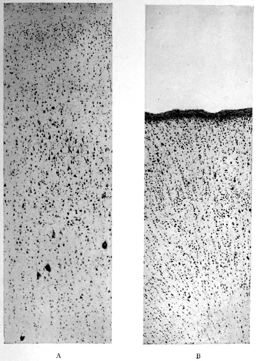
Cortical hemiatrophy—A, relatively normal right precentral (“motor”) cortex; B, atrophic left precentral. Note in B: 1. Absence of giant pyramids of Betz (corticospinal, upper motor neurones). 2. Superficial (subpial) condensation of tissues with sclerosis (gliosis). The tissues in all areas examined on the left side yielded this effect.
207Head: Hair in good quantity. Scalp normal. Calvarium shows diploë. Dura Mater over left cerebral hemisphere inseparably adherent to calvarium, over right hemisphere normal. Arachnoidal Villi moderately developed. Pia Mater shows injected veins, notably in the sulci of the right hemisphere. Pia mater everywhere thin and clear. Vessels at base of normal appearance.
Brain weight 1180 grams. Spread on a board, the right hemisphere tends to flatten so that it measures 1.5 cm. more from side to side than its fellow. Besides more marked venous injection, the right hemisphere shows also flatter and slightly more plastic convolutions. The posterior poles of the hemispheres are a little firmer than the parts anterior. The orbital and hippocampal gyri on the right side are a little firmer than the surrounding parts. On section the gray and white matter shows no lesions, excepting the slight plasticity of the tissues at large on the right side and a well marked induration, with retraction under the knife, of the occipital and hippocampal white matter. The basal ganglia of the left side are normal. On the right side a sagittal section demonstrates a rounded area of induration, with ill-defined borders, measuring perhaps 1.5 cm. from above downwards by 2 × 2 cm., situated largely in the lenticular nucleus and involving the greater portion of the globus pallidus, a small segment of the putamen below and behind and the regionary part of the anterior commissure with surrounding tissues. The most striking feature of this lesion is the occurrence in the middle of a cluster of vacuoles or cystic clefts, with smooth pale interiors, ranging from pinhead to 0.25 cm. or even 0.5 cm. in greatest diameters. There are six to eight clefts to a surface of section. The color of the lesion differs little from that of the globus pallidus itself, but the tissue is a trifle translucent. It is impossible to demarcate the lesion with the eye. Induration is demonstrable several mm. beyond the visible part of the lesion. The consistence of the lesion slightly surpasses the usual consistence of the olivary bodies.
Cerebellum, Pons and Bulb weight 165 grams. Cerebellar tissue a trifle more plastic than usual. The right olive is not so prominent as usual.
Note. Thyroid: Weight 125 grams. Both lobes and isthmus enlarged. One lobe more than the other; lobe on one side measuring 6 × 4 cm.
208Anatomical Diagnoses
Enlargement of thyroid gland.
Exophthalmos with dilated pupils.
Fatty degeneration of thoracic muscles.
Slight aortic sclerosis.
Dilatation of right heart.
Hypertrophy of left ventricle.
Slight tricuspid endocarditis.
Bicuspid aortic valve.
Hypostatic pneumonia.
Acute and chronic splenitis.
Fatty liver (central necroses?).
Acute nephritis.
Chronic gastritis.
Small breasts.
Axillary hair absent.
Petechial eruption of chest.
Varicose veins.
Chronic external adhesive pachymeningitis of left side.
Moderate swelling of right hemisphere with venous injection.
Slight occipital gliosis of both sides.
Slight gliosis of orbital and hippocampal gyri of right side.
Sclerosis with atrophy of occipital and hippocampal white matter of right side.
Gliotic lesion (1.5 × 2 × 2 cm. of right lenticular nucleus involving anterior commissure).
1. Was the exophthalmic goitre in Carrie Pearson due to syphilis? Unfortunately we have no clear proof that Carrie Pearson was syphilitic. She was stated to have been syphilitic by the physician who treated her before her commitment to Danvers Hospital. There is, however, no proof of syphilis, inasmuch as the patient died in the pre-Wassermann period.
2. Is the thalamic lesion probably syphilitic? No lymphocytosis or plasmocytosis characterizes the lesion, which is the only lesion of the sort in the Danvers collection. It would not do to call a lesion syphilitic just because it is sui generis. In any event, the clinical analysis of the case faced the claim of syphilis as an actual factor in the patient’s life and as a possible factor in the goitre.
209
It is well known that the ARGYLL-ROBERTSON PUPIL is characteristic of the so-called “PARA-SYPHILITIC DISEASES” (“general paresis” and “tabes”); does this sign occur in other neurosyphilitic conditions?
Case 54. Julius Kantor was a shoemaker of 35 years, who came to the hospital for treatment because his family physician had found a positive W. R. in Kantor’s blood serum. He had had a cough for a number of years, and during the last year a little blood had been found in the sputum; whereupon Kantor had been placed under active anti-tuberculosis treatment. The enterprising family physician had found the positive W. R. in the first days of his treatment for tuberculosis. There was, in fact, a history of a chancre nine years before, which had not been followed by any secondary or tertiary symptoms, and which had been but scantily treated.
There were no mental symptoms.
Kantor was physically fairly well developed and nourished. There were a few piping râles in the left upper chest, both in front and back, and also a slight dulness with increased vocal and tactile fremitus. No tubercle bacilli, however, could be found on repeated sputum examination.
Neurologically, the pupils were myotic and both showed the Argyll-Robertson reaction. There were no abnormal reflexes whatever, and there was neither ataxia nor speech defect. Not only the blood but also the spinal fluid W. R. proved to be positive; there was a marked increase in the albumin and globulin; there was a gold sol reaction of the syphilitic type, and there were but three cells per cmm.
1. In view of the headache in case Kantor, what other causes of headache are to be considered? It is certain that irritations of the dura mater can produce headache, and the physiological observation of the sensitiveness of the membranes and the non-sensitiveness 210of the brain substance is an ancient and classical observation. Internal hemorrhagic pachymeningitis produces severe headache. The relations of this disease to trauma, to arteriosclerosis, and possibly to syphilis (alcohol perhaps should also be considered) in certain instances have not been entirely cleared up. Syphilitic headaches are, according to Lewandowski, dependent also upon a dural affection or upon a periosteal affection. The headaches of brain tumor are also commonly related to dural conditions, either directly due to the pressure of the tumor itself, or indirectly to the heightened intracranial pressure consequent upon the tumor. It is clear that the tension under which the dura mater lies is not always localized in the region of a brain tumor or a syphilitic lesion. Head has claimed that brain tumor produces headaches of two kinds, according to whether the disease affects the dura mater or is dependent upon an increase of pressure in the brain. It does not appear that the pia mater has any relation to headaches, but meningitis, in which the inflammation is confined to the pia mater, is nevertheless associated with headache; the headache is here supposed to be due to the increase in brain pressure, and thus actually to an effect wrought upon the dura mater. Vasomotor disorders and various types of cephalic hyperemia are thought to produce a kind of headache, but Lewandowski calls this kind of headache somewhat in question. Reflex headaches are stated to be produced indirectly by a process of radiation from interior lesions in the brain. There are certain headaches called nodal headaches (Schwielen-Kopfschmerz). Hypermetropia, caries of the teeth, adenoids, and diseases of the nose and axillary cavities, to say nothing of thoracic and abdominal diseases, are also counted among conditions that may produce headaches. In this connection, Head has claimed differential zones of headache corresponding to certain diseases.
The brain itself may produce headache through intoxications, through conditions produced by a variety of diseases; may follow neuroses. Alcohol may produce headaches in some persons even when it is taken in very small doses. Certain uremic cases yield headaches, as do also gouty and chlorotic conditions. According to Lewandowski, the headaches of arteriosclerotics are due possibly to vasomotor disturbances 211in the membranes, or one may think of nutritive cerebral disorders. A peculiar form of headache is that of fatigue after mental work, allied to which is the neurasthenic headache; constitutional headaches have been assumed to occur, to say nothing of hysterical headaches. There remains also the important question of migraine, for which a vasomotor explanation has been proposed.
2. Was Kantor suffering from tuberculosis of the lungs? The hypothesis of lung syphilis ought certainly to be very seriously considered. Upon repeated sputum examination, no tubercle bacilli have yet been found.
3. Is Kantor a case of general paresis? In the absence of mental symptoms, and in consideration of the mildness of the reactions, it is certainly not easy to make the diagnosis of general paresis. However, the diagnosis of tabes dorsalis is not justified either. Accordingly, we may answer our question: whether the Argyll-Robertson pupil occurs in other neurosyphilitic diseases, by pointing out that in the case of Julius Kantor, as in the case of Henri Lepère (105) and Frederick Stone (106), the Argyll-Robertson pupil has been found in syphilitic conditions that are neither typically paretic nor typically tabetic.
212
Does the Argyll-Robertson pupil necessarily indicate neurosyphilis?
Case 55. Daniel Falvey, 44 years of age, was an almshouse transfer to the Danvers State Hospital in the year 1904, when the principle of state care was adopted in Massachusetts. As in most of the almshouse transfers of that day, little could be discovered as to antecedents. He had been a mill-worker from the time of his immigration in 1890, at 30 years of age. He had been somewhat alcoholic. There was a shock some 17 months before his death, which occurred about seven weeks from the date of transfer.
Not only was he unable to walk unsupported, but when supported there was a slight dragging of the left leg and the gait was noted to be somewhat propulsive. The tongue and hands were tremulous, and the left grasp was somewhat weaker than the right. Both knee-jerks were increased although neither more than the other. There was no sensory disorder.
Although but 44 years of age, Falvey presented the appearance of a much older man. His heart was somewhat enlarged and there was a degree of peripheral arteriosclerosis. On the whole, no special attention was attracted to this case clinically and he was regarded as an example of arteriosclerotic dementia, like many another among the transfers. However, we owe to Dr. H. M. Swift the important observation of the Argyll-Robertson pupils. The case was studied long before the Wassermann method was available, and is here reported merely to call attention to the fact that the stiff pupils may have other neural origin than neurosyphilis.
The autopsy material in the case was worked up by one of the authors.[11] The autopsy had been performed by Dr. A. M. Barrett, who found on section through the brain stem at the 213anterior border of the pons a mass springing from and continuous with the pineal gland, lying in the third ventricle and the aqueduct of Sylvius. Upon further study, this mass was found to begin posteriorly in the pineal body itself, from which the mass could hardly be told in the gross except by an injected border.
This mass proved upon microscopic examination to be a psammoma, which histologically resembled a glioma rather than a sarcoma. Throughout the mass there was a variable content of fibrillary intercellular substance having the histological reactions of neuroglia fibrillæ. The histological details (mitosis, large giant cells with multiple nuclei, etc.) do not here concern us. We deal with a neoplasm springing from the pineal gland growing on the posterior half of the third ventricle, the anterior orifice of the aqueduct of Sylvius, and the space between the velum interpositum as far back as the posterior corpora quadrigemina. There is no evidence in the body of old syphilis; although it is possible that the stiff pupils were neurosyphilitic, it seems probable that they were related to the pineal tumor. At all events, there are in the literature evidences that the pineal-quadrigeminal group of tumors and other lesions may bring about pupillary disturbances. On this account, we here include the case. The tumor hardly led to an error in diagnosis since neither neurosyphilis nor brain tumor was at all expected clinically.
1. Can alcoholism produce identical results? See Case Murphy, (60), one of alcoholic pseudoparesis.
2. What is the nature of stiff pupils? A pupil is called stiff in the sense of the Argyll-Robertson pupil if it fails to react to illumination either of itself or of the other eye and at the same time if it reacts properly in convergence and accommodation. Of course the stiffness of a blind eye must not be regarded as an Argyll-Robertson pupil. In a case of right-sided Argyll-Robertson pupil, therefore, the left pupil reacts properly both to direct illumination of itself and to illumination of the right eye, but the right eye fails to react to illumination of either eye. Such an Argyll-Robertson right pupil will remain of the same width both in darkness and in light. Clinicians agree that the Argyll-Robertson 214is diagnosticated rather too frequently than too seldom, and this by reason of the fact that a sluggishness of light reaction is interpreted as stiffness. The sign, as is well known, has come to be regarded as almost pathognomonic of tabetic or paretic neurosyphilis. Nonne, however, has found among 510 cases of alcoholism, nine instances of Argyll-Robertson pupil and 19 cases of sluggish light reactions. The pathological anatomy of this sign is still doubtful although a number of schematic accounts are available; among hypotheses, one may think of an elective effect of the tabetic or paretic degeneration upon reflex collaterals. The explanation would then resemble that for absent knee-jerks and kindred reflex disorders. We should then hypothesize a loss of the finer processes of the terminal arborizations about the cells of the nucleus of sphincter nucleus iridis. However, the situation of the sphincter iridis has not yet been absolutely determined.
When a pupil is said to be entirely stiff it means that it reacts neither to light nor accommodation. This condition not infrequently follows the partial stiffness or Argyll-Robertson reaction.
3. Is the Argyll-Robertson pupil more tabetic than paretic? This has been claimed at times, but in point of fact, the Argyll-Robertson pupil is very frequent in paresis, and so also are posterior column changes. According to statistics of Bumke, 36% of tabetics fail to show the Argyll-Robertson pupil, and 38% of paretics. When, however, finer methods, such as those standardized by Weiler, with photographic records, are employed, the number of cases without at least a tendency to the Argyll-Robertson pupil becomes much smaller.
In connection with the important question as to the classical Argyll-Robertson pupil and pupillary sluggishness to light, it may be inquired what are the ocular signs in neurosyphilis? Joffroy has tabulated the signs in 300 general paretics as follows:
Sign. No. of cases. Per cent.
Alterations of light reflex 235 78
Inequality 205 68
Abolition of light reflex (bilateral or unilateral) 156 52
Abolition of light reflex (bilateral) 133 44
Irregularity of pupil 117 39
215Irregularity of both pupils 109 36
Diminution of light reflex 108 36
ditto (bilateral) 79 26
Alteration in accommodation reflex 79 26
Diminution of accommodation reflex 52 17
Mydriasis 41 13
Myosis 40 13
Diminution of light reflex (unilateral) 35 11
Abolition of accommodation reflex 35 11
Diminution of accommodation reflex (bilateral) 29 9
Abolition of accommodation reflex (bilateral) 26 8
Diminution of accommodation reflex (unilateral) 23 7
Fundus changes 21 7
Vascular changes 16 5
Abolition of accommodation reflex (unilateral) 12 4
Paresis of the third nerves 10 3
Ptosis 9 3
Irregularity of one pupil 8 3
Nystagmus 7 2
Visual acuity lost 7 2
Atrophy of disc 6 2
Total blindness 5 2
Paralysis of the fourth nerves 1 1
216
Can neurosyphilis exist in the absence of positive findings in the spinal fluid?
Case 56. There was no great difficulty in setting up a diagnosis of general paresis in the case of James Burns, a mechanic of 31 years of age, who came voluntarily to the Psychopathic Hospital for treatment. The point in Burns’ case was that the spinal fluid proved entirely negative in all respects despite the fact that the serum W. R. was positive, and despite the following facts of history and mental examination.
The patient claimed syphilitic infection seven years before, namely, at 24 years of age, and also claimed that he had infected his wife, who was in fact at the time undergoing antisyphilitic treatment. He complained of insomnia, worry, depression, hypersensitivity to noises (such as those made by his own children), thoughts of suicide, and amnesia. The amnesia, however, might be regarded as subjective since our tests failed to show amnesia. Nor was there any diminution in arithmetical ability. Despite the patient’s claim that he had been “way off in his way of thinking,” there appeared to be no delusions. Beyond a certain flightiness in conversation, we could hardly get any evidence of psychosis unless of the neurasthenic order.
Physically, however, the left pupil failed to react to light though it was found to react to distance, and the right pupil exhibited a diminution of its reaction to light. There was no ataxia of gait, yet there was a complete Romberg reaction. There was a moderate tremor of the hands and of the tongue. Otherwise there were no reflex disorders upon systematic examination, nor was there any demonstrable disorder in the rest of the physical examination.
1. What is the diagnosis in the case of James Burns? On the whole we agree with Nonne, that negative spinal fluid findings (of course, in the absence of treatment) preclude the diagnosis of general paresis. The symptoms 217might possibly be explained, however, by means of a localized syphilitic involvement of the cerebrum, no cells or products of inflammation having penetrated to the spinal fluid. According to Head and Fearnsides, this condition may be found especially in the anterior or middle fossa. Accordingly, going upon these views of Nonne and of Head and Fearnsides, we should be entitled to make, perhaps, a diagnosis of cerebral syphilis.
2. What is the significance of the Argyll-Robertson pupil in James Burns? Nonne states that if one follows cases with Argyll-Robertson pupil over a sufficient period of years, they one and all eventuate in active symptoms of cerebrospinal syphilis (not necessarily of the cortical type), and this despite the fact that the pupillary change may have been present a number of years before any other symptom had developed.
218
Neurosyphilis (“DISSEMINATED ENCEPHALITIS”) within seven months of initial infection. Autopsy.
Case 57. We borrow the main features of a remarkable case examined at the Danvers State Hospital clinically by Dr. H. W. Mitchell and reported elaborately by Dr. A. M. Barrett. This case, whom we shall call John Summers, acquired syphilis at about the end of the third week in May, 1902, and consulted a physician on June 12, at which time a characteristic initial lesion of syphilis was plain. Summers was excessively alcoholic at times and was not seen by a physician again until July 2, just after an alcoholic debauch. At this time there was ulceration of the primary lesion, and a papillary eruption had developed over the arms, chest, abdomen, and legs. Mercurial treatment and mixed treatment were given. Arthritis occurred but disappeared with increased dosage.
About six months after infection, the patient developed severe headaches, hardly controllable by treatment. Amnesia and a certain stupidity, with neglect of personal habits, and even of eating, developed, whereupon Summers was admitted to the Danvers Hospital, December 11, 1902. He weighed 124 pounds, was extremely feeble, with dull and expressionless face, coarse purposeless movements of arms; left pupil larger than right; right external strabismus and ocular ptosis; increased knee-jerks, crossed adductor reflex, coarse tremors of arms and hands; and extreme clouding of consciousness. It was doubtful whether the pupils were stiff to light or not.
The patient died on the ninth day, December 18, in a state of coma. After admission, his stupor had become more marked; there had been incontinence of urine and fæces, and the patient could be aroused only by loud tones. Difficulty in swallowing had developed; the right-sided ptosis had become more marked, and muscular twitchings had developed on the right side. When the left leg was pinched, there was twitching of the left leg and arm. There was slight spasticity of the right arm and leg. An examination upon the day of death definitely showed a lack of reaction of the pupils to light.
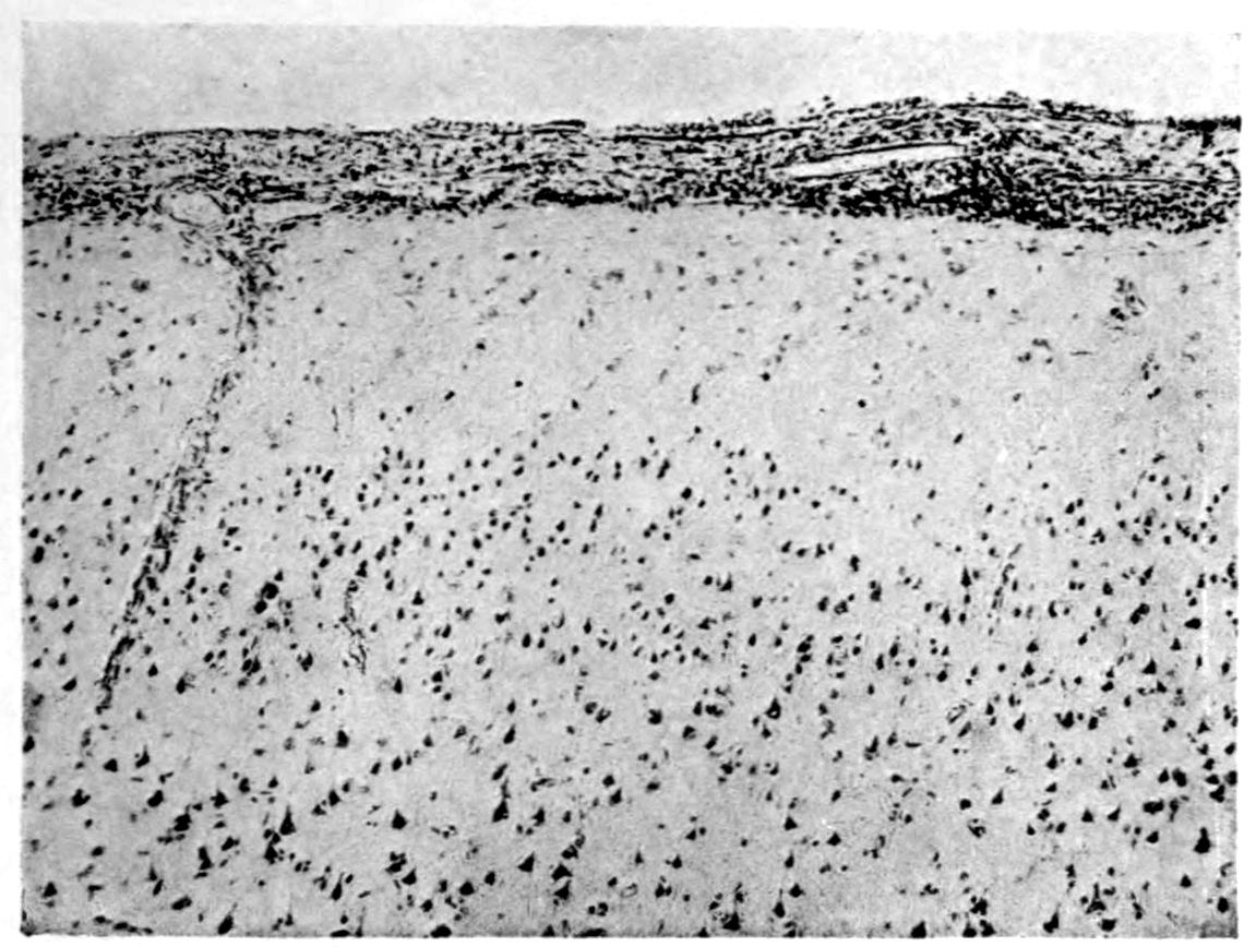
1. Exudate in pia mater—mononucleosis.
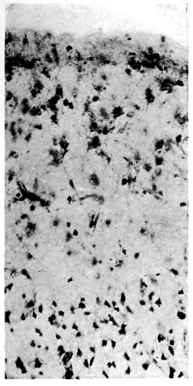
2. Superficial (subpial) cellular reaction of neuroglia tissue (expanded cell bodies).
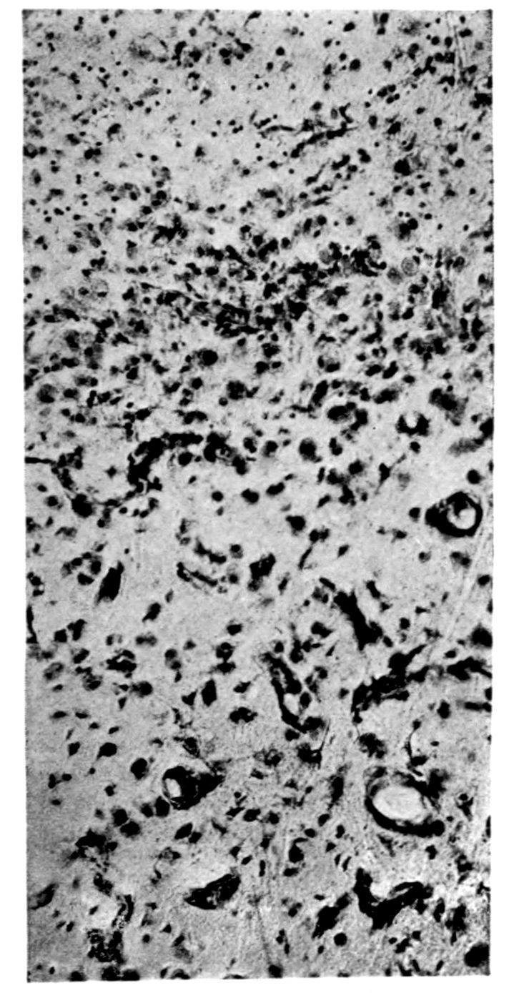
3. Cellular gliosis of deeper layers of cortex. Apparent increase in capillary supply, possibly relative to loss of neural elements.
Case 57. Neurosyphilis (“disseminated syphilitic encephalitis” of A. M. Barrett), fatal seven months from initial infection. (Photographs by A. M. Barrett.)
219Dr. Barrett was able to find in the literature a case of Bechterew which histologically resembled his own case, but though in the instance reported by Bechterew the first symptoms developed within the year following infection, death did not occur until two years later.
In view of a total duration of symptoms clearly not over seven months, it is interesting to inquire how far microscopic brain changes could have proceeded. Neither calvarium nor dura mater showed changes. There was a slight haziness of the pia mater over the convexity, but the pia mater over the base (especially below the cisterna and from thence spreading out over the pons and into the fissure of Sylvius) was not only hazy but definitely thickened and hyperæmic. The thickening was most marked about the root of the right third nerve (corresponding with the eye findings in life). There was also a macroscopic thickening of the left Sylvian artery. Section of the brain showed nothing abnormal except a small area among the pyramidal fibres of the right side of the pons, where there was a single hemorrhagic area about 7 mm. in diameter around which there were small punctiform hemorrhages. (Compare twitchings of left leg and arm upon stimulation of left leg, and note also the muscular twitchings and slight spasticity of right leg and arm noted just before death.) This case was examined and reported upon in 1905. We learn from Dr. Barrett that a re-study of the case with modern methods has failed to demonstrate a spirochetosis.