II. THE SYSTEMATIC DIAGNOSIS OF THE MAIN FORMS OF NEUROSYPHILIS (continued 2)
Reading edition
4. What are the prodromal symptoms of cerebrospinal syphilis? According to Nonne, headache, dizziness, sleeplessness, mental symptoms of the irritability group, loss of capacity as to mental work, whether severe or not, and loss of capacity for difficult thinking; also impairment of memory. Nonne does not regard these phenomena as characteristic of syphilitic vascular disease, and calls attention to the fact that in every organic disease the same subjective symptoms occur. The triad—headache, dizziness, and impairment of memory—is for example now counted as a prodromal symptom complex for arteriosclerotic apoplexy (Cramer). Of course, apoplectic attacks occur without such preliminary symptoms: particularly, according to Nonne, the nocturnal attacks.
5. Can the fleeting paralysis be of service in differentiating the diffuse from the paretic form of neurosyphilis? Probably not. In both forms transient paralyses occur as well as the permanent ones. In general, however, the transient paralyses are more frequent in paretic neurosyphilis, whereas the permanent ones occur more often in diffuse neurosyphilis.
126
There are cases of NEUROSYPHILIS in which the laboratory signs are positive but in which there are no clinical signs or symptoms (PARESIS SINE PARESI?).
Case 25. Richard Lawlor[6] was admitted to the Psychopathic Hospital, October 29, 1914, being sent there from a general hospital where he had gone on account of a self-inflicted wound of the wrist, apparently made in a period of depression with suicidal intent. Routine notes follow.
Family History. Paternal grandparents both died of heart disease. Maternal grandfather died at seventy-two of dropsy. Moderately alcoholic. Maternal grandmother died of shock at fifty-six. Father died at age of forty, after an illness of eight years, from heart disease. Father all his life was subject to fainting spells and headaches. The only paternal cousin died at thirteen months of brain fever. Mother, aged forty-seven, is, to say the least, eccentric. Says “she has several times been given up from tuberculosis.” Two maternal uncles died of tuberculosis, one from rupture, one from heart disease. One uncle who “doesn’t know anything after he has a teaspoonful of liquor.” Several other uncles and aunts whose history is not obtained. Patient is mother’s only child. Mother was twice married. There were several miscarriages by both husbands; patient child by first marriage.
Past History. Patient born thirty-two years ago, full term, normal delivery and development. Measles, mumps, and chickenpox in childhood. Subject to headaches since seven or eight years old. Kicked in the face by horse at seventeen or eighteen, not considered serious. Hit by a baseball three or four years ago, leaving him hard of hearing 127on left side. Married ten years ago; no children because he says his wife needed an operation. He denies venereal disease by name and symptoms. For past ten years has had attacks of depression lasting but a short time, but quite severe. Never caused him to quit work as a barber and he felt better when working. His married life he says was fairly happy except for his wife’s extravagances, and on this account he left her a little over a year ago, and she has applied for a divorce, which he is willing that she should have, but does not wish to give her alimony. He admits moderate alcoholism.
Present Trouble. Patient states that since he left his wife a year ago he has felt sorry a number of times. He has wished he had her back. He has felt lonely. He has had six or eight periods of depression in that time similar to those he has had for many years, lasting two or three days, and sometimes a week. These were always precipitated by some cause for worry. In these attacks he feels nervous, sleeps poorly, has little or no appetite, sweats during his work and everything looks black. Several times in these attacks he has had suicidal ideas. Ten months ago he considered taking corrosive sublimate. For a little over a week before entrance to hospital he had been out of work and had been “sporting.” The day before entrance he had a telephone message from his lawyer which upset him somewhat and he walked the floor all night. He had just been shaving when the idea of suicide came to him. He sat down a minute when suddenly the thought “to hell with the world” came to him; he took the razor and slashed his wrist. He does not remember drawing the razor across his wrist. As soon as he saw the blood he felt sorry, called his mother, and was taken to an emergency hospital and then sent to the Psychopathic Hospital.
Physical Examination. Patient is a well developed and nourished man thirty-two years of age. Head is normal as to size and shape; there are no scars or marks of injury. Hair and skin not remarkable in any way. Ears negative to external examination. Teeth well kept; two missing, several gold fillings. Tongue very slightly coated. Throat negative. Tonsils easily visible without evidence of inflammation 128or exudation. Neck, no thyroid enlargement, no abnormal pulsations, no adenopathy. Chest, symmetrical, expansion good, resonant throughout. Breath sounds transmitted normally. No râles or rubs heard. Heart, no enlargement or cardiac dulness. Sounds of good quality, no murmurs heard. Rate regular. Pulses equal, regular and synchronous, and of good volume and tension. Systolic blood pressure 130, diastolic 65. Abdomen, flat, soft and tympanitic throughout; no masses; no tenderness. Liver edge not felt, below costal margin. Spleen not palpable. Extremities negative, except for incised wound on left wrist.
Neuromuscular Examination. Pupils are large, round, regular, equal and react readily to light and accommodation. No nystagmus, strabismus or ptosis. No weaknesses or paresis of facial muscles. The tongue projects medially and shows no tremor. The triceps and biceps reflexes are readily elicited, and are quite active, as are the knee-jerks and ankle-jerks. On one occasion it was thought that the tendon reflexes were slightly more active on the left than on the right. This was never confirmed; always afterwards found equal. There was no tremor of extended hands. Abdominal reflexes not elicited. Cremasteric present on both sides. The plantar response is flexor. There is no Babinski, Gordon or Oppenheim. No Romberg. Coördination tests well performed. No speech defect. No sensory disturbances. Urine examination negative.
Wassermann reaction in the serum: Positive, with cholesterinized antigen; negative, with syphilitic fetal liver antigen.
Wassermann reaction in fluid positive on two occasions. Examination of spinal fluid, November 4: globulin +++, albumin ++, 100 cells per cubic millimeter; large lymphocytes, 8 per cent; small lymphocytes, 90 per cent; plasma cells, 0.7 per cent; endothelial cells, 1.3 per cent. November 11, globulin +++, albumin +++, cells 18 per cubic millimeter. November 26, globulin ++, albumin ++, cells 92 per cubic millimeter; large lymphocytes, 13.1 per cent; small lymphocytes, 82.1 per cent; plasma, 1.2 per cent; endothelial, 3.6 per cent.
Gold sol, November 4, 5555432100.
Gold sol, November 26, 3332100000.
129Mental Examination. On entrance to hospital patient seemed slightly depressed and a bit irritable. This condition lasted two days, after which he was agreeable and apparently entirely over his depression. Even during his mild depression, however, he talked freely. There was no evidence of retardation. He told his story readily. Orientation was intact. Memory excellent. Educational knowledge well retained. There was no evidence of any hallucinations or delusions.
1. Was Richard Lawlor insane?
There was, then, on the mental and physical examination nothing to make a definite suggestion of a psychosis, and the most one could think of was a psychoneurosis or a cyclothymia of at least ten years’ duration. The findings in the cerebrospinal fluid and the Wassermann reactions, however, give us material for thought. Certainly one cannot call the man insane; all who saw him agreed on this point.
2. If Richard Lawlor should some day develop mental symptoms, what would be the genesis of the new psychosis? Though writers such as Fildes and McIntosh, and Swift, have suggested an anaphylactic or hyperallergic explanation for the development of symptoms after a normal interval; such a hypothesis could hardly obtain in the present case. The hyperallergic hypothesis for the development of tertiary neurosyphilis would run to the effect that in the secondary stages there had been a definite disease of the nervous system, which, however, absolutely cleared up, leaving no inflammatory vascular or parenchymatous relics of its existence. Nothing would on this hypothesis remain except a hypersensitisation of the tissues. In some later period of the now clinically normal person, one or more spirochetes from a lesion outside the nervous system are carried into the nerve tissues and there set up an anaphylactic or hyperallergic reaction. It is obviously difficult to prove the correctness or incorrectness of the hyperallergic theory without numerous examinations of the spinal fluid, in clinically normal persons after the secondaries have passed. The present case, so far from demonstrating a normal fluid, demonstrates a highly pathological fluid, even though there are absolutely no clinical symptoms which could be regarded as of 130nervous origin. The burden of proof at the present time would seem to lie with those who claim hyperallergy in neurosyphilis. We prefer on present evidence to think that at the conclusion of the secondaries a disease process often remains in the nerve tissues despite clinical quiescence.
3. What is the prognosis in the case of Richard Lawlor? The prognosis re neurosyphilis is doubtful. We have, however, boldly termed the condition PARESIS SINE PARESI, meaning thereby to suggest that the patient is in considerable danger of the efflorescence of a true diffuse or paretic neurosyphilis. We have no means of telling, however, whether the positive symptoms would be those of a paretic or a non-paretic neurosyphilis. As data accumulate regarding these cases of paresis sine paresi, we may be able finally to come upon some case in which trauma shall bring out the clinical symptoms of neurosyphilis. For discussion of this matter, see the case of Bessie Vogel (52) in Part III of this book.
4. Should Lawlor have been brought to a psychopathic hospital? It is a safe working rule to have any person who attempts suicide observed. A large percentage of suicides occur in psychotic individuals and a suicidal attempt is not infrequently the first recognized abnormality. Immediate observation is a necessary safeguard against another more successful attempt.
131
Demonstrates SYMPTOMS and LESIONS of PARETIC NEUROSYPHILIS (“general paresis”). Autopsy.
Case 26. John Morrill, 49, an operative in a mill town in Essex County, Mass., was described as a “Saturday night and Sunday drinker,” with a history of very serious long sprees at the age of 43. It seems that he had had what was called “sciatica” at 35, and was treated in hospital for seven weeks at that time. The nature of this sciatica is in doubt, but there was a history of syphilitic infection at 36 years (scar of glans).
Morrill had been married twice, and two of the children were dead; one daughter was described as “very nervous,” but there were four children under ten years of age, all regarded as perfectly healthy.
Morrill had been a mill operative of average capacity, was industrious, and had supported his family despite alcoholism. The syphilis had been treated with reasonable thoroughness.
Aside from alcoholism, there had been no symptoms up to two months before admission to Danvers Hospital. Then there had been insomnia, fatigue, agitation, eruption on foot, loss of ten pounds in weight, hypochondriacal fears, apprehensiveness for the future of the children, incoherent talk; and just before admission, his talk was described as foolish. He had taken to running away and hiding in bushes by a pond and in the cellars of other people’s houses.
The patient was of medium height and weight, with thin grayish hair and grayish irides; musculature was slender. The face was blank in expression, the teeth poorly preserved with atrophy of gums, the tongue coated, and the breath foul. There was a gummy secretion of the eyelids, an area of brownish branny eruption over both clavicles, a number of depressed scars over the limbs and back, and another area of scaly eruption on the right heel and the sole of the foot. The heart area was increased, and the sounds were faint at 132the base, with the first sound accentuated at the apex. The urine showed a trace of albumin.
Neurologically, the Romberg position was maintained with a general tremor and fluttering of the eyelids. In complicated movements, the patient was slightly ataxic. The pupils were irregular, the left being much larger than the right. There were no light reactions to be obtained in window light. The reaction to accommodation was present, though slight. Vision was poor, ¼-inch capitals could not be read by left eye at reading distance. The knee-jerks were diminished equally; the Achilles jerks were absent; the other reflexes were normal. Upon the sensory side, the patient gave a history of pains in the legs at irregular intervals for several years. These pains he described as of a darting character. There was little or no sensory disorder, although the outer surface of the right leg required a deeper pressure to elicit sensation. There were no disorders of muscle sense.
If Morrill was to be trusted, he had been born in Ireland, and had come to the United States at the age of 17. He married at 18; there had been seven pregnancies by the first wife, with one stillborn child; one child had died at five weeks. The four children by the second wife were healthy. The first signs of neuritis had occurred at 45 and had received the diagnosis neuritis, although no connection between the neuritis and the syphilis had been noted.
The patient entered the hospital July 26, 1904, and was discharged, improved, January 5, 1905. He returned a little more than a year later, January 15, 1906, and died March 21, 1906. The total duration of the disease from the onset of mental symptoms may therefore be stated as somewhat under two years. When the patient appeared at the hospital the second time, he showed a positive Romberg sign, an unsteady gait, an ataxia that still was moderate, and somewhat more marked tremors, involving fingers, tongue, and face. He was now unable to read ½-inch type with the left eye. The knee-jerks, formerly diminished, were both exaggerated, the left slightly more so. The Achilles reaction, not obtained formerly, now appeared on the right side. The pupils reacted as before. The sensory loss had become more marked, since 133sharp and dull points could hardly be distinguished. Deep pinpricks were not felt in the leg, and heat could not be told from cold.
The speech in 1904 had been somewhat defective (“truly rural” rendered as “tooly lualal,” “sifted soft thistles” as “thoft thsistles”), and there had been little further development of the speech defect. The handwriting had lost appreciably in legibility and had become much more tremulous. During the first period of hospital observation Morrill had what might possibly have been visual hallucinations, but it was impossible to tell whether his story of seeing his wife and children trying to get in through the window was hallucinatory or a matter of fabrication. Memory was decidedly imperfect and few details of recent events could be produced. The association of ideas was almost a so-called “flight” of apprehensive, fearful ideas, loosely connected, incoherently expressed, and dealing chiefly with his work and his children. Judgment was imperfect; the height of the room was estimated as 24 feet, but the height and weight of persons were estimated with fair accuracy, and also the length of small objects, whose lengths were doubtless remembered rather than estimated. The estimate of time elapsing during a medical examination was accurate, but the estimate of longer durations involving over-night memories was hopelessly imperfect. Emotionally, there was a dulling of sensibility, an appearance of suspicion and apprehensiveness; the patient fancied himself to be in a hopeless condition as a result of syphilis, but at the same time accompanied his statement of his hopelessness with laughter. A sample of his hypochondriacal ideas: “I am all gone; I am good for nothing; I am all gone now; I can’t drink now; can’t write or talk at all; worse than when you saw me first; nothing in my inside; all wrong through me again; I aint got no swallow now; I can’t die even; my heart aint much good; I can’t hear it beat; I don’t think it flutters; no life in these hands; they are all cold and dead” (pointing to his arms and moving them about). During such a portrayal the patient laughed in a silly way.
During the second hospital stay, Morrill was at first restless, 134sleepless, profane, imperfectly oriented for time, possibly for place, and also for the attendants. A few weeks later he became stuporous and confused, and his feebleness and physical exhaustion were finally ended by death, March 21, 1906. Death was preceded by a semi-comatose condition; a left otitis media had developed.
At the autopsy, it appeared that death was due to an early bronchopneumonia associated with acute splenitis and doubtless related to the otitis media of the left side. The body at large showed, aside from these acute lesions, a few chronic lesions, including slight scars of the left apex, and chronic adhesive pleuritis, chronic diffuse nephritis, and aortic and coronary syphilis. The aorta showed slight linear and nodular markings, with a single small dark ulcer in the upper thoracic region, but the aorta did not show the characteristic scarring which syphilitic aortas often show. The femoral marrow was of a dark red chocolate color. The thyroid appeared to be smaller than normal. A slight sacral decubitus had developed.
The description of the head (E.E.S.) is given in full on account of the encephalitic lesions shown. These encephalitic lesions may be summed up as follows:
Local cerebral atrophy and sclerosis of the frontal, orbital, and central regions, especially of the left operculum and left supramarginal gyrus.
Extension of sclerosis to hippocampal gyri with effacement of substantia reticularis alba.
Slight chronic internal hydrocephalus.
Granular ependymitis (especially of floor of 4th ventricle).
Compensatory edema of frontal and central pia mater.
Cerebellar sclerosis (culmen monticuli, lobus culminis, lobus cacuminis).
Spinal sclerosis (grossly evident in the posterior columns of the upper thoracic region and of the lumbar enlargement).
The details are as follows:
Head:—Bald on top. Hair gray. Scalp normal. Calvarium thin, deeply excavated by arachnoidal villi to right of vertex. Diploë absent. Dura closely adherent in bregmatic region. Dura of usual thickness. 135Sinuses contain cruor clot. Arachnoidal villi slight. Pia mater hazy and over sulcal veins porcelain white over all of vertex except occipital poles and over flanks (notably left). Thickened also around circle of Willis, over culmen monticuli and in posterior cerebellar notch. Edema of pia corresponding to atrophy of frontal and central regions. Cerebral atrophy most marked in orbital surfaces of both frontal lobes, in left area of Broca, and in left supramarginal region. The ascending branch and the ascending ramus of the posterior limb of the left Sylvian fossæ both readily admit the thumb by reason of atrophy of adjacent substance. Induration corresponds closely with atrophy, but is not more marked about the left Sylvian fossa. There is sclerosis of both hippocampal gyri, with loss of the substantia reticularis alba. The culmen monticuli and lobus culminis are firmer than the clival regions, and the lobus cacuminis is again slightly firmer than the clival region. Cerebellum a little softer than usual. Pia strips with usual readiness from all regions. The subpial region of the frontal lobes is a trifle grayer than that of the rest of cerebrum. Ventricles slightly dilated. Surfaces evenly sanded. Floor of fourth ventricle shows numerous coarse, closely set granules. Brain wt. 1200 grms. Cord shows a slight increase of consistence over one or two upper thoracic segments and in lumbar enlargement corresponding with a slight graying out of posterior columns. In places there is a suggestion of graying out also in lateral columns. A few calcified plaques in posterior lumbar pia.
Analysis of these details shows a number of lesions that characterize paretic neurosyphilis (among others, granular ependymitis, frontal atrophy, chronic leptomeningitis), but the lesions are more than merely frontal, extending as they do back as far as the postcentral regions on both sides, and even as far as the left supramarginal gyrus. The cerebellar involvement although frequent, can hardly be said to be characteristic in paretic neurosyphilis. The spinal involvement is characteristic of a case which is probably to be regarded as one of taboparesis; that is, of paretic neurosyphilis following a number of years after the establishment of tabetic neurosyphilis. The aorta is almost constantly affected by sclerosis in paretic neurosyphilis. The absence of diploë in 136the skull is not infrequent and the adherent dura mater is often found.
Microscopically, the tissues showed the characteristic lesions of Paretic Neurosyphilis; nerve cell destruction, fibrillar and cellular gliosis, lymphocytic and plasma cell deposits about the small vessels.
1. What are the clinical evidences of syphilis outside the nervous system? The brownish branny eruptions of the skin, the depressed scars and the scaly eruption on right heel and sole are very suggestive of syphilis. Such clinical evidences of syphilis are very important in systematic examination. Although the laboratory tests are of the utmost assistance in the diagnosis of syphilis, the clinical signs should not be neglected, and no physician should rest satisfied with laboratory signs alone. X-ray diagnosis of bone conditions sometimes succeeds when all other methods have failed.
137
GUMMA of cerebral cortex verified by operation; death.
Case 27. The presenting picture in the case of David Tannenbaum was that of deep dementia, in which condition the patient was brought to the hospital. There was a meagre history to the effect that about four months before admission, he had lost his job in a hotel through lack of further work. We heard that at this time he had begun to suffer with excruciating pains in the head; at first, worse at night, later, worse by day. It appeared that this pain, though it came and went, was chiefly localized on the left side of the head. For a fortnight, Tannenbaum had been dragging his legs, until finally he had become unable to walk at all.
Pari passu with these developments, Tannenbaum had become mentally confused and irritable, and his memory had become untrustworthy. For several days before admission, an appearance of marked dementia was presented, with slow incoherent, or at all events, irrelevant words, and a complete disorientation for person. However, his vision had become so poor that it would have been hard for him to have recognized any one.
It appeared that the family history was entirely negative; that the patient was without education but had been physically very strong, and had been fairly successful at first in the junk business, and later in the clothing business; but latterly he had been less fortunate in the clothing business, and finally had to resort to work as a laborer around a hotel.
His wife had had eleven pregnancies with but one miscarriage. Nevertheless, out of the eleven pregnancies, there were now but four living children.
Physically, Tannenbaum was a rather small man; he was flabby and looked as if he had recently lost weight. The skin showed areas of pigmentation on the face and sides of the neck, and some dark copper-colored circular areas, marble-size, in the neck (syphilitic?). There was a slight radial 138arteriosclerosis. The heart was slightly enlarged with distant and indistinct sounds. There was a small pedunculated growth on the right side of the abdomen.
The pupils failed to react to flash-light but they reacted to sunlight. They both were slightly irregular but were equal in size, and reacted in accommodation. There was apparently almost complete blindness and extreme deafness. Arm-jerks and knee-jerks were absent; there was an occasional slight response of the left ankle-jerk, but the right ankle-jerk was absent; the left abdominal reflex was very feeble; the right absent; the cremasteric reflexes were absent, but there were no other abnormalities in the systematic examination. Hand grips weak; gait awkward, with right leg held somewhat flaccidly.
It was significant that percussion over the left frontal and parietal regions was able to elicit great pain. Either through the patient’s deafness or through sensory aphasia, spoken language was not understood. The serum W. R. was positive, the fluid W. R. negative.
Diagnosis: The clinical symptoms seem clearly to indicate syphilis. The local skull tenderness and impairment of vision might well suggest intracranial pressure. Uniting these suggestions, we might automatically arrive at a diagnosis of cerebral gumma. We have learned to be rather cautious of making a diagnosis of gumma of the brain through its mere rarity.
Decompression was suggested and executed. A deep growth resembling a Gumma, in the view of the surgeon, was discovered. No attempt could be made to remove it. The patient died without recovering consciousness.
1. What is the significance of the negative fluid W. R. in this case of cerebral gumma? The W. R. producing substances not infrequently fail to appear in the spinal fluid from a gumma of the brain. The serum W. R. was positive in this case, but even the serum W. R. may be negative in cases of gumma, both of the brain and of the body at large. It must be remembered that the serum W. R. may be negative in paretic neurosyphilis (general paresis); the serum W. R. is even more apt to be negative in cases of gumma.
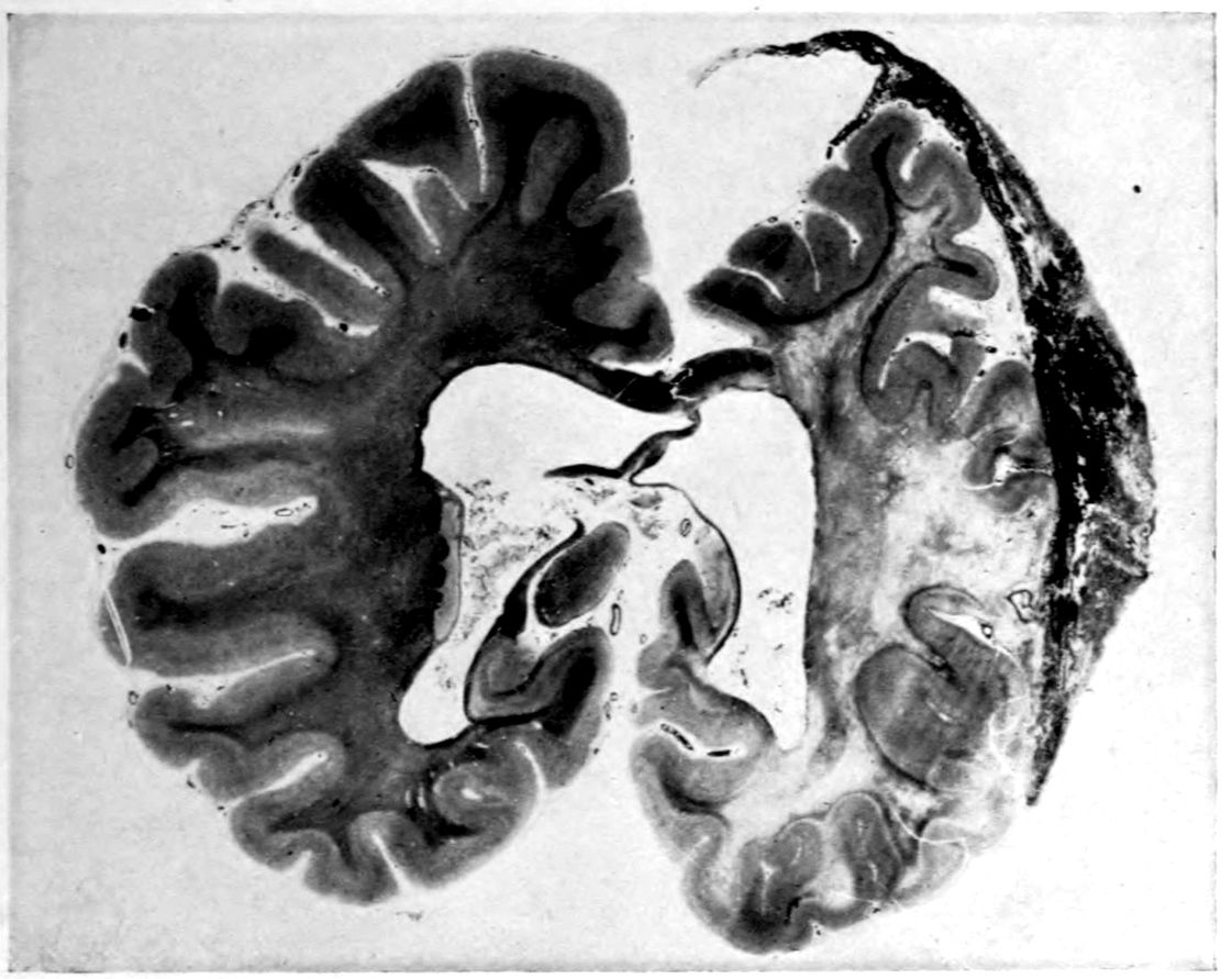
Gummatous meningitis. Compression of hemisphere. Tissue destruction of underlying cortex.
1392. Is operative procedure to be advised in cerebral gumma? There are cases in which the acute and threatening symptoms of heightened intracranial pressure require operative treatment simply because the therapeutist cannot wait for the effect of antisyphilitic treatment. Moreover, antisyphilitic treatment of cerebral gumma is not always as successful as that of most syphilitic lesions.
3. Could the intracranial pressure be caused by other syphilitic lesions than gumma? A heavy meningitis may cause symptoms such as produced by an intracranial tumor. In such a case one will usually find evidences of inflammation in the spinal fluid. Cysts caused by syphilitic lesions may also produce identical symptoms.
4. What is the significance of cranial tenderness? Where sensitiveness to cranial percussion is not due to a scalp lesion it is very suggestive of a tumor underlying this point. A gummatous lesion of the cranium itself, may occur without causing pain or increased sensitiveness.
140
CRANIAL NEUROSYPHILIS (focal syphilitic extraocular palsy) without mental symptoms.
Case 28. A chef, Paolo Marini, 28 years of age, reported that on awaking one morning, everything appeared double to him and that his right eyelid had begun to drop. In the following month Marini had begun to feel weak and to have difficulty in swallowing, as well as at times difficulty in breathing. The diplopia was found to develop when Marini looked to the right. Mentally, the patient was in all respects normal, and no other physical signs were found except the diplopia and ptosis above mentioned. The W. serum test was positive, but the tests of the spinal fluid were negative.
Diagnosis: “Cerebral syphilis.”
1. What is the anatomical cause of this condition? It is thought to be due in a number of cases to a small diffuse gummatous lesion at the basis cerebri. In the case of Marini this lesion appears to have been a little more extensive and to have interfered with the tenth and twelfth nerves also.
2. Why is the spinal fluid negative in such a case as that of Marini? Head and Fearnsides believe that intracerebral lues is characterized by a negative spinal fluid, under which circumstance one has always to consider the possibility of brain tumor or migraine in addition to the suspicion of syphilis.
3. What other causes besides syphilis should one consider for the sudden diplopia? Brain tumor, multiple sclerosis, cerebral arteriosclerosis, tuberculous meningitis, trauma and migrainous ophthalmoplegia, are not infrequently at the bottom of this condition. Cases also occur in which the etiology remains obscure, even at autopsy.
Under antisyphilitic treatment, Marini slowly improved.
141
The SIX TESTS in TABETIC NEUROSYPHILIS (“tabes dorsalis”) may run milder than in paretic neurosyphilis (“general paresis”) and characteristically run somewhat like those of diffuse (meningovascular) neurosyphilis; in particular, the fluid Wassermann Reaction and the gold sol reaction are apt to run milder. The clinical course of tabes dorsalis is protracted and the prognosis as to life is good.
Case 29. Mario Sanzi, 55 years of age, had been having what he called rheumatism since his 43d year. This rheumatism affected only the hips and legs, had at times been very severe, and for two years past had been almost constant. Before that time, pains had come at intervals, lasted a variable period, and suddenly disappeared. They were of knife-thrust character, and could probably be called “lancinating.” In a given attack, these pains would come at intervals of seconds or more. There was also a certain unsteadiness in locomotion and inability to control the vesical sphincter.
Physically, the patient was entirely normal so far as could be made out except neurologically. Argyll-Robertson pupils, absence of knee-jerks, and ankle-jerks, Romberg sign, and characteristic gait, left no cause for doubting the diagnosis of Tabes Dorsalis. The blood and spinal fluid both proved positive to the W. R., though the W. R. in the fluid gave a negative reaction with 0.1 cm. and became positive with 0.3 cm. or more. The globulin was somewhat increased though less markedly so than in paresis. The gold sol reaction was “syphilitic” but weak. It is to be noted that the disease had run a 12–years’ course before a doctor had been consulted. The primary infection occurred at 32 years, namely, 11 years before the symptoms began. At the time of his primary infection, Sanzi had received several years of treatment, chiefly in the form of mercury by mouth.
1421. What is the value of mercurial treatment of syphilis in the prevention of tabetic or other forms of neurosyphilis? “Fournier strove for many years to convince the medical profession that a syphilitic patient should be treated for at least two years after his infection, whether the syphilis seemed latent or patent. The method of treating only the symptoms he characterized as the opportunist method; treatment in the absence of definite symptoms the preventive method, as preventing the later manifestations. That prolonged treatment does prevent is shown by Fournier’s figures analyzing 2396 cases presenting tertiary signs. These he divides into three groups: Group I, comprising 1878 cases, or 78 per cent of the whole number, having no treatment or inadequate treatment—that is mercury for less than one year; Group 2, comprising 455 cases, or 19 per cent, having moderate treatment—that is, mercury for one to three years; and Group 3, comprising the remaining 19 cases which represent only 3 per cent of the whole number, having treatment for more than three years.”[7]
In the light of what we now know concerning latent neurosyphilis, it would seem well for patients to be followed from time to time with the W. R. on blood and spinal fluid after the supposed completion of the treatment of primary and secondary syphilis. The examination of the spinal fluid is not superfluous, as our experience with the so-called paresis sine paresi abundantly shows. At the present day it is not good practice to assure a patient that he is cured after two years of ordinary mercurial treatment without resort to frequent spinal fluid tests, even though the serum W. R. be negative.
143
TABETIC NEUROSYPHILIS (“tabes dorsalis”) is often quite ATYPICAL clinically and may even show no single symptom warranting the old clinical name “locomotor ataxia.”
Case 30. Stephen Green is a case of Tabes Dorsalis with active knee-jerks and without locomotor or muscle-sense disorder. When observed at the age of 45, it appeared that there were but two complaints: lack of control of the vesical sphincter and shooting pains in the legs. It appeared that the urinary disorder dated back ten years, when there had been difficulty in passing the urine. Sounds had been passed at the time; occasionally there had been incontinence during after years, ascribed by Mr. Green to the passing of the sound. However, the physician at that time stated that the incontinence was a symptom of tabes dorsalis. The incontinence had recently become worse, especially marked at night, though also occurring in the day; much worse during excitement, and very much worse after taking alcoholic drinks. Besides incontinence, there is also difficulty at times in passing the urine, as well as dysuria.
As for the pains in the legs, they had been first noticed some three or four years ago and considered to be mild rheumatic effects. Now, however, they have grown progressively worse and have been the effective cause of giving up business. The pains are sharp, darting, pinching, and burning, and last, say, about a second with an interval of about the same length. The attack will continue sometimes for many hours.
There is a strabismus of the left eye, ascribed by the patient to an accident with an umbrella (there had been operation without relief). The pupils showed the Argyll-Robertson effect and were markedly irregular. Despite the divergent strabismus with diplopia, the eye movements were well performed although not in parallel axes. Ankle-jerks could not be obtained even on reinforcement, but the knee-jerks 144were lively, and the other deep and skin reflexes proved normal. The blood and spinal fluid tests were characteristic of tabes dorsalis.
It appears that the syphilis was acquired by this patient 15 years before; that is, 5 years before neurological symptoms began. Three courses of treatment had been taken at a well-known watering-place, and mercury pills had been taken for two years by mouth. The patient is married; has no children; there have been no pregnancies.
1. What causes may be assigned for the absence of children in the family of a tabetic? There may be lesions of the genital apparatus (orchitis, or more specialized toxic lesions). But impotence such as characterized the present case must also be taken into account.
2. What is the therapy for tabetic pains? Pyramidon is nowadays much in favor; morphine may be used; some authors recommend that the patients be instructed to chloroform or etherize themselves slightly for relief of the pain. Surgery of the nerve roots may be resorted to in extreme cases. Intraspinous therapy, suggested by various authors, seems to exert beneficial effect in many cases.
3. Is the lack of control of the vesical sphincter an unusual initial symptom? On the contrary, the more careful the clinical observation, according to some observers, the more likely is the examiner to find that vesical symptoms were the earliest or among the earliest complaints of the patient. Baldwin Lucke found sphincter disturbances to be initial in 8¼% of his long Blockley series. He found sphincter disturbance to occur in some stage of the disease in 67.6%, being exceeded in frequency only by staggering gait (87.2%) and lancinating pain (71.6%). According to Lucke, the most frequent initial symptom is lancinating pain in the lower extremity, which, it will be noticed, occurred also in our case of Stephen Green as an initial symptom along with vesical disturbance. Lucke’s figures show that paresthesia of the lower extremities (17.6%) and weakness of the extremities (16.4%) are the next initial symptoms in frequency.
4. Could the early treatment in the case of Stephen Green be considered as adequate? No better answer can be given to this question than by quoting from Dr. Joseph 145Collins,[8] who probably has done more than any other one man in this country in insisting on the need of proper treatment of syphilis. As to the adequate treatment of syphilis he says:
“It consists in the proper use of salvarsan and mercury begun at the earliest possible moment after infection and kept up till all biochemical evidence of the disease has ceased, while the metabolism of the individual is maintained as nearly normal as possible. But the physician does not do his whole duty when he has accomplished this. He must solicitously watch the individual to see that no evidence reappears for months and even years after the apparent cure. As an index of such reappearance the Wassermann test of the blood serum and of the cerebrospinal fluid is the safest guide.
“Until there is a definite unanimity of belief among physicians as to when the treatment of syphilis shall be begun, and some concert of action as to what constitutes the adequate treatment of syphilis, we cannot hope to make any considerable progress in the prevention of syphilis of the nervous system, save by educating the individual toward infection.”
146
TABETIC NEUROSYPHILIS may produce symptoms chiefly if not entirely in the region supplied by the CERVICAL plexus (“cervical tabes”).
Case 31. Paul Halleck, 35, was a salesman who had begun to find it hard to carry his sample case, since he was unable to tell whether or not he had it in his hand. There was not only an anesthesia of the hands, but they felt numb and there was often a tingling sensation. Of late it had become hard for Halleck to dress himself or to write, and these symptoms had been slowly growing worse. There was no other complaint. There was, however, a history of a chancre about 7½ years before, which had been followed by a rash and a sore throat. There had been treatment with mercury and potassium iodid alternating for a period of two years.
Physically, there was no evidence of disease except neurologically. The pupils were unequal (the right larger than the left) and reacted slowly to accommodation and not at all to light. A marked ataxia of the hands was shown in coat-buttoning. The finger-to-nose test showed a marked dysmetria. Arm-jerks as well as knee- and ankle-jerks were absent. There was a slight swaying in the Romberg position but no true Romberg sign. There was no difficulty in locomotion. Both blood and spinal fluid proved positive to the W. R.; globulin and albumin were increased. The gold sol reaction was syphilitic, and there were 85 cells per cmm.
This case is probably not a pure example of Cervical Tabes, since the knee-jerks are also absent, and we may suppose a degree of lumbar spinal cord changes in addition to the cervical changes. It well illustrates, however, that the tabetic involvement of the cord may be quite generalized and that it may strike high as well as low.
147
ERB’S SYPHILITIC SPASTIC PARAPLEGIA.
Case 32. Margaret Neal, a maid-of-all-work, 36 years of age, was committed to a home for inebriates on account of her excessive alcoholism, but she was shortly transferred to the Psychopathic Hospital on account of difficulty with locomotion. We found a very marked spasticity in walking, with a characteristic scissors gait. The pupils were somewhat irregular, and although both reacted to light, the left reacted far more slowly than the right and the reaction failed to hold well. The arm reflexes were very active, and the knee-jerks and the ankle-jerks were particularly exaggerated. There was a double Babinski reaction, as well as Oppenheim and Gordon reflexes and a bilateral ankle clonus. There seemed to be tenderness over the nerve trunks in the back of the leg, below the knee. There was no evidence of incoördination, no Rombergism, no disturbance of sensation, no disorder of the special senses, and not even a tremor of the tongue or hands.
Mentally, the patient was entirely negative.
Diagnosis: Symptomatically, it is entirely clear that the patient was suffering from Spastic Paraplegia. One would have to consider besides spinal syphilis, also amyotrophic lateral sclerosis, syringomyelia, and spinal cord tumor. However, there appeared to be no definite wasting of muscles, and the fact that the sensations were intact seems to rule out also syringomyelia. There was none of the characteristic pain associated with a cord tumor. There was, in fact, a strong clinical premonition that the case was one of spinal syphilis, simply because syphilis is the most common cause of spastic paraplegia in the adult. The pupillary anomalies were also highly suggestive.
The serum W. R. proved to be weakly positive, as was also the gold sol reaction in the zones characteristic of syphilis. The spinal fluid examination yielded 14 cells per cmm. There 148was a positive globulin test and a moderate increase in albumin. The W. R. of the spinal fluid was negative.
1. Why was the spinal fluid W. R. negative in this case of spinal syphilis? The explanation of negative W. R.’s in spinal syphilis is not easy. Possibly, however, in the course of years the intensity of the process has been reduced and possibly the W. R. has been one of the first tests to disappear.
2. How shall we explain the nerve trunk tenderness? We might consider this to be due possibly to an inflammation about the posterior roots. On the whole, partly on account of the situation of the pains below the knee, it seems probable that the nerve trunk tenderness of this case is the residuum of an alcoholic neuritis.
Treatment: Under injections of mercury salicylate, there was a rapid improvement. In fact, in the course of several months, the patient regained an ability to walk long distances. There still remains a certain spasticity, but the abnormal spinal reflexes above mentioned are no longer present.
149
SYPHILITIC MUSCULAR ATROPHY, probably due either to spinal parenchymal lesions, or to root neuritis, or to both.
Case 33. Joseph Graham, now 50 years of age, seemed no longer to be able to do good work as a teamster. His arms had become weak and the muscles had become tremulous and apparently wasted. There was also pain in the left leg and hip. It appears that this latter symptom had been thought to be rheumatism, having begun about 8 years before with a sudden sharp shooting pain in the left hip, about the region of the sciatic notch. Graham had rubbed the hip with liniment, but without reducing the so-called rheumatism. The trembling of the hands had begun some years later, but no wasting had been noticed except during the past year. The pain in the leg had suddenly become so severe that a month before medical observation he had quit work. The question immediately arose whether Graham was not suffering from some familial form of muscular atrophy; but according to his representations, there was nothing of the sort in the family.
Physically, there was little to note. Neurologically, there was more. The pupils were somewhat irregular in outline, and the right was larger than the left. The left pupil failed to react to light, and the right pupil reacted very slowly and with but a slight excursion. There was no tremor of the tongue and no evidence of facial palsy nor was there smoothing of the nasolabial folds. It was somewhat remarkable, that in the absence of these signs, there was a marked speech defect. The atrophy of arms, forearms, and hands was well marked, especially the atrophy of the thenar and hypothenar eminences of the right hand. The extended hands, especially the right, showed a marked coarse tremor. Fibrillation was found in the muscles of the hands, forearms, arms, and pectoral muscles. There was no dysmetria, and the diadochokinesia was normal. Strength was diminished (dynamometer 150right hand, 32 kg., left 31 kg.). There was little or no atrophy of the legs, although the left thigh was perhaps slightly atrophic and the gluteal muscles of the left side were somewhat flabby. The patellar and Achilles reflexes were absent on both sides. There was a slight swaying in Romberg position. Gait was normal. There was a marked tenderness on the left side of the sciatic notch, as well as over the entire distribution of both external and internal popliteal nerves. This area of skin was also hyperesthetic. There were no other neurological signs on systematic examination.
Diagnosis: The sensory disorder, the speech defect, and the pupillary abnormalities seem to render the diagnosis of progressive muscular atrophy doubtful. Nor was there any dissociation of sensations to suggest a syringomyelia. Under such circumstances, one must fall back upon the question of syphilis. Both blood and spinal fluid proved to be positive to the W. R.; the globulin was increased and the albumin markedly so; there were 61 cells per cmm., and the gold sol reaction read 4 4 4 4 3 2 1 0 0 0.
1. Is there a relation of Syphilitic Muscular Atrophy to amyotrophic lateral sclerosis? Spiller, some years since, claimed such a relation, and it would seem with some justice.
2. How shall the present case be classified? There is evidence of root pains (left hip). We may naturally suppose that these root pains are reasonably good clinical evidence of a meningitic lesion, of which the spinal fluid clinically gave a confirmation. The fibrillation in this case somewhat suggests, however, a central origin for the muscular atrophy. Accordingly, it would be difficult to definitely classify the present case as either one of meningovascular syphilis or one of central syphilis. It will be remembered that Head and Fearnsides classify muscular atrophy under both these headings.
151
The period of SECONDARY SYPHILIS is frequently (over a third of all cases?) MARKED BY approved signs of NEUROSYPHILIS precisely like those of full-blown paretic or diffuse (meningovascular non-paretic) neurosyphilis. These signs occur sometimes in association with severe clinical symptoms, sometimes without clinical symptoms.
Case 34. John Bennett, 28, was brought to the Psychopathic Hospital much confused. His brother, who came with him, said that he had been a very heavy drinker but had given up drinking about four months before. He had recently had a cold but was otherwise in good health up to the night before admission. On this night, Bennett had become suddenly excited and went into his mother’s room, at the common home, and began to curse her. However, he was put to bed safely, but on the next morning began to moan continuously. After some hours of moaning, he was brought to the hospital. Here he remained difficult to manage, being irritable, noisy, and resistive. Questions he either would not or could not answer, and there was even no evidence that he understood questions. However, within a few hours, it was clear that he was slowly coming out of the confused state. On the following day, it was possible even to rouse him and get his name. The confusion gradually cleared still further and, by the end of three days, he had become mentally absolutely well so far as could be determined.
He then informed us that he had had a chancre about five or six months before, followed by a secondary skin eruption; that he had received four injections of salvarsan (the last, a month before admission) and three injections of mercury. At about the time of the last injection of salvarsan, he had developed headache with pain and slight stiffness in the back of his neck; and a fortnight later, he began to have dizzy spells, followed during the last week by difficulty in hearing. There was amnesia for everything that happened after his 152spell of sudden excitement on the evening before admission, and this amnesia was never lifted for the four days that followed.
Physically, Bennett was very well built and muscular. Nor were there any evidences of disease outside the nervous system. There was some slight stiffness of the neck and slight pain on movement of the head, which probably ought to be attributed to meningitis. The neurological examination showed tendon reflexes all normal, and normal sensations. There were, in fact, no neurological signs except that both pupils were dilated; the left was larger than the right. Both pupils reacted to light but reacted very poorly. They reacted much better to accommodation.
The W. R. proved to be positive, as might well be expected in a man whose infection had taken place less than six months before. The globulin and albumin of the cerebrospinal fluid were in great excess, of a degree which we clinically express by ++++. The W. R. of the fluid also was strongly positive down to 0.1 of a cmm. The gold sol reaction was the “paretic” type, and there were 228 cells per cmm.
1. How early may clinical evidence of neurosyphilis set in after infection? Craig found one case of “brain syphilis” occurring one month after infection. Frye claims a case of tabes dorsalis developing six weeks after infection. Craig states that he has had three cases of brain syphilis occurring within six months, and six within a year of infection.
2. What effect did the salvarsan injections have in causing or preventing the symptoms in this case? Nonne sums up the neurorecidive question as follows: Since the introduction of salvarsan therapy for neurosyphilis, paralyses of various cranial nerves are seen more frequently. This higher frequency is in part only apparent since more attention has been paid of late to auditory and labyrinthine disorders. On the whole, however, it must be considered that salvarsan does mobilize spirochete foci which without salvarsan therapy would perhaps have remained latent. Probably we are here dealing in some instances with fresh infections of neurosyphilis, in other cases with a Herxheimer reaction. 153Ehrlich believed that these latent foci occur particularly in places with stagnant blood current; as, for instance, in the narrow bony canals. This hypothesis, sufficient in some instances, is less satisfactory for cases of peripheral neuritis, for example.
3. What treatment is indicated? Intensive antisyphilitic treatment is strongly indicated. Whatever may be the truth concerning the production of neuro-recurrences (“neurorecidives”) it is certain that the symptoms usually vanish with a continuance of salvarsan therapy. The important point is to give efficient treatment, and in a case like Bennett’s improvement is fairly certain unless some serious insult occurs before the remedial efforts have been given time. It is still an open question whether intraspinous treatment is more efficient in such cases than intensive intravenous injections of salvarsan. In Bennett’s case diarsenol was injected intravenously twice a week in 0.6 gm. doses, reënforced with intramuscular injections of mercury salicylate and potassium iodid by mouth. Under this treatment improvement began slowly and in a few months he was symptomatically well and after three months his tests were practically negative.
154
JUVENILE PARETIC NEUROSYPHILIS (“juvenile paresis”) with OPTIC ATROPHY.
Case 35. Mary Coughlin, a blind girl of 16 years, was brought to the hospital in a state of great excitement, laughing and crying alternately. The neurologist is entitled to think of blindness, and particularly of the optic atrophy which Mary showed, as probably due to syphilis. However, there was no history of syphilis in the father, who died in an accident at the age of 40, or the mother, who died at 45, of heart trouble. An elder sister was married and well; two younger sisters were living and well. The fifth sibling, a boy, had died in infancy. There had been no miscarriages. In fact, the only point in favor of syphilis was the somewhat far-fetched point that the younger brother of the patient had died in infancy.
The patient’s history was rather suggestive of some other diagnosis. Her birth had been normal, she walked and talked at 13 months, was at school from six to twelve, reaching the seventh grade, and was considered bright. At three years of age, she had been run down by a car and dragged under the fender for a considerable distance. Her head was hurt but the patient did not lose consciousness in the accident. Fainting spells began at 11, in which spells the patient would lose consciousness for a minute or two. About this time, the patient’s eyesight had begun to fail, and for some four years she had been entirely blind. Headaches had come on of late.
The Coughlin case, except for the above-mentioned suspicion of syphilitic optic atrophy, might be regarded as an unusual example of a post-traumatic disease.
We found her to be fairly well developed and nourished; there was a deformity of the lower half of the sternum and of the third and fourth ribs on the right side. There were no other physical phenomena found upon systematic examination. The left pupil still reacted to light; the right failed to react, but this lack of reaction could not be regarded as of 155Argyll-Robertson nature on account of the finding of optic atrophy with the ophthalmoscope.
Mentally, it appeared that the patient’s retention of school knowledge was poor, though her blindness for four years had doubtless given her little opportunity to keep such information fresh. Rather strangely, Mary gave utterance to many delusions: first, expecting to receive her sight by an operation on the head; second, to write a book of her doings; third, to buy a house for the children; fourth, would pay $3000 for the house, earning the money by working at a tailor’s or as a trained nurse; fifth, to go on the stage to earn money by dancing; sixth, will have lots of money.
One of Mary’s characteristic statements is as follows: “Won’t it be lovely when I can see Dr. H.’s face in heaven or some other lovely place? Dr. H. was a grand doctor to me, and when we get together again we are going to Tremont Temple and keep us together. I am going to do some dancing and play the piano. I am going to graduate at the high school and go to Trinity College in Washington, and I hope I shall be a faithful keeper of mother’s tomb.”
The patient was at times euphoric and expansive.
At this stage, what with optic atrophy, euphoria, and expansive delusions, we should perhaps be entitled, had Mary been an adult, to offer the diagnosis General Paresis. In fact, on the whole, any other than a syphilitic cause for the optic atrophy was exceedingly doubtful. Brain tumor of a nature to produce optic atrophy might very improbably last so long as five years. There was no evidence of any intoxication at the time when the blindness occurred.
The W. R. was positive in the blood and spinal fluid; there was a positive globulin test, and an excess albumin as well as 15 cells per cmm.
1. What is the significance of Mary’s trauma at three years? So far as we are aware, none.
2. What light could be thrown by a W. R. study of the family? In some instances, much light is thrown; in the present case all three living sisters of the patient have been examined and their serum W. R.’s have been found negative.
1563. What is the prognosis of juvenile general paresis? Death within a few years, as in general paresis in adults. The patients live rarely more than four or five years after the onset of symptoms. Mary Coughlin died a year and a half after the above examination, namely, in her eighteenth year, some seven years after the onset of symptoms.
4. What can be said of treatment? A few favorable results have been reported after intraspinous therapy (Swift-Ellis). Too little work has been done with systematic treatment of juvenile neurosyphilis, both paretic and non-paretic, to permit important conclusions at this time.
5. How can we explain the infection of this sibling whereas the others, both younger and older, escaped? It would seem that we would have to discard the hypothesis of a congenital infection and consider that it was acquired accidentally during the lifetime of the patient. Considering the prevalence of syphilis it is rather to be wondered that more such cases of “innocent” infection do not occur in children. We may recall how many instances of juvenile gonorrhea occur. In a case as this where the symptoms calling attention to syphilis necessarily occur so long after the original infection it is practically impossible to trace the origin of the infection.
157
The diagnosis of JUVENILE PARESIS is often easy.
Case 36. Theresa Mullen, an under-sized girl of 12 years, presented a remarkable appearance due to congenital amputations of the fingers and toes. She lay in bed, drivelling and making unintelligible cries. It appeared that the patient weighed about 12 pounds at birth and was very fat; that she had been fed on condensed milk, had survived cholera infantum, whooping cough, and, as the parents said, “two kinds of measles.”
Theresa had gone to school at 5 years, reaching the third grade at the age of 9; but at this time, she began to lose ground and was put in a class for backward children. Moreover, at about this time, the teachers noticed spells of causeless laughter and meaningless twisting back and forth. Theresa would also scream at night, looking about the room; once, rising and crying, “Take him away, that black thing,” though no appropriate object was present. There had been little or no complaint of headache. Theresa had been deteriorating for some time, and for a year past had been having increased difficulty in walking. For two months the child had not spoken intelligible words; for the last week, she had been incontinent.
The diagnosis was almost obvious from the manual and pedal deformities taken in connection with the saddle-back deformity of the nose. It was interesting in connection with the contentions of W. W. Graves, that the scapulae were scaphoid in type.
Accordingly, the history given by the parents seemed consistent enough. The parents were both 36 years of age, having married at 23. The first pregnancy was a miscarriage at two months, of unknown cause. Theresa came next; thirdly, came a miscarriage at three months; fourthly, a girl, who is not strong or well physically, has suffered much from headaches and sore throat, but is fairly bright. The 158fifth pregnancy resulted in a boy, who is bright but of under-size. Three more pregnancies resulted in miscarriage.
Taking into account the above-mentioned physical characteristics, the personal history, and the family history of Theresa, the diagnosis could hardly be in doubt even in the absence of a lack of pupillary reaction to light on the right side, infantilism of genitalia, positive W. R.’s of serum and spinal fluid, positive globulin, and excess albumin, 34 cells per cmm. and the paretic type of gold sol reaction which were found.
The prognosis of this case appears to be rapid deterioration, terminating in death within a few months. Now and again, however, some such cases spontaneously improve. Such a case as that of Theresa Mullen is always disheartening in itself but suggests the social value of Wassermann tests in the other members of the family. The other children of the Mullen family proved to be suffering also from syphilis, since their blood sera all showed a positive W. R.
1. What is the characteristic age of onset in Juvenile Paresis? An impression has prevailed in some quarters that the typical onset of juvenile paresis is in the adolescent years, and Clouston’s first case (1877) developed in a boy of 16. Thierry’s 58 cases, developing from the 8th to the 20th year, averaged 14 years of age at onset. Mott’s 22 cases from the 8th to the 23d year, averaged 17 years at onset. According to Clouston, juvenile paresis develops most often at puberty (15 to 17 years). It is sometimes claimed that cases developing symptoms early live longer, and that juvenile cases developing symptoms after the 20th year run a short course. For a case developing in the 5th year, see John Friedreich, Case No. 77.
2. What may be concluded from the physical signs (congenital amputations) present in this case before the development of mental symptoms? Some cases of juvenile paresis appear to show no physical signs whatever in childhood. While these amputations might be the accidental result of a difficult delivery, it is more probable that they are due to a syphilitic process.
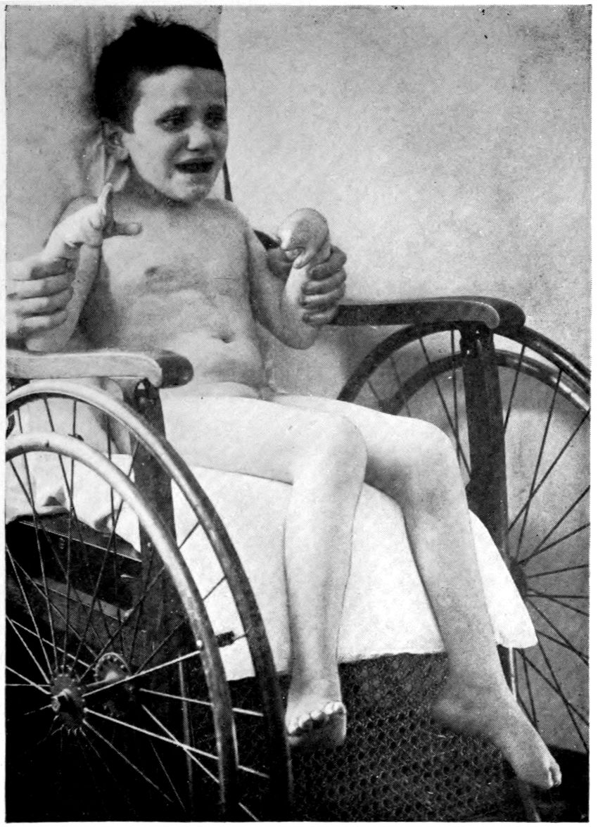
Juvenile paresis—congenital amputation of digits. This case reached fourth grade in school before deterioration.
159
CONGENITAL SYPHILIS is apparently capable of producing simple FEEBLEMINDEDNESS (that is, a form of disease non-paretic, non-tabetic, without special tendency to progression, and without tendency to vascular insults).
Case 37. Isaac Goldstein was a small boy of six years and seven months, with a father known to be suffering from general paresis. The child was very irritable and nervous and very difficult to manage, but would hardly have been the subject of medical attention except in a family study suggested by the paresis of the father.
The child had been born at term and had apparently undergone a normal development. Physically, he showed no definite signs of congenital syphilis. In fact, the physical examination was to all intents and purposes negative. The W. R. of the serum, however, proved to be positive. Mental tests showed that his mental age was that of a child of a little over five years. Taking all things into account, it is probable that he should be regarded, therefore, as somewhat retarded mentally.
1. Is syphilis answerable for the mental retardation in this case? Provided that the family is free from feeblemindedness and mental disease, it would seem that the retardation of a congenital syphilitic should perhaps be regarded as syphilitic in origin. Of course, the institutions for the feebleminded have not shown exceedingly high percentages of syphilitic children in various W. R. surveys; still, the percentage of positive reactions in institutions for the feebleminded is clearly higher than the incidence of congenital syphilis shown in the population at large. Hence, we may conclude that syphilis is one of the etiological factors in the production of feeblemindedness. Dr. W. E. Fernald, of the Waverley School for the Feebleminded, has recently pointed out that the syphilitic cases belong rather in the lower grades (idiots and imbeciles) of feeblemindedness than in the higher (morons).
1602. Can we guess what the pathological anatomy and histology of the brain may be in such cases? The Waverley studies now in process seem to indicate that some cases have little or no gross alterations, but show a few slight traces of lymphocytic accumulations discovered upon extended search, and a certain tendency to the appearance of rod cells in various foci. But the whole matter is still sub judice. It is a question whether these traces of chronic inflammation are the residuals of a more active process or the beginnings of a process that is about to be more active.
3. How characteristic is a positive W. R. in the serum of a child without physical stigmata of congenital syphilis? If we limit the term stigmata to the major and more important signs, we must reply that it is not unusual to find positive W. R.’s in sera of physically normal-looking children. Except in family studies, such cases will often escape notice, either because there are no stigmata whatever, or because such stigmata as exist are of a minor nature and regarded as unimportant anomalies. Some of these cases occur in the clinics later in life as so-called syphilis hereditaria tarda. If one wishes to discover these cases with late development of symptoms before their full bloom, the most obvious method is to examine carefully the children of known syphilitics.
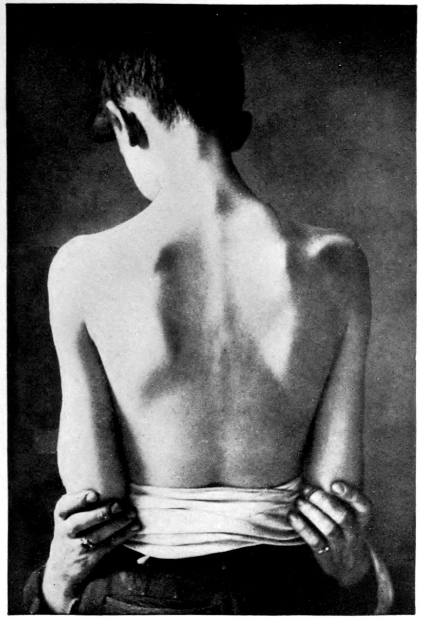
Scaphoid Scapulae.
161
JUVENILE TABETIC NEUROSYPHILIS (“juvenile tabes”); TREATMENT.
Case 38. The point in presenting Archibald Sherry, a Juvenile Tabetic of 12 years on admission, is perhaps to exhibit pride in therapeutic results.
There was little or no doubt of the diagnosis; in an adult, the phenomenon would be called tabes dorsalis with a question of general paresis. The right pupil was larger than the left and reacted neither to light nor to distance. There was a slight tremor of the tongue and of the outstretched hands. The knee-jerks and ankle-jerks could not be obtained, nor could the periosteal reflexes in the legs. There was a slight unsteadiness in the gait and in various finer movements, and a slight ataxia of the legs. There was not a classical Romberg sign but there was slight swaying in Romberg position. The teeth were Hutchinsonian. For the rest, the physical examination was practically negative.
The family history was of interest. On the paternal side there was nervousness as well as alcoholism and degeneracy. The maternal grandmother had cancer. Archibald’s father was immoral and alcoholic. There was a girl four years older than Archibald, who, though nervous and unstable, has shown no signs or symptoms of syphilis and does not yield a W. R. in blood or spinal fluid.
Archibald himself was born at term, a large child, who, however, lost weight rapidly, developing a marked skin eruption on head and back three weeks after birth. This skin disease lasted for a month and a half and then spontaneously disappeared. Archibald remained weak and sickly, not walking until three years of age. However, he did well in school up to the end of his 11th year, when he failed to keep up with the children. He had been an amiable child and had gotten on well with his playmates. Some time in his 10th year physical disability had begun; there was numbness in the legs with weakness; at times, actual inability to 162walk. The right pupil was noticed by the mother to have increased in size; the eyelashes had turned white. There was pain over the left eye and a feeling of weight on top of the head. Speech became difficult or even confused.
Consistently enough, the W. R. both in blood and spinal fluid was positive. Globulin and albumin were present in large amounts; there were 150 cells per cmm.
Granting that this be in some sense a case of juvenile tabes we may raise a doubt whether the case is one of congenital syphilis. The W. R.’s of the blood of both father and mother are negative. Syphilis is denied by them. The nervous and unstable older sister failed to show definite symptoms of syphilis or a positive W. R. There had been no miscarriages or stillbirths. The question arises whether the Hutchinsonian teeth do not indicate congenital syphilis. It appears, however, that it is possible to develop Hutchinsonian teeth if syphilis is acquired before the teeth are formed. We have no data as to how or why this particular baby should have acquired syphilis, if he did so acquire it, at the age of three weeks. On the whole, sceptics may doubt our suggestion that the case is one of acquired juvenile tabes. Possibly the question is academic so far as treatment is concerned.
Prognosis: The rarity of juvenile tabes is such that little can be said as to prognosis. Three and a half years have passed since a few injections of salvarsan were made. The pains above mentioned rapidly disappeared, the gait became steadier, the attacks of confusion ceased, and the speech improved. Unfortunately, on account of a lack of coöperation on the part of Archibald’s mother, we have been unable to continue treatment. However, we have from time to time followed the patient in his home and he seems to have shown no falling back after the initial improvement. It would be of great value could we know the situation in the spinal fluid at the present time.
1. Is there any explanation why paresis should occur in some juveniles and tabes in others? There is no available explanation for this difference nor any for the characteristic early optic atrophy of juvenile tabetics.
164
Be frustrate, all ye stratagems of Hell,
And, devilish machinations, come to nought!
Paradise Regained, lines 180–181
165