Front matter (continued 2)
Reading edition
San José: San Isidro el General, KU 32249, UMMZ 75025; 10 km. N San Isidro el General, MCZ 29099-103; 13 km. WSW San Isidro el General, KU 86517; 15 km. WSW San Isidro el General, KU 68487 (tadpoles), 68488 (young), 68495 (young); 20 km. WSW San Isidro el General, KU 32248.
Panama: No province: Cano Saddle, USNM 69588; Punta de Pena, USNM 38733; Quipo, AMNH 18925-6. Bocas del Toro: Almirante, KU 80080, 91835-6; 1.6 km. W Almirante, KU 91837; 3 km. W Almirante, KU 91824 (skeleton), 91838-43, 91906-7; 11 km. NW Almirante, CNHM 67853-61; 13 km. W Almirante, KU 91825-7 (skeletons), 91844-9; Fish Creek, KU 92329; Isla Popa, KU 91850-1. Canal Zone: Barro Colorado Island, CNHM 6007, 13316, 13325, 13331, 13360-2, 13377-8, MCZ 24191-5, UF 7523, UMMZ 63547-60, 64457, 69497 (3); 3.7 km. W Cocoli, KU 67916; Fort Sherman, MCZ 10139; Gatun, MCZ 35644; Junction roads C25B and C16, TNHC 23839; Madden Forest Preserve, TNHC 23837-8. Coclé: El Valle, KU 77521-4, 77649 (tadpole), TNHC 23369. Comarca del Barú: Progreso, UMMZ 61085-9. Colón: Achiote, KU 77516-20, 77648 (young); Río Candelaria, CNHM 67851-2. Darién: Río Esnápe, Sambú Valley, MCZ 8539; Río Sucubti, Chalichiman's Creek, AMNH 40512; Camp Creek, AMNH 40758-9; Camp Creek, Camp Townsend, AMNH 40988. Panamá: NW slope Cerro Prominente, KU 80459; Finca La Sumbadora, KU 91914 (skeleton). Chiriquí: 2 km. W Concepción, AMNH 68910.
Columbia: Antioquia: Puerto Berrio, CNHM 30805 (Goin); Turbo, USNM 39899. Caldas: Pueblorrica, Santa Cecilia, CNHM 54768-71 (Goin). Choco: No specific locality, AMNH 3984-6; Andagoya, BMNH 1915.10.21. 69-70, CNHM 81857 (Goin); Golfo de Urabá, CNHM 63881 (Goin); Peña Lisa, Condoto, BMNH 1913.11.12. 118-125, 1913.11.12. 137-146 (Goin); Pizarro, CNHM 4451-3, 4455-61 (Goin); Río San Juan, Playa del Oro, CNHM 54772 (Goin); Río Quesada, AMNH 13615-77; 37 km. up Río Puné, AMNH 13688; 48 km. up Río Puné, AMNH 13689. Narino: Tumaco, Río Rosario, CJG 2310-13 (Goin). Valle: Buenaventura, BMNH 1895.11.16.82 (Goin); Raposa, WAT 166, 346-47, 388 (Goin); Río Calima above Córdoba, CJG 2249-57 (Goin).
Ecuador: No province: Bulun, AMNH 10620. Esmeraldas: Cachabé, AMNH 10625-8; Río Capayas, CNHM 35712; Río Sapaya, UMMZ 58910 (5); Salidero, AMNH 10623-4; San Javier, AMNH 10618. Guayas: Hacienda Balao Chico, UMMZ 123904. Imbabura: Pambelar, AMNH 10629, 10631. Pichincha: Hacienda Espinosa, 9 km. W Santo Domingo de los Colorados, KU 40220.
Smilisca puma (Cope), new combination
Hyla puma Cope, Proc. Amer. Philos. Soc., 22:183, 1885 [Holotype.—USNM 13735 from Nicaragua; J. F. Moser collector]. Günther, Biologia Centrali-Americana: Reptilia and Batrachia, p. 270, Sept., 1901. Nieden, Das Tierreich, Amphibia, Anura I, p. 251, June, 1923. Cochran, Bull. U. S. Natl. Mus., 220:58, 1961.
Hyla wellmanorum Taylor, Univ. Kansas Sci. Bull. 25(1):843, July 1, 1952 [Holotype.—KU 30302 from Batán, Limón, Costa Rica, Edward H. Taylor collector]; Univ. Kansas Sci. Bull., 36(1):626, June 1, 1954. Duellman and Berg, Univ. Kansas Publ. Mus. Nat. Hist., 15:194, Oct. 26, 1962.
Smilisca wellmanorum, Starrett, Copeia, 4:303, Dec. 30, 1960.
Diagnosis.—Size small ([M] 38.0 mm., [F] 46.0 mm.), differing from other species in the genus by the following combination of characters: skull about as long as broad; frontoparietal fontanelle keyhole-shaped; supraorbital flanges absent; squamosal small, not in contact with maxillary; bony portion of ethmoid terminating at anterior edge of orbit; tarsal fold weak, two-thirds length of tarsus; inner metatarsal tubercle small, low, flat, elliptical; snout rounded in dorsal profile; lips thin and flaring; fingers having only vestige of web; toes one-half webbed; diameter of tympanum about two-thirds that of eye; narrow labial stripe white; pair of dark brown (sometimes interconnected) stripes on tan dorsum; no blue spots on flanks or thighs; vocal sac in breeding males pale brown. (Foregoing combination of characters distinguishing S. puma from other species in genus.)
Description and variation.—Ten breeding males from the vicinity of Puerto Viejo, Heredia Province, Costa Rica, have snout-vent lengths of 32.5 to 37.9 mm. (34.8 mm.). In these specimens, the length of the tibia to the snout-vent length is 0.48 to 0.53 (0.51), and the tympanum/eye ratio is 0.52 to 0.72 (0.65). Seven females have snout-vent lengths of 40.8 to 45.8 mm. (43.9 mm.). No individual has more than a vestige of a web between the second and third and fourth fingers. None has a web between the first and second fingers. Breeding males lack nuptial excrescences on the thumbs. The vocal sac is moderately large and bilobed.
In preserved specimens the dorsal ground color varies from yellowish tan to grayish brown. All specimens have dark brown dorsal markings in the form of a pair of dorsal stripes, variously modified (Pl. 7A). In some specimens, such as KU 91716, the stripes are discrete and extend from the postorbital region nearly to the vent. In most specimens the stripes are connected by a transverse mark in the scapular region and in many others also by a transverse mark in the sacral region. In some specimens the stripes are fragmented posteriorly; fragmentation is extreme in KU 30300, in which the dorsal pattern consists of two series of dark longitudinal dashes. The other extreme is a nearly complete fusion of the stripes, as in KU 91714. A dark brown interorbital bar usually extends onto the eyelids, but in some specimens this is reduced to a short V-shaped mark or small spot between the eyes. There is no dark post-tympanic mark, but dark brown pigment forms a venated pattern from the axilla to the mid-flank; the inguinal region is white, finely mottled with dark brown. The dorsal surfaces of the hind limbs are colored like the body and have two or three dark brown transverse marks on the thighs, three to five marks on the shanks, and one or two marks or irregularly arranged dark flecks on the tarsi. The anterior and posterior surfaces of the thighs are pale tan to brown. The webbing of the feet is tan to grayish brown. A narrow white labial stripe, white anal stripe, and narrow white stripes on the tarsi and outer edges of the forelimbs are invariably present. The ventral surfaces are creamy white.
In life the dorsum is tan or pale brown with dark brown markings. Some individuals have scattered metallic green flecks on the dorsum. The flanks are mottled dark brown and creamy white. The posterior surfaces of the thighs are dark brown. The vocal sacs are grayish brown, and the iris is a deep bronze color.
Natural History.—Smilisca puma inhabits humid lowland tropical forests having more or less evenly distributed rainfall throughout the year. The equable climatic conditions seemingly permit these frogs to be active throughout most of the year. Taylor (1952:846) found calling males at Batán, Costa Rica, on July 20, 1951. We found the species breeding near Puerto Viejo, Costa Rica, on February 19, June 18, July 13, and July 31. Specimens of calling males from Costa Rica in the collection at the University of Southern California were obtained in February at La Fortuna, on August 22 at Los Diamantes, on August 30 at Jabillos, and on September 5 at La Lola. Gravid females were collected in June, July and August.
Males call from shallow water. All breeding congregations of this species that we have found were in a grassy marsh, 7.5 kilometers west of Puerto Viejo, Costa Rica. Tadpoles were found in water-filled depressions in the marsh at night. When first observed, tadpoles were near the surface of the water; they responded to light by quickly taking refuge in the dense grass. No tadpoles were observed by day.
The breeding call consists of a low squawk, usually followed by a series of one or more rattling secondary notes (duration of primary notes, 0.06-0.35 seconds; of secondary notes, 0.10 to 0.47 seconds), repeated at intervals of 5 to 55 seconds. The primary notes have 187 to 240 pulses per second and major frequencies of about 740 to 1870 cycles per second (Pl. 11A).
Only six tadpoles are available for study. Four of them in stage 34 of development have body lengths of 9.0 to 9.5 mm., tail lengths of 14.0 to 15.0 mm., and total lengths of 23.0 to 24.5 mm. One tadpole in stage 38 and one in stage 40 have total lengths of 31.0 mm. A typical tadpole in stage 34 of development (KU 91807 from 7.5 km. W Puerto Viejo, Heredia Province, Costa Rica) has a body length of 9.5 mm., tail length of 15.0 mm., and total length of 24.5 mm.; body about three-fourths as deep as wide; snout rounded dorsally and laterally; eyes widely separated, directed dorsolaterally; nostril about midway between eye and tip of snout; mouth anteroventral; spiracle sinistral, about two-thirds distance from snout to posterior end of body and slightly below midline; anal tube dextral; caudal musculature slender, barely curved upward distally; dorsal fin extending onto body; at mid-length of tail, depth of caudal musculature equal to that of dorsal fin and ventral fin; body grayish brown, palest ventrally; caudal musculature pale creamy yellow with bold gray reticulations; caudal fins transparent with gray reticulations anteriorly and black flecks posteriorly on both fins (Fig. 14A). Median part of upper lip bare; rest of mouth bordered by two rows of short blunt papillae; lateral fold present; tooth-rows 2/3; upper rows equal in length; second upper row broadly interrupted medially; three lower rows complete, first and second rows equal in length, slightly shorter than upper rows; third lower row noticeably shorter; upper beak shallow, forming broad, continuous arch with slender lateral processes; lower beak slender, broadly V-shaped, both beaks finely serrate (Fig. 15B).
All six tadpoles are colored alike, except that in the larger specimens scattered white flecks are present on the ventral surface of the body, and the dark reticulations continue farther posteriorly on the caudal fins than in the smaller tadpoles. In two specimens the third lower tooth-row is only about one-half the length of the other lower rows, and in one specimen the second lower tooth-row is shorter than the first. Coloration of tadpoles in life: "Body olive-brown with silvery green flecks laterally. Caudal musculature olive-brown with greenish tan flecks. Fins brown with greenish gold flecks. Iris deep bronze." (Duellman, field notes, February 19, 1965).
One recently metamorphosed young (KU 91808) has a snout-vent length of 12.4 mm. In life this frog had a pale tan dorsum with dark brown markings, yellowish tan posterior surfaces of thighs, grayish brown throat, and bronze iris.
Remarks.—The identity of Cope's Hyla puma has not been known. The name has appeared in various compilations, but no workers have referred any of their specimens to that species. Examination of the holotype (USNM 13735), an adult female, revealed the presence of the following combination of characters: snout-vent length 45.8 mm., snout blunt above and rounded laterally, nostrils close to tip of snout, lips thin and flaring, a vestige of a web on the hands, feet about one-half webbed, tarsal fold weak and extending about two-thirds length of tarsus, dorsal markings consisting of a faded dark interorbital bar and a pair of faded longitudinal brown marks connected by a transverse band in the scapular region. The type agrees well with specimens of Smilisca wellmanorum (Taylor, 1952); the vestigial webbing on the hands and the dorsal coloration are especially significant. Consequently, we consider Hyla wellmanorum Taylor, 1952, to be a synonym of Hyla puma Cope, 1885. Cope gave only "Nicaragua" as the locality for Hyla puma. The specimen was part of a collection received at the United States National Museum from Lt. J. F. Moser. Among the species in the collection are Dentrobates pumilio, Phyllomedusa helenae, Corythophanes cristatus, Pliocercus dimidatus, Tretanorhinus nigroluteus, and others characteristically found on the Caribbean lowlands of Central America. Thus, it seems reasonable to assume that the type specimen of Hyla puma came from the Caribbean lowlands. Though no other Nicaraguan specimens have been found by us, numerous specimens are known from the Caribbean lowlands of Costa Rica.
Cochran (1961:58), in her catalogue of type specimens in the United States National Museum, listed Hyla puma Cope, 1885, as a synonym of Hyla molitor Schmidt, 1857. She made no qualifying statements. Schmidt (1858:246), in his descriptions of the species in the year following his publication of the names and Latin diagnoses, stated: "Dorsum uniformly gray, more intensive on back, fading away laterally and on extremities; in every-day-life this blue would be called Mueller's Blau. A delicately dotted black line runs on the canthus rostralis from the opening of the nose to the corner of the eye. In the armpits, on the flanks and the thighs two of our three specimens have black marblings." [Free translation] Certainly on the basis of coloration Hyla puma is distinctly different from Hyla molitor.
Distribution.—This species lives in the wet, forested regions of the Caribbean lowlands of Costa Rica and presumably southern Nicaragua (Fig. 3). All specimens are from low elevations; the highest known elevation for the occurrence of this frog is 285 meters at Laguna Bonilla.
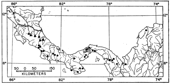
Fig. 3. Map showing locality records for Smilisca puma (triangles) and Smilisca sila (circles).
Specimens examined.—62, as follows: Nicaragua: No specific locality, USNM 13735.
Costa Rica: Alajuela: Jabillos, 5 km. N Santa Clara, USC 8058 (6); 5 km. W La Fortuna, USC 8078 (2); Río La Fortuna at La Fortuna, USC 7151 (3). Cartago: Laguna Bonilla, tunnel camp near Peralta, KU 32171. Heredia: Puerto Viejo, KU 86521; 5.9 km. W Puerto Viejo, KU 64307; 7.5 km. W Puerto Viejo, KU 64308-10, 64311 (skeleton), 64312-15, 68635-6 (skeletons), 85001-2 (skeletons), 86520, 87770-1 (skeletons), 91709-16, 91791-2, 91807 (tadpoles), 91808 (young). Limon: Batán, KU 30300-2; La Lola, KU 32169, USC 141, 201, 8067; Los Diamantes, KU 32170, UMMZ 118470 (6), USC 212; 2.4 km. E Los Diamantes, USC 8049 (5).
Smilisca sila new species
Hyla gabbi, Noble, Proc. Biol. Soc. Washington, 37:66, Feb. 21, 1924. Dunn, Occas. Papers Boston Soc. Nat. Hist., 5:413, Oct. 10, 1931. Schmidt, Smithsonian Misc. Coll., 89(1):6, March 16, 1933.
Hyla sordida, Dunn, Copeia, 3:166, Nov. 19, 1937. Cooper, Copeia, 2:121, June 30, 1944. Breder, Bull. Amer. Mus. Nat. Hist., 86(8):417, Aug. 26, 1946.
Hyla phaeota, Breder, Bull. Amer. Mus. Nat. Hist., 86(8): pl. 55, Aug. 26, 1946.
Holotype.—Adult male, KU 91852 from a small stream at the north edge of the village of El Volcán, Chiriquí Province, Panamá, elevation 1280 meters; obtained on Feb. 5, 1965, by William E. Duellman.
Paratypes.—KU 91853-74, collected with the holotype.
Diagnosis.—Size moderate ([M] 45.0 mm., [F] 62.2 mm.); skull wider than long, having large, ovoid frontoparietal fontanelle; supraorbital flanges absent; squamosal small, not contacting maxillary; bony section of ethmoid extending anteriorly between nasals; tarsal fold weak, full length of tarsus; inner metatarsal tubercle low, flat, elliptical; lips thick, rounded, not flaring; fingers one-third webbed; toes three-fourths webbed; diameter of tympanum about one-half that of eye; margin of upper lip faintly marked by interrupted creamy white stripe; dark spots on dorsum; pale flecks on flanks and posterior surfaces of thighs; vocal sacs in breeding males dark brown. (Foregoing combination of characters distinguishing S. sila from any other species in genus.)
Description of holotype.—Snout-vent length 36.6 mm.; tibia length 19.8 mm., 54.1 per cent of snout-vent length; foot length 15.5 mm., 42.3 per cent of snout-vent length; head length 12.7 mm., 34.7 per cent of snout-vent length; head width 13.3 mm., 36.8 per cent of snout-vent length; snout short, in lateral profile truncate, only slightly rounded above, in dorsal profile rounded; canthus rounded; loreal region noticeably concave; lips thick, rounded, not flaring; nostrils not protuberant, directed laterally; internarial distance 3.0 mm.; internarial area flat; top of head flat; interorbital distance 3.5 mm., 26.3 per cent of head width; diameter of eye 4.2 mm., thrice distance (1.4 mm.) from tympanum to eye, and half again distance (2.8 mm.) from orbit to nostril; pupil horizontally ovoid; width of eyelid 2.8 mm., 21.1 per cent of head width; dermal fold from posterior corner of orbit covering upper edge of tympanum to point above insertion of forelimb; diameter of tympanum 2.3 mm., 54.7 per cent of diameter of eye; no axillary membrane; arms moderately robust; weak fold on wrist; faintly scalloped fold along ventrolateral margin of forearm; fingers short, slender; fingers from shortest to longest, 1-2-4-3; vestige of web between first and second fingers; others about two-fifths webbed; discs moderate, diameter of that on third finger about one-third diameter of eye; triangular outer palmar tubercle; elliptical inner palmar tubercle on base of pollex; subarticular tubercles large, conical, none bifid; supernumerary tubercles few, large, conical; brown nuptial excrescence on prepollex; heels overlap by about one-fifth length of shank when hind limbs adpressed; tibiotarsal articulation extending to nostril; tarsal fold weak, extending nearly full length of tarsus; inner metatarsal tubercle elliptical, flat; outer metatarsal tubercle absent; toes moderately long; toes from shortest to longest, 1-2-3-5-4, third and fifth about equal in length; discs about same size as those on fingers; webbing extending to middle of penultimate phalanx on all toes, except only to distal end of antepenultimate phalanx of fourth toe; subarticular tubercles round; supernumerary tubercles large, round, present only on proximal digits. Anal opening directed posteriorly at level of upper edge of thighs; no noticeable anal sheath; flat tubercles ventrolateral to anal opening large; skin of chest, belly, and posterior surfaces of thighs granular; other surfaces smooth; tongue broadly cordiform, shallowly notched posteriorly, and barely free behind; vomerine teeth 4-4, situated on ventral surfaces of separated rounded prominences between posterior margins of small, ovoid inner nares; vocal slits long, each situated along inner margin of ramus; color (in preservative) pinkish tan above with irregular olive-brown markings forming interconnected spots on back; four bars on dorsal surface of each thigh; five bars on shank, and three on tarsus; inguinal region white with black mottling; posterior surfaces of thighs yellowish tan proximally, dark brown distally; margins of lips grayish white with brown markings; ventral surfaces of hands and feet grayish brown; belly and posterior part of throat creamy white; anterior part of throat brown.
Description and variation.—Ten breeding males from Finca La Sumbadora, Panamá, have snout-vent lengths of 40.0 to 44.8 mm. (42.3 mm.). In these specimens the tibia/snout-vent length ratio is 0.50 to 0.57 (0.54), and the tympanum/eye ratio is 0.48 to 0.58 (0.53). There is a geographic gradient in size; specimens from the western part of the range (southern Costa Rica) are smaller than those from the eastern part of the range (eastern Panamá). Five males from the Pacific lowlands of southern Costa Rica have snout-vent lengths of 31.6 to 38.2 mm. (34.7 mm.); ten males from El Volcán, Chiriquí, Panamá, 32.6 to 37.9 mm. (36.4 mm.), and eight males from Barro Colorado Island, Canal Zone, 38.2 to 42.0 mm. (35.6 mm.). These are smaller than the males from Finca La Sumbadora, which is east of the Canal Zone. Ten females from El Volcán have snout-vent lengths of 44.2 to 55.6 mm. (49.2 mm.), as compared 56.1 to 62.2 mm. (58.2 mm.) in three females from Finca La Sumbadora.
Large females have scattered small tubercles on the head and back; tubercles occur in males from Costa Rica and in some males from western Panamá. The truncate snout is characteristic of both sexes.
The coloration of Smilisca sila consists of a gray, tan, or pale reddish brown dorsal ground color and a creamy white venter. The dorsum is marked by dark brown, olive-brown, or dark reddish brown spots or blotches (Pl. 7B). Usually the blotches are discrete, but in some individuals they are interconnected and form an irregular dark mark on the dorsum. There is no tendency for the blotches to form transverse bars as in Smilisca sordida. In one specimen (KU 80467) the blotches are fused and form two wide irregular longitudinal stripes, as in Smilisca puma. In some females the dorsal markings are reduced to a few small spots or are nearly absent (KU 92332), whereas in other females the dorsal markings are bold. In one female (KU 91894) the dorsal markings are narrowly bordered by pale blue, and numerous pale blue flecks are present on the pale brown dorsum. In many individuals of both sexes small white flecks are present on the dorsal surfaces.
Usually the flanks and posterior surfaces of the thighs have black mottling enclosing pale blue spots and flecks, respectively. The dorsal surfaces of the limbs are marked by dark brown transverse bars; usually three or four bars are present on each forearm, thigh, and shank. The coloration of the flanks and limbs varies geographically. Specimens from southern Costa Rica and western Panamá have distinct bars on the limbs; the posterior surfaces of the thighs have brown reticulations enclosing small blue flecks in specimens from Costa Rica and bolder, black reticulations enclosing large pale blue spots in specimens from western Panamá. In specimens from Costa Rica the flanks are brown with pale blue flecks, whereas in those from Chiriquí, Panamá, the flanks are pale blue with dark brown mottling in the inguinal region. Frogs from El Valle and Cerro la Campana usually have distinct bars on the limbs; the posterior surfaces of the thighs are colored as in frogs from Chiriquí, and the inguinal region is pale blue with coarse brown mottling. Specimens from Barro Colorado Island are marked like those from El Valle and Cerro la Campana, except that on the posterior surfaces of the thighs fine black reticulations enclose many dark blue spots. In specimens from Darién and from Panamá Province east of the Canal Zone (Altos de Pacora, Cerro Jefe, Finca La Sumbadora, and Río Pacora), the markings on the dorsal surfaces of the limbs are indistinct or absent in males, but distinct in some females. Intense brown and black pigment forms fine reticulations delimiting bold blue spots on the flanks; this coloration extends to the axilla in many specimens. Fine black reticulations enclose many dark blue spots on the posterior surfaces of the thighs.
In females, the throat is creamy white; in some specimens scattered brown flecks are present on the chin and throat. In breeding males the anterior part of the throat is dark gray or dark brown.
The coloration in life is as variable as it is in preservative. In life the holotype had a tan dorsum with dark olive-green irregular markings and small green flecks. The limbs were tan with dark brown transverse bars. The flanks were grayish tan anteriorly; the inguinal region and posterior surfaces of thighs were blue with black mottling. The belly was creamy white, and the throat was brown with creamy yellow flecks. The iris was a dull bronze color. Among the paratypes, some individuals had green flecks, others did not. The inguinal region and posterior surfaces of the thighs were pale blue, pale yellowish green, or grayish tan with black mottling. The blue was most noticeable in females.
Colors of a male from Finca La Sumbadora, Panamá, were described as follows: "Dorsum olive-brown; irregular dark brown blotches, pale green flecks, and raised creamy yellow spots on dorsal surfaces; belly creamy white; throat grayish brown; undersides of limbs grayish tan; groin, anterior and posterior surface of thigh, inner surface of shank, anterior edge of tarsus, and proximal parts of third and fourth toes pale blue marbled with dark brown and black; webbing brown; iris pale bronze, finely reticulated with black." (Duellman, field notes, January 28, 1964.)
A female (now KU 91894) from Altos de Pacora, Panamá, was described as follows: "An irregular dark brown, green-bordered figure on head and back; dark brown, green-bordered bands on limbs—all on a lighter brown and heavily green-spotted background. These markings are more vivid at night than during the day. Lower sides, from midbody onto front of thighs and rear of thighs onto venter of shanks to heels and thence dorsally onto basal portions of toes heavily blue spotted on a light brown (front of thighs and venter of shanks) to blackish brown background. Venter cream. Iris gray-brown, finely veined with dark brown." (Charles W. Myers, field notes, December 14, 1964.) Note that in the earlier discussion of coloration of preserved specimens, the green spots and borders have changed to pale blue after six months in alcohol.
In living individuals from Costa Rica and Panamá west of the Canal Zone, the blue coloration on the flanks and thighs is much less conspicuous than in specimens from eastern Panamá. The color of the iris is variable, even in frogs from one locality. The coloration of the iris in 13 living frogs (now KU 92333-45) from Valle Hornito, Chiriquí, Panamá, was described as follows: "Iris variable—from pale to dark brown; in a few the iris has a golden cast to the brown; in a few others the lower half of the iris is pale gray with the upper half being light brown." (Charles W. Myers, field notes, April 24, 1965).
Natural history.—Smilisca sila inhabits the Pacific slopes of lower Central America where a pronounced dry season occurs. We have records of males calling in December through May and also in August (latter date from El Volcán, Chiriquí, Panamá). The breeding season seems to be correlated with the time of the year when the water is clear and at a low level in the streams where these frogs breed.
Males call from the edges of small, shallow streams, from rocks in the streams, or less frequently from vegetation overhanging the streams. Females are most frequently found on the banks of streams, and clasping pairs usually are in shallow pools in streams. One individual was found in a bromeliad about three meters above the ground in the daytime.
The breeding call consists of a low squawk, usually followed by a series of one or more rattling secondary notes (duration of primary notes, 0.06 to 0.28 seconds; of secondary notes, 0.14 to 0.48 seconds), repeated at intervals of 4 to 20 seconds. The primary notes have 97 to 120 pulses per second and major frequencies of about 900 to 2220 cycles per second (Pl. 11B).
Eggs were obtained artificially in the field; the average length of ten embryos in the neural groove stage is 2.4 mm., and the average diameter of the outer envelope is 4.9 mm. Hatchlings have large, conical oral discs, heavy gills, and a large amount of yolk; their average total length is 6.3 mm.
Tadpoles have been found in pools in clear streams; some tadpoles have been observed to cling by their mouths to rocks in the stream; others were found on the bottom where they seek refuge among pebbles or under rocks and leaves. A complete developmental series of tadpoles is not available. Eleven tadpoles in stage 25 of development have body lengths of 8.3 to 10.2 mm. (9.3 mm.), tail lengths of 17.3 to 21.0 mm. (18.8 mm.), and total lengths of 25.9 to 31.0 mm. (28.1 mm.). One tadpole in stage 41 and one in stage 42 have body lengths of 11.5 and 12.5 mm., tail lengths of 27.2 and 29.5 mm., and total lengths of 38.7 and 42.0 mm., respectively. The snout-vent lengths of two specimens in stage 43 and one in stage 45 are 12.7, 13.0, and 13.6 mm., respectively.
A typical tadpole in stage 25 of development (KU 80620 from Finca La Sumbadora, Panamá) has a body length of 9.5 mm., tail length of 19.0 mm., and a total length of 28.5 mm.; body only slightly wider than deep, nearly flat dorsally; snout broadly rounded in dorsal view, bluntly rounded in lateral view; eyes widely separated, directed dorsolaterally; nostril slightly closer to eye than to tip of snout; mouth ventral; spiracle sinistral, located about two-thirds distance from snout to posterior edge of body; anal tube dextral; caudal musculature moderately heavy, straight; dorsal fin not extending onto body; fins deepest at about two-fifths length of tail, where depth of caudal musculature about equal to depth of dorsal and depth of ventral fin; musculature extending nearly to tip of tail; body dark grayish brown above and pale grayish tan below with small dark brown spots dorsally and white flecks laterally; caudal musculature pale tan with dark brown flecks over entire surface and dark brown streaks on posterior one-half of ventral fin and on all of dorsal fin (Fig. 14B). Median one-third of upper lip bare; rest of mouth bordered by a single row of conical papillae; lateral fold present; tooth rows 2/3; upper rows cone-shaped, about equal in length, broadly ∧-shaped; second upper row narrowly interrupted medially; lower rows complete, about equal in length, but slightly shorter than upper rows; upper beak moderately massive, its inner surface forming a continuous arch with short lateral processes; lower beak broadly ∨-shaped; both beaks finely serrate (Fig. 15D).
Tadpoles from El Volcán, Chiriquí (KU 91833), are more heavily pigmented than those from Finca La Sombadora; the spots on the tail are larger. In life these tadpoles had dark brownish black bodies with golden and green lichenous flecks; the tail was tan with dark brown markings, and the iris was a grayish bronze color. In life tadpoles from Finca La Sumbadora were olive-tan above and dark gray with pale bluish gray irridescent spots ventrally. The caudal musculature was creamy tan with brown flecks and streaks, and the iris was pale bronze.
Metamorphosing young have been found on vegetation at the edge of streams and have been raised in the laboratory. Seven recently metamorphosed young have snout-vent lengths of 13.6 to 15.6 mm. (14.6 mm.). A living juvenile (KU 91913) raised in the laboratory from a tadpole obtained at Finca La Sumbadora had a brown dorsum with darker brown markings, a white spot below the eye, and a narrow white labial stripe. The belly was white; the flanks were brown with white spots, and the posterior surfaces of the thighs were yellowish tan. The iris was a golden bronze color with much black reticulation.
Remarks.—This species has been confused with Smilisca sordida; most authors have referred both species to Hyla (Smilisca) gabbi. Examination of the types of Hyla sordida, gabbi, salvini, and nigripes revealed that all of the names were referable to a single species (S. sordida), and that the small, blunt-snouted species in Panamá and southern Costa Rica probably was without a name. Possibly Hyla molitor Schmidt (1857) is based on the species that we have named S. sila, but several discrepancies in his description, plus the unknown provenance of the type, have led us to discount the applicability of that name to the species under consideration.
Distribution.—Smilisca sila ranges along the Pacific slopes and lowlands of southern Costa Rica and Panamá at elevations from sea level to about 1300 meters; in northern South America the species occurs in the Caribbean lowlands and in the valleys of the northward draining rivers of Colombia (Fig. 3).
Specimens examined, 234, as follows: Costa Rica: Puntarenas: 6 km. E Golfito, KU 91717; Quebrada Boruca, 22 km. E Palmar Norte, KU 64265-6; Río Zapote, 7 km. E Palmar Norte, USC 7100 (2). San José: San Isidro el General, KU 28200; 14 km. NW San Isidro el General, USC 7098 (2); 15 km. WSW San Isidro el General, USC 7097.
Panama: Canal Zone: Barro Colorado Island, AMNH 62320-3, CNHM 13324, 13326-8, 13330, 13338, 13359, 13423-5, KU 80460-6, 80619 (young), 80625 (skeleton), UMMZ 63542-6, USC 7051. Chiriquí: Boquete, AMNH 69815, UMMZ 58441-5; El Volcán, KU 77413, 91828-31 (skeletons), 91852-74, 91832 (eggs), 91833 (tadpoles); 6 km. S El Volcán, CNHM 60442; 16 km. NNW El Volcán, KU 91879-90; Finca Palosanto, 6 km. WNW El Volcán, KU 77406-12, 77692 (skeleton), 91875-7, 92330-1; Río Colorado, 17 km. NNW El Volcán, KU 91878, 92332; Valle Hornito, 19 km. NE Gualaca, KU 92333-45. Coclé: El Valle, AMNH 55440-5 (13), 59607-14, CNHM 48140, 60349-2, 60387-92, 60401-4, 60443, 67842-5, KU 91834 (young), 91902-4, TNHC 23751-2, USNM 140653. Colón: Río Candelaria, AMNH 53708-15, CNHM 67826-36. Darién: Camp Creek, Camp Townsend, AMNH 40756-7, 40936-9, 40992; Río Chico, AMNH 39784, 40986-7; Río Pita, CNHM 67823-5; Tacarcuna, USNM 141796-802; Three Falls Creek, AMNH 41684, 51788. Los Santos: Cerro Hoya, USNM 148213-4; Lajamina, Río Puria, KU 67915. Panamá: Altos de Pacora, KU 91894; Cerro Jefe, KU 91895-6; Cerro La Campana, CNHM 67846, KU 91897-900, USNM 139689; Finca La Sumbadora, KU 80467-81, 80620 (tadpoles), 91910 (eggs), 91911-2 (tadpoles), 91913 (young), 91908-9 (skeletons); Río Calobra, USNM 53722, Río Pacora, 9 km. NNE Pacora, KU 91901. Veraguas: Cerro Carbunco, USNM 129066; Cerro Tute, CNHM 67837-41; Isla Cebaco, Río Platanal, KU 91891-3.
Colombia: Antioquia: Urabá, Villa Arteaga, CNHM 63893 (Goin). Atlantico: Sabanalarga, Río Causa, AMNH 14506.
Smilisca sordida (Peters), new combination
Hyla sordida Peters, Monatsb. Konigl. Akad. Wissen. Berlin., p. 460, 1863 [Syntypes.—ZMB 3141 (two specimens) from "Veragua," Panamá; J. von Warszewicz collector]. Brocchi, Mission scientifique au Mexique ..., pt. 3, sec. 2, Études sur les batrachiens, p. 42, 1881. Boulenger, Catalogue Batrachia Salientia in British Museum, p. 393, Feb. 1, 1882. Günther, Biologia Centrali-Americana: Reptilia and Batrachia, p. 273, Sept. 1901. Nieden, Das Tierreich, Amphibia, Anura, I, p. 258, June, 1923.
Hyla gabbi Cope, Jour. Acad. Nat. Sci. Philadelphia, new ser., 8, pt. 2:103, 1876 [Syntypes.—USNM 30658-9 from near Sipurio, Limón, Costa Rica; William M. Gabb collector]. Brocchi, Mission scientifique au Mexique ..., pt. 3, sec. 2, Études sur les batrachiens, p. 37, 1881. Boulenger, Catalogue Batrachia Salientia in British Museum, p. 372, Feb. 1, 1882. Cope, Bull. U. S. Natl. Mus., 32:32, 1887. Günther, Biologia Centrali-Americana: Reptilia and Batrachia, p. 274, Sept. 1901. Werner, Abhand. Konigl. Akad. Wissen. München., 22:351, 1903. Nieden, Das Tierreich, Amphibia, Anura I, p. 252, June, 1923. Taylor, Univ. Kansas Sci. Bull., 35(1):840, July 1, 1952. Cochran, Bull. U. S. Natl. Mus., 220:54, 1961.
Hyla nigripes Cope, Jour. Acad. Nat. Sci. Philadelphia, new ser., 8, pt. 2:104, 1876 [Syntypes.—USNM 30685-6, from Pico Blanco, Costa Rica; William M. Gabb collector]. Brocchi, Mission scientifique au Mexique ..., pt. 3, sec. 2, Études sur les Batrachiens, p. 38, 1881. Boulenger, Catalogue Batrachia Salientia in British Museum, p. 394, Feb. 1, 1882. Cope, Bull. U. S. Natl. Mus., 32:32, 1887. Günther, Biologia Centrali-Americana: Reptilia and Batrachia, p. 278, Sept., 1901. Nieden, Das Tierreich, Amphibia, Anura I, p. 253, June, 1923. James, Copeia, 3:147, Sept. 30, 1944. Taylor, Univ. Kansas Sci. Bull, 35(1):853, July 1, 1952. Cochran, Bull. U. S. Natl. Mus., 220:56, 1961.
Hyla salvini Boulenger, Catalogue Batrachia Salientia in British Museum, p. 372, Feb. 1, 1882 [Syntypes.—BMNH 1947.2.24.13-14 from Cartago, Costa Rica; Osbert Salvin collector]. Günther, Biologia Centrali-Americana: Reptilia and Batrachia, pl. 71, Fig. B., Sept., 1901. Werner, Abhand. Zool.-Bot. Gesell. Wien, 46:8, Sept. 30, 1896.
Smilisca gabbi, Starrett, Copeia, 4:303, Dec. 30, 1960.
Diagnosis.—Size moderate ([M] 45 mm., [F] 64 mm.); skull slightly wider than long, having large and elongate frontoparietal fontanelle; supraorbital flanges absent; squamosal small, not contacting maxillary; bony section of ethmoid terminating just anterior to anterior edge of orbit; tarsal fold weak, full length of tarsus; inner metatarsal tubercle long, low, flat, elliptical; lips thin and flaring; fingers one-half webbed; toes four-fifths webbed; diameter of tympanum about one-half that of eye; no white labial stripe; dorsal dark markings irregular, sometimes forming broad transverse bars; pale flecks on flanks and usually on posterior surfaces of thighs; vocal sacs in breeding males white. (Foregoing combination of characters distinguishing S. sordida from any other species in genus.)
Description and variation.—Ten breeding males from 15 to 20 kilometers west-southwest of San Isidro el General, San José, Costa Rica, have snout-vent lengths of 38.1 to 42.6 mm. (40.5 mm.). In these specimens, the tibia/snout-vent length ratio is 0.50 to 0.54 (0.52), and the tympanum/eye ratio is 0.45 to 0.57 (0.49). Specimens from the Pacific slopes of Costa Rica are larger than those from the Meseta Central and the Caribbean lowlands. Ten males from 6 kilometers east of Golfito, Puntarenas, have snout-vent lengths of 38.4 to 44.6 mm. (41.8 mm.), and five males from Rincón, Peninsula de Osa, have snout-vent lengths of 38.8 to 41.6 mm. (40.3 mm.). Snout-vent lengths of ten males from La Fortuna, Alajuela, are 31.9 to 36.0 mm. (34.4 mm.), of ten males from Pandora, Limón, 33.8 to 37.6 mm. (35.9 mm.), and of ten males from Escazú and Río Jorco on the Meseta Central, 34.3 to 37.6 mm. (36.0 mm.). Eight females from the Río Jorco on the Meseta Central have snout-vent lengths of 48.8 to 53.8 mm. (50.4 mm.), and six females from various localities on the Pacific slopes of Costa Rica have snout-vent lengths of 56.5 to 64.0 mm. (59.8 mm.). The only noticeable differences in proportions between males and females is in the tympanum/eye ratio; for example, this ratio is 0.47 to 0.53 (0.49) and 0.54 to 0.68 (0.61) in ten males and eight females, respectively, from the Meseta Central.
The shape of the snout and the associated cranial elements of S. sordida vary geographically and ontogenetically. Specimens from the Caribbean lowlands have blunt snouts in lateral view; those from the Pacific lowlands have longer, more slender snouts that are pointed in lateral view, and those from the Meseta Central are intermediate in snout shape between the two lowland populations (Fig. 4). These differences in shape of the snout are dependent on the nature of the underlying cranial bones, principally the maxillaries and nasals. In specimens from the Caribbean lowlands the nasals are long, wide, and narrowly separated from the ethmoid; the anterior edge is just posterior to the nostril. The maxillary flanges are nearly vertical. In specimens from the Pacific lowlands the nasals are relatively shorter, narrower, and rather widely separated from the ethmoid; the anterior edges of the nasals do not extend so far forward as in specimens from the Caribbean lowlands. The maxillary flanges slant medially. In these cranial characters, specimens from the Meseta Central are intermediate between the two lowland populations.
Superimposed on this geographic variation are ontogenetic changes, which are most noticeable in males. In smaller, and presumably younger, specimens the snouts are more pointed than in larger specimens; consequently some small males from the Caribbean lowlands resemble larger males from the Pacific lowlands, since the nasals and maxillaries of the former are not fully ossified. In addition, in small breeding males the ethmoid is only about one-half ossified, a large frontoparietal foramen is present, the anterior arm of the squamosal extends only about one-fourth the distance to the maxillary (two-thirds the distance in larger specimens), and the tegmen tympani are short, as compared with the long, thin elements in larger specimens.
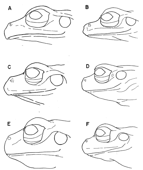
Fig. 4. Variation in the shape of the snout in Smilisca sordida; left column females, right column males; all from Costa Rica: (A) Camp Seattle, Rincón de Osa, Puntarenas Prov. (UMMZ 123684); (B) Quebrada Agua Buena, 3 km. SW Rincón de Osa, Puntarenas Prov. (USC 7236); (C) Río Oro, 28.5 km. NW Villa Neily, Puntarenas Prov. (KU 91742); (D) Río Jorco, near Desamparados, San José Prov. (KU 91765); (E-F) Bambú, Limón Prov. (USC 7183). ×3.
The dorsal ground-color of Smilisca sordida is gray to pale tan or reddish brown; the venter is white. The dorsum is variously marked with dark gray, dark brown, reddish brown, or olive-green spots or blotches (Pl. 7C). A dark interorbital bar usually is present. The dorsal markings on the body usually consist of a blotch, or two or more spots, on the occiput, in the scapular region, and in the sacral region. In many specimens, especially females, these markings are in the form of broad transverse bars. A female (USC 7164) from Las Cañas, Guanacaste, Costa Rica, has a tan dorsum with many black flecks and round brown spots bordered by darker brown. One female (KU 91763) from the Río Jorco, San José, Costa Rica, has a unicolor tan dorsum. Some individuals have scattered, small white spots on the dorsum; these are most evident in a male (USC 7153) from La Fortuna, Alajuela. White labial stripes and anal stripes are absent in all specimens.
The limbs are marked by dark brown transverse bars; these are indistinct in some specimens from the Meseta Central and Caribbean lowlands, whereas they are distinct in all specimens from the Pacific lowlands. Specimens from the Caribbean lowlands have two to six bars on each shank, whereas specimens from the Pacific slopes have four to six bars on each shank, and specimens from the Meseta Central have as many as eight bars on each shank. A narrow, sometimes broken white line is present on the ventrolateral edge of the forearm. The webbing on the hand is tan or pale gray, and the ventral surfaces of the tarsi and the webbing on the feet are dark gray or brown. Breeding males have dark brown nuptial excrescences on the prepollex.
The flanks and posterior surfaces of the thighs usually are marked by bluish white and creamy tan flecks, respectively, but vary considerably. In specimens from the Caribbean lowlands a small amount of flecking is present in the inguinal region, and on the posterior surfaces of the thighs flecks are few or absent. In specimens from the Meseta Central, numerous large flecks or small, round spots (pale bluish white in life) are on the posterior half of the flanks; small flecks are on the posterior surfaces of the thighs. Specimens from the Pacific slopes and lowlands of southern Costa Rica (Puntarenas and San José Provinces) have bold mottling of black and bluish white on the flanks and many bluish white flecks on the posterior surfaces of the thighs. The flanks are reticulated from the axilla to the groin in two females (UMMZ 123684 and USC 7236) from Rincón, Peninsula de Osa. In specimens from the Pacific slopes of Guanacaste in northwestern Costa Rica, flecks are present in the inguinal region; indistinct flecks are on the posterior surfaces of the thighs.
The throat is immaculate in specimens from the Caribbean lowlands in Limón Province; the throats are dusky laterally in most other specimens except some from the Meseta Central, in which the throats are heavily flecked with black. This variation occurs in males and females.
The color and pattern in life are highly variable. A composite description of living individuals (now KU 91718-41) from 6 kilometers east of Golfito, Puntarenas, Costa Rica, illustrates the variability: "Dorsum pale olive-green, fading to tan posteriorly, or tan all over with dark olive-green or dark brown spots on back and bars on limbs. Flanks dark brown with cream, greenish gray, or bluish gray mottling. Posterior surfaces of thighs dark brown with pale blue, pale green, or tan flecks. Iris creamy silver. Throats white with some brown flecks peripherally." (Duellman, Field notes, February 15, 1965.) A male from the Río Jorco, San José, Costa Rica, was dull olive-tan above with olive-green marks; the flanks were brown with pale tan flecks, and the posterior surfaces of the thighs were pale brown with cream-colored flecks. Six females from the same locality were reddish brown above with olive-brown or dark brown markings; one was uniform orange-tan, and another was dull olive-green with darker markings.
The color of the iris in living frogs varies from creamy silver to grayish yellow or bronze with a variable amount of black reticulation.
Natural History.—Smilisca sordida is not associated with any one type of vegetation; instead it lives in the vicinity of rocky streams having low gradients. Breeding takes place primarily in the dry season, when the water in the streams is clear and at a low level. Through most of the range of S. sordida showers, or even short heavy rains, occur in the dry season. After such rains the breeding activity is maximal. Breeding congregations have been found from December through April, but a few calling males and gravid females have been taken in June, July, and August. In the rainy season non-breeding individuals are found sitting on bushes near streams at night. Taylor (1952:843) found specimens in bromeliads by day.
Males usually call from rocks or gravel bars in, or at the edge of, streams. Some individuals perch in low bushes overhanging the streams, and some sit in shallows in the streams. Clasping pairs have been found on the banks of streams and in shallow water in streams.
The breeding call consists of one to six moderately short, rather high-pitched notes (duration 0.18 to 0.45 seconds) repeated at intervals of 12 seconds to several minutes. Each note is a vibrant rattle having 78 to 135 pulses per second and major frequences of about 1200 to 2600 cycles per second (Pl. 11C).
The tadpoles live in shallow parts of the streams, where they cling to the surfaces of small rocks and hide beneath leaves and rocks. A complete developmental series of tadpoles is not available; measurements of those stages examined are summarized in Table 12.
A typical tadpole in stage 36 of development (KU 68475 from 15 km. WSW of San Isidro el General, Costa Rica) has a body length of 11.7 mm., tail length of 22.8 mm., and a total length of 34.5 mm.; body about three-fourths as deep as wide; snout broadly rounded in dorsal view, sloping and rounded in lateral view; eyes widely separated, directed dorsolaterally; nostril slightly closer to eye than to tip of snout; mouth ventral; spiracle sinistral, about two-thirds distance from snout to posterior end of body and slightly below midline; anal tube dextral; caudal musculature heavy, straight; dorsal fin not extending onto body; fins deepest at about mid-length of tail; there depth of caudal musculature equal to depth of dorsal fin and half again as deep as ventral fin; musculature extending nearly to tip of tail; body reddish brown above and pale grayish brown with white flecks below; caudal musculature pale tan with brown flecks; a series of reddish brown dashes at base of caudal fin separated from others in series and from dashes on other side by creamy white; fins transparent with reddish brown flecks on posterior one-half of ventral fin and on all of dorsal fin (Fig. 14C). Mouth bordered by two rows of short, pointed papillae; lateral fold present; tooth-rows 2/3; upper rows equal in length; second upper row narrowly interrupted medially; three lower rows complete, nearly as long as upper rows, deeply indented medially; upper beak robust, inner surface not forming continuous arch with short lateral processes; lower beak deep, V-shaped; both beaks bearing short serrations (Fig. 15F).
Little variation occurs in structure. In some specimens the second upper tooth-row is complete; no individuals were found to have the row broadly interrupted medially.
The series of dark dashes on the dorsal edge of the caudal musculature is diagnostic of all stages studied. In life, tadpoles from 15 and 20 kilometers west-southwest of San Isidro el General, Costa Rica, had a tan body, often with an olive-tan tinge; the caudal musculature was tan; the flecks and dashes were dull red or reddish brown. Tadpoles from 6 kilometers east of Golfito, Costa Rica, had bodies with olive-green flecks. The caudal musculature was brown with bluish green flecks; the fins were transparent with reddish brown flecks. The belly was a silvery golden color. Tadpoles from Bajos de Jorco, Costa Rica, had brown bodies with bluish green flecks; the tail and fins had reddish brown flecks and dashes. The iris was a bronze color in specimens from all three localities, as well as in the young mentioned in the following paragraph.
Nine recently metamorphosed young were found on vegetation at the edges of streams in April. These specimens have snout-vent lengths of 13.1 to 15.7 mm. (14.9 mm.) and in life were pale greenish tan or olive-tan above and white below. The hands, feet, and thighs were pale yellowish tan.
Remarks.—The foregoing synonymies indicate that confusion has existed in the application of various names, to this species, as well as in use of the names sordida and gabbi to include the species that we describe and name Smilisca sila. Correct allocation of the names involved was possible only after studying and comparing the type specimens, for the descriptions given by the various authors are not sufficiently explicit to determine the nature of many essential features.
The presence of a rounded snout and a long white throat in males distinguishes S. sordida from S. sila, which has a high truncate snout and short dark throat in males. The two syntypes of Hyla sordida Peters, 1863, (ZMB 3141) are males having snout-vent lengths of 36.9 and 37.0 mm. The two syntypes of Hyla gabbi Cope, 1876 (USNM 30658-9), are females having snout-vent lengths of 52.8 and 53.7 mm., respectively. Also included in the collections made by Gabb is eastern Costa Rica are two males (USNM 30685-6), which Cope (1876) named and described as Hyla nigripes. These specimens are soft and faded, but are recognizable as the same as Hyla sordida Peters; the syntypes of Hyla nigripes have snout-vent lengths of 37.6 and 37.7 mm. We have examined one of the syntypes of Hyla salvini Boulenger, 1882 (BMNH 1947.2.24.13), a female having a snout-vent length of 54.6 mm. We are convinced that all of these type specimens are representatives of one species, the earliest name for which is Hyla sordida Peters, 1863. The type localities for three of the named species are in Costa Rica—H. gabbi from Sipurio on the Caribbean lowlands, H. nigripes from the Caribbean slopes of Pico Blanco, and H. salvini from Cartago on the Meseta Central. The type locality of H. sordida was given as "Veraguas" by Peters (1863). At that time Veraguas was often considered to be most of western Panamá. Though we have not seen Panamanian specimens other than the types of S. sordida and one specimen from the Pacific lowlands of western Panamá, the species probably occurs on the Caribbean slopes of western Panamá. The species has been taken on the Caribbean lowlands of Costa Rica within a few kilometers of Panamá; collecting on the Caribbean slopes in the provinces of Bocas del Toro and Veraguas should reveal the presence of Smilisca sordida there.
Distribution.—Smilisca sordida is found along the Pacific slopes and lowlands from Guanacaste, Costa Rica, southeastward to extreme western Panamá, to elevations of about 1200 meters on the Meseta Central in Costa Rica, and on the Caribbean slopes and lowlands of Costa Rica and probably adjacent Panamá (Fig. 5). One specimen purportedly comes from "Río Grande, Nicaragua."
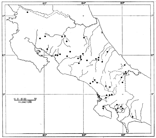
Fig. 5. Map showing locality records for Smilisca sordida.
Specimens examined.—412, as follows: Nicaragua: "Río Grande" (? Depto. Zelaya), MCZ 2634.
Costa Rica: Alajuela: Between Atena and Salto de San Mateo, USC 6185; 8 km. N Ciudad Quesada, USC 7155 (4); La Fortuna, USC 7153 (20); 3 km. E La Fortuna, USC 7150; San Carlos, USNM 29969; Sarchi, KU 32990-9, 36792-3.
Cartago: Cartago, BMNH 1947.2.24.13; headwaters of Río Pacuare, USC 119; Instituto Interamericano de Ciéncias Agricolas, Turrialba, KU 37012, USC 420, 437; Río Reventazón, Turrialba, MCZ 29268: 10 km. N Río Reventazón bridge, USC 7073; 5 km. SW Río Reventazón bridge on Paraiso-Orosi road, USC 669; Turrialba, UMMZ 118405, USC 455, USNM 29936-9.
Heredia: Puerto Viejo, KU 36791.
Guanacaste: Las Cañas, USC 7164; Santa Cecilia, MCZ 7924-5; Tilarán, USC 7161 (5).
Limón: Bambú, USC 7171 (2), 7183 (13); La Lola, USC 820 (6), 6083-94, 8064, 8071; Pandora, USC 7188 (7), 7189, 7190 (3), 7191 (5); Pico Blanco, USNM 30685-6; Río Larí, 14-16 km. SW Amubre, USC 7179, 7180 (10); Sipurio, USNM 30658-9; Suretka, KU 36764, 36765 (skeleton), 36766-78.
Puntarenas: 6 km. N Dominical, KU 91749-50, 91811 (young), 91812 (tadpoles); Esparta, MCZ 8028; 6 km. E Golfito, KU 91718-41, 91809 (young), 91810 (tadpoles), 91816-9 (skeletons), USC 7103 (23); Quebrada Agua Buena, 3 km. SW Rincón de Osa, USC 7236 (6); Quebrada Boruca, 22 km. E Palmar Norte, KU 64264; Rincón de Osa, Camp Seattle, UMMZ 123680-5, S-2792 (skeleton), USC 705 (5), 6023, 7254; Río Barranca, USC 7119 (2); Río Ceiba, 6 km. NW Buenos Aires, KU 91747-8, USC 7112 (7); Río Ciruelitas, 16 km. NW Esparta, USC 7121 (3); Río Claro, 14.2 km. NW Villa Neily, USC 7110 (4); Río Ferruviosa, 7 km. S Rincón de Osa, USC 7235 (4); Río Lagarto at Pan-American Hwy. (Guanacaste Border), USC 7122 (4); Río La Vieja, 30 km. E Palmar Norte, KU 87684 (tadpoles), 91743-6, USC 7083 (2); Río Oro, 28.5 km. NW Villa Neily, KU 91742; Río Volcán, 10 km. W Buenos Aires, USC 7113; Río Zapote, 7 km. E Palmar, USC 7100 (4); 3-5 km. W Palmar, USC 7101 (18); 7 km. SE Palmar, KU 64261-3; 1.2 km. NW Villa Neily, USC 8032; 3 km. NW Villa Neily, USC 7109 (20); 5 km. NW Villa Neily, USC 6176, 8035.
San José: Bajos de Jorco, KU 91813 (tadpoles); Escazú, KU 34863, 34869-75, USC 813; between Monrovia and La Hondura, ± 0.5 km. N Santa Rosa, USC 302 (2); Paso Ancho, Río Jorco, UMMZ 122649 (6), USC 530 (3); Río Jorco, near Desamparados, KU 91757-65, 91796-7, 91820-3 (skeletons), USC 228, 513, 7117 (7); Río Peje, 10 km. SSE San Isidro el General, USC 7115 (3); Río Tiriví, MCZ 7972; San Isidro el General, CNHM 101096, KU 28201, 32989, UMMZ 72024; 15 km. WSW San Isidro el General, KU 64245-56, 68473 (tadpoles), 68474 (young), 68475 (tadpoles), 86516, 91754-6, 91793-5, USC 7097 (6); 17.1 km. WSW San Isidro el General, USC 6047; 18 km. WSW San Isidro el General, USC 689; 20 km. WSW San Isidro el General, KU 64257-9, 64260 (skeleton), 68468 (young), 68469 (tadpoles), 68470 (young), 68471-2 (tadpoles), 68476 (young), 68633-4 (skeletons), 91751-3; San José, AMNH 7501-4, USC 298; Santa Rosa, Río Virilla, USC 7145.
Panama: Chiriquí: Río Jacu, 5.8 km. ESE Paso Canoas, KU 91905. "Veraguas," ZMB 3141 (2).
ANALYSIS OF MORPHOLOGICAL CHARACTERS
Osteology
In attempting to assay the taxonomic significance of skeletal differences we are faced with a dearth of data on the skeletons of frogs in general and hylids in particular. Recent reviews by Brattstrom (1957) and Hecht (1962, 1963) have been concerned with general salientian classification and phylogeny, principally at the family level. Savage and Carvalho (1953), Griffiths (1959), and Baldauf (1959) used osteological characters in determining the taxonomic status of the families Pseudidae, Brachycephalidae, and Bufonidae, respectively. Carvalho (1954) presented osteological evidence for the generic separation of New World microhylids. Zweifel (1956) and Tihen (1962) used osteological characters at the levels of the species-group and species in their respective studies on Scaphiopus and Bufo. Little has been recorded about the skeletons of the hylids. Goin (1961) mentioned dentigerous elements and cranial co-ossification in his synopsis of the genera of hylids. Copland (1957) in his review of the Hyla of Australia, Funkhouser (1957) in her revision of Phyllomedusa, and Zweifel (1958) in his review of Nyctimystes did not consider skeletal characters.
Some osteological studies on hylids have yielded worthwhile information. Mittleman and List (1953) used osteological characters in defining the genus Limnaoedus: Starrett (1960) used cranial characters in combination with jaw musculature in defining the genus Smilisca, and Duellman (1964) used cranial characters in delimiting the Hyla bistincta group. Brief descriptions of cranial structure were given for Phrynohyas (Duellman, 1956) and Ptychohyla (Duellman, 1963a); specific and sexual differences in the skulls of Hyla chaneque and Hyla taeniopus were pointed out by Duellman (1965). Stokely and List (1954) described early cranial development in the hylid Pseudacris triseriata triseriata.
Because our knowledge of the skeleton in hylids is so incomplete, we are not attempting to place Smilisca in the general scheme of hylid phylogeny on the basis of skeletal characters. Instead, our purposes are to describe the skeleton and its ontogenetic development in one member of the genus (S. baudini), and to make comparisons that show taxonomic differences in osteological characters among species of Smilisca.
The study of 68 dried skeletons and 25 cleared and stained preparations, including an ontogenetic series of S. baudini, has resulted in an understanding of the progressive development of skeletal elements and a knowledge of interspecific and intraspecific variation in these elements. Furthermore, investigations of the osteology have provided correlations between some cranial characters and certain aspects of external morphology.
Descriptive Osteology of Smilisca baudini
The following description is based primarily on an adult female (KU 68184):
Skull.—The skull is large, solid, and broader than long; the greatest width is between the sutures of quadratojugal and maxillary on either side of the skull (Pls. 2-3). The maxillaries bear well-developed dorsal flanges, curve gently, join the moderately convex premaxillaries anteriorly and form a slightly truncate snout. The combined premaxillary width is about one-fourth the width of the skull. The premaxillaries are separated medially, and laterally from the maxillaries by sutures. Each premaxillary bears a dorsomedial alary process, which is anteriorly convex and four times as high as the depth of the lateral wing of premaxillary; each premaxillary also has a ventromedial palatine process that projects dorsally from the lingual edge of the premaxillary. The septomaxillaries are closely associated dorsally with the premaxillaries immediately lateral to the prenasal processes.
The nasals are large, widest anteriorly and narrowing posteriorly, parallel to maxillaries, and not separated from the ethmoid by cartilage. The nasals bear long, delicate maxillary processes extending nearly to the maxillaries. Anteriorly, the nasals are widely separated by the partially ossified internasal septum, which is in contact with the premaxillaries between the prenasal processes; the anterior points of the nasals lie approximately one-half the distance between the anterior ends of the ethmoid and the premaxillaries. The ethmoid is large and completely ossified; the margins are smooth. The trunate anterior edge lies between the nasals and is in contact with the internasal septum. The frontoparietals are large, smooth-margined, and bear large supraorbital flanges curving posterolaterally at the rear of the orbit. A small, oval foramen involves the posterior part of the ethmoid and anterior portion of frontoparietals; continued ossification in older specimens fills in the foramen, thereby resulting in a solidly roofed cranium. The auditory regions are relatively massive and bear narrow tegmen tympani; the distal ends of the tegmen tympani are medial to the lateral edge of the pterygoids in dorsal view. The squamosals are large; the long anterior arm is separated from the maxillary by a suture. The delicate, spindle-shaped columellae lie ventral to the tegmen tympani and squamosals, are spatulate distally, and have a broad basal attachment to the auditory region.
The vomers are moderately large and are in contact anteriorly with the premaxillaries and posteriorly with the ethmoid. Each vomer has two wide serrated flanges laterally. The tooth-bearing parts of the vomers are widely separated and at a slight angle to one another; the vomers terminate medially in two pointed processes on the ethmoid. The palatines are edentate, but bear strong ridges throughout their lengths. They are broadly in contact with the maxillary, are narrow medially, and are attached by pointed processes to the medial part of the ethmoid. The pterygoids are large, attached to the maxillaries immediately anterior and medial to the squamosal-maxillary connection, bear well-developed pedicles, which are broadly attached to the proötic, and a wide wing is in contact posteriorly with the distal two-thirds of the quadrate.
The angular makes up most of the lower jaw, bears a broad articular surface posteriorly, and has a small coronoid process on the lingual edge; anteriorly the angular is separated from the dentary and mentomecklian by Meckel's cartilage. The dentary lies external to the angular and extends from the mentomecklian to approximately the mid-length of the angular. The mentomecklians are ossified, but separated by cartilage medially.
Hyoid.—The hyoid plate is curved, thin, and mostly cartilaginous, but calcined posteriorly (Fig. 6). The anterior cornua are slender, cartilaginous, and curve anteromedially from the hyoid plate and thence laterally and posteriorly, to attach to the posterior surface of the proötics. The lateral cornua are broad, flat, cartilaginous lateral extensions from the bases of the anterior cornua. The posterior cornua are bony, except distally.
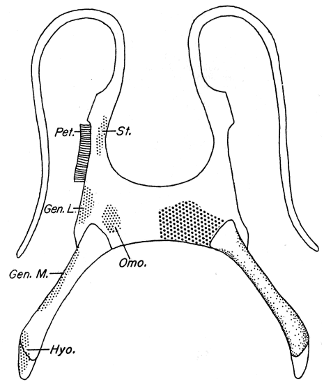
Fig. 6. Ventral view of hyoid apparatus of an adult male Smilisca baudini showing areas of muscle attachment: Gen. L., attachment of geniohyoideus lateralis; Gen. M., attachment of geniohyoideus medialis; Hyo., attachment of hyoglossus; Omo., attachment of omohyoideus; Pet., petrohyoideus; St., attachment of sternohyoideus. KU 64220, ×5.
Vertebral Column.—The atlas lacks transverse processes and a neural crest, whereas transverse processes are present on the other seven presacral vertebrae, and knoblike neural crests are present on the second, third, and fourth vertebrae; a faint neural ridge is visible on the fifth vertebra. The transverse processes are directed laterally on the second and sixth vertebrae, ventrolaterally on the third, posterolaterally on the fourth and fifth, and anterolaterally on the seventh and eighth. The processes are slightly expanded on the fourth, and more so on the fifth, vertebra. The sacral diapophyses are expanded and have a border of calcified cartilage laterally. There are two sacral condyles. The slender coccyx has a thin dorsal ridge on the anterior three-fourths of its length.
Pectoral Girdle.—The omosternum is large, ovoid, and cartilaginous; the sternum is a thin cartilaginous sheet deeply notched posteriorly and is not differentiated into episternal and xiphisternal elements. The coracoids are robust, twice as stout as the clavicles. The epicoracoidal cartilages overlap in the usual arciferal manner, except that they are fused anteriorly between the slender clavicles. The clavicles are strongly arched. The clavicle, coracoid, and scapula on each side form a bony articulation at the glenoid fossa. A bifurcation of the ventral end of the scapula results in a large glenoid foramen. The scapula is flat and expanded dorsally; the suprascapula is broad, flat, and calcified in large adults. In young specimens no distinct ossification of the cleithrum or ossification of endochondral centers are evident.
Arm and Hand.—The humerus is equally well-developed in both sexes and has a prominent lateral crest. The radius and ulna are completely fused. A bony prepollex is present in both sexes. The metacarpals are about equal in length. The phalangeal formula is 2-2-3-3; the terminal phalanges are claw-shaped.
Pelvic Girdle.—The ilia are long, slender, and slightly curved. A thin ridge projects laterally from the dorsal edge of the posterior one-half of each ilium. The ilial prominence is large and knoblike when viewed from above. The anterior edge of the ilial prominence is at the level of the anterior edge of the acetabular border. The dorsal acetabular expansion is small. The pubis is slender, and the ischium is elevated and robust.
Leg and Foot.—The slightly curved femur has a distinct crest proximally on the posterior surface. The nearly straight tibio-fibula is slightly longer than the femur. The tibial and fibial elements are completely fused but have a distinct cleft between them. A small foramen exists at the mid-length of the tibio-fibula. The fibulare (calcaneum) is much more robust than the tibiale (astragalus). The prehallux is large and flat. The metatarsals of the third, fourth, and fifth digits are equal in length; the metatarsal of the second is somewhat shorter, and that of the first is much shorter. The phalangeal formula is 2-2-3-4-3; the terminal phalanges are claw-shaped.
Developmental Cranial Morphology of Smilisca baudini
The following description of development of the skull of Smilisca baudini is based on the examination of 12 cleared and stained specimens. In table 3 the cranial bones are listed in the left hand column in the approximate order of their appearance in the young frogs. Across the top of the table selected specimens designated by developmental stage or snout-vent length are listed. It should be noted that although each individual, from left to right, has an increasing number of ossified bones, the correlation with increasing size is imperfect; the precise ages of the individuals are unknown.
The first bones to appear are the septomaxillaries, frontoparietals, part of the exoccipital, and the parasphenoid in developmental stage 40. The frontoparietals are represented by two slender ossifications dorsomedial to the orbits; the septomaxillaries are present as small ossifications anterior to the nasal capsules (Pl. 1A). The parasphenoid is present as a faint median ossification, and the exoccipital shows some ossification.
Table 3.—The Order of Occurrence of Cranial Ossifications in the Skull of Smilisca baudini. Where Numbers Are Divided by a Slash Mark, the Left and Right Symbols Correspond to the Left and Right Sides of the Skull, Respectively.
Bone Stage 40 Stage 44 12.6 mm. 13.9 mm. 32.0 mm. 27.0 mm. 20.1 mm.
Frontoparietal X X X X X X X
Parasphenoid X X X X X X X
Septomaxillaries X X X X X X X
Exoccipitals X X X X X X X
Squamosals — X X X X X X
Premaxillaries — X X X X X X
Maxillaries — X X X X X X
Nasals — — X X X X X
Pterygoids — — X X X X X
Vomers — — — X X X X
Palatines — — — X X X X
Quadratojugals — — — X X X X
Ethmoid — — — — X X X
Columellas — — — — X X X
Supraorbital Flanges — — — — — X X
Proötics — — — — — — X
Vomerine Teeth — — 1/1 4/3 5/5 3/3 5/4
Maxillary Teeth — 0/7 3/5 6/5 30/31 30/26 37/36
Premaxillary Teeth — 2/4 3/3 5/5 7/6 8/6 8/7
The dentigerous bones are among the most rapidly developed, although not the first to appear. They are present in developmental stage 44 before metamorphosis is completed. The maxillaries bear a few teeth anteriorly and are ossified posteriorly to a point one-third of the distance from the anterior to the posterior edge of the orbit. Ossification lengthens the posterior termini of the maxillaries to the posterior edge of the orbit. In front of the anterior margin of the orbit, bone is proliferated dorsal to the main axes of the maxillaries and forms moderate dorsal maxillary flanges. The premaxillaries appear simultaneously with the maxillaries. Initially they are widely separated medially from each other, and laterally from the developing maxillaries; each bears two or three teeth, large dorsally blunt alary processes, and small palatine processes. The median and lateral edges of the prenasal processes lengthen heterochronously, causing the median edges to be longest and to lie slightly dorsal to the level of the septomaxillaries. After the maxillaries and premaxillaries develop, the vomers appear as small horizontal ossifications anterior to the parasphenoid. Ossification begins in the lateral flanges, then in the prevomerine processes, and lastly in the posterior dentigerous parts of the bones; the prevomerine processes are the last parts of the vomers to ossify completely.
Initially the frontoparietals are present as thin rods of ossification dorsomedial to the orbits; the frontoparietals extend from the anterior to the posterior end of the orbit by developmental stage 44. The anterior ends of the bones remain thin and pointed; ossification progresses medially from the midpoint of the length of the orbit and posteriorly to the level of the exoccipital; a median center of ossification joins the frontoparietals posteriorly, thereby forming the posterior border of the frontoparietal fontanelle. The supraorbital flanges of the frontoparietals do not appear until all other cranial bones are ossified, or nearly so. The most rapid ossification begins laterally at the posterior edge of the orbit and decreases anteriorly over the posterior half of the orbit. This differential rate of proliferation of bone results in the pattern of development of the supraorbital flanges shown in figure 7. The nasals appear as thin slivers of bone half way between the anterior ends of the frontoparietals and the end of the snout. As ossification proceeds the nasals assume a triangular shape in dorsal view. The anterior ends are pointed; the lateral margins are parallel to the maxillaries. The posteromedial points do not reach the lateral margins of the ethmoid, and the maxillary processes extend about three-fourths the distance from the bodies of the nasals to the maxillaries. Following the union of the frontoparietals posteriorly, the nasals widen anteriorly and are narrower at the midpoints of their long axes than anteriorly or posteriorly. With further ossification the maxillary processes extend to the maxillaries and form complete bony anterior margins to the orbits; the mid-parts of the nasals widen (Pl. 1B).
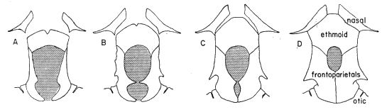
Fig. 7. Developmental sequence of the frontoparietal fontanelle and associated bony elements in Smilisca baudird: (A) KU 60026, ×5; (B) KU 85438, ×4; (C) KU 26328, ×3; (D) KU 68184, ×2.3.
The parasphenoid is the first of the palatal bones to appear. At metamorphosis the bone is well developed; the anterior tip is situated just in front of the anterior edge of the orbit, and posteriorly the lateral processes extend laterally beyond the ossified parts of the auditory region. The pterygoids do not appear until metamorphosis, when ossification is evident in only the mid-parts of the posterolateral arms. Ossification follows in the mid-parts of the anterolateral arms and occurs last in the pterygoid pedicles. The palatines do not appear until all three arms of the pterygoids are at least partly ossified. Ossification proceeds rapidly from the maxillaries medially to the unossified ethmoid, which is the last of the cranial bones to appear. Initially it is extremely shallow; dorsally it is widely separated from the nasals, and ventrally the posterior margin meets the anterior point of the parasphenoid. In dorsal view, ossification proceeds anteriorly between the nasals and posteriorly, ventral to the frontoparietals; ventrally, ossification proceeds posteriorly dorsal to the parasphenoid.
The ventral arms of the squamosal and the supraoccipital region of the exoccipital are the first occipital bones to appear. Ossification follows in the regions of the semicircular canals and occipital condyles. The dorsal end of the ventral arm of the squamosal and the posterior arm of the squamosal ossify as a unit at the same time the quadratojugal appears. Shortly thereafter the anterior arm of the squamosal ossifies, the distal part of the columella appears, and the anterior and lateral parts of the auditory region ossify.
The angular and dentary of the lower jaw appear concurrently with the dentigerous bones. Initially, the angular is short and broad; the articular surface is absent, and the anterior end is slightly overlapped by the dentary. The mentomecklians do not ossify until approximately the same time that the quadratojugal appears in the upper jaw.
Comparative Osteology
The genus Smilisca is characterized by the following combination of cranial osteological characters: (1) A large amount of bone is involved in the skull and a minimal amount of cartilage and/or secondarily ossified cartilage; co-ossification is absent. (2) The skulls are uniformly broad with angular lateral margins, and truncate anteriorly. (3) An internasal septum and quadratojugals are present. (4) A well-developed squamosal minimally extends one-fourth the distance from the dorsal end of the quadrate to the maxillary, and maximally is separated from the maxillary by a suture. (5) The ethmoid is large; the distance between the anterior end of the ethmoid and the anterior edge of the premaxillary varies between 15 and 20 per cent of the total length of the skull.
On the basis of cranial osteology two species-groups can be recognized within the genus Smilisca. The sordida group, comprising S. sordida and puma, is characterized by a broad skull in which the lateral margins of the maxillaries are relatively straight anterior to the orbit. The moderate-sized nasals are rounded anteriorly, and bear relatively short, sometimes blunt, maxillary processes. The long axes of the nasals are not parallel to the maxillaries. The ethmoid is proportionately small in the sordida group. The bony part of the ethmoid terminates near the anterior edge of the orbits and does not extend anteriorly between the nasals; the entire anterior margin of the ethmoid is separated from the nasals by cartilage. The squamosals are generally small. They are narrow in dorsal view, and minimally extend one-fourth the distance from the dorsal end of the quadrate to the maxillary, and maximally, two-thirds the distance. The tegmen tympani are relatively small (Fig. 8).
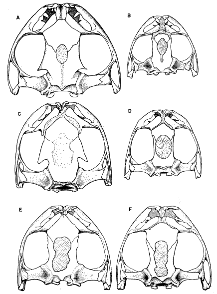
Fig. 8. Dorsal views of the skulls of the species of Smilisca: (A) S. baudini (KU 68184); (B) S. puma (KU 68636); (C) S. phaeota (KU 41090); (D) S. sila (KU 80625); (E) S. cyanosticta (KU 55938), and (F) S. sordida (KU 36765). ×1.5.
In contrast to the tendency for reduction of cranial parts in the sordida group, the baudini group, constituted by S. cyanosticta, phaeota, and baudini, is characterized by more ossification of the cranial elements. The skull is broad; the lateral margins are less angular and are gently curved, rather than straight as in the sordida group. The nasals tend to be larger with the long axes parallel to the maxillary. Anteriorly the nasals are pointed, and posteriorly they bear long, delicate palatine processes extending to the maxillary. The ethmoid is fully ossified, extends anteriorly between the nasals, and laterally is separated by a suture from the nasals if the latter are fully ossified. The squamosals are large, and wide in dorsal view. They minimally extend one-fourth the distance from the dorsal end of the quadrate to the maxillary, and maximally are sutured to the maxillary. The tegmen tympani are massive.
Smilisca sila is intermediate between the two species-groups described. The skull is broad; the lateral margins are gently curved, and have a pronounced angularity just anterior to the palatines which results in a broad, truncate snout. The nasals are moderate in size; because of the anterior angularity of the lateral margins, the long axes of the nasals lie parallel to the maxillary. The nasals are only slightly pointed anteriorly, and posteriorly they bear short, blunt palatine processes and medial processes in contact with the lateral corners of the ethmoid. The ethmoid is fully ossified, but does not extend anteriorly between the nasals. The squamosals are moderate in size and extend one-fourth the distance from the dorsal end of the quadrate to the maxillary. The tegmen tympani are relatively large, but proportionately short.
The cranial characters utilized in the analysis of species groups (general shape, nature of the nasals, ethmoid, squamosals, and tegmen tympani), together with other characters, such as the relative height and shape of the prenasal processes, the extent of the internasal septum, and the nature of the vomers, frontoparietals, maxillaries and pterygoids are useful in distinguishing the various species (Table 4, Fig. 8), as well as in establishing relationships within the species-groups.
Within the sordida group, S. sordida and S. puma can be distinguished by the following characters: The bony part of the ethmoid terminates posterior to the anterior edge of the orbit and is thus widely separated from the nasals by cartilage in S. puma. In S. sordida the bony part of the ethmoid always terminates at a level equal to, or slightly in front of the anterior edge of the orbit; therefore, less cartilage exists between the ethmoid and nasals in S. sordida than in S. puma. The width of the premaxillary comprises about 30 per cent of the width of the skull in S. sordida and 20 per cent in S. puma. The proportion of the length of the skull anterior to the bony part of the ethmoid in S. sordida is approximately 21 per cent, as compared with about 29 per cent in S. puma. The prenasal processes are convex in S. sordida and straight in S. puma.
The marked ontogenetic variation in S. sordida is considered in more detail in the account of that species, but it is pertinent to the present discussion to note that with respect to some features of the skull some young breeding specimens of S. sordida are intermediate in appearance between large females of S. sordida and adults of S. puma. In some breeding males (usually the smaller individuals) of S. sordida the bony part of the ethmoid terminates at the anterior edge of the orbit and is widely separated from the nasals by cartilage. In small individuals S. sordida, especially in males, and in adults of S. puma the tegmen tympani are relatively short, whereas in adult females of S. sordida these elements are long and slender. In the smaller specimens of S. sordida and in S. puma the squamosal is small; it extends only about one-fourth of the distance to the maxillary in the smaller S. sordida and about one-half the distance in S. puma. The more massive squamosal in large adult females of S. sordida extends at least two-thirds of the distance to the maxillary.
Table 4.—Comparative Cranial Osteology of Smilisca.
Character S. baudini S. cyanosticta S. phaeota S. puma S. sila S. sordida
Alary Processes Four times as high as lateral wing of premaxillary; anteriorly convex. Three times as high as lateral wing of premaxillary; anteriorly convex. Two and one-half times as high as lateral wing of premaxillary; anteriorly convex. Two times as high as lateral wing of premaxillary; straight. One and one-half times as high as lateral wing of premaxillary; straight. Two and one-half times as high as lateral wing of premaxillary; slightly convex anteriorly.
Nasals Long, wide anteriorly, narrowing posteriorly; attached to ethmoid. Long, widest posteriorly; attached to ethmoid. Long, widest anteriorly and posteriorly, bearing posteromedial process; not attached to ethmoid. Short, narrow, not attached to ethmoid. Short, wide, bearing small posteromedial processes; not attached to ethmoid. Moderately long narrowest anteriorly and posteriorly; not attached to ethmoid.
Ethmoid Long; entirely ossified; smooth margins. Long, entirely ossified; smooth margins. Long, entirely ossified; smooth margins. Short, about two-thirds ossified; irregular margins. Moderately long; entirely ossified; smooth margins Short; one-half to entirely ossified; irregular margins.
Frontoparietal Small, ovid fontanelle present or absent; long, pointed postorbital processes curving along posterior border of orbit. Large fontanelle, two and one-half times as long as wide; narrow supraorbital flanges with irregular margins. Fontanelle absent; large supraorbital flanges having straight edges and extending posterolaterally. Keyhole-shaped fontanelle; smooth margins; flanges absent. Large, ovoid fontanelle; smooth margins; flanges absent. Large, elongate fontanelle; smooth margins; flanges absent.
Squamosal Large: anterior arm in contact with maxillary. Large; anterior arm in contact with maxillary. Large; anterior arm extending 1/2-2/3 way to maxillary. Small; anterior arm extending 1/2 way to maxillary. Moderately large; anteriorarm extending 1/4 way to maxillary. Moderately small; anterior arm extending 1/4-2/3 way to maxillary.
Within the baudini group, the skull of S. cyanosticta is the most generalized of the three species; the cranial characters are intermediate between S. phaeota and S. baudini. The lateral margins of the skull in S. cyanosticta are gently curved, and have an angularity anterior to the palatine-maxillary suture; the anterior margins are less angular in S. phaeota, which has a broader snout. Posteriorly in S. baudini the margins are slightly curved medially, and the greatest width of the skull is between the quadratojugal-maxillary sutures on either side of the skull. The frontoparietals of S. cyanosticta bear slightly irregular lateral margins and a large fontanelle. There is a tendency for obliteration of the fontanelle with increasing age in both S. baudini and S. cyanosticta; the lateral margins of the frontoparietals bear large supraorbital flanges in both of these species. In S. phaeota the flanges are most prominent; they extend posterolaterally with straight margins along two-thirds of the length of the orbit and terminate in rather blunt points. The broad interorbital flanges result in a relatively broad external interorbital distance. In S. baudini the flanges are curved posterolaterally around the orbit and terminate in sharp, thin points. The tegmen tympani of all three species are massive. In S. cyanosticta the proötics slope posteriorly, whereas they slope anteriorly in S. baudini and S. phaeota.
The skulls of S. cyanosticta and S. baudini are alike in certain respects. The squamosals of both species are large and connected to the maxillary by a bony connection; the squamosals of S. phaeota are large, but extend only two-thirds of the distance from the dorsal end of the quadrate to the maxillary. In S. baudini and S. cyanosticta the nasals are separated throughout their lengths from the ethmoid, whereas the nasals of S. phaeota are separated from the ethmoid by cartilage. The latter separation is due to an incomplete ossification of the nasals in S. phaeota. The bony part of each nasal is constricted in the middle of the long axis of the bone, and the nasals are widest anteriorly; posteriorly each nasal bears a medial process, which is narrowly separated from the lateral edge of the ethmoid.
Table 5.—Variation in the Number of Teeth in the Species of Smilisca. (All Are Males; N = Number of Jaws, or Twice the Number of Individuals; Means Are Given in Parentheses After the Observed Ranges.)
Species N Maxillary Premaxillary Vomerine
S. baudini 20 49-65 (56.0) 9-16 (13.6) 5-9 (7.2))
S. cyanosticta 8 50-64 (57.9) 10-12 (10.8) 4-11 (7.1))
S. phaeota 20 50-68 (58.1) 10-15 (12.1) 5-9 (7.3))
S. puma 6 60-67 (63.6) 11-13 (12.0) 4-7 (5.3))
S. sila 8 48-60 (52.9) 10-14 (11.3) 5-7 (5.7))
S. sordida 12 39-55 (44.2) 7-11 (9.3) 4-6 (5.2)
The teeth of all species of Smilisca are spatulate and bifid. The numbers of maxillary, premaxillary, and vomerine teeth are summarized in Table 5. Smaller and presumably younger specimens of all species of Smilisca have fewer teeth than do larger specimens of the same species. This correlation between size and number of teeth does not exist as an interspecific trend within the genus; for example, the smallest species in the genus, S. puma, has the highest number of maxillary teeth. In small specimens of a given species wide gaps are present between the maxillary teeth posteriorly; in large specimens the gaps are filled by teeth, beginning anteriorly and progressing posteriorly, until the maxillary dentition is continuous.
Musculature
No extensive study of the muscular system was undertaken, but certain muscles know to be of taxonomic importance were studied.
Jaw Musculature.—Starrett (1960) pointed out the unique jaw musculature in Smilisca. In this genus M. depressor mandibulae consists of two parts, one arising from the dorsal fascia and one from the posterior arm of the squamosal. Two muscles arise from the anterior arm of the squamosal and insert on the lateral face of the mandible. Of these muscles, M. adductor mandibulae posterior subexternus lies medial to the mandibular branch of the trigeminal nerve; the other, M. adductor mandibulae externus superficialis, lies lateral to the same nerve (Fig. 9). In most other hylids the latter muscle is absent. No significant variation in the position of the muscles was noted in the various species of Smilisca, though M. adductor mandibulae originate somewhat more anteriorly in S. baudini and S. cyanosticta than in the other members of the genus, all of which have a shorter anterior arm of the squamosal that does not reach the maxillary. The two separate parts of M. depressor mandibulae are not so widely separated in members of the sordida group as in the baudini group.
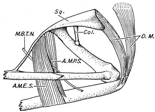
Fig. 9. Lateral view of the left jaw of Smilisca baudini; A. M. E. S., adductor mandibulae externus superficialis; A. M. P. S., adductor mandibulae posterior subexternus; Col., columella; D. M. depressor mandibulae; M. B. T. N., mandibular branch trigeminal nerve; Sq., squamosal. KU 64214, ×5.
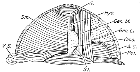
Fig. 10. Ventral view of throat musculature in an adult male Smilisca baudini (Superficial musculature on left, deep musculature on right); A. C. anterior cornua of hyoid; Gen. L., geniohyoideus lateralis; Gen. M., geniohyoideus medialis; Hyo., hyoglossus; Omo., omosternum; Pet., petrohyoideus; S., submentalis; Sm., submaxillaris; St., sternohyoideus; V. S., vocal sac. KU 64220, × 2.5.
Throat Musculature.—The frogs that comprise the genus Smilisca are characterized by paired subgular vocal sacs, essentially the same as those in Triprion (Duellman and Klaas, 1964). The following description is based on Smilisca baudini (Fig. 10).
M. submentalis lies in the anterior angle of the lower jaw, is thick, and consists of transverse fibers extending between the dentaries. M. submaxillaris is thin and arises from the whole of the inner surface of the lower jaw, except for the anterior angle occupied by M. submentalis. Anteriorly M. submaxillaris is broadly attached by fascia to M. hyoglossus and M. geniohyoideus, which lie dorsal to M. submaxillaris. Medially this attachment continues posteriorly for about one-half the length of the hyoglossus. Posteriorly M. submaxillaris is folded and attached to M. sternoradialis of the pectoral girdle. The vocal sacs are formed by a pair of posterolateral evaginations of M. submaxillaris; a broad connection between the pouches allows free passage of air between the pouches.
The deeper throat musculature is essentially the same as that described for Phrynohyas spilomma by Duellman (1956), except for slight differences in the place of attachment on the hyoid.
SKIN
Structure
The skin of Smilisca is typical of that of most hylids in organization and structure. Smilisca sila is distinguished from other members of the genus by the presence of small wartlike protrusions and peculiar white, pustular spots on the dorsum. The wartlike structures are composed of three or four epidermal cells, which protrude from the surface of the epidermis; the structures are covered by a slightly thickened layer of keratin. The white pustules are slightly elevated above the surrounding skin. Internally they consist of aggregations of swollen, granular, pigment-cells (perhaps lipophores) lying between the epidermis and the melanophores.
Biochemical Variations
Dried skins of all species of Smilisca were sent to José M. Cei, Instituto Nacional de Cuyo, Mendoza, Argentina, for biochemical screening by means of the chromatographic techniques described by Erspamer and Cei (1963). The species in the baudini group have detectable amounts of penta-hydroxi-trypatamine, whereas only a trace is present in the other species. Furthermore, species in the baudini group differ from S. sila and the sordida group in lacking, or having only a trace of, tryptophan-containing polypeptides. These superficial biochemical tests support the arrangement of species as ascertained by conventional taxonomic characters.
External Morphological Characters
The features of external morphology that were studied in connection with the taxonomy of the genus Smilisca are discussed below.
Size and Proportions
The frogs of the genus Smilisca are medium to large tree frogs. The three species comprising the baudini group (S. baudini, cyanosticta, and phaeota) are notably larger than S. puma, sila, and sordida (Table 6). The largest specimen that we examined is a female of S. baudini having a snout-vent length of 90 mm. Smilisca puma is the smallest species; the largest male has a snout-vent length of 38 mm. and the largest female, 46 mm.
Table 6.—Comparison of Sizes and Certain Proportions of the Species of Smilisca. (Means in Parentheses Below Observed Ranges; Data for Males Only.)
Species N Snout-vent length Tibia length/ snout-vent Tympanum/ eye
S. baudini 140 47.3-75.9 42.1-53.6 56.1-94.4
(58.7) (47.8) (73.5)
S. cyanosticta 40 44.6-56.8 51.9-59.7 62.7-88.4
(50.7) (56.0) (71.4)
S. phaeota 50 40.8-65.5 50.9-60.2 62.7-85.5
(53.9) (55.5) (76.6)
S. puma 20 31.9-38.1 48.2-53.1 52.1-72.2
(34.7) (51.3) (64.9)
S. sila 33 31.6-44.8 49.7-58.1 47.6-58.3
(37.7) (54.8) (53.2)
S. sordida 55 31.9-44.6 50.5-57.1 46.5-57.1
(37.9) (53.4) (49.1)
No outstanding differences in proportions exist between species, although certain proportions are sufficiently different in some species to warrant mention. Smilisca baudini is a more squat and stocky frog than other members of the genus; this is reflected in the somewhat shorter hind legs (Table 6). The size of the tympanum relative to that of the eye is highly variable within samples of a given species. Even so, noticeable differences in the tympanum/eye ratio are apparent. Members of the baudini group have the largest tympani, whereas S. sila and sordida have the smallest, and S. puma is intermediate (Table 6).
Shape of Snout
Although all members of the genus have rather truncate snouts, subtle differences exist among the species (Pl. 12). Smilisca sila has the shortest snout; that of S. baudini is only slightly longer. The snouts of S. cyanosticta and puma are nearly square in lateral profile, whereas those of S. phaeota and sordida are slightly inclined. The shape of the snout is relatively uniform within each species and displays no noticeable sexual dimorphism, except in S. sordida, in which there are sexual differences and geographic variation (see p. 324).
Hands and Feet
The characters of the hands and feet are among the most taxonomically important external features in Smilisca. Consistent differences exist in relative lengths of the digits, size of subarticular tubercles, size and number of supernumerary tubercles, size and shape of the inner metatarsal tubercle, and amount of webbing (Pls. 4 and 5). In the baudini group the series of species (baudini-phaeota-cyanosticta) show a progressive increase in amount of webbing in the hand and a decrease in number, and corresponding increase in size, of supernumerary tubercles. The amount of webbing in the feet of S. baudini and phaeota is about the same, but the webbing is slightly more extensive in S. cyanosticta. Smilisca puma is unique in the genus in lacking webbing in the hand; furthermore, this species is distinctive in having many large subarticular tubercles on the hand and a relatively small inner metatarsal tubercle. The two stream-inhabitants, S. sila and sordida, have shorter and stouter fingers than the other species. The webbing is most extensive in both the hands and feet of these species, which also are distinctive in having many small supernumerary tubercles on the feet.
Ontogenetic Changes
Minor ontogenetic changes in structure involve the shape of the snout, relative size of the eye, development of the tympanum, and amount of webbing in the hand. In recently metamorphosed young the snout is more rounded than in adults; the canthus and loreal concavity are not evident. Usually the tympanum is not differentiated in recently metamorphosed young, and the eye is proportionately large. The webbing in the feet is completely developed at metamorphosis, but young individuals have noticeably less webbing in the hand than do adults of the same species.
Coloration
Some of the most distinctive characters of the species of Smilisca are color and pattern of the living frogs. Although many chromatic features are lost or subdued in preserved specimens, the patterns usually persist.
Metachrosis
Change in color, well known in frogs, is common in hylids, especially in species having green dorsal surfaces (Phyllomedusa is a notable exception). The non-green Smilisca (puma, sila, and sordida) changes color, but this mostly is a change in intensity of color. In these species the markings usually are most distinct at night; frequently by day the frogs become pallid. The most striking examples of metachrosis in Smilisca are found in the baudini group, in which the dorsal ground-color changes from green to tan; correlated with the change in ground-color may be a corresponding change in the dorsal markings, but the dorsal markings may change to the opposite color.
Chromosomes
Chromosomes of all six species of Smilisca were studied by means of the propriono-orcein squash technique described by Duellman and Cole (1965). Karyotype analysis was attempted for several species by means of intraperitoneal injections of colchicine, which affected the mitotic cells as desired, but the testes examined contained too few mitotic cells to allow accurate determination of karyotypes.
Haploid (n) chromosome numbers were determined from cells in diakinesis, metaphase I, and metaphase II of meiosis. Diploid (2n) chromosome numbers were determined from cells in late prophase and metaphase of mitosis. Chromosome counts from as few as 23 meiotic cells of S. phaeota and as many as 80 cells of S. sordida reveal a constant haploid (n) number of 12; counts of chromosomes in one to five mitotic cells in all species, except S. sila, reveal that the diploid (2n) number is 24.
NATURAL HISTORY
Breeding
Like most hylid frogs Smilisca is most readily collected and observed when individuals congregate for breeding.
Time of Breeding
Smilisca breeds primarily in quiet water and reaches its height of breeding activity at times of plentiful rainfall,—usually from May through October. Through most of its range Smilisca baudini breeds in those months, but in some places where abundant rain falls in other seasons, the species breeds at those times. For example, in southern El Petén and northern Alta Verapaz, Guatemala, Smilisca baudini has been found breeding in February and March. The other pond-breeding species (S. cyanosticta, phaeota, and puma) live in regions lacking a prolonged dry season, and possibly they breed throughout the year, but breeding activity seems to be greatest in the rainiest months.
The two stream-breeding species (S. sila and sordida) breed in the dry season when the streams are low and clear, principally in December through April. At high elevations the species sometimes breed in the rainy season; also, individuals sometimes breed in the short dry season (summer canicula) in July and August.
At several localities species have been found breeding at different times of the year: S. baudini in March and July at Chinajá, Guatemala; S. phaeota in April and August at Palmar Sur, Costa Rica; S. puma in February and July at Puerto Viejo, Costa Rica; and S. sila in February, April, and August at El Volcan, Panamá. These observations indicate only that the population breeds at more than one time in the year, but do not provide any evidence on the breeding cycles of the individual frogs. This is one important aspect of the natural history of Smilisca for which we lack data.
Breeding Sites
All members of the genus Smilisca presumably deposit their eggs in water.
Smilisca baudini usually breeds in temporary rain pools; often these are nothing more than shallow, muddy puddles. In other instances the sites are extensive ditches or large flooded areas (Pl. 8, Fig. 1). This species is an opportunistic breeder, and males gather at any of a wide variety of suitable breeding sites that are formed by torrential rains in the early part of the rainy season. Smilisca baudini nearly always breeds in open pools having bare earthen edges. Frequently congregations of S. baudini are found at such small pools, but are absent from nearby large ponds surrounded by vegetation.
Little is known of the breeding habits of S. cyanosticta, which inhabits humid forests on foothills and lowlands. Apparently its breeding sites are not unlike those of S. phaeota, which usually are pools surrounded by vegetation (Pl. 8, Fig. 2), although sometimes males of S. cyanosticta call from open muddy puddles. In uplands, where standing water is uncommon, this species breeds in quiet pools in streams.
Smilisca puma breeds in grass-choked ponds and marshes, where the males call from bases of dense clumps of grass in the water (Pl. 9, Fig. 1).
Smilisca sila and S. sordida differ noticeably from other species in the genus by breeding exclusively in streams, where males usually call from rocks or gravel bars in or at the edges of streams (Pl. 9, Fig. 2); sometimes individuals perch on bushes overhanging streams. In the streams, or parts of streams, utilized by these frogs the water is clear, shallow, and has a slow gradient; occasional males have been found calling along cascading mountain streams.
Breeding choruses composed of ten or more species of frogs are not uncommon in Middle America, but Smilisca usually breeds alone or with one or two other species and at the most five others. This tendency towards solitary breeding possibly is the result of selection of breeding sites that are unsuitable to many other species of frogs. Nevertheless, many other species of frogs have been found at the breeding sites with the various species of Smilisca; these breeding associates (Table 7) are most numerous for S. baudini, which has a broad geographic range, including a variety of habitats.
Breeding Behavior
Calling sites.—All species of Smilisca usually call from the ground, including rocks and gravel bars; some individuals sit in shallow water near the edge of the pool or stream. Sometimes males of S. baudini, sila, and sordida call from low bushes or trees near the breeding site. One S. baudini was observed calling while it was floating on the surface of a pond. Smilisca cyanosticta, phaeota, and puma call from secluded places at the edge of the water or in the water, whereas S. baudini, sila and sordida call from open situations.
Table 7.—Breeding Associates of the Various Species of Smilisca.
Associate S. baudini S. cyanosticta S. phaeota S. puma S. sila S. sordida
Rhinophrynus dorsalis X — — — — —
Leptodactylus bolivianus — — X — — —
Leptodactylus labialis X — X — — —
Leptodactylus melanonotus X — X X X —
Leptodactylus occidentalis X — — — — —
Leptodactylus quadrivittatus — — X — — —
Leptodactylus pentadactylus — — X X — X
Engystomops pustulosus X — X — — —
Bufo canaliferus X — — — — —
Bufo cavifrons — X — — — —
Bufo coccifer X — — — — —