Chapter XXIX · THE LARYNX, TRACHEA, AND BRONCHI[7]
Reading edition
CHAPTER XXIX THE LARYNX, TRACHEA, AND BRONCHI[7]
Examination of the larynx
—Cardinal Symptoms of Laryngeal Affections:
(1) Interference with the voice:
Hoarseness;
Aphonia
—(2) Dysphagia
—(3) Interference with respiration:
Diphtheritic laryngitis;
Acute œdema of the larynx;
Intubation of the larynx;
Tracheotomy;
Bilateral abductor paralysis;
Syphilitic affections;
Tuberculosis
—Tumours:
Papilloma;
Epithelioma;
Sarcoma
—Foreign bodies in the air-passages:
In the pharynx, larynx, trachea, bronchi.
Examination of the Larynx.—For this purpose the examiner requires a laryngeal reflector with forehead attachment, one or two sizes of laryngeal mirror, a tongue cloth, and the means of obtaining good illumination. The source of light should be by preference placed opposite to and on the same horizontal plane as the patient's left ear. The forehead reflector is placed over the observer's right eye so that he may look through the central aperture, while at the same time he throws a good circle of light into the patient's mouth. The patient should be seated with the head thrown slightly back; the tongue is protruded and covered with the cloth, and held lightly but firmly between the finger and thumb of the left hand. A full-sized mirror, warmed so as to prevent the condensation of the breath upon it, is inserted with the reflecting surface turned downwards, and pressed gently against the soft palate so as to push that structure upwards. The handle of the instrument is carried towards the left angle of the mouth, and by slightly altering the plane of the reflecting surface of the mirror the different parts of the larynx are in turn brought into view. The movements of the vocal cords should be observed during both respiration and phonation, and for the latter purpose the patient should be directed to phonate the vowel sound “eh.”
In the upper part of the mirror the epiglottis usually comes first into view: it is of a pinkish yellow colour, and presents a thin, sharply defined free margin. In front of the epiglottis are the median and lateral glosso-epiglottic folds passing forwards to the base of the tongue, and enclosing the two valleculæ. Extending backwards and downwards from the lateral margins of the epiglottis are the two ary-epiglottic folds which reach the arytenoid cartilages posteriorly. Between the two layers of mucous membrane of which the ary-epiglottic folds are composed are the cartilages of Wrisberg and Santorini. In the interval between the two arytenoid cartilages is the inter-arytenoid fold of mucous membrane, which forms the upper margin of the posterior wall of the larynx. The upper aperture of the larynx is bounded by the epiglottis in front, the ary-epiglottic folds laterally, and the inter-arytenoid fold behind. In the interior of the larynx the vocal folds (true vocal cords) form the most prominent features, being conspicuous as two flat white bands, which form the boundary of the rima glottidis or glottic chink. Above each true cord, and parallel with it, the ventricular fold or false cord is evident as a pink fold of mucous membrane. Between the ventricular fold and the vocal fold on each side is a linear interval, which indicates the entrance to the ventricle of the larynx.
Direct Laryngoscopy.—The larynx may also be examined by the direct method by means of Jackson's or Killian's spatulæ. After cocainisation of the base of the tongue, the soft palate, and the posterior surface of the epiglottis, the patient is seated upon a low stool and his head supported by an assistant. The light is obtained from a small lamp in the handle of the instrument or reflected from a forehead mirror. The spatula is warmed and introduced under the guidance of the eye, its end being passed over the epiglottis, and pressure exerted so as to draw the latter structure forward. In children a general anæsthetic is required, and the examination is made with the head hanging over the end of the table. Killian's “suspension laryngoscopy” affords the best method of examining the larynx in young children.
Tracheoscopy and Bronchoscopy.—Direct examination of the trachea and larger bronchi may be carried out in a similar way, by passing through the mouth and larynx metal tubes, after the method devised by Killian. This procedure is described as direct upper tracheoscopy and bronchoscopy. The examination may also be made through a tracheotomy wound—direct lower tracheoscopy. These procedures have proved of great service in the recognition of foreign bodies in the lower air-passages, and in their extraction; in the diagnosis of stenosis of the trachea, and of aneurysm pressing on the trachea.
Cardinal Symptoms of Laryngeal Affections
The cardinal symptoms of laryngeal affections are interference with the voice and with respiration, and pain on swallowing. Laryngeal cough of a croupy or barking character may be present, and is usually associated with a lesion of the posterior wall or inter-arytenoid fold. Hæmoptysis is seldom of laryngeal origin, and unless the bleeding spot is visible in the mirror, the source of the bleeding is much more likely to be in the bronchi or lungs.
Interference with the Voice.—Hoarseness results from some affection of the vocal cords: it may be simple laryngitis, some specific cause such as tuberculosis or syphilis, or some condition which prevents the proper approximation of the cords, as in tumours and certain forms of paralysis. Huskiness of voice occurring in a middle-aged person, lasting for a considerable period, and unattended by any other local or constitutional symptom, should always arouse suspicion of malignant disease, and calls for an examination of the larynx. Should this reveal a congested condition of one vocal cord, associated with some infiltration, and should the mobility of the cord be impaired, suspicion of the malignant character of the affection is still further increased. The hoarseness in these cases is sometimes greater than the local appearances would seem to account for.
Aphonia, or loss of voice, sudden in origin, and sometimes transient, occurs more often in women, and is usually functional or hysterical in nature. Although the patient is unable to speak, she is quite able to cough. In these cases there is a bilateral paralysis of the adductor muscles, so that the cords do not approximate on attempted phonation; or the internal tensors may be paretic, leaving an elliptical space between the cords on attempted phonation. If the arytenoideus muscle alone is paralysed, a triangular interval is left between the cords posteriorly. There is no inflammation or other evidence of local disease.
The treatment of functional aphonia should be general and local; tonics such as strychnin, iron, and arsenic should be administered; the intra-laryngeal application of electricity usually effects a sudden cure. In obstinate cases the use of the shower-bath and cold douching, the administration of chloroform, and even hypnotism may be tried.
An examination of the lungs should be made in all cases of adductor paralysis, as this functional condition may be met with in early pulmonary tuberculosis.
Dysphagia.—Pain on swallowing, due to causes originating in the larynx, is usually associated with ulceration of the mucous membrane covering the epiglottis, ary-epiglottic folds, or arytenoid cartilages, that is, in connection with those parts with which the food is brought into direct contact.
The most frequent causes of such ulceration are tuberculosis, syphilis, and malignant disease. The differential diagnosis is often difficult from local inspection alone. The Wasserman test, the previous history, the state of the lungs and sputum, and the results of anti-syphilitic treatment may clear it up.
The treatment of dysphagia, apart from that of the disease associated with it, resolves itself into the use of local sedative applications, such as a weak cocain or eucain spray before meals, insufflations of acetate of morphin and boracic acid, and the use of a menthol spray. One of the best anæsthetic applications is orthoform powder, introduced by means of the ordinary laryngeal insufflator. Its action is more prolonged than that of any of the others, often lasting for from twenty-four to forty-eight hours.
Injection of the superior laryngeal nerve with a 60 per cent. solution of alcohol has been found satisfactory where other means have failed.
Interference with Respiration.—It is only necessary here to refer to such causes of interference with respiration as may call for surgical treatment.
The chief forms of laryngitis to be considered in connection with the production of dyspnœa, are membranous or diphtheritic laryngitis and acute inflammatory œdema.
Diphtheria of the larynx is described on p. 110, Volume I.
Acute Œdema of the Larynx.—Œdema of the larynx may be inflammatory or non-inflammatory in origin. The former is the more common, and may arise in connection with disease of the larynx, such as tuberculosis or syphilis, or it may be secondary to acute infective conditions at the base of the tongue, or in the fauces or pharynx; more rarely it results from infective conditions of the cellular tissue or glands of the neck. The non-inflammatory form may be a local dropsy in renal or cardiac disease, may be induced by pressure on the large cervical veins, and in some cases it appears to follow the administration of potassium iodide in the treatment of laryngeal affections.
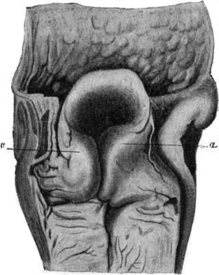
The œdema consists of an exudation into the loose submucous areolar tissue, which may be of a simple serous character or may become sero-purulent. The situations mainly involved are the glosso-epiglottic fossæ between the base of the tongue and the epiglottis, the ary-epiglottic folds (Fig. 287), and the false cords. If the infective process commences in front of the epiglottis this structure becomes swollen and rigid, and often livid in colour—points which are readily discerned on examination with the mirror, or even without its aid in some cases. The patient complains of great pain on swallowing, and has the sensation of a foreign body in the throat. Should the œdema spread to the ary-epiglottic folds, either from the interior of the larynx or from the fauces and pharynx, dyspnœa becomes a prominent and grave symptom. The patient may rapidly become cyanosed, the inspirations assume a noisy, stridulous character, and great distress and imminent suffocation supervene. If laryngoscopic examination is possible, the ary-epiglottic folds may be found greatly swollen and the upper aperture of the larynx partly occluded. Digital examination may reveal the swollen condition of the parts. The urine should be examined for albumin and tube casts.
Fig. 287.—Larynx from case of sudden death, due to œdema of ary-epiglottic folds, a, a. (From drawing lent by Dr. Logan Turner.)
Treatment.—In the milder forms, the sucking of ice, the inhalation of medicated steam, or spraying with a solution of adrenalin, and the application of poultices to the neck, may suffice to relieve the condition. Scarification of the epiglottis and ary-epiglottic folds with a knife, followed by free bleeding, may give complete relief. Diaphoretic and purgative treatment should not be neglected. If suffocation is imminent, tracheotomy or intubation is called for.
In performing tracheotomy, a roller pillow is placed beneath the neck to put the parts on the stretch, and an incision is carried from the lower margin of the cricoid cartilage downwards for about 2 inches. The sterno-hyoids and sterno-thyreoids are separated; the cross branch between the anterior jugular veins, and any other veins met with, secured with forceps before being divided; and the trachea exposed by dividing transversely the layer of deep fascia which passes from the cricoid to the isthmus of the thyreoid. If the isthmus cannot be pulled downwards sufficiently, it may be divided in the middle line. All active bleeding having been arrested, the larynx is steadied by inserting a sharp hook into the lower edge of the cricoid cartilage, and the trachea is opened by thrusting a short, broad-bladed knife through the exposed rings. The back of the knife should be directed downwards, and the opening in the trachea enlarged upwards sufficiently to admit the tracheotomy tube. In children it is sometimes found necessary to divide the cricoid for this purpose (laryngo-tracheotomy). The slit in the trachea is then opened up with a tracheal dilator, and the outer tube inserted and fixed in position with tapes. The inner tube is not fixed, so that it may be coughed out if it becomes blocked, and that it may be frequently removed and cleaned by the nurse. The tube should be discarded as soon as the patient is able to breathe by the natural channel.
Intubation of the Larynx.—This procedure is employed as a substitute for tracheotomy, especially in children suffering from membranous and œdematous forms of laryngitis. As experience is required to carry out the manipulations successfully, and as its use is attended with certain risks which necessitate that the surgeon should be constantly within call, the operation is more adapted to hospital than to private practice. O'Dwyer's apparatus is that most generally employed. The operation consists in introducing through the glottis, by means of a specially constructed guide, a small metal or vulcanite tube furnished with a shoulder which rests against the false vocal cords. The part of the tube which passes beyond the true vocal cords is bulged to prevent it being coughed out.
In an emergency a gum-elastic catheter with a terminal aperture may be passed, as recommended by Macewen and Annandale.
Bilateral Abductor Paralysis.—Both recurrent nerves may be interfered with by such conditions as enlargement of the thyreoid, tumour of the œsophagus, or intra-thoracic tumour, or by injury in the course of operations for goitre. A gradually increasing inspiratory dyspnœa is developed, which at first is only noticed on exertion, when the desire for air is increased; later it becomes permanent, and even during sleep the stridor may be marked. Suffocation may become imminent. When the larynx is examined with the mirror, the vocal cords are seen to lie near each other, and on inspiration their approximation is still greater.
The treatment is directed to removing the cause of pressure on the nerves. In the majority of cases tracheotomy is called for and the tube must be worn permanently.
Syphilitic Affections of the Larynx.—Secondary syphilitic manifestations in the form of congestion of the mucous membrane, mucous patches, or condylomata, are occasionally met with, and give rise to a huskiness of the voice. These conditions usually disappear rapidly under anti-syphilitic treatment.
In tertiary syphilis, whether inherited or acquired, the most common lesion is a diffuse gummatous infiltration, which tends to go on to ulceration and to lead to widespread destruction of tissue. It usually attacks the epiglottis, the arytenoids, and the ary-epiglottic folds, but may spread and implicate all the structures of the larynx. Syphilitic ulcers are usually single, deep, and crateriform; the base is covered with a dirty white secretion, and the surrounding mucosa presents an angry red appearance. When the perichondrium becomes invaded, necrosis of cartilage is liable to occur.
Hoarseness, dyspnœa, and, when the epiglottis is involved, dysphagia, are the most prominent symptoms.
Cicatricial contraction leading to stenosis may ensue, and cause persistent dyspnœa.
The usual treatment for tertiary syphilis is employed, but on account of the tendency of potassium iodide to increase the œdema of the larynx, this drug must at first be used with caution. Intubation or tracheotomy may be called for on account of sudden urgent dyspnœa or of increasing stenosis. The stenosis is afterwards treated by gradual dilatation with bougies, which, if a tracheotomy has been performed, may conveniently be passed from below upwards. An annular stricture causing occlusion may be excised, and the ends of the trachea sutured.
Tuberculosis.—The larynx is seldom the primary seat of tubercle. In the majority of cases the patient suffers from pulmonary phthisis, and the laryngeal mucous membrane is infected from the sputum. The disease may take the form of isolated nodules in the vicinity of the arytenoid cartilages, of superficial ulceration of the vocal cords and adjacent parts, or of a diffuse tuberculous infiltration of all the structures bounding the upper aperture of the larynx. The mucous membrane becomes œdematous and semi-translucent. The nodules coalesce and break down, leading to the formation of multiple superficial ulcers. The parts adjacent to the ulcers are pale in colour. Perichondritis may occur and be followed by necrosis of cartilage and the formation of abscesses in the submucous tissue of the larynx or in the cellular tissue of the neck.
The voice becomes hoarse or may be lost, there is persistent and intractable cough, and in some cases dyspnœa supervenes. When the epiglottis is involved there is pain and difficulty in swallowing.
In the presence of advanced pulmonary phthisis the treatment is chiefly palliative, but if the disease in the lungs is amenable to treatment, and the laryngeal lesion limited, the electric cautery may be used. Tracheotomy may be called for on account of urgent dyspnœa.
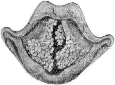
Tumours.—The commonest form of simple tumour met with in the larynx is the papilloma. It may occur at any age, and is comparatively common in children. It most frequently springs from the vocal cords and adjacent parts, forming a soft, pedunculated, cauliflower-like mass of a pink or red colour, which may form a fringe hanging from the edge of the cord (Fig. 288), or may spread until it nearly fills the larynx. In children, the growths are frequently multiple and show a marked tendency to recur after removal. They sometimes disappear spontaneously about puberty.
Fig. 288.—Papilloma of Larynx. (From drawing lent by Dr. Logan Turner.)
The most prominent symptoms are hoarseness, aphonia, and dyspnœa, which in children may be paroxysmal.
The treatment consists in removing the growth by means of laryngeal forceps or the snare, under cocain and adrenalin anæsthesia. For the removal of multiple papillomata, the removal of the growths through Killian's tubes or by suspension laryngoscopy has now taken the place of the external operation in children. In a certain number of cases it has been found that the tumour disappears after the larynx has been put at rest by the operation of tracheotomy.
Cancer.—Epithelioma of the larynx is almost always primary, and usually occurs in males between the ages of forty and seventy. It is important to distinguish between those cases in which the growth first appears in the interior of the larynx—on the vocal cords, the ventricular bands, or in the sub-glottic cavity (intrinsic cancer)—and those in which it attacks the epiglottis, the ary-epiglottic folds, or the posterior surface of the cricoid cartilage (extrinsic cancer).
Clinical Features.—In the great majority of cases of intrinsic cancer the first and for many months the only symptom is huskiness of the voice, which may go on to complete aphonia before any other symptoms manifest themselves. When the larynx is examined in an early stage, the presence of a small warty growth on the posterior part of one vocal cord, or a papillary fringe extended along the free edge of the cord, should raise the suspicion of malignancy, especially if the affected cord is congested and moves less freely than its fellow. Early diagnosis is essential in intrinsic cancer, and the absence of enlargement of lymph glands, or of fœtor and cachexia, must in no way influence the surgeon against making a diagnosis of malignancy. The impaired mobility of the affected cord is an important point in determining the malignant nature of the growth.
Intrinsic cancer may spread over the upper boundaries of the larynx and become extrinsic, or the disease may be extrinsic from the outset.
In cases of extrinsic cancer the early symptoms are much more marked, pain and difficulty in swallowing, and the secretion of frothy, blood-stained mucus being among the earliest manifestations. The cervical glands are infected early, sometimes even before there are any symptoms of laryngeal disease. Difficulty of breathing is also an early symptom on account of the growth obstructing the entrance of air. Tracheotomy may therefore be called for. In other respects the course and terminations are similar to those of intrinsic cancer.
When the growth spreads into the tissues of the neck the patient's sufferings are greatly increased. The œsophagus may be invaded with resulting dysphagia; the nerve-trunks may be pressed upon, causing intense neuralgic pains; the lymph glands become infected and break down, and the growth fungates through the skin. The general health deteriorates and death results, usually from septic pneumonia set up by the passage of food particles into the air-passages, from absorption of toxins, or from hæmorrhage. The duration of this form of the disease varies from one to three years.
The treatment consists in removing the growth. In early and limited forms of intrinsic cancer laryngo-fissure (thyreotomy) gives good results; in more advanced cases the entire larynx must be removed—complete laryngectomy—and at the same time, or after an interval, the associated lymph glands are removed from the anterior triangle of the neck on both sides.
In cases in which excision is impracticable, the sufferings of the patient may be alleviated by performing low tracheotomy, and by feeding with the stomach tube or by nutrient enemata. In some cases the difficulty of feeding the patient may make it necessary to perform gastrostomy.
Sarcoma of the larynx gives rise to the same symptoms as cancer, and can seldom be diagnosed from it before operation.
Foreign Bodies in the Air-Passages.—Foreign bodies impacted in the pharynx usually consist of unmasticated pieces of meat or large tooth-plates, and they occlude both the food and the air-passages, frequently causing sudden death. They are considered with affections of the pharynx.
The bodies most frequently impacted in the larynx are small tooth-plates in the case of adults, and buttons, beads, sweets, coins, and portions of toys in children. These are drawn from the mouth into the air-passage during a sudden inspiratory effort, for example while laughing or sneezing. If the glottis is completely blocked, rapidly fatal asphyxia ensues. If the obstruction is incomplete, the patient experiences severe pain, difficulty of breathing, and a terrifying sensation of being choked. The irritation of the foreign body causes spasmodic coughing and retching, and may induce spasm of the glottis, with threatening suffocation.
Small round bodies may lodge in the upper aperture or in one of the ventricles, and give rise to hoarseness and repeated attacks of dyspnœa and spasmodic cough. Wherever the body is situated, the symptoms may suddenly become urgent from its displacement into the glottis, or from the onset of œdema. The position of the body may often be ascertained by the use of the X-rays.
Treatment.—If the symptoms are urgent, laryngotomy, which consists in opening the larynx below the glottis by dividing the crico-thyreoid membrane, or tracheotomy must be performed at once, and an attempt made to remove the foreign body thereafter. In less severe cases in adults, the throat should be sprayed with cocain, and the larynx examined with the mirror; in children, the direct method must be employed. In both instances an attempt should be made to extract the body by the direct method. As these manipulations are liable to induce sudden spasm of the glottis, the means of performing tracheotomy must be at hand. If it is found impossible to remove the body through the mouth, laryngotomy or tracheotomy should be performed, and the body extracted through the wound, or pushed up into the pharynx and removed by this route. In the case of small bodies, a strand of gauze pushed up from the tracheotomy wound, through the larynx and out of the mouth, catches the foreign body and carries it out (Walker Downie).
The foreign bodies that are most likely to become impacted in the trachea are tooth-plates with projecting hooks, and small coins. The position of the foreign body may be ascertained by the use of Killian's tracheoscope, or by means of the X-rays. If the body remains movable in the trachea, it is apt to be displaced when the patient moves or coughs, and it may be driven up and become impacted in the glottis, setting up violent attacks of coughing and spasmodic dyspnœa.
Tracheotomy should be performed at once, and the edges of the tracheal wound held widely open with retractors, the patient being inverted, or coughing induced by tickling the mucous membrane with a feather. The foreign body is usually expelled, but it may be inhaled into one of the bronchi. One of Killian's tracheal tubes may be introduced through the tracheotomy wound and the body extracted by means of suitable forceps.
Foreign Bodies in the Bronchi.—Rounded objects, which pass through the larynx, usually drop into one or other of the bronchi, usually the right, which is the more vertical and slightly the larger. The body may act as a ball-valve, permitting the escape of air with expiration, but preventing its entrance on inspiration, with the result that the portion of lung supplied by the bronchus becomes collapsed. The physical signs of collapse of a portion or of the whole lung may be recognised on examination of the chest. In some cases the body is dislodged and driven up into the larynx, causing severe dyspnœic attacks and spasms of coughing. The irritation caused by the foreign body in the bronchus may set up bronchitis or pneumonia, and abscess of the lung may supervene. This has frequently followed the entrance of an extracted tooth into the air-passage, and it may be a considerable time before pulmonary symptoms arise. Sometimes the tooth is ultimately coughed up and the symptoms disappear. In some cases the physical signs closely simulate those of pulmonary phthisis.
The treatment consists in removing the body by the aid of Killian's or Jackson's tube passed through the mouth. If this is not successful, low tracheotomy is performed and the tube is passed through the tracheotomy opening.