LAMENESS IN THE HIND LEG.
Reading edition
Anatomo-Physiological Consideration of the Pelvic Limbs.
The pelvic bones as a whole constitute the analogue of the scapulae with respect to their function as a part of the mechanism of locomotive and supportive apparatus of the horse. The manner of attachment or connection between the ilia and the trunk is materially different from that of the scapulae, however, and the angles as formed by the long axes of the ilia in relation to the spinal column are maintained by two functionally antagonistic structures—the sacrosciatic ligaments, and the abdominal muscles by means of the prepubian tendon. The sacro-iliac articulations are such that a very limited amount of movement is possible; free movement, however, is unnecessary because of the enarthrodial (ball and socket) femeropelvic joint.
The various muscles which exert their effect upon the pelvis in changing their relationship between the long axes of the ilia and spinal column, are concerned but little more in propulsion and weight bearing than are the pectoral muscles. A general treatise on the subject of lameness does not properly include such structures any more than it does the various affections of the dorsal, lumbar and sacral vertebrae or inflammation of the abdominal parietes. Involvement of such parts cause manifestations of lameness but the matter of establishing a diagnosis is difficult in many instances and in some cases impossible.
The femeropelvic articulation is formed by the hemispherical head of the femur and the acetabulum; the latter constituting a cotyloid cavity which is deepened by the cotyloid ligament.
The round ligament (ligamentum teres) is the principal binding structure of the hip joint and it arises in a notch in the head of the femur and is attached in the subpubic groove close to the acetabular notch. Another ligament, peculiar to Equidae—the accessory (pubiofemoral)—is attached to the head of the femur near the round ligament and passes through the cotyloid notch and along the under side of the pubis. It is inserted or blends with the prepubic tendon. This ligament prevents extreme abduction of the leg. The joint capsule encompasses the articulation and is attached to the brim of the acetabulum and the edge of the head of the femur.
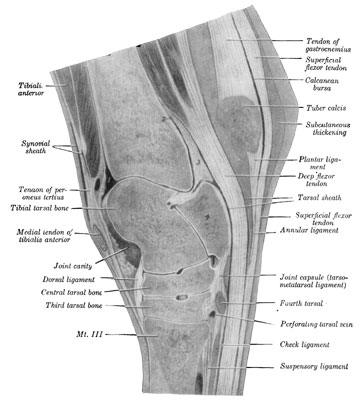
Fig. 40—Sagital section of right hock. The section passes through the middle of the groove of the trochlea of the tibial tarsal bone. 1 and 2. Proximal ends of cavity of hock joint. 3. Thick part of joint capsule over which deep flexor tendon plays. 4. Fibular tarsal bone (sustentaculum). A large vein crosses the upper part of the joint capsule (in front of 1). (From Sisson's "Anatomy of the Domestic Animals.")
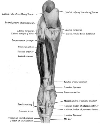
Fig. 41—Muscles of right leg; front view. The greater part of the long extensor has been removed. 1, 2, 3. Stumps of patellar ligaments. 4. Tuberosity of tibia. (From Sisson's "Anatomy of the Domestic Animals.")
The stifle joint is analagous to the knee joint of man and is to be considered an atypical ginglymus (hinge) articulation formed by the femur, tibia and patella. The ligaments are femerotibial, femeropatellar and capsular.
In addition to the usual provision for articulation of bones there are situated cartilaginous menisci between the condyles of the femur and the head of the tibia. These discs surround the tibial spine and are otherwise shaped to fit perfectly between the articular portions of the femur and tibia.
Collateral ligaments (internal and external lateral) pass from the distal end of the femur to the proximal portion of the tibia. The mesial (internal) arises from the internal condyle of the femur and is attached to a rough area below the margin of the medial (internal) condyle of the tibia. The lateral (external), shorter and thicker, arises from the depression on the lateral epicondyle and inserts to the head of the fibula.
The crucial or interosseus, anterior and posterior, are situated between the femur and tibia, and according to Smith,[34] the crucial ligaments are necessary to properly join the two bones, because of the character of the structure of the articular ends of the femur and tibia.
The femeropatella ligaments are two thin bands which reinforce the capsular ligament. They arise from the lateral aspects of the femur, just above the condyles and are inserted to the corresponding surfaces of the patella.
The patellar ligaments are three strong bands which arise from the antero-inferior surface of the patella, and are inserted to the anterior aspect of the tuberosity of the tibia.
Taken as a whole, the tarsal bones, interarticulating and articulating with the tibia and metatarsal bones form the hock joint and this articulation is analagous to the carpus. As with the carpus, there is less movement in the inferior portion of the joint than in the superior part of the articulation. The chief articulating parts are the tibia with the tibial tarsal bone (astragulus).
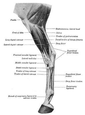
Fig. 42—Muscles of lower part of thigh, leg and foot; lateral view, o', Fascia lata; q, q', q", biceps femoris; r, semitendinosus; 21', lateral condyle of tibia. The extensor brevis is visible in the angle between the long and lateral extensor tendons. (After Ellenberger-Baum, Anat. für Künstler.) (From Sisson's "Anatomy of the Domestic Animals.")
The capsular ligament is attached around the margin of the articular surfaces of the tibia, to the tarsal bones, the collateral ligaments (internal and external lateral) and to the metatarsus.
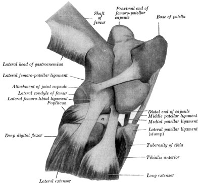
Fig. 43—Right stifle joint; lateral view. The femoro-patellar capsule was filled with plaster-of-Paris and then removed after the cast was set. The femoro-tibial capsule and most of the lateral patellar ligament are removed. M. Lateral meniscus. (From Sisson's "Anatomy of the Domestic Animals.")
The common ligaments of the tarsal joint are the collateral, the plantar (calcaneo-metatarsal and c. cuboid) and dorsal ligaments (oblique).
The medial (internal lateral) ligament serves to join the medial (internal) tibial malleolus with tibial tarsal (astragalus) and other tarsal bones.
The lateral (external lateral) ligament is inserted to the lateral (external) tibial malleolus and its distal portions are attached to the tibial tarsal (astragalus), fibular tarsal (calcaneum) bone, fourth tarsal (cuboid) and metatarsus bones.
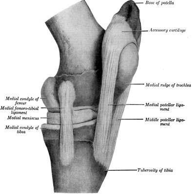
Fig. 44—Left stifle joint; medial view. The capsules are removed. (From Sisson's "Anatomy of the Domestic Animals.")
The plantar ligament (calcaneo-cuboid) is a strong flat band which is attached to the plantar surface of the fibular and fourth tarsal bones (calcaneum and cuboid) and the head of the lateral metatarsal (external small) bone.
The dorsal (oblique) ligament is attached above to the distal tuberosity on the inner side of the tibia. It is inserted below to the central (cuneiform magnum) and third (c. medium) tarsal bones, to the proximal ends of the large and outer small metatarsal bones.
The tarsus is a true hinge joint and because of the great strain which it sustains, is subject to frequent injury. About seventy-five percent of cases of lameness affecting the hind leg may be said to arise from disease of the hock.
As members of locomotion the legs receive strains of two kinds: those of concussion and weight-bearing and strains of propulsion; the latter are the greater. In the horse as a work animal, the hind legs are probably subjected to greater strains than are the front but the manner of construction of the various parts of the pelvic limbs with the possible exception (according to some authorities) of the tibial tarsal joint, offsets this condition.
The femur may be considered analagous to the humerus in that it bears a similar relationship to the ilium, that exist between the humerus and scapula. Further flexion during repose is prevented chiefly by the glutens medius (maximus) muscle and its tendons. The larger tendon inserts to the summit of the trochanter major of the femur and corresponds to the biceps brachii in the action of the latter on the scapulohumeral joint, except that the gluteus medius, in attaching to the femoral trochanter, exerts its effect as a lever of the first class. Because of the relationship between the long axes of the femur and iliac shaft it is evident that the angle formed by these two bones is maintained chiefly by the gluteus muscles during weight bearing. Contraction of muscular fibers of the gluteus medius causes extension of the femur and muscular strain is prevented to a great degree by the inelastic portion of this muscle. The chief physiological antagonistics of the glutei are the quadriceps femoris and tensor fascia lata.
While the leg is supporting weight the stifle joint is fixed in position mainly by the quadriceps femoris group of muscles which are attached to the patella. Tendinous fibres intersect this muscular mass and relieve muscular strain during weight bearing. Because of the manner in which the patella functionates with the trochlea of the femur, comparatively little energy is required to prevent further flexion of the stifle joint. The patella, according to Strangeways, may be considered a sesamoid bone.
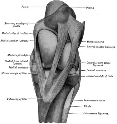
Fig. 45—Left stifle joint; front view. The capsules are removed. 1. Middle patellar ligament. 2. Stump of fascia lata. 3. Stump of common tendon of extensor longus and peroneus tertius. (From Sisson's "Anatomy of Domestic Animals.")
The quadriceps group of muscles is assisted by the anterior digital extensor (extensor pedis) peroneus tertius and tibialis anticus (flexor metatarsi) muscles. The latter pair (flexor metatarsi, muscular and tendinous portions, because of their attachment to the external condyle of the femur and to the metatarsal bone) are enabled to automatically flex the tarsal joint when the stifle is flexed.
The hock is kept fixed in position by the gastrocnemius and the superficial digital flexor (perforatus). The latter structure, which is chiefly tendinous, originates in the supracondyloid fossa of the femur and has an insertion to the summit of the fibular tarsal (calcis) bone. It relieves the gastrocnemius of muscular strain during weight bearing.
Smith[35] styles the function of the stifle and hock joints a reciprocating action, and we quote from this authority the following:
From what has been said, it is evident that flexion and extension of stifle and hock are identical in their action. When the stifle is extended, the hock is automatically extended, nor can it under any circumstances flex without the previous flexion of the stifle. There is no parallel to this in the body. The two joints, though far apart, act as one, and they are locked by the drawing up of the patella, and in no other way. The so-called dislocation of the stifle in the horse is a misnomer. That the patella is capable of being dislocated is beyond doubt, but the ordinary condition described under that term, when the stifle and hock are rigid while the foot is turned back with its wall on the ground, is nothing more than spasm of the muscles which keeps the patella drawn up. The moment they relax the previously immovable limb and useless foot have their function restored as if by magic, but are immediately thrown out of gear in the course of a few minutes as a recurrence of the tetanus of the petallar muscle takes place. The fascia of the thigh, like that of the arm, is a most potent factor in giving assistance to the constant strain imposed on the muscles of the limbs during standing.
Below the hock the hind limb is arranged like that of the fore, the deep flexor (perforans) receiving its additional support from the "check ligament," as in the fore leg.
The natural attitude of standing adopted by the horse is to rest on three legs—one hind and two fore. If he is alert, he stands on all four limbs; but if standing in the ordinary manner, he always rests on one hind leg. He does not remain long in this position without changing to the other. Hour by hour he stands, shifting his weight at intervals from one to the other hind leg, and resting its fellow by flexing the hock and standing on the toe. He never spares his fore-limbs in this manner in a state of health, but always stands squarely on them.
Hip Lameness.
Fortunately, because of the heavy musculature which goes to form a part of the locomotive apparatus of the rear extremity, hip lameness is comparatively rare. While the term is in itself ambiguous and signifies nothing more definite than does "shoulder lameness," yet diagnosis of almost any condition that may be classed under the head of "hip lameness" is not easy except in cases where the cause is obvious, as in wounds of the musculature and certain fractures. To the complexity which the gait of the quadruped contributes, because of its being four-legged, there is added the complicated manner of articulation of the bones of the hind leg. This involves the hip in the manner of diagnostic problems and because of the inaccessibility of certain parts, owing to the bulk of the musculature of these parts, diagnosis of some hip ailments becomes an intricate problem. Consequently, in some instances, before one may arrive at definite and enlightening conclusions, repeated examinations are necessary as well as a knowledge of reliable history and recorded observations of the subject over a considerable period.
Rheumatic affections, when present, usually cause recurrent attacks of lameness; myalgia, due to subsurface injury occasioned by contusion, generally produces an ephemeral disturbance; and while these are examples of cases where occult causes are active, they are by no means unprecedented. In cases where the cause of lameness is not definitely located, and when by the process of exclusion one is enabled to decide that the seat of trouble is in the hip, a tentative diagnosis of hip lameness is always appropriate.
In one instance a Shetland pony evinced a peculiar form of intermittent lameness which affected the left hip, and repeated examinations did not disclose the cause of the trouble. After about a year there was established spontaneously an opening through the integument overlying the region of the attachment of the psoas major (magnus), through which pus discharged. With the occurrence of this fistula, lameness almost entirely disappeared, but the emission of a small amount of pus persisted for more than a year. The subject was not observed thereafter and the outcome in this case is not a matter of record. Whether there existed a psoic phlegmon due to metastatic infection or necrosis of a part of a lumber or dorsal vertebra is a matter for speculation. Thus the presence of some anomalous conditions which affect the pelvic region and cause lameness may be discovered, yet both in hip and shoulder regions causes may not be definitely located by means of practical methods of examination.
Injuries of all kinds are the more frequent causes of hip lameness. In such cases, lameness may result directly and resolution be prompt, or the claudication become aggravated in time, due to muscular atrophy or degenerative changes affecting the hip joint or nerves. Rheumatism or metastatic infection may be the cause of hip lameness as well as affections of the pelvic bones, lumbar and sacral vertebrae. Hip lameness may also be provoked by melanotic or other tumors.
In the diagnosis of hip lameness, one is guided in a general way by the character of the impediment manifested. Swinging-leg lameness is often present and the impediment is more accentuated when the animal is caused to step backward. In many cases lameness is mixed, being about equally noticeable during weight bearing and while the member is being swung. By exclusion of causes which might affect other parts; one may definitely locate the cause of the trouble or determine that a certain region is affected.
The sudden manifestation of lameness is indicative of injury; thermic disturbances may signalize metastatic infection; history, if dependable, is always helpful. Repeated observations, taking into account the course which the affection assumes during a period of a few days, often serve to afford a means of establishing a diagnosis in baffling cases.
Fractures of the Pelvic Bones.
The os innominatum may be so fractured that the pelvic girdle is broken, as in fracture of the iliac shaft, or in a manner that the girdling continuity of the innominate bones is not interrupted. It naturally follows that greater injury is done when the pelvic girdle is broken than when it is not, except in cases where the acetabulum is involved and its brim not completely divided.
Etiology and Occurrence.—Pelvic fractures are usually caused by falls or other manner of contusion. Cases are reported where it would seem that fracture of the iliac angle resulted from muscular contraction, but it is certain that most fractures of this kind are due to collisions with door jambs or similar injuries. In old horses especially, fracture of pelvic bones occurs frequently. This form of injury is of more frequent occurrence in animals of all ages that work on paved streets. The country horse is not subjected to the uncertain footing of the slippery pavement, nor to injuries which compare with those caused by contusions sustained in falling upon asphalt or cobble-stones.
Symptomatology.—While in many cases of pelvic fracture lameness or abnormal decumbency are the salient manifestations, yet the pathognomic symptoms are crepitation or palpable evidence which may be obtained by rectal or vaginal examination. In fractures of the angle of the ilium and the ischial tuberosity, perceptible evidence always exists.
In cases where fracture of some portion of the pelvic girdle is suspected and the subject is able to walk, crepitation is sought by placing one hand on an external angle of the ilium and the other on the ischial tuberosity and the animal is then made to walk. Or, by placing the hands as just directed, an assistant may grasp the horse's tail and by alternately exerting traction on the tail and pushing against the hip in such manner that weight is shifted from one leg to the other, crepitation may be detected.
Fracture of the pubis near its symphysis constitutes a grave injury, as there is danger of the bladder becoming caught in the fissure and perforation of its wall may result. Such a case is reported by Bauman[36] wherein a three-year-old gelding bore the history of having been lame for ten days. Upon rectal examination the bladder was found to be hard and tumor-like and about the size of a baseball. The body of the ischium in this case was fractured and a rent in the bladder was caused by a sharp projecting piece of bone. Autopsy revealed, in addition to the fracture and rent of the bladder wall, a large quantity of urine in the peritoneal cavity.
In other instances hemorrhage caused death and not infrequently infection was responsible for a fatal issue. Moller,[37] quoting Nocard, describes a case where fracture occurred through the region of the foramen ovale and paralysis of the obturator nerve followed.
Fractures which include the acetabular bones cause great pain. This is manifested by marked lameness, both during weight bearing and when the member is swung. Such cases terminate unfavorably—complete recovery is impossible.
Where small portions of the angle of the ilium are broken, and the skin is left intact, there exists the least troublesome class of pelvic fracture. If large portions of the ilium are fractured, considerable disturbance results. There eventually occurs more or less displacement in such cases, if such displacement does not take place at the time of injury. The same may be said of fracture of the tuber ischii, but when these bones are fractured a more serious condition results.
Treatment.—When a case is found to be uncomplicated, that is, if the fracture is such that recovery seems possible and after having determined that treatment may be practicable, the first consideration is that of confining the subject in suitable slings. In many cases of pelvic fracture, the affected animal will need to be kept in slings from six weeks to three months, and it becomes a difficult problem to minimize the distress during this long period of confinement in the peculiar manner required for favorable outcome.
The pattern of sling employed should be the best that is obtainable and the matter of its adjustment is quite important lest unnecessary chafing or even necrosis of skin result. Frequent readjustment may be necessary, and time is well spent in this manner since this contributes materially toward a favorable termination by encouraging the subject to remain quiet so that coaptation of the broken bones may be maintained. Aside from slings, mechanical appliances that are helpful in the treatment of these cases are not yet in use.
A regimen that is nutritive and at the same time laxative is essential and in some cases cathartics and enemata are necessary. Also, during the first few days, if there is retention of urine, catheterization is imperative. In a word, the handling of such cases consists largely in keeping the subject inactive, as comfortable as possible, and giving attention to suitable diet.
Simple fracture of the external iliac angle needs no particular attention, except that the subject is kept quiet until lameness subsides. In all cases where much of the bone is broken, the animal is blemished, but interference with function does not follow. If infection results because of a compound fracture, loose pieces of bone must be removed surgically and drainage provided for.
In fracture of the ischial tuberosity, infection is more apt to result than in like injury of the ilium, and greater displacement of bone occurs. This displacement, due to contraction of the attached muscles, is in some instances a contributing cause to the infection which often follows in these cases. In females where the body of the ischium is fractured, lacerations of the vagina may be present, and this constitutes a serious complication which usually terminates fatally.
After-care in fracture of the pelvic girdle consists principally in allowing a protracted period of rest before subjects are put to work.
Fractures of the Femur.
Etiology and Occurrence.—This is a comparatively rare injury in the horse because of the protection afforded the femur by the heavy musculature. Fragilitas of the bone probably exists in many cases when fracture of its diaphysis occurs. It is generally conceded that the neck of the femur is rarely broken because of a lack of constriction in this part, but fracture of the trochanters has been recorded rather frequently. However, Lienaux and Zwanenpoete[38] state that fracture of the neck of the femur is of frequent occurrence in Belgian colts. Tapley[39] reports in the Veterinary Journal (English) fracture of the head and internal trochanter of the femur and patellar luxation occurring simultaneously affecting a mule. In this case the mule was found decumbent on a concrete floor. After three weeks, the subject was destroyed and autopsy revealed rupture of the left pubiofemoral ligament, tearing with it a portion of the articular surface of the femur. The internal trochanter was also fractured in four small pieces. In this case it is fair to suppose that the mule in trying to regain footing on a slippery floor violently abducted the legs and fracture resulted. It is possible also that a temporary luxation of the patella took place first and caused the animal to struggle in such manner that fracture followed.
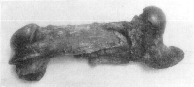
Fig. 46—Oblique fracture of the femur of a 1,500 six-year-old draft horse. Showing shortening of bone, owing to a lateral approximation of the diaphysis because of muscular contraction. Photo by Dr. Edward Merillat.
Symptomatology.—According to Cadiot and Almy,[40] "regardless of the location of femoral fractures, the subject is usually intensely lame, the animal frequently walking on three legs—fractures of the diaphysis are characterized by an abnormal mobility."
As a rule, crepitation is to be recognized in fractures of the shaft of the bone, by passively moving the leg to and from the medial plane (adduction and abduction).
Fracture of the trochanter major is signalized by local swelling and evidence of pain; the forward stride is shortened because this movement tenses the tendon of the gluteus major (maximus) which is attached principally to the trochanter.
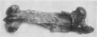
Fig. 47—Same bone as in Fig. 46 after about six months' treatment. In this case Dr. Merillat employed a weight to counteract muscular contraction. It is noticeable that very little provisional callus has formed in this case, and in spite of unusual ingenuity and good facilities for caring for the subject, union of bone did not occur.
Treatment.—Reduction of femoral fracture in the horse is practically impossible, and retaining the broken bones in coaptation is not possible by means of mechanical appliances. Consequently, prognosis is unfavorable in fracture of the body of the femur. When union of bone occurs, there results shortening of the leg and animals are rendered permanently lame. If the immediate region of the head of the bone is involved as well as in case of fracture of the condyles, an incurable arthritis ensues.
Where the trochanters are broken, chronic lameness and muscular atrophy is the result. Therefore, it is evident that, because of the manner of function of the femur, the leverage afforded by its great trochanter and its heavy muscular attachments, fractures of this bone in the horse do not terminate favorably.
Luxation of the Femur.
Etiology and Occurrence.—Uncomplicated femoral luxation is of less frequent occurrence in the horse than in the other domestic animals. The deep cotyloid cavity renders disarticulation difficult and luxation does not often take place. Complications that usually occur are rupture of the round (coxofemoral) ligament or fracture of the neck of the femur. Falls or violent strains are necessary to produce this luxation. Goubaux is quoted by Cadiot and Almy[41] as having observed the head of the femur in an instance wherein luxation had long existed. In this case autopsy revealed the fact that the inner portion (two-thirds) of the head of the femur had completely disappeared.
Luxation of the femur is observed in old emaciated animals that are worked on slippery pavements. Occasionally, evidence of chronic luxation of the femur is observed in the anatomical laboratory. The chronicity of the condition is obvious when one notes the well formed articulation which Nature provides for the head of the femur, where fracture or other serious complications are not present.
Symptomatology.—In every case there must exist either restriction of movement or an evident abnormal position of the leg, or both conditions may exist at once. Also, the leg may be markedly shortened. Manifestation of this affection varies, depending upon the character of the luxation (position of the head of the humerus with relation to the acetabulum). Lusk[42] cites a case of a mule which had suffered femoral luxation. The animal was destroyed and on autopsy the head of the femur found to be contained within a false articular cavity situated about four inches above the acetabulum. In Dr. Lusk's case as he states it, the following symptoms were presented: "Limb shortened and fixed in a position of adduction. While standing the affected limb hung directly across and in front of the opposite one; upper trochanter very prominent; skin over hip joint very tense. The mobility of the limb was very limited, especially in the forward direction."
Being very prominent when there is an upward luxation and less perceptible in downward displacement, the location of the trochanter major is an indicator of the character of the luxation with respect to the position of the head of the femur. This variation of position causes abnormal tenseness or looseness of the skin over the region of the trochanter major. Rectal examination is of aid in locating the head of the humerus.
Treatment.—When it is evident that a subject should be given treatment and not destroyed, the animal must be cast and completely anesthetized. With complete relaxation thus secured by rotation of the limb, using the hip joint region as a pivot, reduction may be effected. Traction is exerted in the same direction from the acetabulum that the head of the femur is situated and by pressing over the joint, the displaced bone may be returned in position. If luxation is downward, traction on the extremity will tend to dislodge the head of the femur from the inferior acetabular margin making reduction possible.
The same general plan which is ordinarily employed in correcting luxation is indicated here, but because of the heavy musculature of the hip, complete anesthesia is imperative in all such manipulations.
Gluteal Tendo-Synovitis.
The glutens medius (g. maximus) muscle is inserted chiefly by means of two tendons; one to the summit of the trochanter major of the femur and the other passing over the anterior part of the convexity of the trochanter, and being attached to the crest below it. The trochanter is covered with cartilage, and a bursa (the trochanteric) is interposed between the tendon and the cartilage.
Etiology and Occurrence.—This affection is probably caused in most instances by direct injury to the parts, such as may be occasioned by being kicked, falling on pavement, or being struck by the body of a heavy wagon. Strains in pulling or in slipping are undoubtedly causative factors and in draft horses such strains may result in involvement of this synovial apparatus.
Symptomatology.—If pain be severe and inflammation acute, weight may not be borne with the affected member. There is some local manifestation of the condition in acute cases. Swelling of the tissues contiguous to the bursa is present and pain is evinced upon manipulation of the parts. A characteristic gait marks inflammation of the trochanteric bursa, and as Gunther has put it, the subject generally moves or trots as does the dog—the sound member being carried in advance of the affected one and the forward stride of the diseased leg is shortened. In some chronic cases crepitation is discernible by holding the hand on the trochanter while the subject walks.
Treatment.—In the first stages of an acute affection absolute quiet must be enforced; local antiphlogistic applications are beneficial. Later, vesication of a liberal area surrounding the trochanter major is indicated. Where the condition has become chronic in horses that are to be kept at heavy draft work there is little chance for complete recovery. And, naturally, one is not to expect resolution in cases where there exist erosion and ossification of cartilage—where crepitation is discernible.
Paralysis of the Hind Leg.
Aside from paraplegic conditions due to disease of the cord or the lumbosacral plexus, and monoplegic affections resultant from disturbances of this plexus, paralysis of certain nerves are occasionally encountered.
Anatomy.—The lumbosacral plexus results substantially from the union of the ventral branches of the last three lumbar and the first two sacral nerves, but it derives a small root from the third lumbar nerve also. The anterior part of the plexus lies in front of the internal iliac artery, between the lumbar transverse processes and the psoas minor. It supplies branches to the iliopsoas[43] (designated by Girard, the iliacomuscular nerves). The posterior part lies partly upon and partly in the texture of the sacrosciatic ligament. From the plexus are derived the nerves of the pelvic limb (Sisson).
Paralysis of the Femoral (Crural) Nerve.
Anatomy.—The femoral nerve (crural) is derived chiefly from the fourth and fifth lumbar nerves. It runs ventrally and backward, at first between the psoas major and minor, then crosses the deep face of the tendon of the latter and descends under cover of the sartorious over the terminal part of the iliopsoas. It innervates the psoas major (magnus), psoas minor (parvus), sartorious, rectus femoris, vastus lateralis (interims). Branches supply the stifle and the adductor and pectineus muscles.
Etiology and Occurrence.—While paralysis of the femoral nerve, also known as "dropped stifle" occurs as a result of local injuries and melanotic tumors in gray horses, most cases are due to azoturia. So-called crural paralysis or "hip swinney" is occasionally observed but this is not a condition wherein the nerve is affected in the manner that characterizes the marked atrophy of quadriceps femoris (crural) muscles in some cases of hemaglobinuria. This form of paralysis according to Hutyra and Marek is due primarily to diffuse degeneration of the muscles.
Symptomatology.—When muscular atrophy is not extensive no particular evidence of this condition may be manifested while the subject is at rest, but where muscular waste has occurred, the nature of the ailment is at once recognized. Since the femoral nerve supplies the quadriceps femoris muscles, it follows that when the psoic portion of this nerve becomes diseased, the stifle loses its support, and in a unilateral involvement when the subject attempts to walk on the affected member, the stifle sinks down for want of support and the leg collapses unless weight is caught up with the other leg. Often, following azoturia, a bilateral affection is to be observed.
Treatment.—Horses may be restrained in the standing position, and in the average instance, a twitch and hood are all the restraining appliances necessary.
In cases where the disease is unilateral and atrophy is not of too long standing, recovery is possible in vigorous subjects. All affections, however, wherein degenerative changes involve the nerve trunk, whether due to diffuse myositis or pressure from malignant tumors, will not yield to treatment.
The same general plan of treatment is indicated that is described on page 74 in the consideration of atrophy of the scapular muscles. It is especially important to provide for the subject to be exercised when there is atrophy of the quadriceps muscles following azoturia.
In addition to the foregoing, good results have attended the use of intramuscular injections of oxygen. The technic of the operation consists in preparing the area of skin which covers the atrophied muscles as for any operation. The hair is clipped over five or six or more circular areas of about an inch in diameter; the skin is cleansed and then painted with tincture of iodin.
A long heavy sterile needle, which is connected with an oxygen tank by means of six feet of rubber tubing, is thrust into the depths of the affected muscles and the gas is gently introduced into the tissues. One needs exercise extreme care that the gas enter slowly because great pain is produced by the sudden injection of the oxygen. Likewise too much of the gas must not be introduced at one place. When the oxygen is slowly introduced it may be allowed to enter the tissues until the subject gives evidence of experiencing considerable pain, or if the parts are not particularly sensitive, a reasonable amount (enough to cause a mild degree of diffuse inflammation) is introduced at each one of five or six points. In large animals more points of injection may be used.
No infection or other bad results will follow the execution of a good technic and the treatment may be repeated every three or four weeks until either marked regeneration of tissue is evident or the case is obviously proved hopeless.
Paralysis of the Obturator Nerve.
Anatomy.—The obturator nerve, situated at first under the peritoneum, accompanies the obturator artery through the obturator foramen and gaining the muscles on the internal face of the thigh, terminates in the obturator externus, adductors, pectineus and gracilis, also giving twigs to the obturator internus (Strangeways).
Etiology and Occurrence.—This condition occurs upon rare occasions as the result of injury such as falls which cause extreme abduction of the legs, or in pelvic fracture where the nerve is directly injured, or when melanotic tumors or other new growths compress the nerve in such manner that its function is suspended. Paralysis of the obturator nerve or nerves is met with rather frequently, notwithstanding, in mares, following dystocia. The nerves (one or both) may become bruised at the brim of the obturator foramen by being caught between the pelvis and the body of the fetus in some cases of protracted labor.
Symptomatology.—In a unilateral affection there may be little evidence of the trouble while the subject is standing; or there is to be seen some abduction; or the affected member may present abduction of the stifle and stand "toe outward." If the animal is walked there will be manifested more or less abduction and the character of the impediment varies according to the nature of the involvement.
Following protracted cases of labor in some instances where only a unilateral paralysis exists, walking is performed with difficulty; the subject may be unable to support weight with the affected member and is obliged to hop on the one sound hind leg. In bilateral affections, they are unable to rise. If the condition is severe the sling is required to keep the subject standing, and with this care, recovery will follow.
Treatment.—If new growths or callosities or similar conditions affect the nerve, little, if any, hope for recovery exists. In young and vigorous subjects where cause is not definitely known, a course of strychnin may be given. Good nursing, providing for the subject's comfort and allowing moderate exercise, constitute rational treatment. Stimulating embrocations on the abductor muscles resorted to in cases during the incipient stage may prove helpful.
When paralysis of the obturator nerve occurs as a post-partum complication, and other conditions are favorable, the subject should be raised to its feet without unnecessary delay. If the mare is unable to assist in regaining her feet, a sling is required. Usually little else is necessary and after a few days in the sling the subject can get about unassisted. In the meanwhile the well-being of the affected animal is to be considered just as in any other case where the patient is so confined. The foal in such instances constitutes a source of some trouble, but the average mare offers no serious resistance to the confinement occasioned by the sling.
Good hygienic care, a suitable diet and full physiological doses of strychnin are indicated. Cadiot and Almy recommend vaginal douches of cold water and counterirritation of the region of the inner thigh in these cases.
Paralysis of the Sciatic Nerve.
Anatomy.—The great sciatic nerve leaves the pelvis in company with the gluteal nerves, through the great sciatic foramen (notch), passing downward along the posterior face of the femur. Near the stifle it passes between the two heads of the gastrocnemius muscle and continues as the tibial. Branches supply the following muscles—obturator, semimembranosus (adductor magnus), biceps femoris (triceps abductor femoris), semitendinosus (biceps rotator tibialis), lateral extensor (peroneus) and the tibial nerve, its continuation, innervates the digital flexors.
Etiology and Occurrence.—Paralysis of the great sciatic nerve may be caused by central disorders, injury in falling, fractures and new growths. Because of its protected position, this nerve does not often suffer injury, and paralysis of the sciatic nerve is recorded in a few instances owing to its rarity.
Symptomatology.—When consideration is given the number of muscles that are supplied by the sciatic nerve and the function of these muscular structures, it is obvious that the leg cannot be used in sciatic paralysis. However, the limb is capable of sustaining weight when it is fixed in position, but this is done without exertion of muscular fibers which are supplied by the great sciatic nerve. Trotting is impossible and flexion of the affected member is also likewise precluded. The foot is dragged when the subject is caused to advance.
Under the heading "sciatica," Scott[44] has described a case of acute sciatic affection wherein a pacing horse manifested evidence of great pain of a nervous character. There were muscular twitchings and the leg was held off the floor and moved about convulsively. Breathing was very much accelerated, pulse 85 per minute, the temperature was 103° and manipulation of the hips augmented the pain.
This was not a paralytic condition and recovery resulted, yet undoubtedly this was a case which, if not properly cared for, might have terminated unfavorably.
Treatment.—Prognosis is decidedly unfavorable in paralysis of the great sciatic nerve. If treatment is attempted, it is to be conducted along the same general lines as in femoral paralysis. Particular attention should be given to conditions which will make for the patient's comfort, and as soon as it is evident that the affection is not progressing favorably, the subject should be humanely destroyed.
Iliac Thrombosis.
This condition is undoubtedly of more frequent occurrence than we are wont to grant when one considers the comparatively small number of cases that are actually recognized in practice. It does not follow, however, that iliac thrombosis rarely exists. Probably in the majority of instances there is insufficient obstruction of the lumina of vessels to provoke noticeable inconvenience. Or, if circulation is hampered to the extent that function is impaired and manifestations are observed by the driver, the subject may be permitted to rest a few days and partial resolution occurs, so that further trouble is not noticeable.
As judged by lesions of the aorta and iliac arteries in dissecting subjects, the conclusion that arteritis and resultant disorders are of rather frequent occurrence, is logical.
Etiology.—Inflammation of the vessel walls and resultant prolifieration of tissue together with the accumulation of clotted blood becoming organized, serve to obstruct the lumen of the affected artery. The cause of arteritis is unknown in many instances, but parasitic invasion and contiguous involvement of vessels in some inflammatory injuries are etiological factors.
Symptomatology.—A characteristic type of lameness signalizes iliac thrombosis and the following brief abstract from a contribution on this subject by Drs. Merillat[45], clearly portrays the chief symptoms:
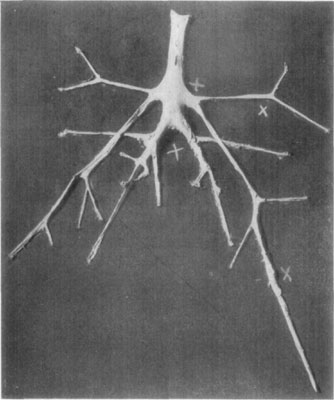
Fig. 48—Exposure of aorta and its branches, showing location of thrombi in numerous places. In this case (same as Fig. 49) Dr. L.A. and Dr. Edward Merillat found the cause of the condition to be due to sclerastomiasis.
The seizures are accompanied with profuse sudation, tremors, dilated nostrils, accelerated respirations and other symptoms of pain and distress, all of which, together with the lameness, disappear as rapidly as they had developed, leaving the animal in an apparently perfect state of health, ready to fall with another attack of precisely the same kind, as soon as enough exercise is forced upon it. The rectal explorations may reveal a pulseless state of one or more of the iliac arteries and a hardness and enlargement of the aortic quadrifurcation, but sometimes this palpation fails to disclose any perceptible diminution of the blood current of these vessels. The obturation being incomplete, it may be impossible by palpation to decide that thrombosis really exists. In this event and, in fact, in all eases, the clinical symptoms are sufficiently characteristic to make a diagnosis without reservation. It cannot be mistaken for any other disease, once properly investigated. Any given seizure may easily be mistaken for azoturia, at first, but a better examination soon excludes that disease.
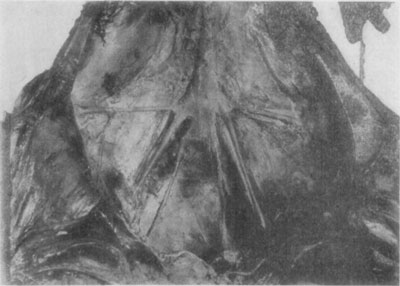
Fig. 49—Illustrative of thrombosis of the aorta, iliacs and branches. Photo by Dr. L.A. Merillat.
Prognosis and Treatment.—In the majority of instances, when there is occasioned serious inconvenience, the outcome is not likely to be favorable, according to Möller. Detachment of a portion of the thrombus, according to Hoare, may result in the lodgment of an embolus in the brain or kidneys. The latter authority also states that muscular atrophy may occur owing to lack of blood supply in some of these cases. Möller states that moderate exercise or work stimulates the establishment of collateral circulation. Massage per rectum is condemned as dangerous by Cadiot.
Fracture of the Patella.
Etiology and Occurrence.—Patellar fractures are rarely met with in the horse but may be caused by falls and heavy contusions. Violent muscular contraction, it is said, may also bring about the same condition.
Symptomatology.—Fracture may be transverse or vertical, and depending on the manner in which the bone is broken, prognosis is either at once rendered favorable or unfavorable. The patella performs a function which is in a way similar to that of the sesamoids and when fractured, complete recovery is improbable in the average instance. When complete, transverse fractures permit of separation of the parts of bone. Tension on the straight ligaments below and contraction of the quadriceps above usually cause insuperable difficulty in the handling of this type of fracture in the horse.
Compound fractures as well as multiple or comminuted fractures occasionally occur and these constitute injuries which are generally considered fatal, although Andrien, according to Cadiot and Almy, succeeded in obtaining complete recovery in a case of compound fracture of the patella and the horse was in service and almost free from lameness two months after treatment was begun.
No difficulty is encountered in recognizing the fracture of the patella because of the exposed position of the bone. Crepitation, and in some cases fissures, may be easily detected.
Treatment.—In simple fracture, when treatment is thought advisable, the subject is put in a sling and kept as nearly comfortable as possible. If little inflammation exists, the application of a vesicant two or three weeks after the injury has been inflicted will be helpful and serve to hasten repair.
Bandages or mechanical appliances are of no practical use in the handling of these cases.
Luxation of the Patella.
Etiology and Occurrence.—This, the most common luxation met with in the equine subject, has been described by writers as existing in many forms. Patellar disarticulation may be more practically considered as momentary and fixed, regardless of the position taken by the patella. Described under the title of false luxation are recorded cases wherein the quadriceps (crural) muscles become contracted in such manner that a condition simulating true disarticulation of the patella obtains. Also, some practictioners report cases of patellar luxation and refer to pseudo-luxations, without clearly defining the conditions which constitute pseudo-luxation. This has contributed to the extant cause of misconception as to actual differences between luxation and conditions simulating dislocation.
Luxation of the patella is a condition wherein the articular portions of the femur and patella assume abnormal relations whether such displacement of the patella be momentary and capable of spontaneous reduction, or fixed and requiring corrective manipulation. Spasmodic contraction of the crural muscles which sometimes retains the patella in such position that the leg is rigidly extended, does not in itself constitute luxation of the patella; and unless this bone becomes lodged on the upper portion of a femoral condyle or laterally displaced out of its femoral groove, luxation cannot be said to exist in the horse. These are sub-luxations.
Occasionally one may observe in suckling colts outward luxation of the patella wherein there is history of navel infection and no marked evidence of rachitis is present. Some of these cases recover. In a unilateral involvement of this kind in a three-month-old mule colt, the author observed a case wherein an unfavorable prognosis was given and destruction of the subject advised, because of the extreme dislocation of the patella. This colt, however, was not destroyed and in three weeks had apparently recovered. No treatment was given in this instance; the colt was allowed the run of a small pasture with its dam and in time it matured, becoming a sound and serviceable animal.
Classification.—Two forms of true patellar luxation in the horse may be considered; one which is due to the patella becoming fixed upon the internal trochlear rim of the femur and the other when the patella slips over the outer rim of the trochlea.
The first form is known as upward luxation and is made possible by rupture of the mesial (internal) femeropatellar ligament. According to Cadiot and Almy, it is only by the rupture of this ligament—the femeropatellar—that upward luxation may occur. This type of luxation is rarely observed and is usually due to violent strain and abnormal extension of the stifle joint.
The second class, outward luxation, occurs in colts and is, in many instances, congenital. This form of luxation is also the one usually seen following debilitating diseases such as influenza and pneumonia.
Upward luxation of the patella is characterized by the stiff-extended position of the leg. When the patella is situated upon the inner trochlear rim, the tibia must be extended because of the traction exerted by the straight ligaments. Since the stifle and hock joints extend and flex in unison, there is presented also an extension of the tarsus. Extension of the stifle joint would increase the distance between the femoral origin of the gastrocnemius and its insertion to the summit of fibular tarsal bone (calcis) were it not for the gastrocnemius and superficial flexor (perforatus). Extension of the hock in upward luxation of the patella, permits of flexion of the phalanges. In upward luxation, then, the leg is extended as if too long, but the phalanges may be in a state of moderate flexion. If the foot rests on the ground when the extremity is not flexed, it is almost impossible for the subject to step backward. Because of immobilization of the stifle and hock joints in upward luxation, the subject can walk only by hopping on the sound leg and then the extremity is flexed, allowing the anterior portion of the fetlock to drag on the ground.
In some cases practitioners are called to attend young animals that are reported to be "stifled" (often in young mules that have made a rapid growth) and upon arrival the only noticeable symptom of preëxisting luxation is the soiled condition of the anterior fetlock region—evidence of its having been dragged. Such cases may be styled momentary luxation, whether they are due to a weakened condition of the patellar ligaments or spasmodic contraction of the crural muscles.
In upward luxation, reduction is effected by attempting further extension of the stifle joint and at the same time the patella is pulled outward, off the internal rim of the trochlea. This is attempted by securing the subject in a standing position; the sound side is kept against a wall if possible and a rope is tied to the extremity of the affected leg. Traction is exerted upon the rope and at the same time force is directed against the stifle joint to produce further extension if possible, so that the straight patellar ligaments may relax sufficiently to allow the patella to be dislodged from its position upon the inner trochlear lip. Failing in this manner of procedure, the affected animal is to be cast and anesthetized with chloroform. The relaxation which attends surgical anesthesia will permit of reduction of the dislocated bone and manipulations such as have just been outlined may be employed.
Following reduction in the average case it is essential that the subject be given vigorous exercise for a few minutes. Reduction having been affected, the application of a vesicant over the whole patellar region is customary.
In cases of habitual luxation, unless the ligaments are so lax that the patella may be displaced laterally over the inner as well as the outer trochler rims, division of the inner straight patellar ligament will correct the condition. This desmotomy has been advocated by Bassi, and good results in appropriate cases have been reported by Cadiot, Merillat and Schumacher. This operation has been found a corrective in cases of outward luxation as well as those of upward dislocation of the patella when resorted to before the trochleae are worn from frequent luxation.
Outward luxation of the patella is occasioned by a lax condition of the internal femeropatellar ligament or a rupture of the same so that the patella slips over the outer femoral trochlear rim and permits of an abnormal flexion of the stifle joint. The outer trochlear rim being the smaller of the two, inward luxation does not occur in the horse. With the patella disarticulated in this manner, the action of the quapriceps femoral group of muscles has no effect on the stifle joint and, therefore, flexion of this articulation occurs as soon as the subject attempts to sustain weight and the leg collapses unless weight is at once taken up by the other member if sound.
As a rule, the reduction of this form of luxation is not difficult. The patella may be pushed inward and into position without manipulation of the leg. Retention of the patella in position is a difficult problem. Bandaging is considered impractical and is not ordinarily done in this country. Benard, according to Cadiot and Almy, recommends bandaging with a heavy piece of cloth in which an opening is made through which the patella is allowed to protrude, and by turning such a bandage snugly about the stifle several times, the patella is held in position. This bandage should be kept in place for about ten days.
In young and rachitic animals outdoor exercise and a good nutritive ration for the subject are indicated. Hypophosphites in assimilable form may be beneficial, and vesication of the patellar region contributes to recovery.
Where extreme luxation is present in both stifles, the prognosis is unfavorable. In such cases, degenerative changes may exist and in some instances the ligaments are so diseased and elongated that regeneration is impossible. Williams[46] reports a case where bilateral "floating" (outward) luxation was present and extensive degeneration changes affected the articulation.
In subjects suffering frequent dislocation of the patella (habitual luxation) it is possible in some cases, to prevent its occurrence or at least to minimize the distress occasioned by momentary luxation, by keeping the animals in wide stalls so that "backing" is unnecessary. In some nervous subjects that seem to be suffering from cramp of the crural muscles, the difficulty and pain of their being backed out of narrow stalls, accentuates the nervousness. Sudation and restlessness are manifested and the subject presents a clinical picture of distress and fear of a painful ordeal. In some cases of this kind, complete recovery takes place by the time animals are five or six years of age. One should avoid keeping such subjects in narrow stalls. Preferably patellar desmotomy should be performed that relief may be obtained at once.
Luxations attending some cases of influenza recover promptly when subjects are kept comfortably confined in roomy box-stalls. The administration of stimulative medicaments such as nux vomica and the application of an active blistering agent to the patella serve to hasten recovery. Dislocations in such cases are often bilateral and they are usually momentary. Reduction occurs spontaneously, as a rule, and the subjects are not occasioned much distress if they are kept quiet for a few days.
Chronic Gonitis.
Etiology and Occurrence.—Chronic inflammation of the stifle joint is met with following acute synovitis due to strains and concussion. It is an ailment which affects heavy horses and particularly animals that are kept at work on paved streets, but this does not explain its existence in animals that are not subjected to work likely to cause concussion. Berns[47] considers rheumatism a probable cause of gonitis and, as he states, the dropsical form of affection of this joint is not ordinarily attended with manifestations of inconvenience to the subject. Gonitis is often bilateral and its onset is insidious in many instances.
Symptomatology.—In unilateral gonitis weight is not borne by the affected member. There is noticeable distension of the joint capsule—a characteristic pendant pouching protrusion. When both stifles are affected the subject frequently shifts the weight from one limb to the other. Lameness comes on gradually and during the incipient stages may be intermittent but it progressively increases so that in time affected animals become useless. In bilateral affections animals drag the toes because of the pain incident to flexing the stifles. This is particularly evident when the subject is made to trot. As the disease progresses, atrophy of the quadriceps femoris muscles becomes pronounced and as destructive changes involving the articular cartilages take place. The subject becomes more lame and eventually is rendered incapable of service.
Upon manipulation of the patellar region, one is impressed with the fact that hyperesthesia does not exist in proportion to the pain manifested during locomotion. In some cases a gelatinous swelling is present and may be detected by palpating between the straight ligaments of the patella. Williams, Hughes, Merillat, Hadley and others have directed attention to the existence of floating masses (corpora oryzoidea) in the synovial capsule of this joint in gonitis, and as with all cases of arthritis, irreparable damage is often done the articular cartilages during the course of the ailment.
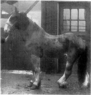
Fig. 50—Chronic gonitis. The knuckling which results from long continued inactivity of the crural muscles in chronic cases is marked in this instance. Photo by Dr. L.A. Merillat.
Treatment.—No effective method is as yet known which will control this condition during its incipiency. The disease progresses, and more or less damage is done the affected parts in the course of months or even years in some cases before subjects are rendered hopelessly crippled. When recognized early (before chronic gonitis exists) aspiration of the synovia and the injection of diluted tincture of iodin might prove beneficial in cases of synovial distension. Chronic gonitis is considered an incurable affection and as soon as subjects manifest evidence of distress from this condition they should by all means be taken from work. Firing and vesication have not been productive of beneficial results.
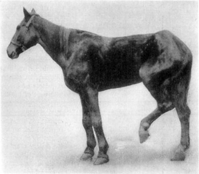
Fig. 51—Gonitis. Showing position assumed in such cases because of pain occasioned. Photo by Dr. C.A. McKillip.
Open Stifle Joint.
Anatomy of the Joint Capsule.—This joint capsule is thin and very capacious. On the patella it is attached around the margin of the articular surface, but on the femur the line of attachment is at a varying distance from the articular surface. On the medial side it is an inch or more from the articular cartilage; on the lateral side and above, about half an inch. It pouches upward under the quadriceps femoris for a distance of two or three inches, a pad of fat separating the capsule from the muscle. Below the patella it is separated from the patellar ligaments by a thick pad of fat, but inferiorly it is in contact with the femerotibial capsules. The joint cavity is the most extensive in the body. It usually communicates with the medial sac of the femerotibial joint cavity by a slit-like opening situated at the lowest part of the medial ridge of the trochlea. A similar, usually smaller, communication with the lateral sac of the femerotibial capsule is often found at the lowest part of the lateral ridge. (Sisson's Anatomy.)
Thus it is seen that because of its frequent communication with the other parts of this large synovial membrane, a wound which opens the external portion of the femerotibial capsule may be the cause of contamination and resultant infectious arthritis of the whole stifle joint. Because of the distance between the most dependent part of the femerotibial articulation and the summit of the patella, one may misjudge the exact location of the lowermost part of this portion of the capsular ligament of the stifle joint and thereby fail at once to appreciate the seriousness of calk wounds in this region.
Etiology and Occurrence.—Wounds to the patellar region are of rather frequent occurrence, and because of the comparatively unprotected position of these structures, the capsular ligaments of the stifle joint may be perforated as a result of violence in some form. Calk wounds which penetrate the tissues in the immediate region of the lower portion of the external part of the femerotibial capsule sometimes result in open joint because of tissue necrosis resulting from the introduction of infection. Contused wounds sometimes destroy the skin and fascia over large areas on the lateral patellar region and because of subsequent sloughing of tissue due to infection as well as to the manner in which such wounds are inflicted, septic arthritis subsequently occurs. Penetrant wounds, such as may be caused by a fork tine may not result in infection; if infectious material is introduced an infectious arthritis does not necessarily follow, though such cases should be considered as serious from the outset.
Symptomatology.—The pathognomonic symptom of open stifle joint is the profuse escape of synovia, indicating perforation of the synovial capsule; by means of a probe the wound may be explored in a way that will clearly reveal the nature of the injury.
After a few days have elapsed in cases where considerable infection has taken place, there is manifestation of pain as in all cases of infective arthritis. Hughes[48] gives an excellent description of the clinical aspect of arthritis which applies here:
Acute arthritis begins like an ordinary attack of synovitis. In joints other than the pedal and pastern, there is sudden and extensive swelling, which at first is intra-articular, succeeded by extra-articular tumefaction, and accompanied by violent lameness. The pain soon becomes intense and agonizing. There is severe constitutional disturbance, the temperature ranging from 104 to 106 degrees and the pulse from 60 to 72. Painful convulsions of the limb occur, shown by involuntary spasmodic elevations due to reflex irritation of the muscles. There is loss of appetite, rapid emaciation, the flank is tucked up and the back arched. In from three to six days, the tumefaction around the joint tends to soften at a particular place, and bursts, and a discharge that is sometimes of a sanious character, mixed with synovia, escapes. Great exhaustion at times supervenes, and if the joint is an important one, the horse lies or falls and is unable to rise.
Treatment.—In small puncture wounds the immediate application of a vesicating ointment has given good results, but when infection has taken place to such extent that the animal manifests evidence of intense pain, and lameness is marked and local swelling and hyperesthesia are great, vesication is contraindicated. In such instances the exterior of the wound and its margins should be prepared as in similar affections of other joints. A quantity of synovia is then aspirated by means of a small trocar and care should be taken to observe all due aseptic precautions. Subsequently the injection of from four to six ounces of a mixture of tincture of iodin, one part to ten parts of glycerin, and gentle massage of the joint immediately after the injection has been made, serves to check the infective process in some cases.
The subject should be cared for as has been previously suggested in arthritis proper provisions for comfort being made. Good nursing is always essential to a successful issue. However, the author cannot view cases of open stifle joint with the same optimism concerning their course and outcome that is expressed by a number of writers on this subject. It is a grave condition wherein the prognosis should be given advisedly.
Fracture of the Tibia.
Etiology and Occurrence.—Because of its exposed position to kicks, and its lack of protection by heavy musculature (especially on its inner surface), there is afforded ample opportunity for frequent injury to the tibia. Fractures are complete and varying as to nature, or incomplete. The heavy tibial fascia affords sufficient protection so that fissures without entire solution of continuity of the bone may occur from violence to which this part is often subjected. Möller classes tibial fracture as ranking second in frequency—pelvic fracture being more often met with in horses. This does not apply in our country as phalangeal and metacarpal and even metatarsal fractures are observed in more instances than are such injuries to the tibia. The tibia is occasionally broken at its middle and lower thirds, but malleolar fractures are not common.
Symptomatology.—When fracture is complete and all support is removed, the leg dangles, and the nature of the injury is so obvious that there is no mistaking its identity. However, in case of incomplete fracture one needs to base all conclusions upon the history of the case, evidence of injury, or other knowledge of the character of violence to which this bone has been exposed. For without the presence of crepitation (even by excluding other possible causes for the pronounced lameness which characterizes some of these cases) we can only resort to the knowledge which experience has taught that fracture may be deemed probable in many injuries to the tibial region. Consequently, we are to look upon all injuries that affect the tibia as being fractures of some sort when there is either local evidence of the infliction of violence or whenever marked lameness attends such injuries, unless there is positive indication that no fractures exist.
A careful examination of parts of the tibia, i.e., noting the amount and painfulness of swellings, exploration with the probe, and observations of the course taken in any given case, will determine the exact nature of injuries. Such examination needs to extend over a period of a week or in some instances two or three weeks may pass before the true state of affairs is apparent. In the meanwhile, cases are to be handled as though tibial fracture certainly existed.
Prognosis.—Prediction of the outcome in tibial fracture is somewhat presumptuous, but in the majority of cases in mature subjects fatality results. Cadiot[49], however, views this condition with more optimism than have American practitioners. While he considers the condition grave, in citing case reports of successful treatment by d'Arboval, Duchemin, Leblanc, and others, his conclusion is that many practitioners erroneously consider fractures of the tibia as incurable.
The method of handling these cases by Leblanc is as follows: The subject is placed in a sling; a pit is excavated below the affected member so that a heavy weight may be attached to the extremity; splints are applied to each side of the leg, which is padded with oakum, and this is kept in position by means of bandages covered with pitch. The outer splint extends from the hoof to the stifle and the inner one from the hoof to the upper third of the leg. This method in the hands of Leblanc has been successful in several instances, according to Cadiot.
In a foal the author has in one instance succeeded in obtaining complete recovery in a simple fracture of the lower third of the tibia where the only support given the broken bone was a four-inch plaster-of-paris bandage which was adjusted above the hock. Below the tarsus a cotton and gauze bandage was applied to prevent swelling of the extremity. In this instance (an emergency case in which materials that are not to be recommended were necessarily employed) recovery took place within thirty days.
As has been mentioned in the consideration of radial fractures, heavy leather is better suited for immobilization of these parts than a cast or other rigid splint materials. Mature animals may be expected to resist the immobilization of the hind legs because of the normal manner of flexion of the tarsal and stifle joints in unison. Therefore, the application of rigid splints to the leg and including the hock is productive of disastrous results in some cases.
The application of cotton and bandages to pad the member and the adjusting of heavy leather splints on either side of the leg, and retaining them in position with four-inch gauze bandages will prove more nearly satisfactory than some other methods employed. Prognosis is unfavorable, however, in most cases of compound fracture and recovery is improbable when the upper portion of the tibia is broken.
Rupture and Wounds of the Tendo Achillis.
Etiology and Occurrence.—Cases are recorded by Uhlrich in which rupture has followed degenerative changes affecting the tendo Achillis. Not infrequently, the result of a trauma, division of the tendo Achillis occurs. Möller states that rupture of this tendon may be due to jumping, in riding horses and in draught horses, in their efforts to avoid slipping. In runaways, it sometimes occurs where sharp-edged implements are bounced against the legs in such fashion that division of the tendon results.
Symptomatology.—With division of the tendo Achillis or of the musculature of the gastroenemii and the superficial flexor (perforatus), there remains nothing to inhibit tarsal flexion except the deep flexor tendon (perforans) and this does not support the leg. When attempt is made to sustain weight with the affected member, abnormal flexion of the tarsus takes place and the hock sinks almost to the ground. The symptoms are so characteristic that recognition is always easy even in case no wound of the skin exists.
Prognosis.—Spontaneous recoveries occur and such cases are reported by Bouley who is quoted by Cadiot as having observed division of the tendo Achillis due to a sword wound wherein at the end of four months recovery was complete. Division of this tendon in brood mares has been practiced by the early settlers of parts of the United States for the purpose of preventing their straying too far from home. In such instances one leg only was so mutilated and in most instances, it is reported that spontaneous recovery took place.
In unilateral involvement without complications, the prognosis is not unfavorable if provisions for giving necessary attention are available.
Treatment.—The subject is to be confined in a sling and the member bandaged and supported by means of leather splints. Immobilization as for fracture is not necessary but, nevertheless, movement is to be restricted as much as possible. In case of open wounds, the exposed tissues are cared for along general surgical lines. Where the divided parts of the tendon are maintained in fairly close and constant relation, granulation of tissue, sufficient to sustain weight takes place in from six weeks to three months.
Spring-Halt. (String-Halt.)
Occurrence.—This condition is a myoclonic affection of the hind leg which is discussed in works on theory and practice under the head of neuroses, but the cause or causes have not been established. Theories that heredity is responsible have their supporters and advocates of hypotheses attributing it to disease of the sciatic nerve, patellar subluxation, fascial contraction of various muscles, "dry spavin" (tarsal arthritis), iliac exostoses, disease of the foot and contraction of the hoof, are on record in veterinary literature. This ailment affects old horses more frequently than it does young and is seen in all breeds of animals including mules.
Fig. 52—Spring-halt.
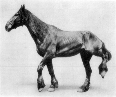
Symptomatology.—This disease develops slowly, and progressively increases in severity as a rule, but does not ordinarily constitute cause for rendering an animal unserviceable. While the affection is sometimes bilateral (occasionally affections of the forelegs are reported) and the extreme flexion of the legs in the spasmodic manner which characterizes spring-halt, cause great waste of energy during locomotion, yet such cases are rare. Usually the ailment is markedly evinced when subjects are first taken from the stable, but as they are exercised the manifestation diminishes, and in many instances it completely subsides. The condition is generally more noticeable when the subject is made to step backward. In some animals there is marked abduction at the time flexion occurs and in singular instances the spasmodic contraction is so violent that the subject falls to the ground as a result of the peculiar flexion of the leg.
In severe cases of "scratches" or chemical irritation of the extremity, the legs are abnormally flexed in a manner which simulates spring-halt, but because of the evident injury of the parts this is not likely to confuse. Since all facts concerning etiological agencies are surrounded with so much obscurity, classification does not lend any particular assistance in the consideration of this ailment.
Prognosis.—One cannot intelligently give a prognosis in these cases if forecast is expected to state the exact course following treatment. However, in a general way, cases of recent affection are thought more favorable than are those of long standing or in old animals where myositis and other muscular and fascial affections exist owing to years of hard service.
Treatment.—No known line of medicinal treatment is of service, nor is any particular surgical operation to be considered dependable for obtaining relief. Operations of almost every conceivable nature have been tried with the hope of securing recovery in spring-halt but under no condition can the practitioner as yet be reasonably certain of effecting permanent relief in any case. Treatment is, therefore, entirely empirical.
Neurectomies have been performed and recoveries following were attributed thereto; fascial divisions in the crural region have been done with good results and this manner of treatment has its favorers. Advocates of tenotomies, likewise, are to be found. Consequently, one may summarize thus: Spring-halt is a disease of unknown origin—the exact cause has not been determined; therefore, all treatment is, in a way, experimental. The recommendation of any given procedure in handling cases must then be a matter of opinion based either upon practical experience or knowledge of the experiences of others. Divisions of the lateral digital extensor (peroneus) below the tarsus near its point of insertion to the extensor of the digit is recommended here because it is followed by a percentage of recoveries that is as large as in any other method of treatment and the operation is not difficult to perform nor is its performance fraught with any dangerous complications. In selected subjects about fifty per cent of cases recover in from two to six weeks following this operation.
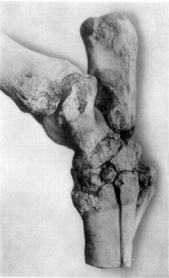
Fig. 53—Lateral (external) view of tarsus showing effects of generalized tarsitis.
Open Tarsal Joint.
Like the tibia the hock is exposed to frequent injuries and in some cases wounds perforate the joint capsule. When due to calk wounds where horses are kicked, the injury is often on the side of the tarsus (medial or lateral) and such wounds not infrequently result in infectious arthritis. Horses sometimes jump over wire fences and wounds are inflicted which constitute extensive laceration of the joint capsule. In firing for bone spavin, where a deep puncture is made very near the tibial tarsal (tibioastragular) joint if infection gains entrance, serious and generalized infection of the open joint cavity supervenes in some cases.
Symptomatology.—There is no marked difference in the constitutional disturbances which are occasioned in this condition and those encountered in other cases of septic arthritis (previously considered herein) except that there is a difference in the degree of resultant derangement and local tissue changes. Chiefly, because of the difficulty encountered in keeping the hock joint in an aseptic condition or securely bandaged, open tarsal joint constitutes a more serious condition than a similar affection of the fetlock. Otherwise, a very similar condition obtains and the same diagnostic principles serve here that have been described on page 110 in considering open fetlock joint.
Treatment.—The same plan that is described in detail for treatment of similar conditions affecting the fetlock joint is indicated in this affection. Exceeding care must be exercised in bandaging the hock, however, lest the animal be so irritated that in the extreme flexion of the tarsus which is often caused by bandaging, the wound dressings may be completely deranged. A wide gauze bandage material is most satisfactory; cotton of long fiber is separated in thin layers and wound about the hock, extending from the site of injury to a point about six inches proximal to the summit of the os calcis. By using an abundance of cotton in this way, it will not be found necessary to apply the bandages very snugly; with a four-inch gauze bandage material, which is supported above the cap of the hock and brought across the anterior face of the tarsus in a diagonal manner, a comfortable and very serviceable protective dressing is provided for. Animals so treated will not ordinarily resist because of pressure from the bandages. Pressure is unavoidable in the use of adhesive dressings or where careful attention is not given the manner of applying cotton to the parts. Such methods are sure to result disastrously. But if subjects are kept quiet after the parts have been properly bandaged, no difficulty is encountered in maintaining asepsis in an uninfected wound. Recovery takes place in favorable cases in from three weeks to three months, depending on the nature and extent of injuries inflicted.
Fracture of the Fibular Tarsal Bone (Calcaneum.)
Etiology and Occurrence.—This condition though rarely met with in the horse, is the result of violent strain upon the os calcis by the gastrocnemius and superficial flexor tendons in efforts put forth by animals in attempts to regain a footing when the hind feet slip forward under the body, or in jumping and in falls or direct contusion by heavy bodies. Hoare[50] reports a case of a mare that had produced fracture in jumping.
Fracture of the other tarsal bones are very seldom observed but may be occasioned by contusions wherein multiple or comminuted fractures are produced, such as are to be seen in small animals. Fracture of the tibial tarsal bone (astragalus) is to be observed as a complication in luxations of the tarsal joint and, according to Cadiot, the other tarsal bones may likewise suffer fracture in luxations of the hock.
Symptomatology.—Great pain attends this accident according to the observations given in recorded cases. In the case cited by Hoare the animal evinced great pain and uneasiness; the hock was unduly flexed; the calcaneum was displaced forward; and marked crepitation was present. A portion of the body of the calcaneum was protruding through the perforated skin. The animal was destroyed and the bone was found broken in three pieces.
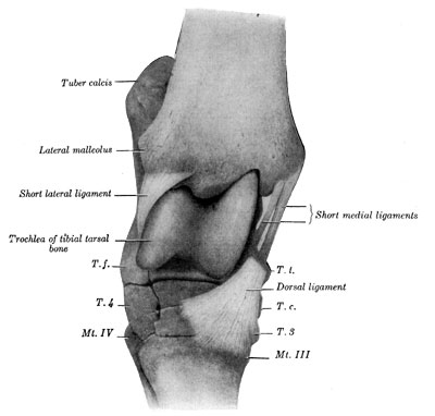
Fig. 54—Right hock joint. Viewed from the front and slightly laterally after removal of joint capsule and long collateral ligaments. T. t., Tibial tarsal bone (distal tuberosity). T. c., central tarsal bone. T. 3. Ridge of third tarsal bone. T. f. Fibular tarsal bone (distal end). T. 4. Fourth tarsal bone. Mt. III, Mt. IV. Metatarsal bones. Arrow points to vascular canal. (From Sisson's "Anatomy of the Domestic Animals.")
Since the support for the tendo Achillis is removed in such fracture and no leverage on the metatarsus obtains, it naturally follows that any attempt to sustain weight must result in extreme flexion of the hock and descent of this part in a manner similar to cases of rupture or division of the Achilles' tendon. The two conditions should not be confused, however, as the parts may be definitely outlined by palpation and the slack condition of the tendon and displaced summit of the calcaneum, which characterize fracture of the fibular tarsal bone, are easily recognized.
Treatment.—Prognosis is unfavorable in the majority of cases, but should attempts at treatment be undertaken in young and quiet mares which might prove valuable for breeding purposes in case of imperfect recovery, they should be put in slings and the member is to be immobilized as in tibial fracture. Authorities are agreed that prognosis is entirely unfavorable in mature animals, when the case is viewed from an economic standpoint.
Tarsal Sprains.
Etiology and Occurrence.—The hock joint is often subjected to great strain because of the structural nature of this part and its relation to the hip as well as the manner in which the tarsus functionates during locomotion. That ligamentous injuries owing to sprain frequently occur and attendant periarticular inflammations with subsequent hypertrophic changes follow, is a logical inference. Fibrillary fracture of the collateral ligaments may take place in falls or when animals make violent efforts to maintain their footing on slippery streets. In expressing opinions concerning the frequency with which the hock is found to be the seat of trouble in lameness of the pelvic members, different writers place the percentage of hock lameness at from seventy-five to ninety per cent. And when one considers the possibility that a goodly proportion of cases of tarsal exostis are the outcome of sprains, the occurrence of tarsal sprains may be more generally admitted.
Symptomatology.—A mixed type of lameness is present and the nature of the impediment varies, depending upon the location of the injury. Sprains of the mesial tarsal ligaments cause lameness somewhat similar to that of spavin. However, in establishing a diagnosis, local evidence in these cases is of greater significance than the manner of locomotion. During the acute stage of inflammation there is to be detected local hyperthermia, some hyperesthesia and a little swelling. Later, when resolution is not prompt, considerable swelling (or perhaps correctly speaking, an indurated enlargement) variable in size is developed. In some cases the entire tarsal region becomes greatly enlarged and this swelling is very slowly absorbed in part or completely. Such sub-acute cases are observed during the winter season and particularly where subjects are kept in tie stalls without exercise for weeks at a time.
Treatment.—Attention should be directed toward relief for the animal in all acute inflammations. Local applications of heat are helpful and, of course, rest is essential. Towels that are wrung out of hot water and held in position by means of a few turns of a loose bandage and this covered with an impervious rubber sheet, will serve as a practical means of application of hydrotherapy. Following this when conditions improve, as in the handling of all similar cases, counterirritation is indicated.
When proper care is given at the onset and where injury does not involve too much ligamentous tissue, recovery takes place in a few weeks but in some cases which occur during the winter season in farm horses, complete recovery does not result until several months have passed.
Curb.
The hock is said to be curbed when the normal appearance, viewed from the side, is that of bulging posteriorly at any point between the summit of the calcaneum and the upper third of the metatarsus. Among some horsemen a hock is said to be "curby" whenever there exists an enlargement of any kind on the posterior face of the tarsus whether it be due to sprain, exostosis or proliferation of tissue as a result of contusion.
French veterinarians consider under the title of "courbe," an exostosis situated on the mesial side of the distal end of the tibia. Cadiot and Almy state that this condition (courbe) is of rare occurrence. Percivall defines curb as "a prominence upon the back of the hind leg, a little below the hock, of a curvilinear shape, running in a direct line downwards and consisting of infusion into, or thickening of, the sheath of the flexor tendons." Möller's version of true curb is a thickening of the plantar ligament (calcaneocuboid or calcaneometatarsal). Hughes and Merillat consider curb as a synovitis having for its seat the synovial bursa which is situated between the superficial flexor tendon (perforatus) and the plantar ligament.
Occurrence.—Certain predisposing factors seem to favor the occurrence of curb. A malformation of the inferior part of the tarsus so that its antero-posterior diameter is considerably less than normal is a contributing cause. Such hocks are known as "tied-in." Another fault in conformation is the existence of a weak hock that is set low down on a crooked leg, especially when such a member is heavily muscled at the hip. Given such conformation in an excitable horse, and curb is usually produced before the subject is old enough for service. It is certain that in cases where conformation is bad, greater strain is put upon the plantar ligament. This structure serves to bind the tibial tarsal (calcis) bone to the metatarsus; traction exerted upon its summit by the tendo Achillis is great when animals run, jump or rear and also at heavy pulling. In animals having curby hocks, sprain is likely to result and curb supervenes.
Symptomatology.—The characteristic swelling which marks curb may develop quickly and lameness occur suddenly or the enlargement comes on gradually and slowly, causing little lameness. Lameness is not proportionate to the size of the swelling and in all cases whether subacute or chronic, the condition improves with rest, but lameness is again manifested upon exertion. A horse which "throws a curb" will go lame until the acute inflammatory condition subsides and depending upon treatment received and conformation of the hock, this requires from three days to two or three weeks.
The character of the swelling varies; in some cases it is not large but rather dense and lacking in evidence of heat and hyperesthesia; in other cases there is considerable swelling, which is hot and doughy, somewhat painful to the touch but not necessarily productive of much lameness. In any event, whether the swelling or enlargement is big or little, its location makes it conspicuous when viewed in profile.
In most cases after the acute inflammatory period has passed, lameness is slight, if at all present, and in time no interference with the subject's usefulness is occasioned because of the curb, but the animals often remain blemished—complete resorption of inflammatory products being unusual when much disturbance has existed.
Treatment.—The handling of curb during the acute inflammatory stage is along the same lines as in sprain—local applications of cold and heat. Subjects must be kept quiet until all inflammation has subsided, for there are no cases wherein a little brisk exercise is more likely to cause a recurrence of lameness before recovery is complete than in curb. Vesication is in order in a week or ten days after the affection has set in; in old stubborn cases that have resisted ordinary treatment for a few months, the use of the actual cautery (line firing) is to be recommended.
Fig. 55—Spavin.
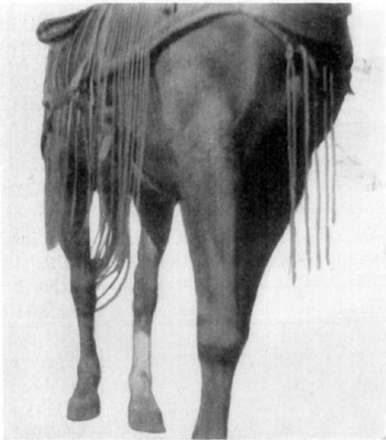
Spavin. (Bone Spavin.)
This term is applied to an affection of the tarsus which is usually characterized by the existence of an exostosis on the mesial and inferior portion of the hock. There is also included under this name, articular inflammation wherein no external evidence is shown. Spavin lameness has long been recognized and much has been written upon this subject. Since authorities are agreed that most cases of lameness in the hind leg are due to hock affection, and because the majority of cases of lameness which have the tarsal region as the seat of trouble are instances of spavin lameness, this disease merits all the attention it has received.
Etiology and Occurrence.—Causes may well be classified as predisposing and exciting, for there are many etiologic factors to be reckoned with in spavin, some of which are widely different in nature.
Considered as predisposing causes, hereditary influences play an important rôle and may, owing to faulty conformation, subject an animal to affections of this kind because of disproportionate development of parts (weak and small joints and heavy muscular hips); or as a consequence of inherited traits, a subject may manifest susceptibility to degenerative bone changes which are signalized by the formation of exostoses of different parts on one or more of the legs. Hereditary predispositions make for the presence of spavin in a large percentage of the progeny of sires so affected. This fact has been repeatedly demonstrated in this country as well as elsewhere according to Quitman, Dalrymple and Merillat.[51] A number of states have passed stallion inspection laws stipulating that animals having such exostoses as spavin and ringbone cannot be registered except as "unsound."
Asymmetrical conformation, particularly where the hock is obviously small and weak as compared with other parts of the leg, constitutes a noteworthy predisposing cause.
Peters' theory is plausible that the screw-like joint between the tibia and the tibial tarsal (astragulus) bones causes these structures to functionate in a manner not in harmony with the provisions allowed by the collateral ligaments of the tarsus, permitting movement only in a direction parallel with the long axis of the body.
Because of the quality of their temperaments, nervous animals possessing no particular congenital structural defects of the hock and having no history of spavined progenitors, are subject to spavin when kept at work likely to produce tarsal sprain. Spavin usually develops early in such subjects and examples of this kind may be frequently observed in agricultural sections of the country. Where spavin develops in unshod colts at three and four years of age, shoeing is not an influencing agency when animals are not worked on pavements.
Exciting causes of spavin are sprain and concussion. Various hypotheses are recorded as to how sprains are influenced and among others may be mentioned that of McDonough[52], which is that the foot is robbed of its normal manner of support by the ordinary three-calked shoe. With such a shoe, little support is given the sides of the foot; hence, undue strain is put upon the collateral ligaments of the tarsus. Moreover, the shoe with its calks increases the length of the leg and adds to the leverage on the hock, by virtue of such added length. This makes for greater strain upon the mesial or lateral tarsal ligaments whenever the foot bears upon a sloping ground surface, so that one side (inner or outer) is higher or lower than the other. But according to McDonough's theory (a good one concerning horses that work on pavements), the chief error in shoeing lies in that the foot is deprived of its normal base or support on the sides—the three-calked shoe being an unstable support—and that this manner of shoeing city horses working on pavements is an "inhumane" practice, a "diabolical method."
Whether spavin has its point of origin within the articulation as a rarefying ostitis of the cancellated structure of the lower tarsal bones as suggested by Eberlein; or, as Diekerhoff asserts, that the cunean bursa may be the initial point of affection, is unsettled; but it is reasonable to consider occult spavin as having its origin within the articulation, and that cases readily yielding to cunean tenotomy are primarily due to affection of the cunean bursa.
Symptomatology.—Where a visible exostosis exists, the presence of spavin is easily detected, yet exostoses that extend over large areas may constitute cause for serious trouble and still be difficult of detection. By observing the internal surface of the hock from various suitable angles, such as from between the forelegs or directly behind the subject, one may note the presence of any ordinary exostosis.
The position assumed by the spavined horse is often characteristic. More or less knuckling is usually present (Liautard, McDonald). There is abduction of the stifle in some cases, or the toe may be worn in unshod horses so that it presents a straight line at the surface. This is manifested to a great degree in some animals and in others the foot is not dragged and there is no wearing of the hoof at the toe.
Spavin lameness is so distinctive that one trained and experienced in the examination of horses that are spavined, should correctly diagnose the condition in practically every instance without recourse to other means than noting the peculiar character of the gait of the subject. Lameness develops gradually in the majority of instances, and an important feature in spavin lameness is that it disappears after the subject has gone a little way, to return again as soon as the animal has rested for a variable length of time—from a half hour to several hours. This "warming out" is marked during the incipient stage, but less pronounced in most chronic cases. A complete disappearance of lameness is observed in some instances, while in others only partial subsidence is evident. Because of the fact that pain is occasioned both during weight bearing and while the leg is being flexed and advanced, there is manifested the characteristic mixed lameness and exaggerated hip action which typifies spavin. By throwing the hips upward with the sound member it is possible to advance the affected leg with less flexion, hence less pain is experienced in this manner of locomotion. When made to step aside in the stall, a spavined horse will flex the affected member abruptly and when weight is taken on the diseased leg, symptoms are evinced of pain, and weight is immediately shifted to the sound limb. This is marked during the incipient stages of spavin. Lameness usually precedes the formation of exostosis, though cases are observed wherein an exostosis is present and no lameness is manifested and no history of the previous existence of lameness is available.
The "spavin test" is of value as a diagnostic measure when it is employed with other means of examination, though reaction to this test is seen in some cases in old "crampy" horses that have experienced hard service. The test consists in flexing the affected leg (elevating the foot from the ground twelve to twenty-four inches) and holding the member in this position for a minute, whereupon the animal is made to step away immediately at a trot. During the first few steps taken directly thereafter, the subject shows pronounced lameness and this constitutes a reaction to the spavin test.
Where no exostosis is present it becomes necessary to exclude other causes for lameness but the characteristic spavin lameness is to be relied upon to a greater extent in such cases than are other means of examination. Such cases are known as occult spavin and may be present for months before any external changes in structure are observable. In some instances no extoses form even during the course of years. The spavin test is of aid in establishing a diagnosis here but the marked "warming out" peculiar to spavin is not so pronounced in such cases.
Prognosis.—An animal having hereditary predisposition to spavin is not likely to recover completely whether this predisposition be due to faulty conformation or susceptibility to bone changes. In predicting the outcome, the temperament of the subject is to be taken into account, as well as the character of service the animal is expected to perform. And finally, a very important feature to be noted, is the location of the exostosis. If situated rather high and extending anterior to the hock, there is less likelihood of recovery resulting than where an exostosis is confined to the lower row of tarsal bones. When situated anterior to the tarsus a large exostosis may by mechanical interference to function, cause lameness when all other causes are absent. In making examinations one must not be deceived by the inconspicuous and seemingly insignificant exostosis which has a broad base. In some cases of this kind, dealers style the condition as "rough in the hock" when as a matter of fact, in some instances, incurable spavin lameness develops.