LAMENESS IN THE FORE LEG. (continued 3)
Reading edition
Treatment.—When the anatomy of the diseased parts is taken into consideration, and an analysis of the lesions which occur in cases where considerable structural change is occasioned by this affection, it is obvious that recovery is impossible. Only in cases where the inflammation is promptly checked before damage has been done the navicular bone or the flexor tendon, is permanent recovery possible. The disease is not frequently treated during this stage, however, and in the majority of instances the condition becomes chronic.
As soon as a diagnosis is made the shoes must be removed, the toe shortened with the hoof pincers and rasp and the subject is put in a well bedded box-stall. If the animal is very lame and the inflammation is acute, ice-cold packs should be applied to the feet. As soon as acute inflammation has subsided the foot may be so pared that all excess of sole and frog is removed without lowering the heels, and the animal may be blistered about the coronet region. The subject may be shod later, with heel calks that raise the heel moderately and a protracted period of rest should be enforced.
In cases where no acute inflammatory condition exists, neurectomy is beneficial. One must discriminate, however, between favorable and unfavorable subjects. This is not a last resort expedient to be employed in cases where extensive lesions of the navicular structures exists. With proper shoeing, and by putting the subject at suitable work, where concussion of fast travel on hard roads is not necessary, the best results are obtainable.
Laminitis.
This disease is primarily a non-infective inflammation of the sensitive laminae which very frequently affects the front feet. Often all four feet are affected, less frequently one foot (when its fellow is unable to sustain weight) and rarely the hind feet alone.
Occurrence.—Probably a greater number of cases of laminitis occur in localities where horses that are worked on heavy transfer wagons are, when in a state of perspiration, allowed to stand exposed to sudden lowering of temperature and to stand in a cool or cold shower of rain such as occurs near the coast of the Great Lakes or the ocean in some parts of this country.
This disease occurs in connection with digestive disorders of various kinds and, because of the frequent association of the two conditions, the common term "founder" has long been employed to designate laminitis. In cases of "over-loading," particularly when a large quantity of wheat has been eaten by animals that are unaccustomed to this diet, laminitis almost constantly results.
Large draughts of cold water, when drunk by animals that are overheated is often followed by laminitis. Concussion, such as attends hard driving, especially in unshod horses or on rough and hard roads, is often succeeded by this affection. Likewise, as has been stated, injury such as is occasioned by long continued standing on the same foot is followed by laminitis. Some horses that are frequently shod, suffer from this affection a few hours after shoes have been reset. Dr. Chas. R. Treadway of Kansas City reports the rather frequent occurrence of such conditions in horses that are in the fire department service in his city.
Age in no way influences the occurrence of laminitis and the general condition of an animal with regard to its vigor or state of flesh has no apparent influence toward predisposing horses to this ailment.
Etiology and Classification.—As it is with some other diseases, one may unprofitably theorize on cause and readily enumerate many conditions which are apparently contributory toward producing the affection. Causes may well be grouped, however, and a more definite understanding of laminitis is possible as a result. Such collocation would include conditions which directly or indirectly affect the digestion, such as puerperal laminitis, drinking of large quantities of cold water and exposure to cold and rain when the body is warm. All of these various conditions might be said to affect the vaso-constrictor nerves in such manner that the natural tendency (because of the peculiar structure of the sensitive laminae and their mode of attachment to the non-sensitive wall) which solipeds have for this affection is indirectly due to this one cause—vaso-constriction. According to Dr. D.M. Campbell, the effect of toxic materials, which may be absorbed from the digestive tract or the uterus in parturient females, upon the vaso-constrictor nerves, is such that a passive congestion of the sensitive laminae occurs and laminitis is the result. He believes that even the chilling of the surface of the body when very warm, by a cold rain, constitutes a condition wherein the effect upon the vaso-constrictors is the same.
This grouping does not include the effect of direct injuries of any and all kinds to which the feet are subjected such as: Concussion in fast road work, injuries occasioned by tight or ill fitting shoes, contusions of any kind resulting in non-infectious inflammation of the sensitive laminae, as well as the causes which produce laminitis where weight is borne by one foot when its fellow is out of function.
A classification which is practical is that of acute and chronic laminitis. To the practicing veterinarian it is this manner of consideration that is essential in the handling of these cases.
Symptomatology.—In the acute attack the condition is so well described by Dr. R.C. Moore[30] that we quote him in part as follows:
The acute form is generally ushered in very suddenly. Often a horse that is perfectly free from symptoms of the disease is found a few hours later so stiff and sore that he will scarcely move. They stand like they were riveted to the ground. If forced to move the evidence of pain subsides to some extent after they have gone a short distance, to return more severe than ever after they have been allowed to stand for a short time. If the disease is confined to the two front feet, the hind feet are placed well under the center of the body to support the weight and the front ones are advanced in front of a perpendicular line so as to lessen the weight they must bear. If they are made to move, the same position of the feet is maintained. If made to turn in a small circle, they do so by using the hind feet as a pivot, bringing the front parts around by placing as little weight on them as possible.
Placing the hind feet so far under the body, arches the back and often leads to errors in diagnosis, the condition sometimes being taken for diseases of the loins or kidneys.
If all four feet are involved, the animal stands in the usual position assumed in health, but if urged to move, the least effort to do so usually brings on chronic spasms of the entire body. In very severe cases, a slight touch of the hand will develop the spasms. At times they are so severe, and have such short intermissions, that the disease has been mistaken for tetanus. However, the clonic nature of the spasm should prevent such an error. If they are lying down, it is difficult to get them to arise, and if they do so, they show marked symptoms of pain for some time after rising.
If the disease is confined to the hind feet, they are placed well forward to relieve the strain on the toe caused by the downward pull of the perforans (deep flexor) tendon, but in place of the front feet being kept in front of a perpendicular line, as they are when the disease is confined to the front ones, they are placed far back under the body, so they will carry the maximum share of the body weight of which they are capable. The position of the feet is of great importance and offers symptoms that should not be overlooked.
When the subject is caused to walk, symptoms of excruciating pain are manifested in all acute cases of laminitis. In some cases where all four feet are affected, no reasonable amount of persuasion will cause the suffering animal to move from its tracks.
There is acceleration of the rate of heart action; the pulse is full and in some cases, bounding. As the affection progresses the pulse becomes rather weak and irregular. The character of the pulse in the region of the extremity is a reliable indicator; but one has to learn to make necessary discrimination because of the condition of the parts, as in some cases of lymphangitis or where the skin is abnormally thick. The characteristic throbbing pulse is, however, easily recognized in most cases. Temperature is variable, though usually elevated from one to four degrees above normal. This symptom varies with the type and stage of the affection. In a subject that has been down, unable to rise for several days, where there is a suppurative and sloughing condition of the laminae, the temperature is high. Whereas, in some other and less destructive cases there may be little thermic disturbance after the first few hours have lapsed.
A constant symptom in bilateral affections of acute laminitis is the difficulty with which the subject supports weight with one foot. It is this which causes the victim to stand as if "rooted to the ground" when all four feet are involved. If one attempts to take up one foot, thus causing the subject to stand on the other, there is much resistance and in many cases the animal refuses to give the foot.
When we consider that the sensitive parts of the foot are encased by a horny, unyielding box and that, when the laminae are congested, a great pressure is brought to bear upon the sensitive structures, it is easy to understand why the condition is so painful.
Chronic laminitis is a sequel of acute inflammation of the sensitive laminae. It varies as to intensity and the exact manner of its manifestation depends upon preëxisting disturbances.
In some mild cases of laminitis there are recurrent attacks wherein no particular structural change exists, and diagnosis is established chiefly by noting the character of the pulse at the bifurcation of the large metacarpal (or metatarsal) artery just above the fetlock. The same manifestation of pain is present when weight is supported by one foot, though in a lesser degree. There is less local heat to be detected by palpation than in the acute cases.
Chronic laminitis as it occurs following acute attacks which have resulted in structural changes of the foot, present the same symptoms just described and, in addition, the peculiar alterations in structure exist. When, owing to acute inflammation of the sensitive laminae, there has resulted necrosis of this sensitive tissue together with infiltration between the anterior surface of the distal phalanx (os pedis) and the contacting hoof, the lower portion of the distal phalanx is turned downward and backward (rotated upon its transverse axis). Because of the traction which is exerted by the deep flexor tendon (perforans), as it attaches to the solar surface of the distal phalanx, this rotation is facilitated. With hyperplasia of lamina, at the anterior portion of the distal phalanx, there results a thick "white line." Rotation of the distal phalanx necessitates a descent of its apical portion and there occurs a "dropped sole."
In time, partly because of excessive wear of hoof at the heel, owing to an altered condition in the normal antagonistic relation between the flexor and extensor tendons, the toe makes an excessive growth, and the concavity of the anterior line is accentuated owing to this abnormal length of hoof. The hoof, because of recurrent inflammatory attacks, is corrugated—elevations of horn in parallel rings are usually present.
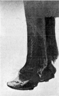
Fig. 33—The hoof in chronic laminitis. Note the concavity. This animal was serviceable for any work that could be performed at a walk.
Animals that are so affected in traveling strike the heel first and the toe is later contacted with the ground surface. Rotation of the distal phalanx upon its transverse axis produces a condition, with respect to this peculiar impediment, that is equivalent to added and excessive length of the deep flexor tendon.
Where there occurs suppuration, by careful inspection of the coronary region, one may early recognize detachment of hoof. In such cases animals remain recumbent and, while the condition is not so painful at this stage, the practitioner must not overlook the real state of affairs. History, if obtainable, will be a helpful guide in such cases. Separation of hoof occurs as a rule in from four to ten days after the initial attack of acute laminitis. Needless to say these cases are hopeless, when the economic phase of handling subjects is considered.
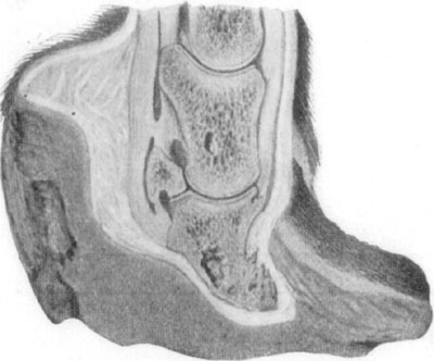
Fig. 34—Showing the effects of laminitis. By permission, from Merillat's "Veterinary Surgical Operations."
Treatment.—Much depends upon the concomitant disturbances (or causes if one is justified in referring to them as such) as to the manner in which laminitis is to be treated. In all cases where digestive disturbances exist, the prompt unloading of the contents of the alimentary canal is certainly indicated. D.M. Campbell[31] in a discussion of laminitis has the following to say regarding the treatment of such cases:
Because superpurgation may be followed by laminitis, the advisability of using the active hypodermic cathartics is questioned. Neither arecolin nor eserin can cause superpurgation. The action of the former does not continue longer than an hour after administration and of the latter not more than eight hours. The action of either is mild after the first few minutes.
I do not think that anyone has recommended either arecolin or eserin where there is severe purgation. Where the intestinal canal is fairly well emptied and its contents fluid, I should be inclined to rely upon intestinal antiseptics to hold in check harmful bacterial growth.
The use of alum in the treatment of laminitis is held to be without reason other than the empirical one that it is beneficial. If laminitis is due chiefly to an autointoxication, good and sufficient reason for the administration of alum can be shown based upon its known physiological action. It is the most powerful intestinal astringent that I know of and has the fewest disadvantages. I have not noted constipation following its use nor diarrhea, nor a stopping of peristalsis, nor indigestion, and in any case its action lasts at most only a few hours, and if it did all these, it could not much matter. Quitman says, that it constricts the capillaries. If this is true, a thing of which I am not certain, is it not reasonable to suppose that as with other vaso-constrictors, e.g., digitalis, there is a selective action on the part of the capillaries (not of the drug) and those that need it most, i.e., those of the affected feet in laminitis, are constricted most? All body cells exert this selective action in the assimilation of food, the tissue needing most any particular kind of food circulating in the blood, gets it.
Our first consideration in laminitis should be to remove the cause—to stop the absorption of the toxin in the intestinal tract that is producing the condition. This we accomplish by partially unloading it by the use of the active hypodermic cathartics and stopping absorption by the surest and most harmless of intestinal astringents. Whether the astonishingly prompt and certain action of alum in this case is due wholly to its astringent action or whether alum combines with the harmful bacterial products chemically and forms an innocuous combination, I can only surmise, and it is unimportant. At any rate, when alum is administered, the onslaught of the disease is promptly stopped. Irreparable damage may already have been done if the case is a neglected one, but whether administered early or late in acute attacks, the progress of the disease is stopped immediately.
The same authority may be profitably quoted in the matter of handling all cases wherein the revulsive effect of agents which diminish vascular tension are chiefly indicated or necessary as adjuvants. In this connection, Campbell says:
The early and vigorous administration of aconitin in laminitis to its full physiological effect, is more logical. Assuming that laminitis is due to absorption of harmful products from the intestinal tract permitted through the deranged functioning of the organs of digestion, or assuming that it is due to an extension of the inflammation from the mucosa to the sensitive lamina, or that it is a reflex from a sudden chilling of the skin, we have in any of these conditions a disturbed circulation, and aconitin is the first and foremost of circulation "equalizers." Furthermore, in laminitis there is an elevation of the temperature, an almost invariable indication for aconitin. A speedy return of the temperature to normal, a very marked diminution of the pain and improved conditions generally, appear coincident with the symptoms of full physiological effect of aconitin when given in cases of laminitis, which constitutes assuredly an important part of its treatment.
Fig. 35—Inferior (convex) surface of Cochran shoe.
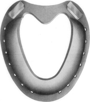
Where lameness is not great as in cases wherein no marked structural change of the foot has occurred, proper shoeing is very beneficial. By keeping the heels as low as possible and shoeing without heel calks a more comfortable position is made possible. Thin rubber pads which do not elevate the heel are of service in diminishing concussion.
Dr. David W. Cochran of New York City has attained unusual success in cases of chronic laminitis with dropped sole by the use of a specially designed shoe.
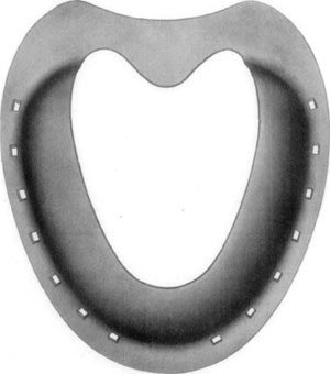
Fig. 36—Superior surface, showing concavity or bowl, as formed by the toe and branches of the shoe, as designed by Dr. David W. Cochran.
Cochran claims that, not only are horses with dropped soles that would otherwise have to be put off the streets enabled to do a fair amount of work by means of this shoe, but that continually wearing it, meanwhile keeping the convexity of the front of the hoof rasped thin, in time brings about a marked improvement, and that after some months or years of use the animals are able to work with ordinary rubber-pad shoes, provided they are arranged to facilitate breaking over.
From having been successfully used on some race horses of high value, the Cochran shoe has attained considerable notoriety and is being used by a number of practitioners. A disadvantage, however, arises from the fact that few horseshoers other than Doctor Cochran seem able to make the shoe, the peculiar shape of which offers considerable difficulty in forging. Concerning the application of the shoe Cochran[32] says:
"The most important primary procedure is the preparation of the foot to receive the shoe. All excess of growth must be removed from the anterior face of the hoof. The outer face must be reduced at the toe (not shortened), but rasped down thin for the lighter the top of the foot is, the more chance the sole and coffin bone will have of resuming their former normal position. The pressure of the wall at the toe upon the exudate between wall and coffin bone, tends to force the coffin bone and sole out of their normal position. Leave the sole alone. You can lower the excess of growth at the heels.
"There are many designs of shoes to relieve this condition. A great deal depends on the judgment of the shoer to meet the conditions presented, depending on the degree of the convexity and strength of the sole. In some cases we use a shoe that admits of a large amount of sole room. Again, we shoe with a shoe of wide cover. In other cases a shoe with even pressure over the whole sole. In some cases a high, narrow shoe, resting only on the wall, or the ordinary plain shoe with side calks welded close to the outside edge and the shoe dished well from these as a foundation. Then we have the air cushion pad designed after the model of the bowl shoe."
In cases when slight and persistent lameness interferes sufficiently to prevent using an animal at any sort of work on hard roads, median neurectomy will relieve all lameness in most instances. This is a safe operation, moreover, in that no bad after effects are to be feared, even though lameness were to continue.
Calk Wounds. (Paronychia.)
Etiology and Occurrence.—Injuries of various kinds are inflicted upon the coronary region but usually they are due to the foot being trampled upon. When the foot that inflicts the injury happens to be unshod, a contusion of the injured member is occasioned, but in the majority of instances, wounds that demand attention are the result of shoe calks which have penetrated the tissues in the region of the coronary band. Often calk wounds are self-inflicted. When animals are excited and in turning crowd one another, they often perform dancing movements which frequently result in deep calk wounds of the coronet. Some horses have a habit of resting the heel of one hind foot upon the anterior coronary region of the other. While sleeping in this position, if they are suddenly awakened, the weight is abruptly shifted to the uppermost foot and the one underneath is (because of the pain attending its being wounded) quickly drawn out from under its fellow. In this way deep cuts may divide the coronary band and inflict extensive injury to the sensitive lamina as well.
An infectious type of coronary inflammation occurs in some localities during the winter months, wherein the condition is enzootic.
Symptomatology.—Depending upon the manner in which the injury has been produced, the appearance of the wound varies and likewise lameness is more or less pronounced. If the tissues are not divided and the wound is chiefly of the subsurface structures, there will not immediately occur pronounced local evidence of the existence of injury; but as soon as the lame animal is made to move, the peculiar character of the impediment (supporting-leg lameness with the affected foot kept well in advance of its normal position) directs attention to the extremity and all of the symptoms of acute inflammation are discovered.
Where a wound is inflicted which divides, in some manner, the surface structures (skin, coronary band, or the hoof wall) one's attention is at once called to the existence of the wound.
Because of the fact that there is every facility for the production of a sub-coronary and podophylous infection, these wounds should receive prompt attention. In some instances, the pastern joint is opened by calk wounds and then, of course, an infectious arthritis succeeds the injury.
Treatment.—In all contused wounds of the coronary region the parts need thorough cleansing; the hair, if long is clipped and a cataplasm is applied. Or preferably, an iodin-glycerin combination of one part of iodin to four parts of glycerin is poured on a layer of cotton, and this is confined in contact with the inflamed parts by means of a bandage.
Where normal resistance to infection obtains, the subject usually suffers no suppurative disturbance when the surface structures are not broken; and daily applications of the antiseptic lotion above referred to stimulates complete resolution. This may be expected in from four to ten days depending upon the extent of the injury.
If a calk wound has been inflicted, the adjoining surface structures are freed of hair and the parts cleansed in the usual manner, (which in wounds recently inflicted, should be done without employing quantities of water) and after painting the wound surface with tincture of iodin and saturating its depths with the same agent, the wound is cleansed, if it contains filth, by means of a small curette. By using a small and sharp curette, one is enabled to cleanse the average wound quickly and almost painlessly.
In such cases, equal parts of tincture of iodin and glycerin are employed. The wound is filled with this preparation and a quantity of it is poured upon a suitable piece of aseptic gauze or cotton and this is contacted with the wound. The extremity is carefully bandaged and this dressing is left in position for forty-eight hours unless there occurs, in the meanwhile, evidence of profuse suppuration—which is unusual.
One is to be guided as to the progress made by the degree of lameness present. If little or no lameness develops, it is reasonable to expect that infection has been checked; that the wound is dry and redressing every second day is sufficiently frequent.
Where cases progress favorably, recovery (unless infectious arthritis results) should occur in from ten days to three weeks. Where extensive sub-coronary fistulae result, either from lack of prompt or proper attention, the condition is then one requiring a radical operation to establish drainage and to disinfect if possible, the suppurating tissues.
Corns.
Etiology and Occurrence.—In horses, because of a tendency toward contraction of the heel in some subjects, together with work on hard roads and pavements, where the feet become dry and brittle, and because of neglect of the matter of shoeing, this affection is of frequent occurrence. Unshod horses are rarely affected. If conformation is faulty and too much weight is borne on the inner or the outer quarter, and the hoof wall at the quarter tends to turn inward, corns are usually present. They occur more frequently on the inner quarters of the front feet, though the outer quarters are occasionally also affected and in rare instances corns are found at the toes. They do not often affect the hind feet.
As soon as injury by pressure, such as is supposed to cause the formation of corns, is brought to bear on the sensitive sole, an extravasation of blood occurs. In time when the cause remains active, this discoloration is evident in the substance of the insensitive sole and consists in a red or yellowish spot which varies in size—this is ordinarily termed dry corn.
In some cases where infection of this extravasation of blood and serum occurs, instead of desiccation and discoloration of the insensitive parts, there is, in time, manifested a circumscribed area of destruction of the insensitive sole and the abscess may, where no provision for drainage exists, burrow between sensitive and insensitive laminae and perforate the tissues at the coronet. If the suppurative material discharges readily by way of the sole, no disturbance of the heel or quarters occurs above the hoof.
Symptomatology.—A supporting-leg-lameness characterizes this condition; and this lameness in most instances varies in degree with the amount of distress which is occasioned by pressure upon the inflamed parts. By an examination of the sole after having removed all dirt, and exposed the horny sole to view, no difficulty is encountered in locating the cause of the trouble.
Treatment.—Before suppuration has taken place and in the cases where suppuration does not occur, the horse-shoer's method of paring out the diseased tissue affords a means of temporary relief; but unless frequently done, in many cases, lameness results within about three weeks after such treatment has been given. In other instances temporary relief is not to be gotten in this manner for any great length of time or until a more rational mode of treatment becomes necessary so that the subject may experience a cessation of the inconvenience or distress.
The general plan which meets with the approval of most practitioners consists in careful leveling of the foot and removing enough of the wall and sole at the quarters to make possible frog pressure by means of a bar shoe. With frog pressure, expansion of the heel follows in time, and permanent relief is obtainable in this manner. Thinning the wall of the quarter is advocated by many practitioners and is undoubtedly beneficial in chronic cases where marked contraction has taken place. The wall must be thinned with a rasp until it is readily flexible by compressing with the thumbs.
There are instances, however, where corns and contraction of the heel have existed so long that they do not yield to treatment. Such cases are found in old light-harness or saddle-horses that have been more or less lame for years and where there exists marked contraction of the heels, rough hoof walls, and hard and atrophied frogs.
Suppurating corns require surgical attention in the way of removal of the purulent necrotic mass and making provision for drainage. Dry dressings, such as equal parts of zinc sulphate and boric acid, may be employed to pack the cavity. After the infectious condition has been controlled, and the wound is dry, the same plan of treatment is indicated that is employed in the non-suppurating corn. Ample time is allowed, however, for the surgically invaded tissues to granulate and, if the subject is to be put in service, a leather pad, under which there has been packed oakum and tar, affords good protection.
Quittor.
This name is employed to designate an infectious inflammation of the lateral cartilage and adjoining structures. The disease is characterized by a slowly progressive necrosis and by a destruction of more or less of the cartilage and by the presence of fistulous tracts.
Etiology and Occurrence.—The disease is due to the introduction of pus producing organisms into the subcoronary region of the foot under conditions which favor the retention of such contagium and extension of infection into contiguous tissues.
Morbific material is introduced into the region of the lateral cartilage by means of calk wounds and other penetrant injuries of the foot. A sub-coronary abscess which, because of lack of proper care or because of virulency of the contagium or low vitality of the subject, is quite apt to result in cartilaginous affection and its perforation by necrosis follows.
Symptomatology.—Quittor is readily diagnosed on sight in many instances. Where there is dependable history or other evidence of the chronicity of an infectious inflammation of the kind, quittor is easily identified. If no positive evidence of the disease exists, by means of careful exploration of sinuses with the probe, one may distinguish between true cartilaginous quittor and superficial abscess formation that is often accompanied by hyperplasia.
Lameness depends upon the extent of the involvement as it affects the structures contiguous to the cartilage. A variable degree of lameness is manifested in different cases.
Treatment.—Two general plans of handling this disease are in vogue. One, the more popular method, consists in the injection of caustic solutions of various kinds into the fistulous openings with the object of causing sloughing of necrotic tissue and the stimulation of healthy granulation of such wounds. The other mode consists in either complete surgical removal of the cartilage or its remaining portions, or removal of the diseased parts of curettage.
When quittor has not extensively damaged the foot and the lateral cartilage is not partly ossified as it is in some old chronic cases, the complete removal of the lateral cartilage by means of the Bayer operation or a modification thereof is indicated. A complete description of the Bayer operation as well as Merillat's operation for this disease (the latter consisting in part, in the removal of diseased cartilage with the curette) are given in Volume three of Merillat's "Veterinary Surgical Operations."
Treatment by injection of caustic solutions has many advocates and because of the fact that, in many instances the condition is such that they are not desirable surgical cases and also because some animals may be put in service before treatment is completed, the injection method is popular.
The mode of treatment advocated by Joseph Hughes, M.R.C.V.S., constitutes a very successful manner of handling quittor and we can do no better than quote Dr. J.T. Seeley [33] on his manner of using this particular treatment.
Fig. 37—Hyperplasia of right fore foot, due to chronic quittor.
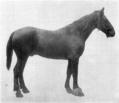
Preparation.—First remove the shoe, have the foot pared very thin and balanced as nicely as possible. Moreover, all loose fragments of horn must be detached and all crevices cleaned thoroughly.
Next, have the leg brushed and hair clipped from the knee or hock to the foot and scrubbed with ethereal soap and warm water, after which the foot must be scrubbed in like manner. The foot is then placed in a bichlorid bath several hours daily, for from two to five days, depending upon whether or not soreness is shown. The bichlorid solution is 1 to 1,000 strength.
On removing the horse from the bath a liberal layer of gauze is soaked in 1 to 1,000 bichlorid solution and placed so as to cover the entire foot. On discontinuing the bath, cover the foot with gauze saturated with a 1 to 1,000 bichlorid solution. This is to be covered with absorbent cotton and a gauze bandage, and over all is placed an oil cloth or silk covering. This pack is kept moist with bichloride solution for forty-eight hours. The foot is then ready for injection.
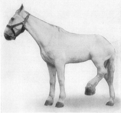
Fig. 38—Chronic quittor, left hind foot. Showing position assumed because of painfulness of the affection.
Preparation of the Injection Fluids.—Have on hand a pint of a one per cent aqueous solution of formaldehyd made under cleanly conditions, even to a clean bottle and cork, and a clean container when ready to use the liquid. Prepare also a bichlorid of mercury solution as follows: Hydrarg. Chlor. Corros. 3IV; Acid Hydrochlor. 3Iss.; Aqua Bulliens, Oij. This should be thoroughly triturated, and then filtered into a clean bottle, when it is ready for use.
Injection.—The patient should be laid on a table, if one is available, or cast, and the foot securely fixed. Then, with an ordinary one-ounce hard rubber syringe, with a good plunger (tried first to note whether or not any fluid works around between the barrel and the plunger), introduce one syringe full of the formaldehyd solution, then thoroughly probe the quittor to determine the number of sinuses. This done, inject each sinus. If two sinuses open on the surface, close one with cotton while filling the other so that if there is a connection the solution will come in contact with all tissues involved. Irrigate with the full pint of formaldehyd solution first, then follow with six or eight ounces of the bichlorid solution. Never probe the foot nor allow it to be tampered with except in the manner prescribed.
After-Treatment.—Put on a pack saturated with a solution of bichlorid of mercury 1 to 1,000 and let it remain two days. Remove pack, and once daily afterwards wipe off with cotton the secretion which accumulates on the outside, and apply a dry dressing or healing oil composed of phenol, camphor gum and olive oil.
When Dangerous to Inject.—Never inject a quittor in the acute stage. Never inject a quittor if considerable lameness is present. On injecting a solution of formalin, hold cotton tightly around the nozzle of the syringe, when the plunger is down, then withdraw the syringe gently and note particularly if the fluid returns through the opening; if none returns cease operations at once, as it is dangerous to proceed farther, it indicates that the sinus is not well defined and the fluid retained will cause much trouble and often the death of the patient.
Experience has taught that, if extensive destructive changes of the foot exist, the Bayer operation is not indicated. In the country, where quittors are not so frequently met as in urban practice, the Merillat operation is preferable in all cases. However, the cost of the protracted period of idleness, which convalescent surgical patients require, renders the Hughes method more satisfactory in the hands of the general practitioner, especially in the city.
Nail Punctures.
Nail punctures, as herein considered, embrace all penetrant wounds of the solar surface of the horse's foot due to trampling upon street nails. This does not include accidental nail pricks occasioned in shoeing. In city practice, in some stables, these cases are of frequent occurrence; and, generally speaking, nail punctures are observed more frequently in urban horses than in animals that are kept in the country.
Occurrence and Method of Examination.—This condition, then, is a rather common cause of lameness and in no case, where cause of the claudication is not obvious, is the practitioner warranted in concluding his examination without careful search for the possible existence of nail puncture of the solar surface of the foot.
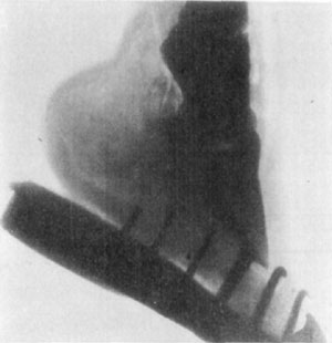
Fig. 39—Skiagraph of foot. The X-ray offers very limited possibilities in the diagnosis of lameness. The location of a "gravel" or a nail that had worked its way some distance from the surface, or of an abscess of some proportion, deep in the tissues, might be facilitated under some circumstances by the aid of the X-ray. Its use in the detention of fractures is very limited, owing to the difficulty encountered in getting a view from the right position—many trials being necessary in most cases. The case shown above was diagnosed clinically as incipient ringbone. The X-ray revealed no lesions. (Photo by L. Griessmann.)
In occasional instances there co-exists an obvious cause for supporting-leg-lameness and an occult cause—a nail puncture. Where such complications are met, the practitioner is not necessarily guilty of neglect or carelessness when the nail puncture is not discovered at once, nevertheless, an examination is not complete until practically every possible cause of lameness has been located or excluded in any given case.
In a search for nail puncture it is necessary to expose to view every portion of the sole and frog in such manner that the existence of the smallest possible wound will be revealed. This necessitates removal of the shoe, if, after a preliminary examination, a puncture is not found, when there is good reason to suspect its presence. However, where it is readily possible to locate and care for a wound without removal of the shoe, allowing the shoe to remain materially facilitates retaining dressings in position and relieves the solar surface of contact with the ground. If extensive injury or infection exists, it is of course necessary to remove the shoe and leave it off. By removing a superficial portion of all of the sole and frog, thus carefully and completely exposing to view all parts of the solar surface of the foot, and with the aid of hoof-testers one is enabled to positively determine the existence of nail punctures. Because of the tendency of puncture wounds of the foot to close, and since the superficial portion of the solar structures are usually soiled, it is absolutely necessary to conduct examinations of this kind in a thorough manner.
Symtomatology.—Not all cases of nail puncture cause lameness during the course of the disturbance and in many instances no lameness is manifested for some time after the injury has been inflicted—not until infection has been the means of causing considerable inflammation of sensitive structures. Nevertheless, this lack of manifestation occurs only in cases where serious injury has not taken place and the degree of lameness is a constant and reliable indicator of the character and extent of nail punctures within twenty-four hours after injury has been inflicted.
The position assumed by the affected animal inconstantly varies with the location and nature of the injury and is not of particular importance in establishing a diagnosis. The subject may support some weight with the affected member and stand "base-wide" or "base-narrow," or no weight may be borne with the foot or the animal may point or keep the extremity in a state of volar flexion. In cases where extensive injury has been inflicted, and great pain exists, the foot is kept off the ground much of the time and it may be swung back and forth as in all painful affections of the extremity.
Nail punctures cause typical supporting-leg-lameness and in some cases certain peculiarities of locomotory impediment are worthy of notice. Punctures of the region of the heel, which directly affect or involve the deep tendon sheath, cause a type of lameness wherein pain is augmented, when dorsal flexion of the extremity occurs as well as when weight is borne. Wounds in the region of the toe of the hind feet sometimes cause the subject to carry the extremity considerably in advance of the point where it is planted and, just before placing the foot on the ground, it is carried backward a little way—ten or twelve inches.
However, diagnosis of nail puncture is based on the finding of the characteristic wound or resultant local changes.
Course and Prognosis.—The nature of the progress and the manner of termination of these cases are variable. If the coffin joint has been invaded, and a septic arthritis exists, the condition is at once grave. An open and infected tendon sheath, while not so serious, constitutes a condition which is distressing, and recovery is slow even under the most favorable conditions. Where a heavy, rigid and sharp nail enters the foot, in such manner that fracture of the third phalanx (os pedis) occurs, this complication makes for a protraction of the condition. Experience teaches that the natural course and termination in these cases are modified by the location and depth of the injury, virulency of the contagium and resistance of the subject to such infection.
Prevention.—In all horses which are kept at such work that exposure to nail punctures is frequent, a practical means of prevention of such injuries consists in the employment of heavy sole leather or suitable sheet metal to cover the sole of the foot and, at the same time, confine oakum and tar in contact with the solar surface to prevent the introduction of foreign material between the foot and such protecting appliances. Further, if drivers and owners could be impressed with the serious complications which so frequently attend wounds of this kind, undoubtedly many cases which are now lost, because of ignorance or neglect on the part of the teamsters or proprietors of horses, would be saved by prompt and rational treatment.
Treatment.—The treatment of this condition falls so largely within the dominion of surgery that we can give little more than an outline here.
In cases where there exists no evidence of open joint or open tendon sheath as judged by the site of the puncture and degree of lameness present (after having thoroughly cleansed the solar surface of the foot and enlarged the opening in the nonsensitive sole) a little phenol is introduced into the wound. In such cases, where it is possible for the antiseptic to contact every part of wound surface to the extreme depths of the puncture, infection is prevented when such treatment is promptly administered. This may be considered as first aid, or emergency care, and is indicated in all wounds of the foot whether the injury be serious or almost insignificant.
Subsequently one of two general courses may be pursued in the treatment of cases of nail puncture. One, by the employment of means to keep the wound patent and injection of suitable antiseptics, or agents that are more or less caustic in conjunction with strict observance of asepsis and wound protection. The other method consists in prompt establishment of drainage by surgical means and includes exploration and curettage.
The first method is better adapted to the use of the average general practitioner and he would do well to keep the opening in the nonsensitive structures patent. By introducing equal parts of tincture of iodin and glycerin daily, good results will follow in most instances. The wound is protected in unshod horses, either by completely bandaging the foot and retaining, in contact with the wound, cotton that is saturated with iodin and glycerin, or, if a minor injury exists, the moderately enlarged opening in the nonsensitive sole or frog, which has been moistened with the antiseptic, is packed with a very small quantity of cotton. A little practice in this mode of closing benign puncture wounds will enable the practitioner to successfully protect the sensitive parts in the treatment of such cases in unshod country horses.
When the condition progresses favorably the wound may be dressed every second day or twice weekly, and in the course of from two to six weeks recovery should be complete.
If the practitioner is somewhat proficient as a surgeon, and has at his command facilities for doing surgery, the second method is preferable in many cases. By using a local anesthetic on the plantar nerves and confining the subject on an operating table, restraint should be perfect. The solar surface of the foot is first thoroughly cleansed, the puncture wound is enlarged in the nonsensitive structures and the parts are then moistened with phenol or other suitable antiseptics. By means of a small probe the puncture is explored and, depending on the character of the wound and the structures involved, surgical intervention is varied to suit the case. If necessary, all of the insensitive frog is removed, and in wounds affecting the region of the heel the tissues may be incised from the puncture outward dividing all of the tissues outward and backward to the surface. A suitable surgical dressing is then applied.
If, on the other hand, the puncture extends into the navicular bursa, the radical operation is perhaps indicated, though not until one is sure that infection of the bursa and serious consequences are to follow if this operation is not performed. Detailed description of the technic of this operation belongs to the realm of surgery and a good discussion of it is to be found in William's work on veterinary surgical and obstetrical operations.
One may summarize the discussion of treatment of nail puncture by saying that emergency care as herein described is of first consideration. In every case an immunizing dose of anti-tetanic serum should be given. Subsequently, the method employed must suit the character of the wound, existing facilities for handling the subject and the skill and aptitude of the practitioner.