Volume 11 — The Central Nervous System of Nucula and Malletia
Reading edition
WILLIAM A. HILTON
These bivalve forms are grouped among the simplest of the molloscs. It is especially from the condition in Nucula as described by Pelseneer '91, that the conception of the most anterior ganglion being composed of four ganglia, has its chief support. Drew '01, who has also studied Nucula, believes that the lobes of the ganglion in Nucula are superficial and that the four connectives coming from the ganglion may be interpreted in another way. That is, that one pair of nerves may represent an otocystic branch partly fused with the connective. This view seemed reasonable to him as Stempel '99 in Solenyma found the otocystic nerves arose directly from the cerebral ganglion.
The two species of this group used for study were collected at Laguna Beach. Nucula castrensis Hinds, occurs abundantly at low tide under rocks. It is rather small for dissection, but very good complete series were obtained and stained in hematoxylin. Malletia faba Dall, was much less abundant. Specimens were obtained from holdfasts or from dredging. Although this was a larger species, gross dissection was not very easily carried out on any of the specimens, but good series were made.
The ganglia of Nucula are easily studied in section. The cerebral mass seems composed of one main mass, partly divided into four subdivisions, the two central most completely fused, and the lateral quite distinct in places. The central portion might represent the cerebral ganglia and the lateral, the pleural if we take that interpretation. The pedal ganglion is made of right and left parts quite completely fused except at the margins. The pedal mass is the smallest of the three chief ganglionic areas. The visceral ganglia are quite widely separated and a little larger than the pedal mass.
The ganglia of Malletia are in general plan similar to those of Nucula, the greatest differences being in the cerebral mass. The cerebro-pleural mass seems almost one. In most sections it is very compact and a little more complicated in structure than the ganglion of Nucula. However there are two small ventral ganglionic branches or small ganglia attached to the ventral side of the cerebral mass. These small ganglia may represent the visceral. Farther back in a cross section series as the cerebral mass disappears two other small branches take origin and run parallel to the nerves from the ganglionic cords. These two branches on each side seem to run together before the pedal ganglia are reached. Neither of these pairs of nerves seems connected with an otocyst.
At the cephalic end of the cerebro-pleural ganglion the large ganglionic cords are in evidence. A little distance from the cephalic end on the dorsal side there are quite large groups of cells down from the surface and surrounded by nerve fibers. The course of the fibers here is quite complex. On the ventral lateral sides of the ganglia are paired light areas of fibers which may be traced into the fibers of the ganglionic cords.
The pedal ganglion is small and much as in Nucula. The visceral ganglia are larger and widely separated.
In both Nucula and Malletia young specimens were used for study. In Nucula there was more the appearance of four ganglia in the cerebro-pleural mass, and the ganglia seem less complex than in Malletia. This last species has more separate pleural ganglia, if the ganglionic cords can be so regarded.
In neither of the species studied were all parts of the connectives easy to follow, so it was impossible to test the suggestions of Drew, but in both species there is some indication of two lateral lobes of the cerebral mass, and in Nucula there is good evidence of two central ganglia as well as the smaller lateral ones. The lateral ganglia of the cerebral mass are most clearly separated in Malletia. In Nucula the lateral ganglia are larger in proportion and the distribution of the gray and white matter is more irregular.
REFERENCES
Drew, G. A.1901 The life history of Nucula delphinodonta. Quart, jour. sc. vol. 44, pt. 3.
Pelseneer, P.1891 Contribution á l'étude des Lamellibranchs. Arch. d. biol. xi.
Stempell1899 Zur Anatomie von Solrmya togata. Zool. Jahrb. Bd. xiii.
(Contribution from the Zoological Laboratory of Pomona College)
EXPLANATION OF FIGURES
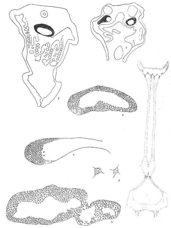
Fig. 1. Diagram of the ganglia of Nucula castrensis, reconstructed from serial sections. The probable position of the connectives is shown and the proportionate distances between ganglia are given. The upper ganglion is the cerebro-pleural with large nerves leading off from the ganglion which is itself lobed into four chief lobes. The pedal ganglion is next. In section the pedal ganglion at one place seems to be made up of four parts which may correspond to four connectives from the cerebro-pleural although only one pair of connectives was clearly determined. The visceral ganglion is connected with the pedal below. ×70.
Fig. 2. Cross section of cerebro-pleural ganglion. On the right side one of the lateral ganglia is shown. The one of the other side does not show because the section is not straight across. The dorsal side is up. ×300.
Fig. 3. Section of the pedal mass of Nucula, through the center. The dorsal side is up. ×300.
Fig. 4. Left side of the visceral mass of Nucula. Dorsal side up. ×300.
Fig. 5. Nerve cells from the central nervous system of Nucula. ×450.
Fig. 6. Section through the body of Nucula showing the position of the cerebro-pleural ganglion cut through the center. Dorsal side up. The cellular portion of the ganglion is black. ×70.
Fig. 7. Section through the body of Nucula at the level of the visceral nerves which are shown on either side of the section. The area of nerve cells is shown in black. ×70.
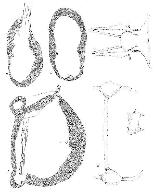
Fig. 8. Reconstruction from serial sections of the cerebro-pleural mass nerves and connectives of Malletia faba. The drawing is a ventral view, the cephalic side is at the top. ×70.
Fig. 9. Reconstruction of pedal ganglion of Malletia from the ventral side. Cephalic side at the top. ×70.
Fig. 10. Reconstruction of visceral ganglia of Malletia. ×70.
Fig. 11. Section through cerebro-pleural mass of Malletia. The dorsal side is up. On the ventral side to the left and right are the beginnings of the lateral lobes or ganglionic cords which may represent the pleural ganglia. In this species the cerebral ganglia are not separated into right and left halves as in Nucula. ×300.
Fig. 12. Section through the central part of the pedal mass of Malletia. The dorsal side is up. ×300.
Fig. 13. Section through one visceral ganglion of Malletia. The dorsal side is up. ×300.