Chapter III · PARASITIC ARTHROPODA AFFECTING MAN
Reading edition
The relation of insects to man as simple parasites has long been studied, and until very recent years the bulk of the literature of medical entomology referred to this phase of the subject. This is now completely overshadowed by the fact that so many of these parasitic forms are more than simple parasites, they are transmitters of other microscopic parasites which are pathogenic to man. Yet the importance of insects as parasites still remains and must be considered in a discussion of the relation of insects to the health of man. In taking up the subject we shall first consider some general features of the phenomenon of animal parasitism.
Parasitism is an adaptation which has originated very often among living organisms and in widely separated groups. It would seem simple to define what is meant by a "parasite" but, in reality, the term is not easily limited. It is often stated that a parasite is "An organism which lives at the expense of another," but this definition is applicable to a predatory species or, in its broadest sense, to all organisms. For our purpose we may say with Braun: "A parasite is an organism which, for the purpose of obtaining food, takes up its abode, temporarily or permanently, on or within another living organism".
Thus, parasitism is a phase of the broad biological phenomenon of symbiosis, or living together of organisms. It is distinguished from mutualism, or symbiosis in the narrow sense, by the fact that only one party to the arrangement obtains any advantage, while the other is to a greater or less extent injured.
Of parasites we may distinguish on the basis of their location on or in the host, ecto-parasites, which live outside of the body; and endo-parasites, which live within the body. On account of their method of breathing the parasitic arthropods belong almost exclusively to the first of these groups.
On the basis of relation to their host, we find temporary parasites, those which seek the host only occasionally, to obtain food; and the stationary or permanent parasites which, at least during certain stages, do not leave their host.
Facultative parasites are forms which are not normally parasitic, but which, when accidentally ingested, or otherwise brought into the body, are able to exist for a greater or less period of time in their unusual environment. These are generally called in the medical literature "pseudoparasites" but the term is an unfortunate one.
We shall now take up the different groups of arthropods, discussing the more important of the parasitic forms attacking man. The systematic relationship of these forms, and key for determining important species will be found in Chapter XII.
Acarina or Mites
The Acarina, or mites, form a fairly natural group of arachnids, characterized, in general, by a sac-like, unsegmented body which is generally fused with the cephalothorax. The mouth-parts have been united to form a beak or rostrum.
The representatives of this group undergo a marked metamorphosis. Commonly, the larvæ on hatching from the egg, possess but three pairs of legs, and hence are called hexapod larvæ. After a molt, they transform into nymphs which, like the adult, have four pairs of legs and are called octopod nymphs. These after a period of growth, molt one or more times and, acquiring external sexual organs, become adult.
Most of the mites are free-living, but there are many parasitic species and as these have originated in widely separated families, the Acarina form an especially favorable group for study of the origin of parasitism. Such a study has been made by Ewing (1911), who has reached the following conclusions:
"We have strong evidence indicating that the parasitic habit has originated independently at least eleven times in the phylogeny of the Ararina. Among the zoophagous parasites, the parasitic habit has been developed from three different types of free-living Acarina: (a) predaceous forms, (b) scavengers, (c) forms living upon the juices of plants."
Ewing also showed that among the living forms of Acarina we can trace out all the stages of advancing parasitism, semiparasitism, facultative parasitism, even to the fixed and permanent type, and finally to endoparasitism.
Of the many parasitic forms, there are several species which are serious parasites of man and we shall consider the more important of these. Infestation by mites is technically known as acariasis.
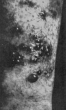
43. Effect of the harvest mites on the skin of man. Photograph by J. C. Bradley.
The Trombidiidæ, or Harvest Mites
In many parts of this country it is impossible for a visitor to go into the fields and, particularly, into berry patches and among tall weeds and grass in the summer or early fall without being affected by an intolerable itching, which is followed, later, by a breaking out of wheals, or papules, surrounded by a bright red or violaceous aureola, (fig. 43). It is often regarded as a urticaria or eczema, produced by change of climate, an error in diet, or some condition of general health.
Sooner or later, the victim finds that it is due to none of these, but to the attacks of an almost microscopic red mite, usually called "jigger" or "chigger" in this country. As the term "chigger" is applied to one of the true fleas, Dermatophilus penetrans, of the tropics, these forms are more correctly known as "harvest mites." Natives of an infested region may be so immune or accustomed to its attacks as to be unaware of its presence, though such immunity is by no means possessed by all who have been long exposed to the annoyance.
44. Harvest mites. (Larvæ of Trombidium). After C. V. Riley.
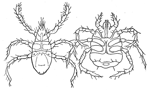
The harvest mites, or chiggers, attacking man are larval forms, possessing three pairs of legs (fig. 44). Their systematic position was at first unknown and they were classed under a special genus Leptus, a name which is very commonly still retained in the medical literature. It is now known that they are the larval forms of various species of the genus Trombidium, a group of predaceous forms, the adults of which feed primarily on insects and their eggs. In this country the species best known are those to be found late in summer, as larvæ at the base of the wings of houseflies or grasshoppers.
There is much uncertainty as to the species of the larvæ attacking man but it is clear that several are implicated. Bruyant has shown that in France the larvæ Trombidium inapinatum and Trombidium holosericeum are those most frequently found. The habit of attacking man is abnormal and the larvæ die after entering the skin. Normally they are parasitic on various insects.
Most recent writers agree that, on man, they do not bore into the skin, as is generally supposed, but enter a hair follicle or sebaceous gland and from the bottom of this, pierce the cutis with their elongate hypopharynx. According to Braun, there arises about the inserted hypopharynx a fibrous secretion—the so-called "beak" which is, in reality, a product of the host. Dr. J. C. Bradley, however, has made careful observations on their method of attack, and he assures us that the mite ordinarily remains for a long time feeding on the surface of the skin, where it produces the erythema above described. During this time it is not buried in the skin but is able to retreat rapidly into it through a hair follicle or sweat gland. The irritation from the mites ceases after a few days, but not infrequently the intolerable itching leads to so much scratching that secondary infection follows.
Relief from the irritation may be afforded by taking a warm salt bath as soon as possible after exposure or by killing the mites by application of benzine, sulphur ointment or carbolized vaseline. When they are few in number, they can be picked out with a sterile needle.
Much may be done in the way of warding off their attacks by wearing gaiters or close-woven stockings extending from ankle to the knee. Still more efficacious is the sprinkling of flowers of sulphur in the stockings and the underclothes from a little above the knee, down. The writers have known this to make it possible for persons who were especially susceptible to work with perfect comfort in badly infested regions. Powdered naphthalene is successfully used in the same way and as Chittenden (1906) points out, is a safeguard against various forms of man-infesting tropical insect pests.
The question of the destruction of the mites in the field is sometimes an important one, and under some conditions, is feasible. Chittenden states that much can be accomplished by keeping the grass, weeds, and useless herbage mowed closely, so as to expose the mites to the sun. He believes that in some cases good may be done by dusting the grass and other plants, after cutting, with flowers of sulphur or by spraying with dilute kerosene emulsion in which sulphur has been mixed. More recently (1914) he calls attention to the value of cattle, and more especially sheep, in destroying the pests by tramping on them and by keeping the grass and herbage closely cropped.
Ixodoidea or Ticks
Until recently, the ticks attracted comparatively little attention from entomologists. Since their importance as carriers of disease has been established, interest in the group has been enormously stimulated and now they rank second only to the mosquitoes in the amount of detailed study that has been devoted to them.
45a. Argus persicus. Capitulum of male. After Nuttall and Warburton.
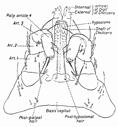
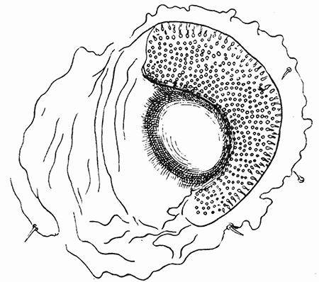
The ticks are the largest of the Acarina. They are characterized by the fact that the hypostome, or "tongue" (fig. 45) is large and file-like, roughened by sharp teeth. They possess a breathing pore on each side of the body, above the third or fourth coxæ (fig. 45b).
45b. Left spiracle of nymph of Argas persicus. After Nuttall and Warburton.
There are two distinct families of the Ixodoidea, differing greatly in structure, life-history and habits. These are the Argasidæ and the Ixodidæ. We shall follow Nuttall (1908) in characterizing these two families and in pointing out their biological differences, and shall discuss briefly the more important species which attack man. The consideration of the ticks as carriers of disease will be reserved for a later chapter.
Argasidæ
In the ticks belonging to the family Argasidæ, there is comparatively little sexual dimorphism, while this is very marked in the Ixodidæ. The capitulum, or so-called "head" is ventral, instead of terminal; the palpi are leg-like, with the segments subequal; the scutum, or dorsal shield, is absent; eyes, when present, are lateral, on supracoxal folds. The spiracles are very small; coxæ unarmed; tarsi without ventral spurs, and the pulvilli are absent or rudimentary.
In habits and life history the Argasidæ present striking characteristics. In the first place, they are long-lived, a factor which counts for much in the maintenance of the species. They are intermittent feeders, being comparable with the bed-bug in this respect. There are two or more nymphal stages, and they may molt after attaining maturity. The female lays comparatively few eggs in several small batches.
Nuttall (1911) concludes that "The Argasidæ represent the relatively primitive type of ticks because they are less constantly parasitic than are the Ixodidæ. Their nymphs and adults are rapid feeders and chiefly infest the habitat of their hosts. * * * Owing to the Argasidæ infesting the habitats of their hosts, their resistance to prolonged starvation and their rapid feeding habits, they do not need to bring forth a large progeny, because there is less loss of life in the various stages, as compared with the Ixodidæ, prior to their attaining maturity."
46. Argus persicus. Dorsal and ventral aspects. (×4). After Hassell.
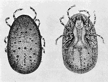
Of the Argasidæ, we have in the United States, several species which have been reported as attacking man.
47. Otiobius (Ornithodoros) megnini, head of nymph. After Stiles.
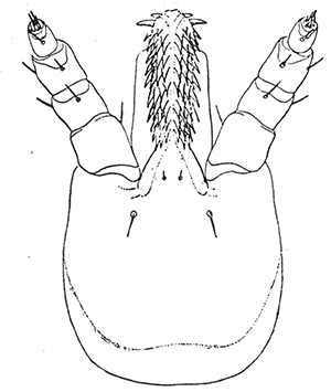
Argas persicus, the famous "Miana bug" (fig. 46), is a very widely distributed species, being reported from Europe, Asia, Africa, and Australia. It is everywhere preeminently a parasite of fowls. According to Nuttall it is specifically identical with Argas americanus Packard or Argas miniatus Koch, which is commonly found on fowls in the United States, in the South and Southwest. Its habits are comparable to those of the bed-bug. It feeds intermittently, primarily at night, and instead of remaining on its host, it then retreats to cracks and crevices. Hunter and Hooker (1908) record that they have found the larva to remain attached for five or eight days before dropping. Unlike the Ixodidæ, the adults oviposit frequently.
The most remarkable feature of the biology of this species is the great longevity, especially of the adult. Hunter and Hooker report keeping larvæ confined in summer in pill boxes immediately after hatching for about two months while under similar conditions those of the Ixodid, Boophilus annulatus lived for but two or three days. Many writers have recorded keeping adults for long periods without food. We have kept specimens in a tin box for over a year and a half and at the end of that time a number were still alive. Laboulliene kept unfed adults for over three years. In view of the effectiveness of sulphur in warding off the attacks of Trombidiidæ, it is astonishing to find that Lounsbury has kept adults of Argas persicus for three months in a box nearly filled with flowers of sulphur, with no apparent effect on them.
48. Otiobius (Ornithodoros) megnini, male. (a) dorsal, (b) ventral aspect. After Nuttall and Warburton.
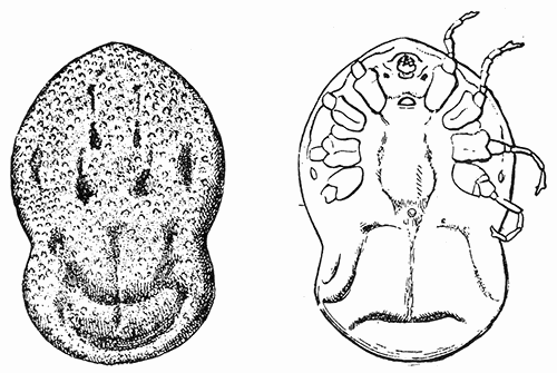
We have already called attention to the occasional serious effects of the bites of this species. While such reports have been frequently discredited there can be no doubt that they have foundation in fact. The readiness with which this tick attacks man, and the extent to which old huts may be infested makes it especially troublesome.
Otiobius (Ornithodoros) megnini, the "spinose ear-tick" (figs. 47, 48), first described from Mexico, as occurring in the ears of horses, is a common species in our Southwestern States and is recorded by Banks as occurring as far north as Iowa.
The species is remarkable for the great difference between the spiny nymph stage and the adult. The life history has been worked out by Hooker (1908). Seed ticks, having gained entrance to the ear, attach deeply down in the folds, engorge, and in about five days, molt; as nymphs with their spinose body they appear entirely unlike the larvæ. As nymphs they continue feeding sometimes for months. Finally the nymph leaves the host, molts to form the unspined adult, and without further feeding is fertilized and commences oviposition.
The common name is due to the fact that in the young stage the ticks occur in the ear of their hosts, usually horses or cattle. Not uncommonly it has been reported as occurring in the ear of man and causing very severe pain. Stiles recommends that it be removed by pouring some bland oil into the ear.
Banks (1908) reports three species of Ornithodoros—O. turicata, coriaceus and talaje—as occurring in the United States. All of these attack man and are capable of inflicting very painful bites.
Ixodidæ
The ticks belonging to the family Ixodidæ (figs. 49 and 50) exhibit a marked sexual dimorphism. The capitulum is anterior, terminal, instead of ventral as in the Argasidæ; the palpi are relatively rigid (except in the subfamily Ixodinæ), with rudimentary fourth segment; scutum present; eyes, when present, dorsal, on side of scutum. The spiracles are generally large, situated well behind the fourth coxæ; coxæ generally with spurs; pulvilli always present.
In habits and life history the typical Ixodidæ differ greatly from the Argasidæ. They are relatively short-lived, though some recent work indicates that their longevity has been considerably under-estimated. Typically, they are permanent feeders, remaining on the host, or hosts, during the greater part of their life. They molt twice only, on leaving the larval and the nymphal stages. The adult female deposits a single, large batch of eggs. Contrasting the habits of the Ixodidæ to those of the Argasidæ, Nuttall (1911) emphasizes that the Ixodidæ are more highly specialized parasites. "The majority are parasitic on hosts having no fixed habitat and consequently all stages, as a rule, occur upon the host."
49. Ixodes ricinus; male, ventral aspect. After Braun and Luehe.
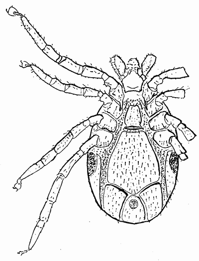
As mere parasites of man, apart from their power to transmit disease, the Ixodidæ are much less important than the Argasidæ. Many are reported as occasionally attacking man and of these the following native species may be mentioned.
50. Ixodes ricinus, var. scapularis, female. Capitulum and scutum; ventral aspect of capitulum; coxæ; tarsus 4; spiracle; genital and anal grooves. After Nuttall and Warburton.
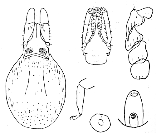
Ixodes ricinus, the European castor bean tick (figs. 49, 50), is a species which has been often reported from this country but Banks (1908) has shown that, though it does occur, practically all of the records apply to Ixodes scapularis or Ixodes cookei. In Europe, Ixodes ricinus is very abundant and very commonly attacks man. At the point of penetration of the hypostome there is more or less inflammation but serious injury does not occur unless there have been introduced pathogenic bacteria or, unless the tick has been abruptly removed, leaving the capitulum in the wound. Under the latter circumstances, there may be an abscess formed about the foreign body and occasionally, serious results have followed. Under certain conditions the tick, in various stages, may penetrate under the skin and produce a tumor, within which it may survive for a considerable period of time.
Ixodes cookei is given by Banks as "common on mammals in the Eastern States as far west as the Rockies." It is said to affect man severely.
Amblyomma americanum, (fig. 158c), the "lone star tick," is widely distributed in the United States. Its common name is derived from the single silvery spot on the scutum of the female. Hunter and Hooker regard this species as, next to Boophilus annulatus, the most important tick in the United States. Though more common on cattle, it appears to attack mammals generally, and "in portions of Louisiana and Texas it becomes a pest of considerable importance to moss gatherers and other persons who spend much time in the forests."
Amblyomma cajennense, noted as a pest of man in central and tropical America, is reported from various places in the south and southwestern United States.
Dermacentor variabilis is a common dog tick of the eastern United States. It frequently attacks man, but the direct effects of its bite are negligible.
The "Rocky Mountain spotted fever tick" (Dermacentor andersoni according to Stiles, D. venustus according to Banks) is, from the viewpoint of its effects on man, the most important of the ticks of the United States. This is because, as has been clearly established, it transmits the so-called "spotted fever" of man in our northwestern states. This phase of the subject will be discussed later and it need merely be mentioned here, that this species has been reported as causing painful injuries by its bites. Dr. Stiles states that he has seen cases of rather severe lymphangitis and various sores and swellings developing from this cause. In one case, of an individual bitten near the elbow, the arm became very much swollen and the patient was confined in bed for several days. The so-called tick paralysis produced by this species is discussed in a preceding chapter.
There are many other records of various species of ticks attacking man, but the above-mentioned will serve as typical and it is not necessary to enter into greater detail.
Treatment of Tick Bites—When a tick attaches to man the first thing to be done is to remove it without leaving the hypostome in the wound to fester and bring about secondary effects. This is best accomplished by applying to the tick's body some substance which will cause it to more readily loosen its hold. Gasoline or petroleum, oil or vaseline will serve. For removing the spinose ear-tick, Stiles recommends pouring some bland oil into the ear. Others have used effectively a pledget of cotton soaked in chloroform.
In general, the treatment recommended by Wellman for the bites of Ornithodoros moubata will prove helpful. It consists of prolonged bathing in very hot water, followed by the application of a strong solution of bicarbonate of soda, which is allowed to dry upon the skin. He states that this treatment is comforting. For severe itching he advises smearing the bites with vaseline, which is slightly impregnated with camphor or menthol. Medical aid should be sought when complications arise.
The Dermanyssidæ are Gamasid mites which differ from others of the group in that they are parasitic on vertebrates. None of the species normally attack man, but certain of them, especially the poultry mite, may be accidental annoyances.
51. Dermanyssus gallinæ, female. After Delafond.
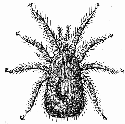
Dermanyssus gallinæ (fig. 51), the red mite of poultry, is an exceedingly common and widespread parasite of fowls. During the day it lives in cracks and crevices of poultry houses, under supports of roosts, and in litter of the food and nests, coming out at night to feed. They often attack people working in poultry houses or handling and plucking infested fowls. They may cause an intense pruritis, but they do not produce a true dermatosis, for they do not find conditions favorable for multiplication on the skin of man.
Tarsonemidæ
The representatives of the family Tarsonemidæ are minute mites, with the body divided into cephalothorax and abdomen. There is marked sexual dimorphism. The females possess stigmata at the anterior part of the body, at the base of the rostrum, and differ from all other mites in having on each side, a prominent clavate organ between the first and second legs. The larva, when it exists, is hexapodous and resembles the adult. A number of the species are true parasites on insects, while others attack plants. Several of them may be accidental parasites of man.
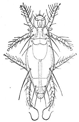
52. Pediculoides ventricosus, female. After Webster.
53. Pediculoides ventricosus, gravid female. (×80). After Webster.
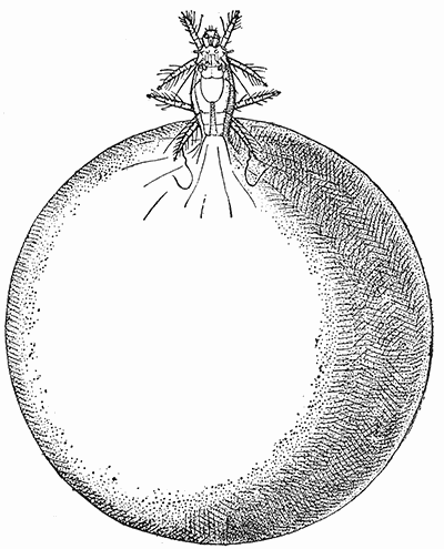
Pediculoides ventricosus (fig. 52 and 53) is, of all the Tarsonemidæ reported, the one which has proved most troublesome to man. It is a predaceous species which attacks a large number of insects but which has most commonly been met with by man through its fondness for certain grain-infesting insects, notably the Angoumois grain moth, Sitotroga cerealella, and the wheat straw-worm, Isosoma grande. In recent years it has attracted much attention in the United States and its distribution and habits have been the object of detailed study by Webster (1901).
54. Pediculoides ventricosus, male. After Braun.
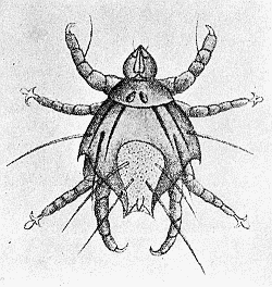
There is a very striking sexual dimorphism in this species. The non-gravid female is elongate, about 200µ by 70µ (fig. 52), with the abdomen slightly striated longitudinally. The gravid female (fig. 53) has the abdomen enormously swollen, so that it is from twenty to a hundred times greater than the rest of the body. The species is viviparous and the larvæ undergo their entire growth in the body of the mother. They emerge as sexually mature males and females which soon pair. The male (fig. 54) is much smaller, reaching a length of only 320µ but is relatively broad, 80µ, and angular. Its abdomen is very greatly reduced.
As far back as 1850 it was noted as causing serious outbreaks of peculiar dermatitis among men handling infested grain. For some time the true source of the difficulty was unknown and it was even believed that the grain had been poisoned. Webster has shown that in this country (and probably in Europe as well) its attacks have been mistaken for those of the red bugs or "chiggers" (larval Trombiidæ). More recently a number of outbreaks of a mysterious "skin disease" were traced to the use of straw mattresses, which were found to be swarming with these almost microscopic forms which had turned their attentions to the occupants of the beds. Other cases cited were those of farmers running wheat through a fanning mill, and of thrashers engaged in feeding unthrashed grain into the cylinder of the machine.
55. Lesions produced by the attacks of Pediculoides ventricosus. After Webster.
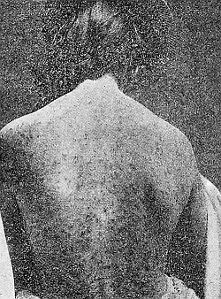
The medical aspects of the question have been studied especially by Schamberg and Goldberger and from the latter's summary (1910) we derive the following data. Within twelve to sixteen hours after exposure, itching appears and in severe cases, especially where exposure is continued night after night by sleeping on an infested bed, the itching may become almost intolerable. Simultaneously, there appears an eruption which characteristically consists of wheals surrounded by a vesicle (fig. 55). The vesicle as a rule does not exceed a pin head in size but may become as large as a pea. Its contents rapidly become turbid and in a few hours it is converted into a pustule. The eruption is most abundant on the trunk, slight on the face and extremities and almost absent on the feet and hands. In severe cases there may be constitutional disturbances marked, at the outset, by chilliness, nausea, and vomiting, followed for a few days by a slight elevation of temperature, with the appearance of albumin in the urine. In some cases the eruption may simulate that of chicken-pox or small-pox.
Treatment for the purpose of killing the mites is hardly necessary as they attach feebly to the surface and are readily brushed off by friction of the clothes. "Antipruritic treatment is always called for; warm, mildly alkaline baths or some soothing ointment, such as zinc oxide will be found to fulfil this indication." Of course, reinfestation must be guarded against, by discarding, or thoroughly fumigating infested mattresses, or by avoiding other sources. Goldberger suggests that farm laborers who must work with infested wheat or straw might protect themselves by anointing the body freely with some bland oil or grease, followed by a change of clothes and bath as soon as their work is done. We are not aware of any experiments to determine the effect of flowers of sulphur, but their efficiency in the case of "red bugs" suggests that they are worth a trial against Pediculoides.
Various species of Tyroglyphidæ (fig. 150f) may abound on dried fruits and other products and attacking persons handling them, may cause a severe dermatitis, comparable to that described above for Pediculoides ventricosus. Many instances of their occurrence as such temporary ectoparasites are on record. Thus, workers who handle vanilla pods are subject to a severe dermatitis, known as vanillism, which is due to the attacks of Tyroglyphus siro, or a closely related species. The so-called "grocer's itch" is similarly caused by mites infesting various products. Castellani has shown that in Ceylon, workers employed in the copra mills, where dried cocoanut is ground up for export, are much annoyed by mites, which produce the so-called "copra itch." The skin of the hands, arms and legs, and sometimes of the whole body, except the face, is covered by fairly numerous, very pruriginous papules, often covered by small, bloody crusts due to scratching. The condition is readily mistaken for scabies. It is due to the attacks of Tyroglyphus longior castellanii which occur in enormous numbers in some samples of the copra.
Sarcoptidæ
The Sarcoptidæ are minute whitish mites, semi-globular in shape, with a delicate transversely striated cuticula. They lack eyes and tracheæ. The mouth-parts are fused at the base to form a cone which is usually designated as the head. The legs are short and stout, and composed of five segments. The tarsi may or may not possess a claw and may terminate in a pedunculated sucker, or simple long bristle, or both. The presence or absence of these structures and their distribution are much used in classification. The mites live on or under the skin of mammals and birds, where they produce the disease known as scabies, mange, or itch. Several species of the Sarcoptidæ attack man but the most important of these, and the one pre-eminent as the "itch mite" is Sarcoptes scabiei.
The female of Sarcoptes scabiei, of man, is oval and yellowish white; the male more rounded and of a somewhat reddish tinge, and much smaller. The body is marked by transverse striæ which are partly interrupted on the back. There are transverse rows of scales, or pointed spines, and scattered bristles on the dorsum.
56a. Sarcoptes scabiei, male. (×100). After Fürstenberg.
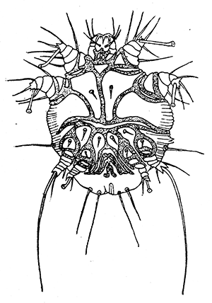
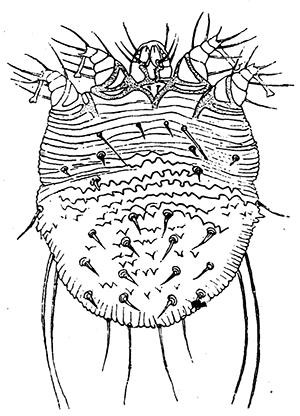
The male (fig. 56) which is from 200-240µ in length, and 150-200µ in breadth, possesses pedunculated suckers on each pair of legs except the third, which bears, instead, a long bristle. The female (fig. 56) 300-450µ in length and 250-350µ in breadth, has the pedunculated suckers on the first and second pairs of legs, only, the third and fourth terminating in bristles.
56b. Sarcoptes scabiei, female. (×100.) After Fürstenberg.
The mite lives in irregular galleries from a few millimeters to several centimeters in length, which it excavates in the epidermis (fig. 57). It works especially where the skin is thin, such as between the fingers, in the bend of the elbows and knees, and in the groin, but it is by no means restricted to these localities. The female, alone, tunnels into the skin; the males remain under the superficial epidermal scales, and seldom are found, as they die soon after mating.
As she burrows into the skin the female deposits her eggs, which measure about 150 × 100µ. Fürstenberg says that each deposits an average of twenty-two to twenty-four eggs, though Gudden reports a single burrow as containing fifty-one. From these there develop after about seven days, the hexapod larvæ. These molt on the sixteenth day to form an octopod nymph, which molts again the twenty-first day. At the end of the fourth week the nymphs molt to form the sexually mature males and the so-called pubescent females. These pair, the males die, and the females again cast their skin, and become the oviparous females. Thus the life cycle is completed in about twenty-eight days.
The external temperature exercises a great influence on the development of the mites and thus, during the winter, the areas of infestation not only do not spread, but they become restricted. As soon as the temperature rises, the mites increase and the infestation becomes much more extensive.
57. Sarcoptes scabiei. Diagrammatic representation of the course in the skin of man.
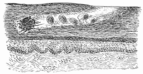
In considering the possible sources of infestation, and the chances of reinfestation after treatment, the question of the ability of the mite to live apart from its host is a very important one. Unfortunately there are few reliable data on this subject. Gerlach found that, exposed in the dry, warm air of a room they became very inactive within twenty-four hours, that after two days they showed only slight movement, and that after three or four days they could not be revived by moisture and warming. The important fact was brought out that in moist air, in folded soiled underwear, they survived as long as ten days. Bourguignon found that under the most favorable conditions the mites of Sarcoptes scabiei equi would live for sixteen days.
The disease designated the "itch" or "scabies," in man has been known from time immemorial, but until within less than a hundred years it was almost universally attributed to malnutrition, errors of diet, or "bad blood." This was in spite of the fact that the mite was known to Mouffet and that Bonomo had figured both the adult and the egg and had declared the mite the sole cause of the disease. In 1834 the Corsican medical student, Francis Renucci, demonstrated the mite before a clinic in Saint Louis Hospital in Paris and soon thereafter there followed detailed studies of the life history of the various itch mites of man and animals.
58. Scabies on the hand. From portfolio of Dermochromes by permission of Rebman & Co., of New York. Publishers.
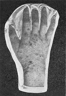
The disease is a cosmopolitan one, being exceedingly abundant in some localities. Its spread is much favored where large numbers of people are crowded together under insanitary conditions and hence it increases greatly during wars and is widely disseminated and abundant immediately afterwards. Though more commonly to be met with among the lower classes, it not infrequently appears among those of the most cleanly, careful habits, and it is such cases that are most liable to wrong diagnosis by the physician.
Infection occurs solely through the passage, direct or indirect, of the young fertilized females to the skin of a healthy individual. The adult, oviparous females do not quit their galleries and hence do not serve to spread the disease. The young females move about more or less at night and thus the principal source of infestation is through sleeping in the same bed with an infested person, or indirectly through bedclothes, or even towels or clothing. Diurnal infestation through contact or clothing is exceptional. Many cases are known of the disease being contracted from animals suffering from scabies, or mange.
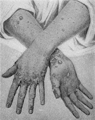
When a person is exposed to infestation, the trouble manifests itself after eight or ten days, though there usually elapses a period of twenty to thirty days before there is a suspicion of anything serious. The first symptom is an intense itching which increases when the patient is in bed. When the point of irritation is examined the galleries may usually be seen as characteristic sinuous lines, at first whitish in color but soon becoming blackish because of the contained eggs and excrement. The galleries, which may not be very distinct in some cases, may measure as much as four centimeters in length. Little vesicles, of the size of a pin head are produced by the secretions of the feeding mite; they are firm, and projecting, and contain a limpid fluid. Figures 58 and 59 show the typical appearance of scabies on the hands, while figure 60 shows a severe general infestation. The intolerable itching induces scratching and through this various complications may arise. The lesions are not normally found on the face and scalp, and are rare on the back.
59. Scabies on the hand. After Duhring.
60. Generalized infection of Scabies. After Morrow.
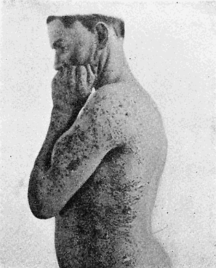
Formerly, scabies was considered a very serious disease, for its cause and method of treatment were unknown, and potentially it may continue indefinitely. Generation after generation of the mites may develop and finally their number become so great that the general health of the individual is seriously affected. Now that the true cause of the disease is known, it is easily controlled.
Treatment usually consists in softening the skin by friction with soap and warm water, followed by a warm bath, and then applying some substance to kill the mites. Stiles gives the following directions, modified from Bourguignon's, as "a rather radical guide, to be modified according to facilities and according to the delicacy of the skin or condition of the patient":
1. The patient, stripped naked, is energetically rubbed all over (except the head) for twenty minutes, with green soap and warm water. 2. He is then placed in a warm bath for thirty minutes, during which time the rubbing is continued. 3. The parasiticide is next rubbed in for twenty minutes and is allowed to remain on the body for four or five hours; in the meantime the patient's clothes are sterilized, to kill the eggs or mites attached to them. 4. A final bath is taken to remove the parasiticide.
The parasiticide usually relied on is the officinal sulphur ointment of the United States pharmacopœia. When infestation is severe it is necessary to repeat treatment after three or four days in order to kill mites which have hatched from the eggs.
The above treatment is too severe for some individuals and may, of itself, produce a troublesome dermatitis. We have seen cases where the treatment was persisted in and aggravated the condition because it was supposed to be due to the parasite. For delicate-skinned patients the use of balsam of Peru is very satisfactory, and usually causes no irritation whatever. Of course, sources of reinfection should be carefully guarded against.
Sarcoptes scabiei crustosæ, which is a distinct variety, if not species, of the human itch mite, is the cause of so-called Norwegian itch. This disease is very contagious, and is much more resistant than the ordinary scabies. Unlike the latter, it may occur on the face and scalp.
Sarcoptes scabiei not only attacks man but also occurs on a large number of mammals. Many species, based on choice of host, and minute differences in size and secondary characters, have been established, but most students of the subject relegate these to varietal rank. Many of them readily attack man, but they have become sufficiently adapted to their normal host so that they are usually less persistent on man.
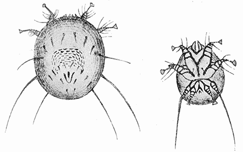
Notoedres cati (usually known as Sarcoptes minor) is a species of itch mites which produce an often fatal disease of cats. The body is rounded and it is considerably smaller than Sarcoptes scabiei, the female (fig. 61) measuring 215-230µ long and 165-175µ wide; the males 145-150µ by 120-125µ. The most important character separating Notoedres from Sarcoptes is the position of the anus, which is dorsal instead of terminal. The mite readily transfers to man but does not persist, the infestation usually disappearing spontaneously in about two weeks. Infested cats are very difficult to cure, unless treatment is begun at the very inception of the outbreak, and under ordinary circumstances it is better to kill them promptly, to avoid spread of the disease to children and others who may be exposed.
61. Notœdres cati, male and female. After Railliet.
Demodecidæ
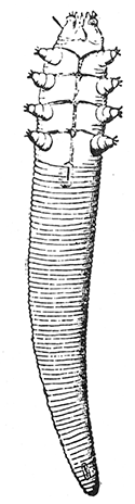
62. Demodex folliculorum. (×200). After Blanchard.
The Demodecidæ are small, elongate, vermiform mites which live in the hair follicles of mammals. The family characteristics will be brought out in the discussion of the species infesting man, Demodex folliculorum.
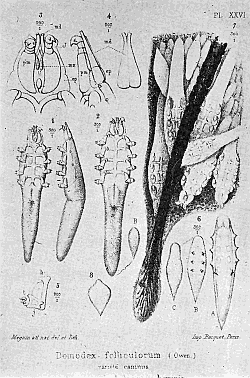
Demodex folliculorum (fig. 62) is to be found very commonly in the hair follicles and sebaceous glands of man. It is vermiform in appearance, and with the elongate abdomen transversely striated so as to give it the appearance of segmentation. The female is 380-400µ long by 45µ; the male 300µ by 40µ. The three-jointed legs, eight in number, are reduced to mere stubs in the adult. The larval form is hexopod. These mites thus show in their form a striking adaptation to their environment. In the sebaceous glands and hair follicles they lie with their heads down (fig. 63). Usually there are only a few in a gland, but Gruby has counted as many as two hundred.
63. Demodex folliculorum. Section through skin showing the mites in situ. Magnification of Nos. 1, 2, 6 and 7, ×150; Nos, 3, 4, 5, ×450. After Megnin.
The frequency with which they occur in man is surprising. According to European statistics they are found in 50 per cent to 60 per cent or even more. Gruby found them in forty out of sixty persons examined. These figures are very commonly quoted, but reliable data for the United States seem to be lacking. Our studies indicate that it is very much less common in this country than is generally assumed.
The Demodex in man does not, as a rule, cause the slightest inconvenience to its host. It is often stated that they give rise to comedons or "black-heads" but there is no clear evidence that they are ever implicated. Certain it is that they are not the usual cause. A variety of the same, or a very closely related species of Demodex, on the dog gives rise to the very resistant and often fatal follicular mange.
Hexapoda or True Insects
The Hexapoda, or true insects, are characterized by the fact that the adult possesses three pairs of legs. The body is distinctly segmented and is divided into head, thorax, and abdomen.
The mouth-parts in a generalized form, consist of an upper lip, or labrum, which is a part of the head capsule, and a central unpaired hypopharynx, two mandibles, two maxillæ and a lower lip, or labium, made up of the fused pair of second maxillæ. These parts may be greatly modified, dependent upon whether they are used for biting, sucking, piercing and sucking, or a combination of biting and sucking.
Roughly speaking, insects may be grouped into those which undergo complete metamorphosis and those which have incomplete metamorphosis. They are said to undergo complete metamorphosis when the young form, as it leaves the egg, bears no resemblance to the adult. For example, the maggot changes to a quiescent pupa and from this emerges the winged active fly. They undergo incomplete metamorphosis, when the young insect, as it leaves the egg, resembles the adult to a greater or less extent, and after undergoing a certain number of molts becomes sexually mature.
Representatives of several orders have been reported as accidental or faculative parasites of man, but the true parasites are restricted to four orders. These are the Siphunculata; the Hemiptera, the Diptera and the Siphonaptera.
Siphunculata
The order Siphunculata was established by Meinert to include the true sucking lice. These are small wingless insects, with reduced mouth-parts, adapted for sucking; thorax apparently a single piece due to indistinct separation of its three segments: the compound eyes reduced to a single ommatidium on each side. The short, powerful legs are terminated by a single long claw. Metamorphosis incomplete.
There has been a great deal of discussion regarding the structure of the mouth-parts, and the relationships of the sucking lice, and the questions cannot yet be regarded as settled. The conflicting views are well represented by Cholodkovsky (1904 and 1905) and by Enderlein (1904).
64. Pediculus showing the blind sac (b) containing the mouth parts (a) beneath the alimentary canal (p). After Pawlowsky.
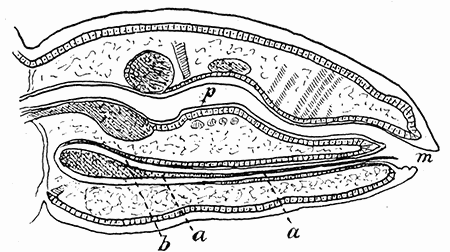
Following Graber, it is generally stated that the mouth-parts consist of a short tube furnished with hooks in front, which constitutes the lower lip, and that within this is a delicate sucking tube derived from the fusion of the labrum and the mandibles. Opposed to this, Cholodkovsky and, more recently, Pawlowsky, (1906), have shown that the piercing apparatus lies in a blind sac under the pharynx and opening into the mouth cavity (fig. 64). It does not form a true tube but a furrow with its open surface uppermost. Eysell has shown that, in addition, there is a pair of chitinous rods which he regards as the homologues of the maxillæ.
When the louse feeds, it everts the anterior part of the mouth cavity, with its circle of hooks. The latter serve for anchoring the bug, and the piercing apparatus is then pushed out.
65. Pediculus humanus, ventral aspect of male. (×10)
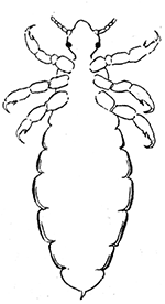
Most writers have classed the sucking lice as a sub-order of the Hemiptera, but the more recent anatomical and developmental studies render this grouping untenable. An important fact, bearing on the question, is that, as shown by Gross, (1905), the structure of the ovaries is radically different from that of the Hemiptera.
Lice infestation and its effects are known medically as pediculosis. Though their continued presence is the result of the grossest neglect and filthiness, the original infestation may be innocently obtained and by people of the most careful habits.
Three species commonly attack man. Strangely enough, there are very few accurate data regarding their life history.
Pediculus humanus (fig. 65), the head louse, is the most widely distributed. It is usually referred to in medical literature as Pediculus capitis, but the Linnean specific name has priority. In color it is of a pale gray, blackish on the margins. It is claimed by some authors that the color varies according to the color of the skin of the host. The abdomen is composed of seven distinct segments, bearing spiracles laterally. There is considerable variation in size. The males average 1.8 mm. and the females 2.7 mm. in length.
The eggs, fifty to sixty in number, stick firmly to the hairs of the host and are known as nits. They are large and conspicuous, especially on dark hair and are provided with an operculum, or cap, at the free end, where the nymphs emerge. They hatch in about six days and about the eighteenth day the young lice are sexually mature.
66. Pediculosis of the head. The illustration shows the characteristic indications of the presence of lice, viz: the occipital eczema gluing the hairs together, the swollen cervical glands, and the porrigo, or eruption of contagious pustules upon the neck. After Fox.
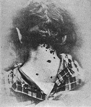
The head lice live by preference on the scalp of their host but occasionally they are found on the eyelashes and beard, or in the pubic region. They may also occur elsewhere on the body. The penetration of the rostrum into the skin and the discharge of an irritating saliva produce a severe itching, accompanied by the formation of an eczema-like eruption (fig. 66). When the infestation is severe, the discharge from the pustules mats down the hair, and scabs are formed, under which the insects swarm. "If allowed to run, a regular carapace may form, called trichoma, and the head exudes a fœtid odor. Various low plants may grow in the trichoma, the whole being known as plica palonica."—Stiles.
Sources of infestation are various. School children may obtain the lice from seatmates, by wearing the hats or caps of infested mates, or by the use, in common, of brushes and combs. They may be obtained from infested beds or sleeper berths. Stiles reports an instance in which a large number of girls in a fashionable boarding school developed lousiness a short time after traveling in a sleeping car.
Treatment is simple, for the parasites may readily be controlled by cleanliness and washing the head with a two per cent solution of carbolic acid or even kerosene. The latter is better used mixed with equal parts of olive oil, to avoid irritation. The treatment should be applied at night and followed the next morning by a shampoo with soap and warm water. It is necessary to repeat the operation in a few days. Xylol, used pure, or with the addition of five per cent of vaseline, is also very efficacious. Of course, the patient must be cautioned to stay away from a lighted lamp or fire while using either the kerosene or xylol. While these treatments will kill the eggs or nits, they will not remove them from the hairs. Pusey recommends repeated washings with vinegar or 25 per cent of acetic acid in water, for the purpose of loosening and removing the nits.
Treatment of severe infestations in females is often troublesome on account of long hair. For such cases the following method recommended by Whitfield (1912) is especially applicable:
The patient is laid on her back on the bed with her head over the edge, and beneath the head is placed a basin on a chair so that the hair lies in the basin. A solution of 1 in 40 carbolic acid is then poured over the hair into the basin and sluiced backwards and forwards until the whole of the hair is thoroughly soaked with it. It is especially necessary that care should be taken to secure thorough saturation of the hair over the ears and at the nape of the neck, since these parts are not only the sites of predilection of the parasites but they are apt to escape the solution. This sluicing is carried out for ten minutes by the clock. At the end of ten minutes the hair is lifted from the basin and allowed to drain, but is not dried or even thoroughly wrung out. The whole head is then swathed with a thick towel or better, a large piece of common house flannel, which is fastened up to form a sort of turban, and is allowed to remain thus for an hour. It can then be washed or simply allowed to dry, as the carbolic quickly disperses. At the end of this period every pediculus and what is better, every ovum is dead and no relapse will occur unless there is exposure to fresh contagion. Whitfield states that there seem to be no disadvantages in this method, which he has used for years. He has never seen carboluria result from it, but would advise first cutting the hair of children under five years of age.
Pediculus corporis (= P. vestimenti) the body louse, is larger than the preceding species, the female measuring 3.3 mm., and the male 3 mm. in length. The color is a dirty white, or grayish. P. corporis has been regarded by some authorities as merely a variety of P. humanus but Piaget maintains there are good characters separating the two species.
The body louse lives in the folds and seams of the clothing of its host, passing to the skin only when it wishes to feed. Brumpt states that he has found enormous numbers of them in the collars of glass-ware or grains worn by certain naked tribes in Africa.
Exact data regarding the life-history of this species have been supplied, in part, by the work of Warburton (1910), cited by Nuttall. He found that Pediculus corporis lives longer than P. humanus under adverse conditions. This is doubtless due to its living habitually on the clothing, whereas humanus lives upon the head, where it has more frequent opportunities of feeding. He reared a single female upon his own person, keeping the louse enclosed in a cotton-plugged tube with a particle of cloth to which it could cling. The tube was kept next to his body, thus simulating the natural conditions of warmth and moisture under which the lice thrive. The specimen was fed twice daily, while it clung to the cloth upon which it rested. Under these conditions she lived for one month. Copulation commenced five days after the female had hatched and was repeated a number of times, sexual union lasting for hours. The female laid one hundred and twenty-four eggs within twenty-five days.
The eggs hatched after eight days, under favorable conditions, such as those under which the female was kept. They did not hatch in the cold. Eggs kept near the person during the day and hung in clothing by the bedside at night, during the winter, in a cold room, did not hatch until the thirty-fifth day. When the nymphs emerge from the eggs, they feed at once, if given a chance to do so. They are prone to scatter about the person and abandon the fragment of cloth to which the adult clings.
The adult stage is reached on the eleventh day, after three molts, about four days apart. Adults enter into copulation about the fifth day and as the eggs require eight days for development, the total cycle, under favorable conditions, is about twenty-four days. Warburton's data differ considerably from those commonly quoted and serve to emphasize the necessity for detailed studies of some of the commonest of parasitic insects.
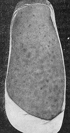
Body lice are voracious feeders, producing by their bites and the irritating saliva which they inject, rosy elevations and papules which become covered with a brownish crust. The intense itching provokes scratching, and characteristic white scars (fig. 67) surrounded by brownish pigment (fig. 68) are formed. The skin may become thickened and take on a bronze tinge. This melanoderma is especially marked in the region between the shoulders but it may become generalized, a prominent characteristic of "vagabond's disease." According to Dubre and Beille, this melanoderma is due to a toxic substance secreted by the lice, which indirectly provokes the formation of pigment.
67. Pediculosis in man caused by the body louse. After Morrow.
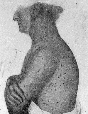
Control measures, in the case of the body louse, consist in boiling or steaming the clothes or in some cases, sterilizing by dry heat. The dermatitis may be relieved by the use of zinc-oxide ointment, to which Pusey recommends that there be added, on account of their parasiticidal properties, sulphur and balsam of Peru, equal parts, 15 to 30 grains to the ounce.
68. Melanoderma caused by the body louse. From Portfolio of Dermochromes, by permission of Rebman & Co., New York, Publishers.
69. Phthirius pubis. Ventral aspect of female. (×12).
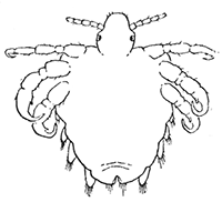
Phthirius pubis (= P. inguinalis), the pubic louse, or so-called "crab louse," differs greatly from the preceding in appearance. It is characterized by its relatively short head which fits into a broad depression in the thorax. The latter is broad and flat and merges into the abdomen. The first pair of legs is slender and terminated by a straight claw. The second and third pairs of legs are thicker and are provided with powerful claws fitted for clinging to hairs. The females (fig. 69) measure 1.5 to 2 mm. in length by 1.5 mm. in breadth. The male averages a little over half as large. The eggs, or nits, are fixed at the base of the hairs. Only a few, ten to fifteen are deposited by a single female, and they hatch in about a week's time. The young lice mature in two weeks.
The pubic louse usually infests the hairs of the pubis and the perineal region. It may pass to the arm pits or even to the beard or moustache. Rarely, it occurs on the eyelids, and it has even been found, in a very few instances, occurring in all stages, on the scalp. Infestation may be contracted from beds or even from badly infested persons in a crowd. We have seen several cases which undoubtedly were due to the use of public water closets. It produces papular eruption and an intense pruritis. When abundant, there occurs a grayish discoloration of the skin which Duguet has shown is due to a poisonous saliva injected by the louse, as is the melanoderma caused by the body louse.
The pubic louse may be exterminated by the measures recommended for the head louse, or by the use of officinal mercurial ointment.
Hemiptera
Several species of Hemiptera-Heteroptera are habitual parasites of man, and others occur as occasional or accidental parasites. Of all these, the most important and widespread are the bed-bugs, belonging to the genus Cimex (= Acanthia).
The Bed-bugs—The bed-bugs are characterized by a much flattened oval body, with the short, broad head unconstricted behind, and fitting into the strongly excavated anterior margin of the thorax. The compound eyes are prominent, simple eyes lacking. Antennæ four-jointed, the first segment short, the second long and thick, and the third and fourth slender. The tarsi are short and three segmented.
It is often assumed in the literature of the subject that there is but a single species of Cimex attacking man, but several such species are to be recognized. These are distinguishable by the characters given in Chapter XII. We shall consider especially Cimex lectularius, the most common and widespread species.
Cimex lectularius (= Acanthia lectularia, Clinocoris lectularius), is one of the most cosmopolitan of human parasites but, like the lice, it has been comparatively little studied until recent years, when the possibility that it may be concerned with the transmission of various diseases has awakened interest in the details of its life-history and habits.
70. Cimex lectularius adult and eggs. Photograph by M. V. S.
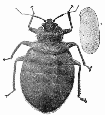
The adult insect (fig. 70) is 4-5 mm. long by 3 mm. broad, reddish brown in color, with the beak and body appendages lighter in color. The short, broad and somewhat rectangular head has no neck-like constriction but fits into the broadly semilunar prothorax. The four segmented labium or proboscis encloses the lancet-like maxillæ and mandibles. The distal of the four antennal segments is slightly club-shaped. The prothorax is characteristic of the species, being deeply incised anteriorly and with its thin lateral margins somewhat turned up. The mesothorax is triangular, with the apex posteriorly, and bears the greatly atrophied first pair of wings. There is no trace of the metathoracic pair. The greatly flattened abdomen has eight visible segments, though in reality the first is greatly reduced and has been disregarded by most writers. The body is densely covered with short bristles and hairs, the former being peculiarly saber-shaped structures sharply toothed at the apex and along the convex side (fig. 159b).
The peculiar disagreeable odor of the adult bed-bug is due to the secretion of the stink glands which lie on the inner surface of the mesosternum and open by a pair of orifices in front of the metacoxæ, near the middle line. In the nymphs, the thoracic glands are not developed but in the abdomen there are to be found three unpaired dorsal stink glands, which persist until the fifth molt, when they become atrophied and replaced by the thoracic glands. The nymphal glands occupy the median dorsal portion of the abdomen, opening by paired pores at the anterior margin of the fourth, fifth and sixth segments. The secretion is a clear, oily, volatile fluid, strongly acid in reaction. Similar glands are to be found in most of the Hemiptera-Heteroptera and their secretion is doubtless protective, through being disagreeable to the birds. In the bed-bug, as Marlatt points out, "it is probably an illustration of a very common phenomenon among animals, i.e., the persistence of a characteristic which is no longer of any special value to the possessor." In fact, its possession is a distinct disadvantage to the bed-bug, as the odor frequently reveals the presence of the bugs, before they are seen.
The eggs of the bed-bug (fig. 70) are pearly white, oval in outline, about a millimeter long, and possess a small operculum or cap at one end, which is pushed off when the young hatches. They are laid intermittently, for a long period, in cracks and crevices of beds and furniture, under seams of mattresses, under loose wall paper, and similar places of concealment of the adult bugs. Girault (1905) observed a well-fed female deposit one hundred and eleven eggs during the sixty-one days that she was kept in captivity. She had apparently deposited some of her eggs before being captured.
The eggs hatch in six to ten days, the newly emerged nymphs being about 1.5 mm. in length and of a pale yellowish white color. They grow slowly, molting five times. At the last molt the mesathoracic wing pads appear, characteristic of the adult. The total length of the nymphal stage varies greatly, depending upon conditions of food supply, temperature and possibly other factors. Marlatt (1907) found under most favorable conditions a period averaging eight days between molting which, added to an equal egg period, gave a total of about seven weeks from egg to adult insect. Girault (1912) found the postembryonic period as low as twenty-nine days and as high as seventy days under apparently similar and normal conditions of food supply. Under optimum and normal conditions of food supply, beginning August 27, the average nymphal life was 69.9 days; average number of meals 8.75 and the molts 5. Under conditions allowing about half the normal food supply the average nymphal life was from 116.9 to 139 days. Nymphs starved from birth lived up to 42 days. We have kept unfed nymphs, of the first stage, alive in a bottle for 75 days. The interesting fact was brought out that under these conditions of minimum food supply there were sometimes six molts instead of the normal number.
The adults are remarkable for their longevity, a factor which is of importance in considering the spread of the insect and methods of control. Dufour (1833) (not De Geer, as often stated) kept specimens for a year, in a closed vial, without food. This ability, coupled with their willingness to feed upon mice, bats, and other small mammals, and even upon birds, accounts for the long periods that deserted houses and camps may remain infested. There is no evidence that under such conditions they are able to subsist on the starch of the wall paper, juices of moistened wood, or the moisture in the accumulations of dust, as is often stated.
There are three or four generations a year, as Girault's breeding experiments have conclusively shown. He found that the bed-bug does not hibernate where the conditions are such as to allow it to breed and that breeding is continuous unless interrupted by the lack of food or, during the winter, by low temperature.
Bed-bugs ordinarily crawl from their hiding places and attack the face and neck or uncovered parts of the legs and arms of their victims. If undisturbed, they will feed to repletion. We have found that the young nymph would glut itself in about six minutes, though some individuals fed continuously for nine minutes, while the adult required ten to fifteen minutes for a full meal. When gorged, it quickly retreats to a crack or crevice to digest its meal, a process which requires two or three days. The effect of the bite depends very greatly on the susceptibility of the individual attacked. Some persons are so little affected that they may be wholly ignorant of the presence of a large number of bugs. Usually the bite produces a small hard swelling, or wheal, whitish in color. It may even be accompanied by an edema and a disagreeable inflammation, and in such susceptible individuals the restlessness and loss of sleep due to the presence of the insects may be a matter of considerable importance. Stiles (1907) records the case of a young man who underwent treatment for neurasthenia, the diagnosis being agreed upon by several prominent physicians; all symptoms promptly disappeared, however, immediately following a thorough fumigation of his rooms, where nearly a pint of bed-bugs were collected.
It is natural to suppose that an insect which throughout its whole life is in such intimate relationship with man should play an important rôle in the transmission of disease. Yet comparatively little is definitely known regarding the importance of the bed-bug in this respect. It has been shown that it is capable of transmitting the bubonic plague, and South American trypanosomiasis. Nuttall succeeded in transmitting European relapsing fever from mouse to mouse by its bite. It has been claimed that Oriental sore, tuberculosis, and even syphilis may be so carried. These phases of the subject will be considered later.
The sources of infestation are many, and the invasion of a house is not necessarily due to neglect, though the continued presence of the pests is quite another matter. In apartments and closely placed houses they are known to invade new quarters by migration. They are frequently to be met with in boat and sleeper berths, and even the plush seats of day coaches, whence a nucleus may be carried in baggage to residences. They may be brought in the laundry or in clothes of servants.
Usually they are a great scourge in frontier settlements and it is generally believed that they live in nature under the bark of trees, in lumber, and under similar conditions. This belief is founded upon the common occurrence of bugs resembling the bed-bug, in such places. As a matter of fact, they are no relation to bed-bugs but belong to plant-feeding forms alone (fig. 19 c, d).
It is also often stated that bed-bugs live in poultry houses, in swallows nests, and on bats, and that it is from these sources that they gain access to dwellings. These bugs are specifically distinct from the true bed-bug, but any of them may, rarely, invade houses. Moreover, chicken houses are sometimes thoroughly infested with the true Cimex lectularius.
Control measures consist in the use of iron bedsteads and the reduction of hiding places for the bugs. If the infestation is slight they may be exterminated by a vigilant and systematic hunt, and by squirting gasoline or alcohol into cracks and crevices of the beds, and furniture. Fumigation must be resorted to in more general infestations.
The simplest and safest method of fumigation is by the use of flowers of sulphur at the rate of two pounds to each one thousand cubic feet of room space. The sulphur should be placed in a pan, a well made in the top of the pile and a little alcohol poured in, to facilitate burning. The whole should be placed in a larger pan and surrounded by water so as to avoid all danger of fire. Windows should be tightly closed, beds, closets and drawers opened, and bedding spread out over chairs in order to expose them fully to the fumes. As metal is tarnished by the sulphur fumes, ornaments, clocks, instruments, and the like should be removed. When all is ready the sulphur should be fired, the room tightly closed and left for twelve to twenty-four hours. Still more efficient in large houses, or where many hiding places favor the bugs, is fumigation with hydrocyanic acid gas. This is a deadly poison and must be used under rigid precautions. Through the courtesy of Professor Herrick, who has had much experience with this method, we give in the Appendix, the clear and detailed directions taken from his bulletin on "Household Insects."
Fumigation with formaldehyde gas, either from the liquid or "solid" formalin, so efficient in the case of contagious diseases, is useless against bed-bugs and most other insects.
Other Bed-bugs—Cimex hemipterus (= C. rotundatus) is a tropical and subtropical species, occurring in both the old and new world. Patton and Cragg state that it is distributed throughout India, Burma, Assam, the Malay Peninsula, Aden, the Island of Mauritius, Reunion, St. Vincent and Porto Rico. "It is widely distributed in Africa, and is probably the common species associated there with man." Brumpt also records it for Cuba, the Antilles, Brazil, and Venezuela.
This species, which is sometimes called the Indian bed-bug, differs from C. lectularius in being darker and in having a more elongate abdomen. The head also is shorter and narrower, and the prothorax has rounded borders.
It has the same habits and practically the same life cycle as Cimex lectularius. Mackie, in India, has found that it is capable of transmitting the Asiatic type of recurrent fever. Roger suggested that it was also capable of transmitting Kala-azar and Patton has described in detail the developmental stages of Leishmania, the causative organism of Kala-azar, in the stomach of this bug, but Brumpt declares that the forms described are those of a common, non-pathogenic flagellate to be found in the bug, and have nothing to do with the human disease. Brumpt has shown experimentally that Cimex hemipterus may transmit Trypanosoma cruzi in its excrement.
Cimex boueti, occurring in French Guinea, is another species attacking man. Its habits and general life history are the same as for the above species. It is 3 to 4.5 mm. in length, has vestigial elytra, and much elongated antennæ and legs. The extended hind legs are about as long as the body.
Cimex columbarius, a widely distributed species normally living in poultry houses and dove cotes, C. inodorus, infesting poultry in Mexico, C. hirundinis, occurring in the nests of swallows in Europe and Oeciacus vicarius (fig. 19i) occurring in swallows' nests in this country, are species which occasionally infest houses and attack man.
71. Conorhinus sanguisugus.
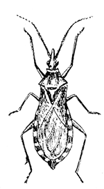
Conorhinus sanguisugus, the cone-nosed bed-bug. We have seen in our consideration of poisonous insects, that various species of Reduviid bugs readily attack man. Certain of these are nocturnal and are so commonly found in houses that they have gained the name, of "big bed-bugs." The most noted of these, in the United States, is Conorhinus sangiusugus (fig. 71), which is widely distributed in our Southern States.
Like its near relatives, Conorhinus sangiusugus is carnivorous in habit and feeds upon insects as well as upon mammalian and human blood. It is reported as often occurring in poultry houses and as attacking horses in barns. The life history has been worked out in considerable detail by Marlatt, (1902), from whose account we extract the following.
72. Beak of Conorhinus sanguisugus. After Marlatt.
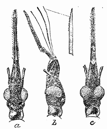
The eggs are white, changing to yellow and pink before hatching. The young hatch within twenty days and there are four nymphal stages. In all these stages the insect is active and predaceous, the mouth-parts (fig. 72) being powerfully developed. The eggs are normally deposited, and the early stages are undoubtedly passed, out of doors, the food of the immature forms being other insects. Immature specimens are rarely found indoors. It winters both in the partly grown and adult stage, often under the bark of trees or in any similar protection, and only in its nocturnal spring and early summer flights does it attack men. Marlatt states that this insect seems to be decidedly on the increase in the region which it particularly infests,—the plains region from Texas northward and westward. In California a closely related species of similar habits is known locally as the "monitor bug."
The effect of the bite of the giant bed-bug on man is often very severe, a poisonous saliva apparently being injected into the wound. We have discussed this phase of the subject more fully under the head of poisonous insects.
Conorhinus megistus is a Brazilian species very commonly attacking man, and of special interest since Chagas has shown that it is the carrier of a trypanosomiasis of man. Its habits and life history have been studied in detail by Neiva, (1910).
This species is now pre-eminently a household insect, depositing its eggs in cracks and crevices in houses, though this is a relatively recent adaptation. The nymphs emerge in from twenty to forty days, depending upon the temperature. There are five nymphal stages, and as in the case of true bed-bugs, the duration of these is very greatly influenced by the availability of food and by temperature. Neiva reckons the entire life cycle, from egg to egg, as requiring a minimum of three hundred and twenty-four days.
The nymphs begin to suck blood in three to five days after hatching. They usually feed at night and in the dark, attacking especially the face of sleeping individuals. The bite occasions but little pain. The immature insects live in cracks and crevices in houses and invade the beds which are in contact with walls, but the adults are active flyers and attack people sleeping in hammocks. The males as well as the females are blood suckers.
Like many blood-sucking forms, Conorhinus megistus can endure for long periods without food. Neiva received a female specimen which had been for fifty-seven days alive in a tightly closed box. They rarely feed on two consecutive days, even on small quantities of blood, and were never seen to feed on three consecutive days.
Methods of control consist in screening against the adult bugs, and the elimination of crevices and such hiding places of the nymphs. Where the infestation is considerable, fumigation with sulphur is advisable.
Parasitic Diptera or Flies
Of the Diptera or two-winged flies, many species occasionally attack man. Of these, a few are outstanding pests, many of them may also serve to disseminate disease, a phase of our subject which will be considered later. We shall now consider the most important of the group from the viewpoint of their direct attacks on man.
Psychodidæ or Moth-Flies
The Psychodidæ or Moth-flies, include a few species which attack man, and at least one species, Phlebotomus papatasii, is known to transmit the so-called "three-day fever" of man. Another species is supposed to be the vector of Peruvian verruga.
The family is made up of small, sometimes very small, nematocerous Diptera, which are densely covered with hairs, giving them a moth-like appearance. The wings are relatively large, oval or lanceolate in shape, and when at rest are held in a sloping manner over the abdomen, or are held horizontally in such a way as to give the insect a triangular outline. Not only is the moth-like appearance characteristic, but the venation of the wings (fig. 163, d) is very peculiar and, according to Comstock, presents an extremely generalized form. All of the longitudinal veins separate near the base of the wing except veins R2 and R3 and veins M1 and M2. Cross veins are wanting in most cases.
Comparatively little is known regarding the life-history and habits of the Psychodidæ, but one genus, Phlebotomus, contains minute, blood-sucking species, commonly known as sand-flies. The family is divided into two subfamilies, the Psychodinæ and the Phlebotominæ. The second of these, the Phlebotominæ, is of interest to us.
The Phlebotominæ—The Phlebotominæ differ from the Psychodinæ in that the radical sector branches well out into the wing rather than at the base of the wing. They are usually less hairy than the Psychodinæ. The ovipositor is hidden and less strongly chitinized. The species attacking man belong to the genus Phlebotomus, small forms with relatively large, hairy wings which are held upright, and with elongate proboscis. The mandibles and maxillæ are serrated and fitted for biting.
According to Miss Summers (1913) there are twenty-nine known species of the genus Phlebotomus, five European, eleven Asiatic, seven African and six American. One species only, Phlebotomus vexator, has been reported for the United States. This was described by Coquillett, (1907), from species taken on Plummer's Island, Maryland. It measures only 1.5 mm. in length. As it is very probable that this species is much more widely distributed, and that other species of these minute flies will be found to occur in our fauna, we quote Coquillett's description.
Phlebotomus vexator, Coq.: Yellow, the mesonotum brown, hairs chiefly brown; legs in certain lights appear brown, but are covered with a white tomentum; wings hyaline, unmarked; the first vein (R1) terminates opposite one-fifth of the length of the first submarginal cell (cell R2); this cell is slightly over twice as long as its petiole; terminal, horny portion of male claspers slender, bearing many long hairs; the apex terminated by two curved spines which are more than one-half as long as the preceding part, and just in front of these are two similar spines, while near the middle of the length of this portion is a fifth spine similar to the others. Length 1.5 mm.
The life-history of the Phlebotomus flies has been best worked out for the European Phlebotomus papatasii and we shall briefly summarize the account of Dœrr and Russ (1913) based primarily on work on this species. The European Phlebotomus flies appear at the beginning of the warm season, a few weeks after the cessation of the heavy rains and storms of springtime. They gradually become more abundant until they reach their first maximum, which in Italy is near the end of July (Grassi). They then become scarcer but reach a second maximum in September. At the beginning of winter they vanish completely, hibernating individuals not being found.
After fertilization there is a period of eight to ten days before oviposition. The eggs are then deposited, the majority in a single mass covered by a slimy secretion from the sebaceous glands. The larvæ emerge in fourteen to twenty days. There is uncertainty as to the length of larval life, specimens kept in captivity remaining fifty or more days without transforming. Growth may be much more rapid in nature. The larvæ do not live in fluid media but in moist detritus in dark places. Marett believes that they live chiefly on the excrement of pill-bugs (Oniscidæ) and lizards. Pupation always occurs during the night. The remnants of the larval skin remain attached to the last two segments of the quiescent pupa and serve to attach it to the stone on which it lives. The pupal stage lasts eleven to sixteen days, the adult escaping at night.
Only the females suck blood. They attack not only man but all warm-blooded animals and, according to recent workers, also cold-blooded forms, such as frogs, lizards, and larvæ. Indeed, Townsend (1914) believes that there is an intimate relation between Phlebotomus and lizards, or other reptiles the world over. The Phlebotomus passes the daylight hours within the darkened recesses of the loose stone walls and piles of rock in order to escape wind and strong light. Lizards inhabit the same places, and the flies, always ready to suck blood in the absence of light and wind, have been found more prone to suck reptilian than mammalian blood.
On hot summer nights, when the wind is not stirring, the Phlebotomus flies, or sand-flies, as they are popularly called, invade houses and sleeping rooms in swarms and attack the inmates. As soon as light begins to break the flies either escape to the breeding places, or cool, dark places protected from the wind, or a part of them remain in the rooms, hiding behind pictures, under garments, and in similar places. Wherever the Phlebotomus flies occur they are an intolerable nuisance. On account of their small size they can easily pass through the meshes of ordinary screens and mosquito curtains. They attack silently and inflict a very painful, stinging bite, followed by itching. The ankles, dorsum of the feet, wrists, inner elbow, knee joint and similar places are favorite places of attack, possibly on account of their more delicate skin.
Special interest has been attracted to these little pests in recent years, since it has been shown that they transmit the European "pappatici fever" or "three day fever." More recently yet, it appears that they are the carriers of the virus of the Peruvian "verruga." This phase of the subject will be discussed later.
Control measures have not been worked out. As Newstead says, "In consideration of the facts which have so far been brought to light regarding the economy of Phlebotomus, it is clearly evident that the task of suppressing these insects is an almost insurmountable one. Had we to deal with insects as large and as accessible as mosquitoes, the adoption of prophylactic measures would be comparatively easy, but owing to the extremely minute size and almost flea-like habits of the adult insects, and the enormous area over which the breeding-places may occur, we are faced with a problem which is most difficult of solution." For these reasons, Newstead considers that the only really prophylactic measures which can at present be taken, are those which are considered as precautionary against the bites of the insects.
Of repellents, he cites as one of the best a salve composed of the following:
Ol. Anisi3 grs.
Ol. Eucalypti3 grs.
Ol. Terebenth3 grs.
Unq. Acid Borac.
Of sprays he recommends as the least objectionable and at the same time one of the most effective, formalin. "The dark portions and angles of sleeping apartments should be sprayed with a one per cent. solution of this substance every day during the season in which the flies are prevalent. A fine spraying apparatus is necessary for its application and an excessive amount must not be applied. It is considered an excellent plan also to spray the mosquito curtains regularly every day towards sunset; nets thus treated are claimed to repel the attacks of these insects." This effectiveness of formalin is very surprising for, as we have seen, it is almost wholly ineffective against bed-bugs, mosquitoes, house flies and other insects, where it has been tried.
A measure which promises to be very effective, where it can be adopted, is the use of electric fans so placed as to produce a current of air in the direction of the windows of sleeping apartments. On account of the inability of the Phlebotomus flies to withstand even slight breezes, it seems very probable that they would be unable to enter a room so protected.
Culicidæ or Mosquitoes
From the medical viewpoint, probably the most interesting and important of the blood-sucking insects are the mosquitoes. Certainly this is true of temperate zones, such as those of the United States. The result is that no other group of insects has aroused such widespread interest, or has been subjected to more detailed study than have the mosquitoes, since their rôle as carriers of disease was made known. There is an enormous literature dealing with the group, but fortunately for the general student, this has been well summarized by a number of workers. The most important and helpful of the general works are those of Howard (1901), Smith (1904), Blanchard (1905), Mitchell (1907), and especially of Howard, Dyar, and Knab, whose magnificent monograph is still in course of publication.
Aside from their importance as carriers of disease, mosquitoes are notorious as pests of man, and the earlier literature on the group is largely devoted to references to their enormous numbers and their blood-thirstiness in certain regions. They are to be found in all parts of the world, from the equator to the Arctic and Antarctic regions. Linnæus, in the "Flora Lapponica," according to Howard, Dyar and Knab, "dwells at some length upon the great abundance of mosquitoes in Lapland and the torments they inflicted upon man and beast. He states that he believes that nowhere else on earth are they found in such abundance and he compares their numbers to the dust of the earth. Even in the open, you cannot draw your breath without having your mouth and nostrils filled with them; and ointments of tar and cream or of fish grease are scarcely sufficient to protect even the case-hardened cuticle of the Laplander from their bite. Even in their cabins, the natives cannot take a mouthful of food or lie down to sleep unless they are fumigated almost to suffocation." In some parts of the Northwestern and Southwestern United States it is necessary to protect horses working in the fields by the use of sheets or burlaps, against the ferocious attacks of these insects. It is a surprising fact that even in the dry deserts of the western United States they sometimes occur in enormous numbers.
Until comparatively recent years, but few species of mosquitoes were known and most of the statements regarding their life-history were based upon the classic work of Reaumur (1738) on the biology of the rain barrel mosquito, Culex pipiens. In 1896, Dr. Howard refers to twenty-one species in the United States, now over fifty are known; Giles, in 1900, gives a total of two hundred and forty-two for the world fauna, now over seven hundred species are known. We have found eighteen species at Ithaca, N. Y.
All of the known species of mosquitoes are aquatic in the larval stage, but in their life-histories and habits such great differences occur that we now know that it is not possible to select any one species as typical of the group. For our present purpose we shall first discuss the general characteristics and structure of mosquitoes, and shall then give the life-history of a common species, following this by a brief consideration of some of the more striking departures from what have been supposed to be the typical condition.
The Culicidæ are slender, nematocerous Diptera with narrow wings, antennæ plumose in the males, and usually with the proboscis much longer than the head, slender, firm and adapted for piercing in the female. The most characteristic feature is that the margins of the wings and, in most cases, the wing veins possess a fringe of scale-like hairs. These may also cover in part, or entirely, the head, thorax, abdomen and legs. The females, only, suck blood.
On account of the importance of the group in this country and the desirability of the student being able to determine material in various stages, we show in the accompanying figures the characters most used in classification.
The larvæ (fig. 73) are elongate, with the head and thorax sharply distinct. The larval antennæ are prominent, consisting of a single cylindrical and sometimes curved segment. The outer third is often narrower and bears at its base a fan-shaped tuft of hairs, the arrangement and abundance of which is of systematic importance. About the mouth are the so-called rotary mouth brushes, dense masses of long hairs borne by the labrum and having the function of sweeping food into the mouth. The form and arrangement of thoracic, abdominal, and anal tufts of hair vary in different species and present characteristics of value. On either side of the eighth abdominal segment is a patch of scales varying greatly in arrangement and number and of much value in separating species. Respiration is by means of tracheæ which open at the apex of the so-called anal siphon, when it is present. In addition, there are also one or two pairs of tracheal gills which vary much in appearance in different species. On the ventral side of the anal siphon is a double row of flattened, toothed spines whose number and shape are likewise of some value in separating species. They constitute the comb or pecten.
73. Culex larva showing details of external structure.
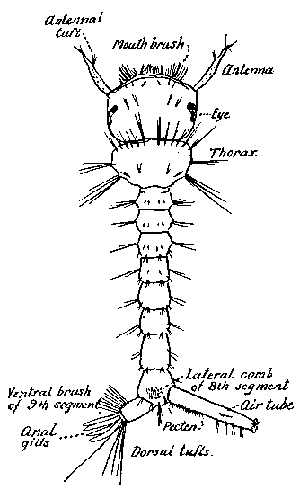
The pupa (fig. 139, b) unlike that of most insects, is active, though it takes no food. The head and thorax are not distinctly separated, but the slender flexible abdomen in sharply marked off. The antennæ, mouth-parts, legs, and wings of the future adult are now external, but enclosed in chitinous cases. On the upper surface, near the base of the wings are two trumpets, or breathing tubes, for the pupal spiracles are towards the anterior end instead of at the caudal end, as in the larva. At the tip of the abdomen is a pair of large chitinous swimming paddles.
As illustrative of the life cycle of a mosquito we shall discuss the development of a common house mosquito, Culex pipiens, often referred to in the Northern United States as the rain barrel mosquito. Its life cycle is often given as typical for the entire group, but, as we have already emphasized, no one species can serve this purpose.
The adults of Culex pipiens hibernate throughout the winter in cellars, buildings, hollow trees, or similar dark shelters. Early in the spring they emerge and deposit their eggs in a raft-like mass. The number of eggs in a single mass is in the neighborhood of two hundred, recorded counts varying considerably. A single female may deposit several masses during her life time. The duration of the egg stage is dependent upon temperature. In the warm summer time the larvæ may emerge within a day. The larvæ undergo four molts and under optimum conditions may transform into pupæ in about a week's time. Under the same favorable conditions, the pupal stage may be completed in a day's time. The total life cycle of Culex pipiens, under optimum conditions, may thus be completed in a week to ten days. This period may be considerably extended under less favorable conditions of temperature and food supply.
Culex pipiens breeds continuously throughout the summer, developing in rain barrels, horse troughs, tin cans, or indeed in any standing water about houses, which lasts for a week or more. The catch basins of sewers furnish an abundant supply of the pests under some conditions. Such places, the tin gutters on residences, and all possible breeding places must be considered in attempts to exterminate this species.
Other species of mosquitoes may exhibit radical departures from Culex pipiens in life-history and habits. To control them it is essential that the biological details be thoroughly worked out for, as Howard, Dyar, and Knab have emphasized, "much useless labor and expense can be avoided by an accurate knowledge of the habits of the species." For a critical discussion of the known facts the reader is referred to their monograph. We shall confine ourselves to a few illustrations.
The majority of mosquitoes in temperate climates hibernate in the egg stage, hatching in the spring or even mild winter days in water from melting snow. It is such single-brooded species which appear in astounding numbers in the far North. Similarly, in dry regions the eggs may stand thorough dessication, and yet hatch out with great promptness when submerged by the rains. "Another provision to insure the species against destruction in such a case, exists in the fact * * * that not all the eggs hatch, a part of them lying over until again submerged by subsequent rains." In temperate North America, a few species pass the winter in the larval state. An interesting illustration of this is afforded by Wyeomia smithii, whose larvæ live in pitcher plants and are to be found on the coldest winter days imbedded in the solid ice. Late in the spring, the adults emerge and produce several broods during the summer.
In the United States, one of the most important facts which has been brought out by the intensive studies of recent years is that certain species are migratory and that they can travel long distances and become an intolerable pest many miles from their breeding places. This was forcibly emphasized in Dr. Smith's work in New Jersey, when he found that migratory mosquitoes, developing in the salt marshes along the coast, are the dominant species largely responsible for the fame of the New Jersey mosquito. The species concerned are Aedes sollicitans, A. cantator and A. tæniorhynchus. Dr. Smith decided that the first of these might migrate at least forty miles inland. It is obvious that where such species are the dominant pest, local control measures are a useless waste of time and money. Such migratory habits are rare, however, and it is probable that the majority of mosquitoes do not fly any great distance from their breeding places.
While mosquitoes are thought of primarily as a pest of man, there are many species which have never been known to feed upon human or mammalian blood, no matter how favorable the opportunity. According to Howard, Dyar, and Knab, this is true of Culex territans, one of the common mosquitoes in the summer months in the Northern United States. There are some species, probably many, in which the females, like the males, are plant feeders. In experimental work, both sexes are often kept alive for long periods by feeding them upon ripe banana, dried fig, raisins, and the like, and in spite of sweeping assertions that mosquitoes must have a meal of blood in order to stimulate the ovaries to development, some of the common blood-sucking species, notably Culex pipiens, have been bred repeatedly without opportunity to feed upon blood.
The effect of the bite varies greatly with different species and depends upon the susceptibility of the individual bitten. Some persons are driven almost frantic by the attacks of the pests when their companions seem almost unconscious of any inconvenience. Usually, irritation and some degree of inflammation appear shortly following the bite. Not infrequently a hardened wheal or even a nodule forms, and sometimes scratching leads to secondary infection and serious results.
The source of the poison is usually supposed to be the salivary glands of the insect. As we have already pointed out, (p. 34), Macloskie believed that one lobe of the gland, on each side, was specialized for forming the poison, while a radically different view is that of Schaudinn, who believed that the irritation is due to the expelled contents of the œsophageal diverticula, which contain a gas and a peculiar type of fungi or bacteria. In numerous attempts, Schaudinn was unable to produce any irritation by applying the triturated salivary glands to a wound, but obtained the typical result when he used the isolated diverticula.
The irritation of the bite may be relieved to some extent by using ammonia water, a one per cent. alcoholic solution of menthol, or preparations of cresol, or carbolic acid. Dr. Howard recommends rubbing the bite gently with a piece of moist toilet soap. Castellani and Chalmers recommend cleansing inflamed bites with one in forty carbolic lotion, followed by dressing with boracic ointment. Of course, scratching should be avoided as much as possible.
Repellents of various kinds are used, for warding off the attacks of the insects. We have often used a mixture of equal parts of oil of pennyroyal and kerosene, applied to the hands and face. Oil of citronella is much used and is less objectionable to some persons. A recommended formula is, oil of citronella one ounce, spirits of camphor one ounce, oil of cedar one-half ounce. A last resort would seem to be the following mixture recommended by Howard, Dyar, and Knab for use by hunters and fishermen in badly infested regions, against mosquitoes and blackflies.
Take 2¼ lbs. of mutton tallow and strain it. While still hot add ½ lb. black tar (Canadian tar). Stir thoroughly and pour into the receptacle in which it is to be contained. When nearly cool stir in three ounces of oil of citronella and 1¼ oz. of pennyroyal.
At night the surest protection is a good bed net. There are many types of these in use, but in order to be serviceable and at the same time comfortable it should be roomy and hung in such a way as to be stretched tightly in every direction. We prefer one suspended from a broad, square frame, supported by a right-angled standard which is fastened to the head of the bed. It must be absolutely free from rents or holes and tucked in securely under the mattress or it will serve merely as a convenient cage to retain mosquitoes which gain an entrance. While such nets are a convenience in any mosquito ridden community, they are essential in regions where disease-carrying species abound. Screening of doors, windows and porches, against the pests is so commonly practiced in this country that its importance and convenience need hardly be urged.
Destruction of mosquitoes and prevention of breeding are of fundamental importance. Such measures demand first, as we have seen, the correct determination of the species which is to be dealt with, and a knowledge of its life-history and habits. If it prove to be one of the migratory forms, it is beyond mere local effort and becomes a problem demanding careful organization and state control. An excellent illustration of the importance and effectiveness of work along these lines is afforded by that in New Jersey, begun by the late Dr. John B. Smith and being pushed with vigor by his successor, Dr. Headlee.