Chapter XII · HOMINOXIOUS ARTHROPODS (continued 2)
Reading edition
gg. With another combination of characters.
h. Costal spine present; body in part black; antennæ noticeably shorter than the epistome, inserted above the middle of the eye and separated from each other by a carina; abdominal segments with marginal macrochætæ; sternopleurals 2:1 or 1:1. Paratricyclea Villen.
hh. Costal spine not distinct, or if present, insect otherwise different.
i. Genæ with several ranges of microchætæ; vibrissal ridges strongly converging; peristome broad; arista moderately plumose; sternopleurals usually 1:1; color entirely testaceous. C. anthropophaga (fig. 87) and grunbergi. Africa. Cordylobia Grünb.
ii. Genæ bare or with but one range of setæ; vibrissal ridges less converging; peristome narrow; arista long plumose.
j. Genæ with a single row of microchætæ.
k. Sternopleurals 2:1; color entirely testaceous. Ochromyia Macq.[P]
kk. Sternopleurals 1:1. P. varia Hough. Africa. Parochromyia Hough
jj. Genæ bare.
k. Basal section of R4+5 bristly only at the immediate base, distal section with a broad curve; distal portion of the abdomen metallic; sternopleurals usually 1:1, rarely 2:1. M. æneiventris Wd. Tropic America. Mesembrinella. G. T.
kk. R4+5 bristly at least nearly half way to the small crossvein; sternopleurals 1:1.
l. Macrochætæ of the abdomen marginal; neither sex with orbitals; no carina between the base of the antennæ; three pairs of presutural inner dorsocentrals. Eastern hemisphere. T. ferruginea. Tricyclea V. d. W. (= Zonochroa B. B. according to Villeneuve 1914).
ll. Abdomen without macrochætæ; wing usually with a marginal streak and gray markings. Brazil Hemilucilia B. B.
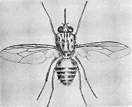
165. Glossina morsitans. (×4.) After Austen.
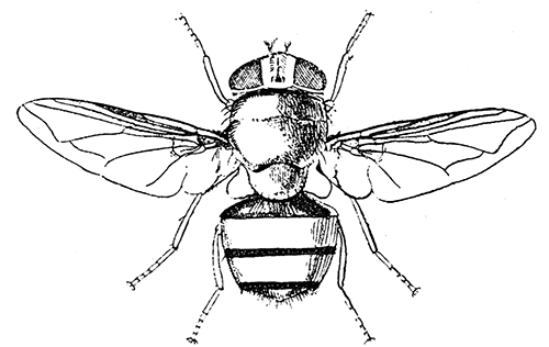
166. Pycnosoma marginale. (×4.) After Graham-Smith.
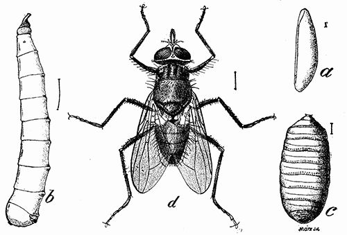
167. Horn fly. (a) egg; (b) larva; (c) puparium; (d) adult. (×4). Bureau of Entomology
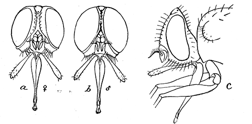
168. Head of horn-fly (Lyperosia irritans); (a) female; (b) male; (c) lateral aspect of female.
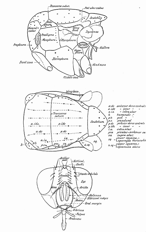
169. Lateral and dorsal aspects of the thorax, and frontal aspect of the head of a muscoidean fly, with designations of the parts commonly used in taxonomic work.
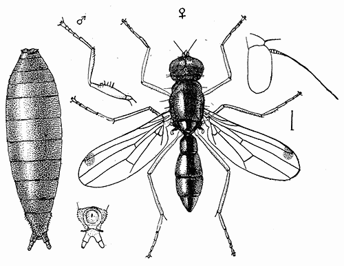
170. Sepsis violacea; puparium and adult. (See page 297.) After Howard.
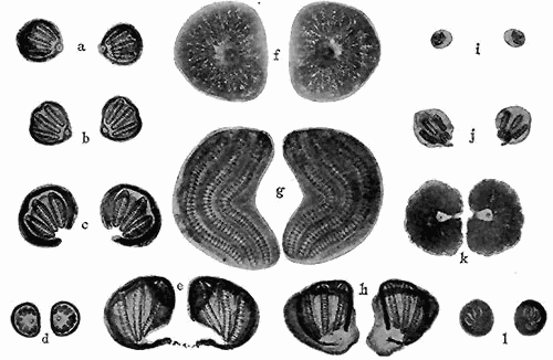
171. Stigmata of the larvæ of Muscoidea. Third instar. (a) Cynomyia cadaverina; (b) Phormia regina; (c) Chrysomyia macellaria; (d) Musca domestica; (e) Sarcophaga sp; (f) Oestris ovis; (g) Gastrophilus equi; (h) Sarcophaga sp; (i) Pegomyia vicina; (j) Protocalliphora azurea; (k) Hypoderma lineata; (l) Muscina stabulans. Magnification for f, g, and k, ×25; all others, ×50.
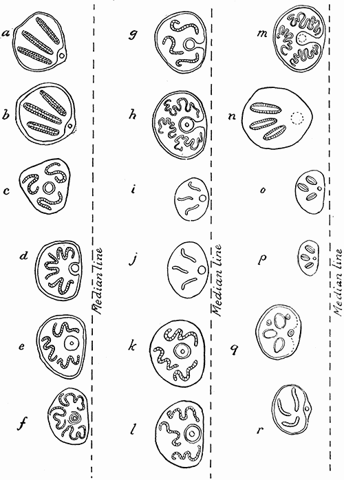
172. Left hand stigmata of the larvæ of muscoidea. Third instar. (a) Lucilia cæsar; (b) Calliphora vomitoria; (c) Stomoxys calcitrans; (d) Pseudopyrellia cornicina; (e) Pyrellia cadavarina; (f) Lyperosia irritans; (g) Mesembrina mystacea; (h) Mesembrina meridiana; (i) Myospila meditabunda; (j) Mydæa umbana; (k) Polietes albolineata; (l) Polietes lardaria; (m) Morellia hortorum; (n) Hydrotæa dentipes; (o) Hebecnema umbratica; (p) H. vespertina; (q) Limnophora septemnotata; (r) Muscina stabulans. (a and b) after MacGregor; (d) after Banks; all others after Portchinsky. Magnification varies. The relative distance to the median line is indicated in each figure.
SIPHONAPTERA. Fleas
Adapted from a table published by Oudemans.
a. Elongated fleas, with jointed (articulated) head, with combs (ctenidia) on head and thorax; with long, oval, free-jointed flagellum of the antenna (fig. 92d). Suborder FRACTICIPATA
b. With ctenidia in front of the antennæ and on the genæ (cheeks); maxillæ with acute apices; labial palpi five-segmented, symmetrical; eyes poorly developed or wanting. On rodents. Hystrichopsyllidæ
c. Abdominal segments without ctenidia.
d. Post-tibial spines in pairs and not in a very close set row; head with ctenidia. Ctenophthalmus Kol.
dd. Post-tibial spines mostly single and in a close set row. Ctenopsyllus and Leptopsyllus. The last genus has recently been erected for L. musculi, a widely distributed species occurring on rats and mice.
cc. Abdominal segments with one or more ctenidia; post-tibial spines in numerous, short, close-set transverse rows on posterior border with about four spines in each row. H. americana. Hystrichopsylla Taschenb.
bb. With only two pairs of subfrontal ctenidia; labial palpi five-segmented, symmetrical; eyes vestigial or wanting. On bats. (= Ischnopsyllidæ). Nycteridipsyllidæ
With more or less blunt maxilla; all tibiæ with notch; a single antepygidial bristle; metepimeron without ctenidium. N. crosbyi from Missouri was found on bats. Rothschild suggests that this is probably the same as N. insignis. (= Ischnopsyllus = Ceratopsyllus), Nycteridiphilus
aa. Head not jointed, i.e. the segments coalescent, traces of the segmentation still being visible in the presence of the vertex tubercle, the falx (sickle-shaped process), and a suture. Suborder INTEGRICIPITA
b. Flagellum of the antennæ long and oval.
c. Usually elongate fleas, with a free-segmented flagellum of the antenna; thorax not shorter than the head, longer than the first tergite.
d. Genæ of the head and the pronotum with ctenidia. Neopsyllidæ
e. Labial palpi four or five-segmented; symmetrical; hind coxæ with patch of spines inside; row of six spatulate spines on each side in front of the antennæ. C. ornate found on a California mole. Corypsylla
ee. Labial palpi two-segmented, transparent, membranous. On hares. Spilopsyllus Baker
dd. No ctenidium on the head.
e. Pronotum with ctenidium. Dolichopsyllidæ
f. Labial palpi five-segmented, symmetrical.
g. Antepygidial bristles one to three; eyes present.
h. Inner side of hind coxæ distally with a comb of minute teeth; falx present. On rodents and carnivores. Odontopsyllus Baker
hh. Inner side of hind coxæ without comb or teeth. Many North American species on rodents. Ceratophyllus Curtis
gg. Antepygidial bristles five on each side; eyes absent; suture white. D. stylosus on rodents. Dolichopsyllus Baker
ff. Labial palpi four or five-segmented; asymmetrical (membranous behind), apex acute. Hoplopsyllus anomalus found on Spermophiles in Colorado. Hoplopsyllidæ
ee. Pronotum without ctenidium. Anomiopsyllus californicus and nudatus on rodents. Anomiopsyllidæ
cc. Very short fleas; flagellum of the antenna with pseudo-segments coalescent; thorax much shorter than the head and than the first tergite Hectopsyllidæ
Flagellum of the antenna with six coalescent pseudo-segments; maxilla blunt. The chigger on man (fig. 93). D. penetrans. (= Rhynchoprion = Sarcopsylla) Dermatophilus Guérin
bb. Flagellum short, round, free portion of the first segment shaped like a mandolin.
c. Thorax not shorter than the head, longer than the first tergite; flagellum either with free segments or in part with the segments coalescent.
d. Head and pronotum with ctenidium; labial palpi asymmetrical. Archæopsyllidæ
With four subfrontal, four genal, and one angular ctenidia. Widely distributed. Ctenocephalus Kol.
e. Head rounded in front (fig. 92a). Dog flea. C. canis
ee. Head long and flat (fig. 92b). Cat flea. C. felis
dd. Neither head nor pronotum with ctenidium. Labial palpi asymmetrical, membranous behind. Pulicidæ
e. Mesosternite narrow, without internal rod-like thickening from the insertion of the coxæ upwards. Human flea, etc. Pulex L.
ee. Mesosternite broad with a rod-like internal thickening from the insertion of the coxæ upwards (fig. 89). X. (Lœmopsylla) cheopis, plague or rat flea. Xenopsylla
cc. Thorax much shorter than the head and than the first tergite. Echidnophagidæ. E. gallinacea, the hen flea also attacks man (fig. 96). (= Argopsylla = Xestopsylla) Echidnophaga Olliff.