Volume 1 — CHAPTER XV.
Reading edition
Arborescent Lycopodiales.
Among the best known plants in the Palaeozoic floras are the genera Lepidodendron and Sigillaria, types which are often spoken of as Giant Club-Mosses or as ancestors of existing species of Lycopodium and Selaginella. Of these genera, but more particularly of Lepidodendron, we possess abundant records in a condition which have made it possible to obtain fairly complete information not only in regard to habit and external features but as to the anatomical characters of both vegetative and reproductive shoots. The structure of Lepidodendron differs too widely from that of recent Club-Mosses (species of Lycopodium) to justify the statement that this prominent member of the Palaeozoic vegetation may be regarded as a direct ancestor of any living plant. There is at least no doubt that Lepidodendron and Sigillaria must be included in the Pteridophyta. The description by Dr Scott[236] of the genus Lepidocarpon, founded on petrified specimens of strobili, demonstrated the existence of a type of lycopodiaceous plant in the Carboniferous period distinguished from all living representatives of the group by the possession of integumented megaspores, which may fairly be styled seeds. Lepidocarpon and another seed-bearing plant Miadesmia are described under a separate heading as lycopodiaceous types characterised by an important morphological feature, which among recent plants constitutes a differentiating character between the Pteridophytes and the Phanerogams.
Lepidodendron.
i. General.
The genus Lepidodendron included species comparable in size with existing forest trees. A tapered trunk rose vertically to a height of 100 feet or upwards from a dichotomously branched subterranean axis of which the spreading branches, clothed with numerous rootlets, grew in a horizontal direction probably in a swampy soil or possibly under water. A description by Mr Rodway[237] of Lycopods on the border of a savannah in Guiana forming a miniature forest of Pine-like Lycopodiums might, with the omission of the qualifying adjective, be applied with equal force to a grove of Lepidodendra. The equal dichotomy of many of the branches gave to the tree a habit in striking contrast to that of our modern forest trees, but, on the other hand, in close agreement with that of such recent species of Lycopodium as L. cernuum (fig. 123), L. obscurum (fig. 124) and other types. Linear or oval cones terminated some of the more slender branches (fig. 188) agreeing in size and form with the cones of the Spruce Fir and other conifers or with the male flowers of species of Araucaria, e.g. A. imbricata. Needle-like leaves, varying considerably in length in different species, covered the surface of young shoots in crowded spirals and their decurrent bases or leaf-cushions formed an encasing cylinder continuous with the outer cortex. The fact that leaves are usually found attached only to branches of comparatively small diameter would seem to show that Lepidodendron, though an evergreen, did not retain its foliage even for so long a period as do some recent conifers.
By the activity of a zone of growing tissue encircling the cylinder of wood the main trunk and branches grew in thickness year by year: the general uniformity in size of the secondary conducting elements affords no indication of changing seasons. As the branches grew stouter and shed their leaves the surface of the bark resembled in some degree that of a Spruce Fir and other species of Picea, in which the leaf-scars form the upper limit of prominent peg-like projections, which, at first contiguous and regular in contour, afterwards become less regular and separated by grooves (fig. 140) and at a later stage lose their outline as the bark is stretched to the tearing point (fig. 140, C). The leafless branches of Lepidodendron were covered with spirally disposed oval cushions less peg-like and larger than the decurrent leaf-bases of Picea, which show in the upper third of their length a clean-cut triangular area and swell out below into two prominent cheeks separated by a median groove and tapering with decreasing thickness to a pointed base, which in some forms (e.g. Lepidodendron Veltheimianum, fig. 185, C, D), is prolonged as a curved ridge to the summit of a lower leaf-cushion.
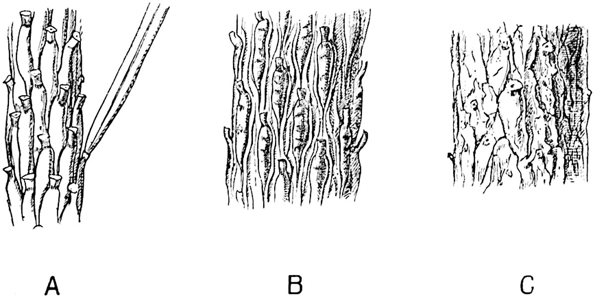
Fig. 140. Picea excelsa. Shoots of different ages showing changes in the appearance of the leaf-cushions: a leaf attached to a cushion in fig. A. (Slightly enlarged.)
A portion of the cushion below the triangular leaf-scar often shows transverse gaping cracks or depressions (fig. 185, C) such as occur on a smaller scale on the older cushions of a Fir twig (fig. 140). Secondary thickening, as in recent trees, is not confined to the vascular cylinder but at an early stage, frequently before there are any signs of secondary wood, the outer region of the broad cortex becomes the seat of active cell-formation which results in the addition of a considerable thickness to the bark. At a later stage of increase in girth, the leaf-cushions are stretched apart and the original surface-features become obliterated by vertical cracks and by the exfoliation of the superficial tissues[238].
Some species of Lepidodendron produced branches characterised by spiral or vertical series of scars; these in older shoots were replaced by depressions having a diameter of several inches and comparable in appearance, as also perhaps in manner of formation, with the scars left on the stem of a Kauri Pine (Agathis australis)[239] on the abscission of lateral branches by a natural process. These shoots, known as Ulodendron, are described in a subsequent section. (page 128.)
A fully-grown Lepidodendron must have been an impressive tree, probably of sombre colour, relieved by the encircling felt of green needles on the young pendulous twigs. The leaves of some species were similar to those of a fir while in others they resembled the filiform needles of the Himalayan Pine (Pinus longifolia). The occasional presence of delicate hyphae in the tissues of Lepidodendron demonstrates susceptibility to fungal pests.
Architecturally, if one may use the term, Lepidodendron owed its power of resistance to the bending force of the wind to its stout outer bark formed of thick-walled elements produced by the activity of a cylinder of cortical meristem (figs. 148, 172, etc.). The vascular axis, of insignificant diameter in proportion to the size of the stem (figs. 152, 153, 172, 181, A), must have played a subordinate part, from a mechanical point of view, as compared with the solid mass of wood of a Pine or an Oak.
•••••
Within the compass of a text-book it is impossible, even if it were desirable, to include an account of the majority of the species of the widely distributed Palaeozoic genus Lepidodendron. In spite of the great number of known species of this common member of Carboniferous floras, our knowledge of the type as a whole is deficient in many points, and such information as we possess needs systematising and extending by comparative treatment based on a re-examination of available data.
In order to appreciate the meaning of certain external features characteristic of Lepidodendron stems it is essential to have some knowledge of the internal structure.
A dual system of terminology has been unavoidably adopted for species of Lepidodendron: the majority of specific names have been assigned to fossils known only in the form of casts or impressions, while petrified fragments, which unfortunately seldom show the surface-features, have received another set of names. A glance at the older palaeobotanical literature reveals the existence of several generic designations, which fuller information has shown to have been applied to lepidodendroid shoots deprived of some of their superficial tissues before fossilisation and differing considerably in appearance from the more complete branches of the same species[240]. It has in some instances been possible to correlate the two sets of specimens, casts or impressions, showing external features, and petrified fragments. We may reasonably expect that future discoveries will enable us to piece together as definite specific types specimens at present labelled with different names.
A well-preserved leaf-cushion of a Lepidodendron—the most obvious distinguishing feature of the genus—is rhomboidal or fusiform and vertically elongated (fig. 146, C, E; fig. 185, C, D): in exceptional cases it may reach a length of 8 cm. and a breadth of 2 cm. The cushion as a whole represents a prominent portion of the stem or branch comparable with the elevation on the twig of a Spruce Fir and the leaf-base of a Lycopodium (cf. fig. 121, A, lower portion) which appears in a transverse section of a branch as a rounded prominence (cf. Lycopodium, fig. 125, A and H). Disregarding differences in detail, a typical Lepidodendron leaf-cushion is characterised by a clearly defined smooth area often situated in the middle region (fig. 146, C, s). This is the leaf-scar or place of attachment of the base of the leaf which was cut off by an absciss-layer while the branch was comparatively young, as in recent forest trees and in some species of Ferns. On the leaf-scar are three smaller scars or cicatricules, the central one is circular or more or less triangular in outline, the two lateral scars being usually oval or circular. The central pit marks the position of the single vascular bundle which constituted the conducting tissue connecting the leaf with the main vascular system of the stem. The two lateral scars (figs. 145, A, p; 146, C, s; 147, p) represent the exposed ends of two strands of tissue, the forked branches of a strand which pass from the middle cortex of the stem into the leaf; this is known as the parichnos, a name proposed by Professor Bertrand in 1891[241].
The specimen shown in fig. 141 shows the linear leaves attached to their respective cushions.
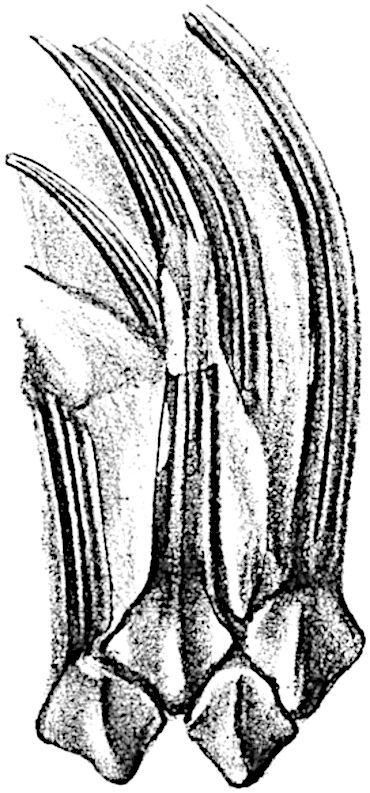
Fig. 141. Lepidodendron Sternbergii. From a specimen in the British Museum (No. v. 1235) from the Coal-Measures of Shropshire. (Nat. size.)
The lamina has a well-defined median keel on the lower surface and on either side a groove in which sections of petrified leaves have demonstrated the occurrence of stomata (cf. fig. 142).
ii. Leaves and Leaf-cushions.
All Lepidodendron leaves, so far as we know, possessed a single median vein only. In some species, as for example in Lepidodendron longifolium Brongn., they have the form of long and slender acicular needles very similar to those of Pinus longifolium; in L. Sternbergii (fig. 141) they are much broader and shorter. In external form as in internal structure it is often impossible to distinguish between the leaves of Lepidodendron and Sigillaria. The distinguishing features enumerated by the late M. Renault cannot be employed, with any great degree of confidence, as diagnostic characters. In transverse section the lamina of a Lepidodendron leaf presents the same appearance as that of the Sigillarian leaves represented in fig. 142. Near the base the free part of the leaf is usually sub-rhomboidal in section with short lateral wings, a ventral keel and two stomatal grooves (fig. 142, A, B, g). The form and arrangement of stomata are shown in fig. 143, A, which was drawn from a piece of a leaf shown in surface-view in a section lent to me by Professor Weiss. It should, however, be pointed out that the leaf cannot be certainly identified with Lepidodendron rather than with Sigillaria, but as the leaves of these two genera are constructed on the same plan the identification is of secondary importance.
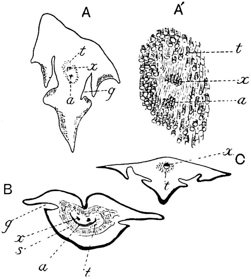
Fig.
142. Leaves of
Sigillaria
in transverse section.
A, A′. Section in the Manchester University Museum (Q. 631).
B, C. Sections in Dr Kidston’s Collection.
The single xylem bundle consists of primary tracheae only, at least in such laminae as have been identified as Lepidodendroid. Surrounding the xylem strand occur delicate parenchymatous cells in some cases accompanied by darker and thicker-walled elements. As in Sigillaria, the leaves of which are more fully described on page 210, a fairly broad sheath of wider and shorter scalariform or spiral transfusion tracheids surrounds the conducting strand (figs. 142, t; 143, B, C, t). As Renault shows in the case of Lepidodendron esnostense[242], the small leaves of which are 1·5–2 mm. broad at the base and several centimetres long, the stomatal grooves and keel die out towards the apex when the lamina assumes a more nearly circular form (fig. 143, C).
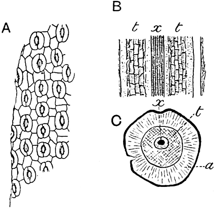
Fig.
143.
A. Stomata in surface-view (Lepidodendron?). a, parenchyma; t, transfusion tracheae; x, xylem. (Manchester University Collection R. 723).
B, C. Lepidodendron esnostense Ren. (After Renault.)
The area of the cushion excluding the leaf-scar is spoken of by some writers as the field. Below the leaf-scar the kite-shaped cushion tapers to a gradually narrowing basal position: in Lepidodendron Veltheimianum, a species characteristic of Lower Carboniferous strata, it is seen to be continuous, as a ridge with sloping sides, with a lower cushion (fig. 185).
Below a leaf-scar the cushion frequently shows a pair of oval areas on which a fine pitting may be detected in well-preserved impressions, these oval scars, as seen in fig. 185, D, are practically continuous at the upper end with the parichnos scars on the leaf-scar area; this is explained by the fact that these infra-foliar scars also owe their existence to patches of lacunar, aerenchymatous tissue in close connexion with the parichnos[243].
Shortly before entering the base of the leaf-lamina the parichnos divides into two arms which diverge in the outer cortical region right and left of the vascular bundle, and passing obliquely upwards they come close to the surface of the leaf-cushion just below the leaf-scar. The diagram—fig. 144, B—shows a leaf-trace, lt, in the leaf-cushion, as seen in a diagrammatic drawing of a vertical radial section of a stem, the dotted lines, p, p′, show the two parichnos arms which are represented as impinging on the surface of the leaf-cushion at p′, and then bending upwards to pass into the leaf-base right and left of the vascular bundle or leaf-trace. For convenience the arms of the parichnos are represented in one plane though actually in different vertical planes.
Fig. 144, A, shows the difference between a view of the original surface of a Lepidodendron, as at a, where a leaf-cushion with a leaf-scar is seen, and a view of an impression representing the outer cortex, b, a short distance below the surface. The surface b, in fig. 144, A, corresponds to the face d-e in the diagrammatic longitudinal section fig. 144, B: the outline of each cushion is clearly visible and in the centre is seen the leaf-trace, lt, with its parichnos.
The surface-features, a (fig. 144, A), have been impressed on the rock, c, (fig. 144, B) in which the specimen was entombed and by the removal of the cast of the stem, that is the thickness b to e in fig. 144, B, the form of the leaf-cushion is revealed. The presence of the two infra-foliar parichnos scars at p′ (fig. 144, A) is explained by the diagram, fig. 144, B, p′.
The relation of the parichnos to the oval scars below a Lepidodendron leaf-cushion has been worked out in detail by Weiss who shows that, at least in some species, the two arms do not bend downwards as shown in the diagram, fig. 144, B, but pursue a straight gradually ascending course as seen in fig. 145, A. Just below the leaf-scar region of the cushion each arm comes into association with a group of lacunar, aerenchymatous tissue, such as occurs in the roots of certain Mangrove plants, and it is this aerenchyma which is exposed on the two oval depressions below the leaf-scar. The structure of this aerenchyma is shown in fig. 145, B; it consists in this species (L. Hickii Wats.) of stellate cells which would constitute an efficient aerating system. Probably, as Weiss suggests, these patches of aerenchyma were originally covered by an epidermis provided with stomata, and it is owing to the destruction of this superficial layer that the two oval scars often form a prominent feature on Lepidodendron leaf-bases[244]. The diagram reproduced in fig. 144, B, may be taken as practically correct, as the patches of aerenchyma described by Weiss do not differ essentially from the parichnos tissue.
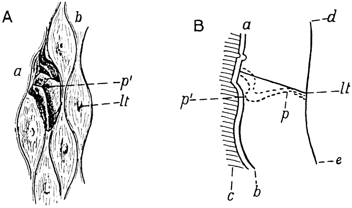
Fig.
144.
Lepidodendron Veltheimianum
Sternb.
Leaf-cushion and leaf-scar seen in surface-view at a; on the rest of the specimen a slightly lower surface is exposed. (After Stur.)
Diagrammatic longitudinal section to explain the differences between its two surfaces a and b shown in fig. A. The shaded portion c represents the rock matrix, the surfaces ab, ed, mark the outer and inner edge of the outer portion of the bark of the Lepidodendron stem. lt, leaf-trace; p, p′, parichnos.
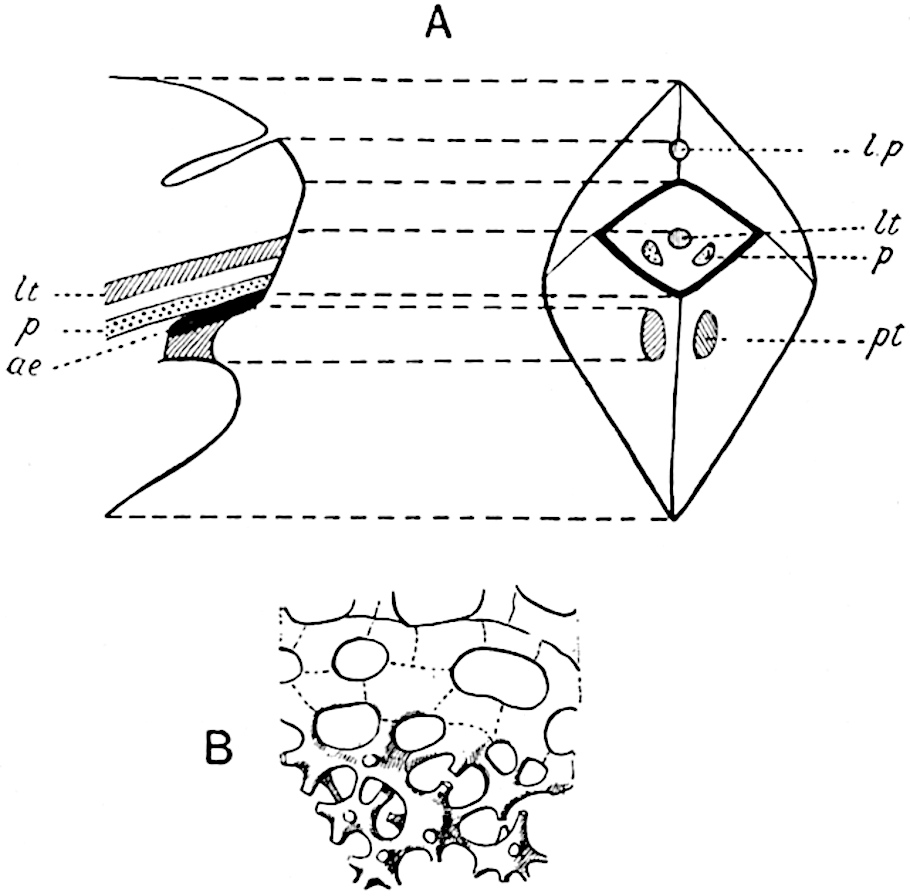
Fig.
145.
Diagrammatic surface-view and longitudinal section of a Lepidodendron leaf-cushion.
Aerenchyma below the leaf-scar. (After F. E. Weiss.)
The parichnos scars are shown on the leaf-scar and cushion in fig. 146, C. In the lower leaf-cushion shown in fig. 146, E, the infra-foliar parichnos scars, p, are clearly seen, but the preservation of the leaf-scar is not sufficiently good to show them on that part of the fossil. In the upper cushion (fig. 146, E) the position of the parichnos arms is shown on the leaf-scar, but the infra-foliar parichnos scars are hidden by two small spiral shells. The genus Spirorbis, to which these shells are referred, appears to have persisted from the Silurian epoch to the present day. The comparatively frequent occurrence of Spirorbis shells on the leaves and other parts of Palaeozoic plants, has recently been dealt with in a paper by Barrois[245] who discusses in detail the habitats of these small animals from the point of view of the conditions under which the plants were preserved. In a note by Malaquin appended to Barrois’ paper the belief is expressed that Spirorbis lived on pieces of Palaeozoic plants which lay under water.
The fact that with one exception all the Spirorbis shells on the specimen of Lepidodendron, of which two leaf-cushions are shown in fig. 146, E, occur on the large parichnos scars on the cheeks of the cushions, suggests the possibility that the escape of gases from the parichnos tissue may have rendered the position attractive to the Spirorbis. It can hardly be accidental that the shells occur on the parichnos strands. This fact recalls the view held by Binney[246] and accepted with favour by Darwin[247] that Lepidodendron and other coal-forest trees may have lived with the lower parts of the stems in sea water.
Above the leaf-scar is a fairly deep triangular or crescentic pit (fig. 146, C, l) known as the ligular pit from the occurrence on younger shoots of a delicate organ like the ligule of Isoetes (fig. 132) embedded in a depression in the upper part of the leaf-cushion. The ligule was first figured in Lepidodendron by Solms-Laubach[248] and described in English material by Williamson under the name of the adenoid organ[249].
In some Lepidodendron stems a second triangular depression may occur above the ligular pit, the meaning of which is not clear: this has been called the triangulum by Potonié[250]. Stur[251] suggested that it may represent the position occupied by a sporangium in Lepidodendron cones.
It is important to remember that as a branch increases in girth the leaf-cushions are capable of only a certain amount of growth: when the limit is reached they are stretched farther apart and thus the narrow groove which separates them is converted in older stems into a comparatively broad and flat channel, thus altering the surface characters.
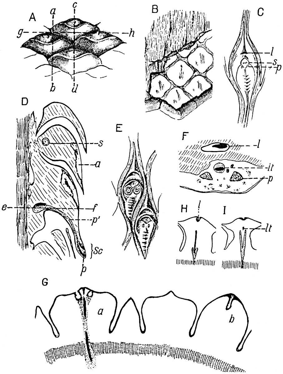
Fig.
146. Lepidophloios and Lepidodendron leaf-cushions.
A, B, D, F, G, H, I. Lepidophloios. (Fig. A should be reversed.)
C, E. Lepidodendron aculeatum.
A, B. From a specimen in the Sedgwick Museum, Cambridge (leaf-cushion 3 cm. broad).
C. From a specimen in the Sedgwick Museum, Cambridge (leaf-cushion 4 cm. long).
D. From a section in the Cambridge Botany School Collection.
E. From a specimen in the Bunbury Collection, Cambridge Botany School, showing Spirorbis shells (leaf-cushion 2 cm. long).
F. From a section in the Williamson Collection, British Museum No. 1, 973.
G, H, I. From sections in the Cambridge Botany School Collection.
Another feature worthy of notice in reference to the leaf-cushions of Lepidodendron is the occurrence in rare instances of alternate zones of larger and smaller cushions. This variation in the size of the leaf-cushions is by no means uncommon in the closely allied genus Sigillaria; in Lepidodendron it has been described by Potonié[252] in L. volkmannianum and more recently by Mr Leslie and myself[253] in a South African species L. vereenigense.
Owing to the natural exfoliation of the superficial layers of the outer bark at a certain stage in the growth of the plant, or in some instances no doubt as the result of post-mortem decay, which destroys the delicate cells of the meristematic zone in the outer cortex, isolated leaf-cushions and strips of the external surface are occasionally met with as carbonised impressions.
The appearance presented by a Lepidodendron stem which has been deprived of its superficial tissues may be dealt with more intelligibly after we have become familiar with the anatomical characters.
iii. Lepidophloios.
Before proceeding further with the genus Lepidodendron a short account may be intercalated of the external features of a lepidodendroid type of stem which it is customary to describe under a distinct generic title Lepidophloios. This name is convenient for diagnostic purposes though it seems clear that apart from the form of the leaf-cushion (fig. 146, A) we are at present unable to recognise any well-defined differences between the two forms Lepidodendron and Lepidophloios. For general purposes the name Lepidodendron will be used as including plants possessing leaf-cushions of the type already described as well as those with the Lepidophloios form of cushion.
The generic name Lepidophloios was first used by Sternberg[254] for a Carboniferous species which he had previously described as Lepidodendron laricinum. In 1845 Corda[255] instituted the name Lomatophloios for specimens possessing the same external characters as those for which Sternberg had chosen the name Lepidophloios. The leaf-cushions of Lepidophloios differ from those of the true Lepidodendron in their relatively greater lateral extension (cf. fig. 146, A and C), in their imbricate arrangement and in bearing the leaf, or leaf-scar, at the summit. In some species referred to Lepidophloios the cushions are however vertically elongated and in this respect similar to those of Lepidodendron: an example of this type is afforded by Lepidophloios Dessorti a French species described by Zeiller[256]. In younger branches the cushions may be directed upwards having the leaf-scar at the top; but in the majority of specimens the cushions are deflexed as in figs. 146, D; 160, A. The shoot of Lycopodium dichotomum shown in fig. 121, B, with the leaves in the reversed position bears a close resemblance to a branch of Lepidophloios.
The photograph of Lepidophloios scoticus Kidst.[257] reproduced in fig. 160, A, illustrates the dichotomous branching of the stem and the form of the cushions with the leaf-scars pointing downwards. In the fertile branch of the same species shown in fig. 160, B, the leaf-scars face upwards.
In most species the cushions are simply convex without a median keel, but in some cases a median ridge divides the cushion into two cheeks as in the genus Lepidodendron. The leaf-scar bears three small scars, the larger median scar marking the position of the leaf-trace, while the lateral scars are formed by the two arms of the parichnos: in some examples of deflexed cushions, though not in all, a ligular pit occurs on the cushion a short distance above the leaf-scar.
The drawing reproduced in fig. 146, A, showing the leaf-scar on the upper edge of the cushion should have been reversed with the leaf-scars pointing downwards. This figure represents part of the surface of a specimen consisting of the outer cortex of a stem with leaf-cushions 3 cm. broad. The thickness of this specimen is 4 cm.: a section through the line ab is represented in fig. 146, D (reproduced in the correct position, with the leaf-scars, sc, pointing downwards): internal to the cushions is a band of secondary cortex (the shaded strip on the outer edge of the section) which was formed on the outside of the phellogen. The phellogen is a cylinder of actively dividing cells in the outer part of the cortex of the stem, often spoken of as the cork-cambium or cortical meristem, which produces a considerable amount of secondary cortical tissue on its inner face and a much smaller amount towards the stem surface. This delicate cylinder frequently forms a natural line of separation between the outer shell of bark and the rest of the stem. In the specimen before us, the thin-walled cells of the phellogen were ruptured before petrification and the outer shell of bark was thus separated as a hollow cylinder from the rest of the stem: this cylinder was then flattened, the two inner surfaces coming into contact. Fig. 146, D, represents a section of one half of the thickness of the flattened shell.
This separation of the outer cortex, and its preservation apart from the rest of the stem, is of frequent occurrence in fossil lycopodiaceous stems. The flattened outer cortical shell of a Lepidophloios, specifically identical with that shown in fig. 146, A and D, was erroneously described by Dr C. E. Weiss in 1881 as a large lepidodendroid cone[258].
Fig. 146, B, affords a view of the inner face of the specimen of which the outer surface is seen in fig. 146, A: the surface shown in the lower part of the drawing, on which the boundaries of the cushions are represented by a reticulum, corresponds to the inner edge of the strip of secondary cortical tissue represented by the vertically shaded band in fig. 146, D.
The shaded surface in fig. 146, B, represents a slightly deeper level in the stem which corresponds to the outer edge of the vertically shaded band of fig. 146, D: the narrow tapered ridges (fig. 146, B) represent the leaf-traces passing through the secondary cortex, and the fine vertical shading indicates the elongated elements of which this strip of secondary cortex is composed.
In the longitudinal section diagrammatically reproduced in fig. 146, D, cut along the line ab of fig. 146, A, the parenchymatous tissue of the stout cushions has been partially destroyed, as at a; at s is seen the section of a Stigmarian rootlet which has found its way into the interior of a cushion. Each leaf-trace is accompanied by a parichnos strand as in the true Lepidodendron; at the base of the leaf-cushion the parichnos branches into two arms which diverge slightly right and left of the leaf-trace, finally entering the base of the leaf lamina as two lateral strands (fig. 147, p). At one point in fig. 146, D the section has shaved a leaf-trace represented by a black patch resting on the parichnos just above the line ef, but it passes through one of the parichnos arms p′ which debouches on to the leaf-scar sc at p. Had the section been cut along the line cd of fig. 146, A the leaf-trace would have been seen in a position similar to that occupied by the parichnos p′ in fig. 146, D.
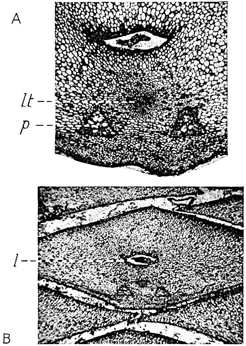
Fig. 147. Lepidophloios leaf-cushion in tangential section. (From a section in the Williamson Collection, British Museum, No. 1973.)
Fig. 147, A, affords a good example of a tangential section through a Lepidophloios leaf-cushion, 1 cm. broad, like that represented in fig. 146, A, showing the vascular bundle lt, the two parichnos strands, p, composed of large thin-walled cells (cf. Isoetes, fig. 133, H, I), and the ligular pit near the upper edge of the section enclosing the shrunken remains of the ligule (fig. 147, B, l).
LEPIDODENDRON
Fig. 147, B, shows the form of the tangentially elongated leaf-cushions of Lepidophloios and their spiral disposition.
Fig. 146, F, represents a section similar to that shown in figs. 147, A and B, but in this case the leaf-trace, lt, and the parichnos strands, p, lie in a cavity formed by the destruction of some of the leaf-cushion tissue. It is worthy of notice that the parichnos cells have resisted decay more successfully than the adjacent tissue of the cushion.
The diagrammatic sketches reproduced in fig. 146, H and I, were made from a transverse section similar to one originally figured by Williamson[259]: fig. 146, H, corresponding in position to the line gh in fig. 146, A, passes through the ligular pit, l, and cuts across the parichnos in the act of branching; the leaf-trace passes outwards beyond the Y-shaped parichnos strand. In the other section, fig. 146, I, the parichnos is shown in a horizontal plane and the leaf-trace, lt, appears in oblique transverse section. In both sections and in fig. 146, G the shaded band at the base represents the secondary cortical tissue external to the phellogen.
The transverse section represented in fig. 146, G, shows in the left-hand cushion, a, the exit of the two parichnos arms and the leaf-trace between them: it illustrates also the various forms assumed by lepidodendroid leaf-cushions when cut across at different levels.
iv. The Anatomy of Lepidodendron vasculare Binney[260]. Figs. 148–155, 168, A.
In the earlier literature dealing with the anatomy of Lepidodendron and Sigillaria the presence or absence of secondary vascular tissue was made the criterion of generic distinction and the distinguishing feature between the classes Pteridophytes and Gymnosperms, Lepidodendron being relegated to the former class because it was supposed to have no power of forming secondary wood, while Sigillaria, characterised by a considerable development of such tissue, was classed by Brongniart and afterwards by Renault as a Gymnosperm. Binney[261] in 1865 recognised that the two types of stem pass into one another, but it was Williamson[262] who provided complete demonstration of the fallacy of the Brongniartian view.
These two undoubted Pteridophytes agree very closely in anatomical structure and both are now recognised as arborescent genera of Lycopodiaceous plants. In a paper published by Lomax and Weiss in 1905[263] a specimen is described from the Coal-Measures of Huddersfield, in which a decorticated stem with the anatomical characters of Binney’s Sigillaria vascularis gives off a branch having the anatomical structure which it has been customary to associate with the species Lepidodendron selaginoides, so-called by Sternberg and founded by him on impressions showing well-preserved external characters.
In 1862 Binney[264] described petrified specimens of vegetative shoots from the Lower Coal-Measures of Lancashire under the names Sigillaria vascularis and Lepidodendron vasculare. These were afterwards recognised as different states of the same species. A few years after the publication of Binney’s paper Carruthers[265] identified Binney’s species Lepidodendron vasculare with Sternberg’s L. selaginoides. The evidence on which this identification rests has not been stated, but many writers have retained this specific designation for the well-defined type of anatomical structure first described by Binney as L. vasculare. The use of the specific name selaginoides is, however, open to objection. The species Lepidodendron selaginoides, as pointed out by Kidston[266], is probably identical with the plant which Brongniart had named L. Sternbergii before the institution of Sternberg’s species, and we are not in possession of convincing evidence as to the connection of L. Sternbergii (= L. selaginoides) with specimens possessing the anatomy of Binney’s type. Binney’s designation is therefore retained for the anatomical type described in the following pages[267].
The most detailed account hitherto published of the anatomy of Lepidodendron vasculare is that by the late M. Hovelacque[268], based on material from the Lower Coal-Measures of England.
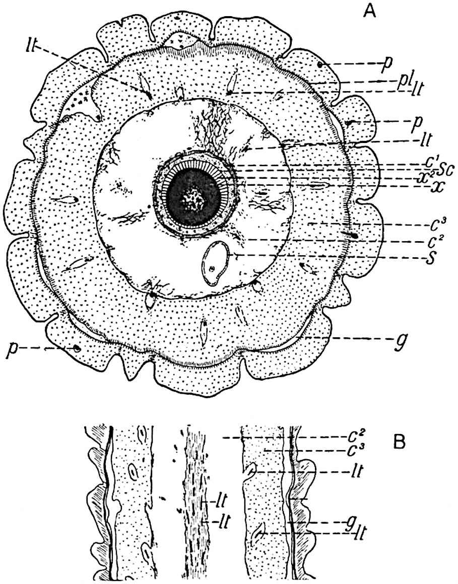
Fig.
148.
Lepidodendron vasculare
Binney.
Transverse section. (Based on a section 2·5 cm. in diameter, in the Cambridge Botany School Collection.)
Longitudinal section. (Drawn from a section in Dr Kidston’s Collection.)
The small shoot, represented somewhat diagrammatically in fig. 148, A, illustrates the anatomical features of a typical example of the species: the shoot has a diameter of 2·5 cm. and its central cylinder (x-sc) is 2·5 mm. in width.
Noticeable features are (i) the small size of the central cylinder (or stele) in proportion to the diameter of the branch, (ii) the production at a comparatively early stage of growth of a zone of secondary wood, x2, which gradually assumes the form of a complete cylinder of unequal breadth, surrounding the primary xylem, x, (iii) the formation of a secondary cortical tissue by a meristematic cylinder (phellogen, pl) situated close to the leaf-cushion region of the outer cortex. On the outer edge the stele consists of narrow tracheae some of which show in longitudinal section the spiral form of thickening characteristic of most protoxylem elements: towards the centre of the stele the diameter of the tracheae gradually increases and parenchymatous cells become associated with the elongated scalariform elements. In the central region the stele is composed of parenchymatous tissue arranged in vertical series of short cells, interspersed with short tracheae distinguished by the greater thickness of their walls and by their scalariform and reticulate thickening bands. Some of these short tracheae are shown in vertical section in fig. 149, B: the fine and broken lines connecting adjacent thickening bands probably represent the remains of the original wall. These delicate bands, which have been figured in various species of lepidodendroid plants[269], are worthy of notice in connexion with the recent work of Mr Gwynne-Vaughan[270] who has shown that in many recent ferns the scalariform bands in the xylem elements are not connected by a thin pit-closing membrane, but are separated from one another by open spaces. In the Lepidodendron tracheae we seem to have a stage in which the intervening membrane is in process of absorption. It is, however, possible that the threads may be the result of contraction and splitting of the membrane during drying or decay.
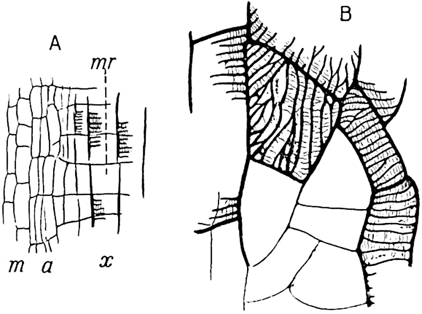
Fig.
149.
Lepidodendron vasculare
.
a
, immature tracheae;
m
, meristem;
mr
, medullary ray;
x
, xylem.
Longitudinal section through the edge of the secondary wood.
Short tracheae in the centre of the stele. (From a specimen from the Halifax Hard bed in Dr Kidston’s Collection.)
The stele of Lepidodendron vasculare, before the addition of any secondary xylem, may be described as a protostele, a term originally proposed by Professor Jeffrey[271], in which the central part of the conducting strand of xylem elements has been converted into rows of parenchyma and short tracheids, the latter being better adapted to storage than to conduction. It is probable that this type of stelar anatomy, which distinguishes L. vasculare from other species, represents a comparatively primitive arrangement forming a transition between the stele of L. esnostense, which consists of a solid rod of tracheids, and the stele of L. Harcourtii (fig. 179, A) and other species in which the xylem forms a cylinder enclosing a large parenchymatous pith.
Parenchymatous cells occur in contact with the outer edge of the xylem-cylinder some of which are distinguished by an irregular reticulate pitting. The tangential section represented in fig. 148, B, illustrates the appearance of a shoot of L. vasculare in which no secondary xylem is present: the central strand of tissue consists of the parenchyma abutting on the xylem with several leaf-traces (lt) passing upwards in an almost vertical course from the outer edge of the stele.
The secondary xylem (fig. 148, A, x2) consists of radially arranged scalariform tracheae with associated rows of parenchymatous cells which form medullary rays (fig. 149, mr). Leaf-traces pass through the medullary rays in the secondary xylem cylinder in a direction at right angles to the primary xylem stele from which they are given off, but at the outer edge of the secondary xylem they bend suddenly upwards and for a time follow a steep and almost vertical course.
In well-preserved longitudinal sections the outermost secondary xylem tracheae are seen to be succeeded by a few narrow and vertically elongated elements (fig. 149, A, a), which represent young unlignified tracheae: these are followed by shorter parenchymatous cells (m) forming part of a meristematic zone from which the secondary xylem receives additions.
Returning to fig. 148, A; the zone of secondary wood, x2, composed of scalariform tracheids and medullary rays, is succeeded by a few layers of parenchymatous cells and beyond this is a broader zone, sc, to which the term secretory zone has been applied[272]; this is made up of small parenchymatous cells varying in size and of larger spaces which appear to have been formed by the disorganisation of thin-walled elements. The whole zone presents a characteristic appearance due to the association of small cells, large clear spaces, and a certain amount of dark-coloured material suggestive of tissue disorganisation and secreted products. The anatomical characters of the secretory zone are shown in the photograph, fig. 168, A, sc. Several leaf-traces are seen in transverse section in the secretory zone (black dots in fig. 148, A, sc; fig. 154, C, lt): each trace consists of a strand of narrow tracheae accompanied by a few encircling layers of small parenchymatous cells. As a trace continues its steeply ascending course through the secretory zone, it becomes associated with a strand of that tissue and assumes the form of a collateral vascular bundle, the outer part of which does not consist of typical phloem but of shorter elements derived from the secretory zone. Beyond the secretory zone we find a more homogeneous tissue composed of parenchymatous elements slightly extended tangentially (figs. 148, A, c1; fig. 168, A, c); this is spoken of as the inner cortical region. In the great majority of sections of L. vasculare as of other species of the genus, the broader middle cortex (fig. 148, c2) is occupied by mineral matter, introduced subsequent to decay of the tissue; or it is represented by patches of delicate tissue composed of loosely arranged parenchymatous cells varying considerably in size and shape, some being small, oval or polygonal elements while others have the form of sinuous hypha-like tubes.
In this middle cortical region may be seen leaf-traces passing outwards in an almost horizontal course (fig. 148, A, lt): after leaving the inner cortex the leaf-traces bend somewhat abruptly outwards to follow a more direct path through the middle and outer cortex. The ring of tissue, s, seen in the middle cortex of fig. 148, A, belongs to a Stigmarian rootlet.
The outer cortex (fig. 148, A and B, c3) consists of homogeneous parenchyma which is stronger and more resistant to decay than the looser middle cortex. The leaf-traces, as shown in fig. 148, B, pass through this region in a rather steeply ascending direction: each is seen to be enclosed by a space originally occupied by a strand of middle cortical tissue which accompanies lepidodendroid leaf-traces on their under side and has already been described as the parichnos, (pp. 97, 100–103; figs. 146, 147).
The surface of the stem shown in section in fig. 148, A, is composed of broad leaf-cushions. A single leaf-trace with its parichnos passes into each cushion, but in the neighbourhood of the base of a cushion the parichnos bifurcates (cf. fig. 146, H, I) and the arms diverge slightly to the right and left finally passing beyond the cushion into the lamina of the leaf, their position being shown, as already explained, by the two small lateral scars on the leaf-scar area.
The diagrammatic sketch of a radial longitudinal section through a leaf-cushion represented in fig. 150 illustrates the relation of the leaf-trace to the leaf-cushion. The trace consists of xylem, x, above and a strand of the secretory zone, st, below; the parichnos tissue was originally present on the under side of the leaf-trace at a. The external surface, bc, marks the limit of the leaf-scar through the middle of which passes the vascular strand lt.
The lower gap a has been formed by the tearing of thin-walled cells of the phellogen, the meristematic tissue from which a considerable amount of secondary cortical tissue or phelloderm has been produced at pd. On the outside of the cushion, c, the cells are somewhat crushed and distinguished by their darker colour from the bulk of the parenchymatous tissue d.
This section also illustrates another characteristic feature of Lepidodendron, namely the presence of a ligule and a ligular pit: the former is represented by a carbonised patch of tissue and the latter extends from the surface of the cushion at b, just above the leaf-scar, almost to the level of the leaf-trace, lt. A comparison of this section with figs. 146 and 147 will make clear the relation of the several parts of the cushion and leaf-scar.
The gaps gg, seen in fig. 148, A and B, mark the position of the delicate meristematic zone or phellogen which arises close to the bases of the leaf-cushions; the phellogen has already produced a few rows of radially disposed elements, represented by short radial lines in the drawing, which constitute secondary cortical tissue.
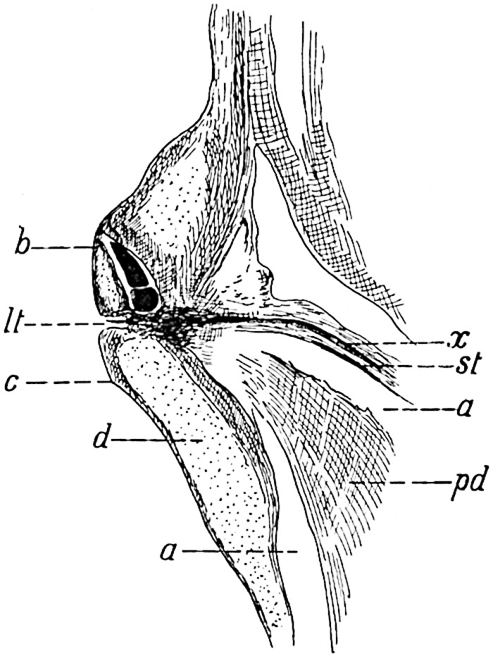
Fig. 150. Lepidodendron vasculare. Leaf-cushions in longitudinal section. (From a specimen in Dr Kidston’s Collection.)
In older shoots the amount of the secondary cortical tissue developed on the inner side of the phellogen is considerable (cf. figs. 152, 153).
The structure of the cortex of a shoot in which secondary growth, both in the stele and in the outer cortex, has progressed further than in the specimen shown in fig. 148 is represented in fig. 151.
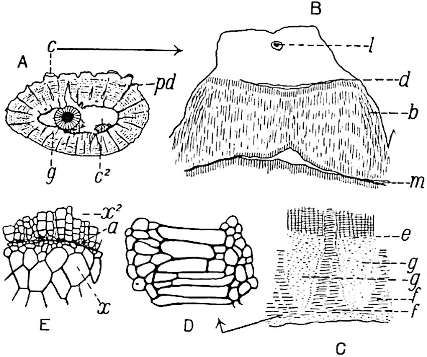
Fig. 151. Lepidodendron vasculare. An older stem than that shown in fig. 148. (From a section in the Manchester Museum. No. 351.)
The section (fig. 151, A) measures 7 × 3·8 cm. in diameter; the primary xylem is surrounded by a fairly broad cylinder of secondary wood (fig. 151, E, x and x2). The almost smooth surface of the primary wood (fig. 151, E, x) is succeeded by the secondary xylem, x2, characterised at its inner edge by the tapered ends of the radial rows of scalariform tracheids between which occur several delicate parenchymatous cells (fig. 151, E, a). The occurrence of such isodiametric elements, often exhibiting a delicate spiral thickening band, is a characteristic feature of the boundary between primary and secondary wood in lepidodendroid stems. The secondary wood is penetrated by numerous medullary rays and in some of them are seen strands of narrow spirally thickened tracheae—the leaf-traces—which are in organic continuity with the exarch protoxylem of the primary wood. The leaf-traces are oval and mesarch. The space, c2, (fig. 151, A) originally occupied by the delicate middle cortex, is succeeded by a shell of outer cortex composed chiefly of secondary tissue (phelloderm, pd) passing towards the inner boundary of this region into the primary outer cortex g (fig. 151, A and C). The radially disposed elements which make up the bulk of the phelloderm are associated with concentric rows of secretory strands, represented by tangentially arranged dots in fig. 151, A: on the outer edge of the phelloderm a few patches of primary cortex are still preserved, as at c, fig. A. One of these is shown on a larger scale in fig. B; at m the phelloderm is interrupted by a gap beyond which the cells have thinner walls and show signs of recent division; this is probably the position of the phellogen. The tissue b, fig. 151, B, consists of secondary cortex succeeded beyond d by the parenchymatous tissue of the leaf-cushion, in which the remains of a ligule, l, are seen in the ligular pit. This section corresponds in position to a line drawn across fig. 150 at the level of b. In this specimen we have two kinds of secondary cortical tissue: that formed external to the phellogen, from m to d in fig. 151, B, is less in amount than that produced internal to the phellogen. We cannot make any satisfactory statement as to the nature of this secondary tissue, whether or not any of it agreed in composition with the cork which is usually formed external to the phellogen in recent plants. As the stem of a Lepidodendron grew in girth the leaf-cushions became separated by intervening depressions composed of the secondary cortex formed external to the phellogen, but at a later stage the cushions were thrown off, leaving the outer edge of the phelloderm as the superficial tissue. This exposed tissue became fissured as growth and consequent stretching continued, producing the appearance seen on the surface of the still older stem represented in fig. 153.
The inner edge of the phelloderm seen at e in fig. 151, C, passes suddenly into the inner primary region of the outer cortex (fig. 151, A and C, g) which comprises two types of parenchymatous tissue, patches of isodiametric cells, g, g, alternating with radially arranged areas consisting of tangentially elongated elements (fig. C, f, f; fig. D) which extend as wedges into the phelloderm.
The longitudinal section represented in fig. 152, B, shows an equal bifurcation of a stem in which no secondary xylem is present; in the lower part of the section the xylem and the outgoing leaf-traces are seen in radial section and at the upper end of each arm the leaf-traces alone, lt, are exposed, as in fig. 148, B. It is interesting to notice the large amount of phelloderm which has been produced in the fork of the branch, at pd, where greater strength is required.
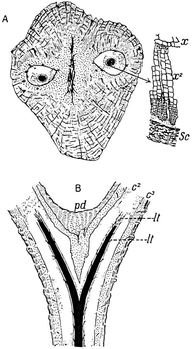
Fig.
152.
Lepidodendron vasculare.
Sections of dichotomously branched shoot.
From a section (10·5 × 9 cm.) in the Cambridge Botany School Collection.
From a section (8 cm. long) in the Cambridge Collection.
The section represented diagrammatically in fig. 152, A, has lost the outermost part of the cortex together with the leaf-cushions; it consists largely of secondary cortex composed of radially disposed phelloderm cells and tangentially placed secretory strands (represented by the discontinuous black lines in the drawing): the dotted region in the central part of the axis is composed of primary cortical parenchyma, and the two spaces surrounding the steles contain portions of the lacunar middle cortex. Each stele possesses a narrow crescentic zone of secondary xylem; the amount is greater in the case of the right-hand stele, of which a small piece is shown on a larger scale; the striking contrast in size between the outer and more internal secondary tracheae is no doubt the expression of some unfavourable condition of growth. The position of the secretory zone beyond the secondary xylem is shown at sc, fig. 152, A.
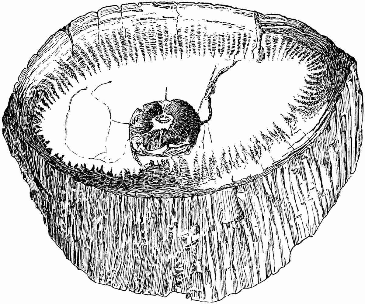
Fig. 153. Lepidodendron vasculare. (From a specimen (16 × 7·5 cm.) in the Manchester Museum.)
An example of a large and partially decorticated stem is afforded by the specimen (16 × 7·5 cm.) shown in fig. 153. The irregularly ribbed surface is formed of rather thick-walled phelloderm, in which occur tangentially arranged rows of secretory strands. The tapered form of the secondary cortex as it abuts internally on the primary cortex is shown very clearly in the drawing (cf. fig. 151, C). The stele in this much older stem consists mainly of secondary wood.
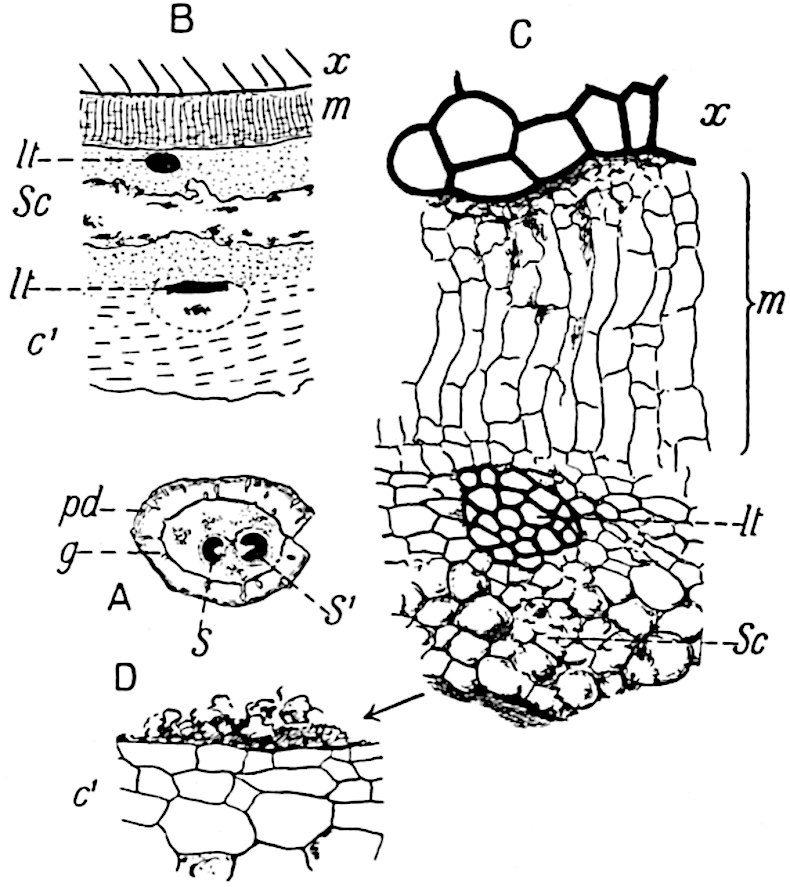
Fig. 154. Lepidodendron vasculare. Shoot (2·8 cm. diam.) with two steles. (From a specimen from Halifax in the Williamson Collection, British Museum, No. 340.)
An interesting example of a small shoot, the largest diameter of which is 2·8 cm., is shown in fig. 154, A: the section was cut a short distance above the bifurcation of the stele into two approximately equal branches. The outer part of the cortex consists of phelloderm, pd, with the usual rows of secretory tracts, and primary outer cortex g; the middle cortex is represented by patches of parenchyma with a few leaf-traces. To one of the steles, s′ (fig. 154, A), a crescent-shaped band of secondary xylem has been added; the other stele, S, possesses no fully developed secondary elements.
Fig. 154, B and C, illustrates the anatomical features immediately external to the primary xylem of the smaller stele, s. The comparatively broad band of radially disposed parenchyma, m, is connected with the outermost elements of the xylem by a few rather dark and small crushed parenchymatous cells. The band m, which we may speak of as the meristematic zone, clearly consists of cells in a state of division; it is in this region that the secondary xylem is produced. Beyond the leaf-trace, (fig. 154, C, lt), occurs a portion of the secretory zone, some of the smaller cells of which show signs of disorganisation; but most of this tissue has been destroyed (fig. 154, B, sc). The outer edge of the secretory zone is shown in fig. 154, D abutting on the cells of the inner cortex, c′. The leaf-trace shown in the inner cortex in fig. 154, B illustrates the more oval or tangentially extended form of the xylem in this region, in contrast to the more circular outline which it exhibits on the inner side of the secretory zone.
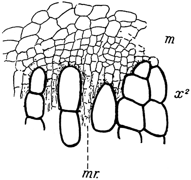
Fig. 155. Lepidodendron vasculare. Outer edge of secondary xylem: m, meristematic zone; mr, medullary ray. (Drawn from the section shown in fig. 168, A).
The transverse section, part of which is reproduced in fig. 168, A, illustrates a characteristic feature, namely the juxtaposition of the outermost tracheae of the secondary xylem and much smaller cells of the meristematic zone. This is seen in fig. 155, which shows a small piece of fig. 168, A, on a larger scale. In plants with a normal cambium the segments cut off from the initial layer fit on to the elements of the xylem or phloem to which they are to form additions, but in Lepidodendron it seems to be a general rule to find each of the most external lignified elements abutting on a group of two or three much smaller cells. It is difficult to believe that the meristem shown in fig. 155, m, could produce secondary xylem elements equal in size to those already formed: in all probability had growth continued there would have been a marked difference between the size of the secondary tracheids, as in fig. 152, A, x2, where there was no doubt some cause which interfered with normal cambial activity. This disparity in size between the secondary xylem elements and the adjacent parenchymatous tissue of the meristematic zone is by no means exceptional and may be described as the general rule. It is at least certain that in Lepidodendron vasculare, as in other species, the secondary xylem was succeeded by a broad band of parenchymatous tissue, from which new tracheae and medullary-ray elements were produced, and not by a narrow cambium such as occurs in recent plants.
v. Lepidodendron stems as represented by casts and impressions of partially decorticated specimens.
The differentiation of the outer cortex of a Lepidodendron into comparatively thin-walled and more resistant tissue has been the cause of unequal decay and the consequent formation of shrinkage cavities. In addition to the unequal resisting power of contiguous tissues, another important factor in determining the nature of casts and impressions is the existence of the cylinder of delicate cells in the outer cortex of stems and branches. As already pointed out, this meristematic cylinder or phellogen constitutes a natural line of separation, as in the case of the cambium layer between the wood and the external tissues in a fresh Sycamore twig. The result of the separation of an outer shell of bark from the rest of the stem and the results of unequal decay in the more superficial tissues, have necessarily led to the preservation of the same specific type under a variety of forms.
Our knowledge of the anatomy of Lepidodendron stems enables us to recognise in fossils of very different appearance specimens in various conditions of preservation of one and the same type. Such names as Knorria, Bergeria and Aspidiaria are examples of generic titles instituted before any adequate knowledge of Lepidodendron anatomy was available.
Differences in age as well as different degrees of decortication have contributed in no small measure to the institution of generic and specific names which more recently acquired knowledge has shown to be superfluous.
a. Knorria.
The designation Knorria, after a certain G. W. Knorr of Nürnberg, was proposed by Sternberg in 1826[273] for casts of Palaeozoic stems of a type figured more than a century earlier by Volkmann[274]. Goeppert, in his earlier works, published drawings of fossil stems which he referred to Sternberg’s genus: one species he at first called Didymophyllum Schollini. He afterwards[275] described some specimens which showed that the features characteristic of Knorria may occur on partially decorticated stems with leaf-cushions of the true Lepidodendron type. His specimens, preserved in the Breslau Museum, demonstrate the accuracy of his drawings and conclusions. Goeppert, and after him Balfour[276], drew attention to the different appearances presented by branches of Araucaria imbricata when preserved with the surface intact and after partial decortication, as illustrating possible sources of error in the determination of fossil stems.
Although it is now a well-established fact that fossils bearing the name Knorria are imperfect lepidodendroid stems, the use of the term may be conveniently retained for descriptive purposes. The specimen from the Commentry coal-field of France, shown in fig. 156, affords some excuse for the institution of several generic names for different states of preservation or decortication of one species. The cortical level exposed at e is characterised by spirally disposed peg-like ridges with truncated apices: it is this form of cast which is usually designated Knorria. The ridges vary in size and shape in different types of stem; they may be narrow as shown at e, fig. 156, or short and broad with rounded distal ends. In some cases they are forked at the apex, as in the partially decorticated specimen of Lepidodendron Veltheimianum represented in fig. 185, A.
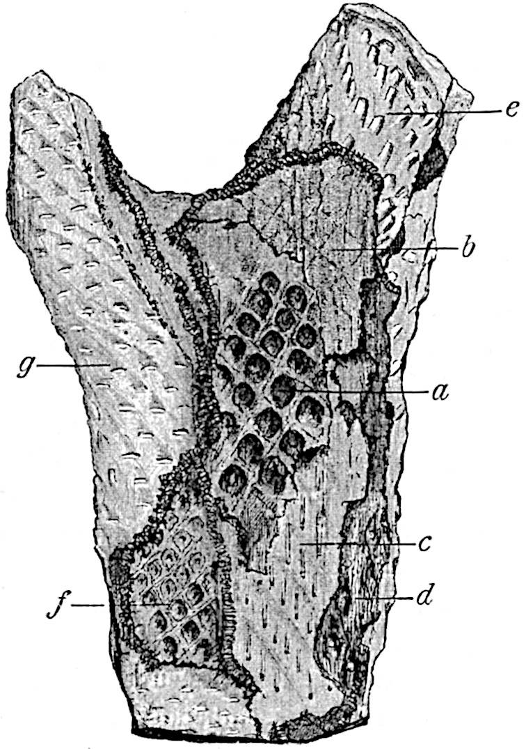
Fig. 156. A dichotomously branched Lepidodendroid stem (Knorria mirabilis Ren. and Zeill.). (After Renault and Zeiller.) (¼ nat. size.) The original specimen is in the Natural History Museum, Paris. a–g, surface features exposed as the result of different degrees of decortication. (See vol. I. p. 102, fig. 23).
The Knorria state represents the impression or cast of the outer cortical region too deep below the leaf-cushion region to retain any indications of the cushion-form; the ridges are the casts of the spaces produced in the cortex by the decay of the sheath of delicate cells surrounding each leaf-trace and by the decay of the thin-walled cells of the parichnos. The occasional forked apex of a ridge is the expression of the fact that the cast was made at the region where the parichnos divides into two arms (cf. p. 100). In certain specimens it is possible to connect the Knorria casts with associated lepidodendroid stems which may be determined specifically; but when we have no evidence as to surface-features the fossils may be designated casts of lepidodendroid stems in the Knorria condition. Such casts are illustrated by numerous drawings in palaeobotanical literature[277].
b. Bergeria.
This is another name first used by Sternberg in his classic work, Die Flora der Vorwelt, for casts of lepidodendroid plants such as Steinhauer[278] had previously figured as Phytolithus cancellatus. Brongniart[279] recognised that the application of the generic title Lepidodendron should be extended to include specimens referred by Sternberg to Bergeria, and a few years later Goldenberg[280] realised that this name does not stand for well-defined generic characters. The correctness of these views was, however, first satisfactorily demonstrated by Carruthers[281] and by Feistmantel[282].
If a Lepidodendron stem loses its superficial layers of outer cortex and in this condition is embedded in sand or mud, the cast is distinguished from that of a perfect stem by the absence of the leaf-scars and by other features. It may, however, still show spirally disposed areas, corresponding approximately to the original leaf-cushions, which are characterised by a small depression or pit either at the apex or near the centre of each oval area: the pit marks the position of the leaf-trace and its parichnos strand. In some cases the exposed surface may be smooth without any indication of leaf-cushions, while narrow spirally arranged grooves represent the obliquely ascending vascular bundles passing through the cortex to the leaves.
Fig. 185, B, shows the Bergeria state of Lepidodendron Veltheimianum, which differs from the Knorria condition in the fact that decortication had not extended below the level at which the form of the leaf-cushions could be recognised. It is clear that no sharp line can be drawn in all cases between the different degrees of decortication as expressed by the terms Knorria and Bergeria.
A list of synonyms of Knorria, Bergeria, and Aspidiaria forms of stem and a detailed treatment of their characteristic features may be found in a recent work by Potonié[283].
c. Aspidiaria.
In one of the earliest English books on fossil plants, the Antediluvian Phytology by Artis[284], a specimen from the Carboniferous sandstone of Yorkshire is figured as Aphyllum cristatum, and a similar fossil is described as A. asperum. These are impressions of Lepidodendron stems in which the characteristic leaf-cushions are replaced by smooth and slightly convex areas with a narrow central ridge. To this type of specimen Presl gave the name Aspidiaria[285], under the impression, shared by subsequent writers, that the supposed external features were entitled to generic recognition.
It is to Stur[286] that we owe the first satisfactory interpretation of fossils included under the name Aspidiaria: he showed that on the removal of the projecting convex areas from some of his specimens a typical Lepidodendron leaf-cushion was exposed (fig. 144, A, a). The Aspidiaria condition (fig. 144, A, b) represents the inner face of the detached shell of outer bark of a Lepidodendron stem, while in the Bergeria casts we have a view of the external face of a stem deprived of its superficial tissues.
In a Lepidodendron stem embedded in sediment the more delicate portions of the leaf-cushions would tend to shrink away from the internal and more resistant tissues of the outer cortex, thus producing spaces between each cushion; further decay would cause rupture of the leaf-traces and the superficial tissues would thus be separated from the rest of the stem. The tendency of Lepidodendron stems to split along the line of phellogen in the outer cortex is seen in fig. 148, A, g. The deposition of sediment on the exposed inner face of this cortical shell would result in the production of a specimen of the Aspidiaria type: the reticulum enclosing the spirally disposed convex areas is formed by the impression of the firmer tissue between the leaf-cushions.
vi. Lepidodendroid axes known as Ulodendron and Halonia.
a. Ulodendron.
This generic name was suggested by Lindley and Hutton[287] for two specimens from the English Coal-measures characterised by leaf-cushions like those of a Lepidodendron, but distinguished by the presence of two vertical rows of large and more or less circular cup-shaped scars. These authors, while recognising the possibility that the fossils might be identical with Lepidodendron, regarded them as generically distinct. The generic title Ulodendron, though no longer denoting generic rank, is still applied to certain shoots of lycopodiaceous plants which may belong to the genera Lepidodendron, Bothrodendron, and according to some authors[288], also to Sigillaria.
The large specimen from the Belgian coal-measures, represented in fig. 211, affords a good example of the Ulodendron form of shoot of the genus Bothrodendron, which is described on page 249. The specimen shown in fig. 157 shows the Ulodendron shoot of Lepidodendron Veltheimianum.
Casts of large Ulodendron scars are occasionally met with as separate fossils bearing a resemblance to an oval shell.
In Steinhauer’s paper on Fossil Reliquiae[289] a drawing is given of a Ulodendron stem under the name Phytolithus parmatus and a similar stem specifically identical with that shown in fig. 157 was figured by Rhode[290], one of the earliest writers on fossil plants, under the comprehensive designation “Schuppenpflanze.”
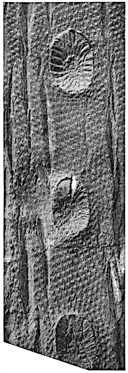
Fig. 157. Lepidodendron Veltheimianum. Ulodendron condition. (From a photograph by Dr Kidston of a specimen from the Calciferous Sandstone series, Midlothian; ⅖ nat. size.) [Kidston (02) Pl. LVII.
There has been no lack of ingenuity on the part of authors in offering suggestions as to the meaning of these large cup-like depressions, and there is still difference of opinion as to their significance. Lindley and Hutton[291] described them as the scars of branches or masses of inflorescence. Sir Joseph Hooker[292] speaks of a specimen of Ulodendron, shown to him by Mr Dawes, on which a large organ, supposed to be a cone, was inserted in one of the depressions, but he was unable to arrive at any conclusion as to the real nature of the fossil. While most authors have seen in the scars pressure-areas formed by the pressure of sessile cones against the surface of a growing branch, others, as for example Geinitz[293], have described the depressions as branch-scars. Carruthers[294] regarded the scars as those of adventitious roots and Williamson referred to them as the scars of reproductive shoots. The depressions vary considerably in size. The Belgian example shown in fig. 211 possesses scars 9 cm. in diameter. A specimen of Bothrodendron in the Manchester Museum from the Lancashire Coal-Measures, to which Williamson[295] has referred, bears two rows of scars 11–12 cm. in diameter on a stem 112 cm. in girth and 233 cm. long. The scars occur in two alternate series, on opposite faces of the axis, the distance between the successive scars in the same row being 29 cm. The surface-features of this large stem are not preserved.
Before considering the nature and origin of the scars it is important to remember the considerable size to which they may attain; other points of importance are the occurrence, either in the centre of each depression or in an excentric position, of an umbilicus or slightly projecting boss, in the centre of which is a pit formed by the decay of an outgoing vascular strand. The sloping sides of the scars sometimes bear elevations resembling leaf-cushions like those on the rest of the stem surface. In the specimen shown in fig. 157 the lower margin of each cup shows indistinctly the outlines of what appear to be leaf-cushions, while the rest of the sloping face is characterised by radial ridges, which may be due to bracts or leaves.
It is obvious that in these cups we have the scars of some lateral organ, but the evidence afforded by specimens of which the depressions contain the remains of such organs is by no means conclusive. A Ulodendron has been figured by D’Arcy Thompson[296], in which the lower part of a lateral organ is attached by a narrow base to one of the scars, but the preservation is not sufficiently good to enable us to decide whether the organ is a cone or a vegetative shoot. Kidston[297] has described other examples showing portions of organs in connexion with the scars, but an examination of the specimens in his collection failed to convince me that his interpretation of them as strobili is correct.
The phenomenon known as cladoptosis, as shown on a stem of the Conifer Agathis[298] and certain Dicotyledonous trees such as Castilloa, suggests a possible explanation of the Ulodendron scars. This comparison was made by Shattock[299] in 1888, but he did not accept the resemblance as a real one. An objection may be urged to the cladoptosis hypothesis that in Ulodendron the branch, whether vegetative or reproductive, was not attached to the whole of the depressed area. On the other hand, a lateral branch originally attached by a narrow base may have continued to increase in diameter until its base became slightly sunk in the bark of the stem, thus producing a cup-like depression which, on the fall of the branch, would retain traces of the original surface-features of the stem.
Mr Watson[300] of Manchester recently published a paper on Ulodendron scars, in which he adduces fresh and, as it seems to me, satisfactory arguments in favour of the branch-scar hypothesis. Fig. 158, from one of Mr Watson’s blocks, illustrates the nature of his evidence. He points out that in the obverse half of a large specimen of Bothrodendron in the Manchester Museum, the umbilicus consists of a cylindrical hole, 18 mm. deep and 8 mm. in diameter, surrounded by a projecting ring of mineral material which doubtless represents some portion of the original plant: on the reverse half of the specimen the continuation of the ring is seen as a prominent cone fitting into the cup-like depression in the obverse half: the conical cast shows that numerous small vascular strands were given off from this ring of tissue, and these strands have the same arrangement and size as the dots which are found on typical Ulodendron scars. He interprets the ring surrounding the umbilicus as the remains of the primary wood and the small strands as leaf-traces supplying the branch.
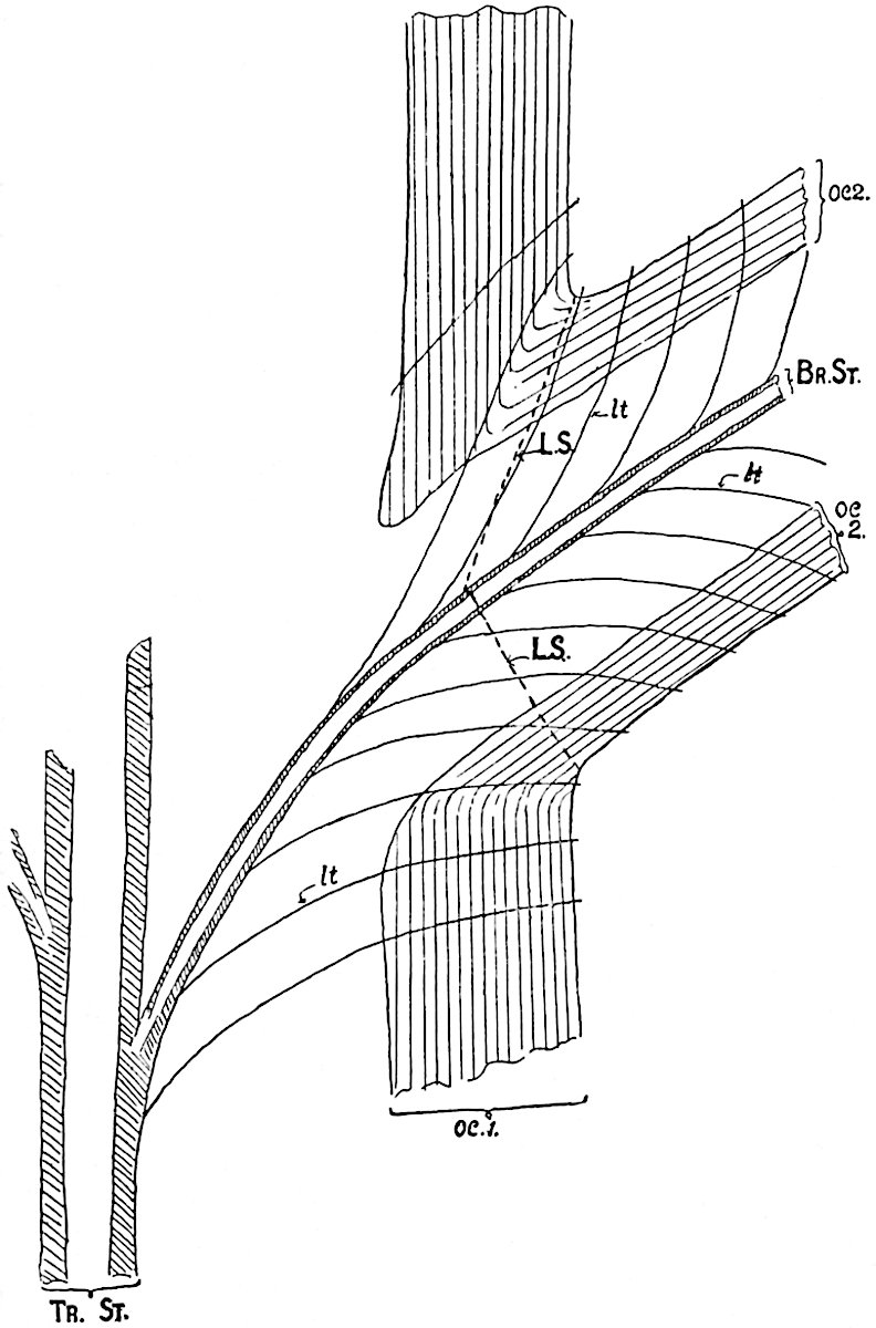
Fig. 158. Diagrammatic section through the base of a branch to illustrate the Branch theory of the Ulodendroid scar. (After Watson.)
In the diagrammatic section shown in fig. 158 the outer cortex of the main stem is represented by oc 1; this consists of secondary tissue. The corresponding tissue in the branch is seen at oc 2. The stele of the stem is shown at Tr. St. and that of the branch at Br. St.; lt, lt, mark the position of the leaf-traces. If we assume the branch to be detached along the line LS, the depression would show numerous spirally arranged dots representing the points of exit of leaf-traces and the vascular axis would be exposed in the umbilicus. This explanation appears to me to be in harmony with the surface-features of Ulodendron scars on both Bothrodendron and Lepidodendron stems. The occasional occurrence of leaf-cushions on a portion of a Ulodendron scar is a difficulty on the cladoptosis hypothesis. Assuming that true leaf-cushions occur, their presence may, as Watson suggests, be due to the folding back of a piece of the outer cortex of the branch which has been “crushed down on to the area of the scar[301].”
Since this account was written a note has been published by M. Renier[302] in which he describes a specimen of Bothrodendron from Liège, one face of which shows a projecting Ulodendroid scar with an excentric umbilicus. On the other face is a dichotomously branched shoot with surface-features corresponding to those on the scar; the evidence that the scar represents the base of the branch is described as indisputable.
Stur[303] held the view that the depressions on Ulodendron stems represent the places of attachment of special shoots comparable with the bulbils of Lycopodium Selago, or, it may be added, with the short branches occasionally produced on Cycas stems. If the depressions were formed by the pressure of the bases of cones, it is clear that the size of the cavity must be an index of the diameter of the cone. The larger Ulodendron scars exceed in diameter the base of any known lepidodendroid strobilus. Another obvious difficulty, which has not been overlooked by Kidston who holds that the scars were produced by sessile cones, is that in Lepidodendron Veltheimianum strobili were borne at the tips of slender branches; the same difficulty is presented by Bothrodendron (Fig. 213). It is unlikely that two types of strobili were produced on the same plant, particularly as the cone of L. Veltheimianum was heterosporous.
The cones of certain species of Pinus remain attached to the tree for many years and their bases become embedded in the stem; this is particularly well shown in the drawing of a cone of Pinus clausa (fig. 159), for which I am indebted to Mr Sudworth, Dendrologist in the United States Forest Service. Mr Sudworth has drawn my attention to P. attenuata and P. muricata in illustration of the same phenomenon[304]. The example shown in fig. 159 cannot, however, be matched by any known specimen of Ulodendron; in the case of the depressions on the stem of a Pine the cone-base fits the circular scar, but in the fossil stems it is practically certain that this was not the case.
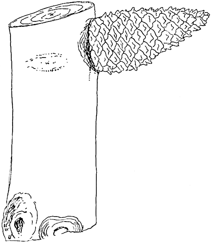
Fig. 159. Pinus clausa. ½ nat. size.
There can be little doubt that certain Palaeozoic Lycopods shed their branches by a method similar to that employed by the Kauri Pine of New Zealand and by some species of Dicotyledons. The evidence adduced in the case of Bothrodendron punctatum is a strong argument in favour of extending the same explanation to other Ulodendron shoots.
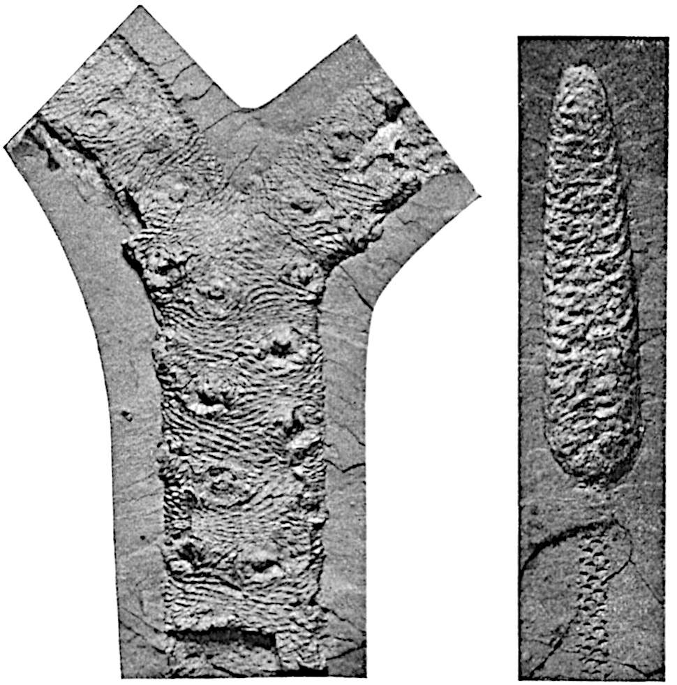
Fig.
160.
Lepidophloios scoticus Kidst. From a specimen from the Calciferous Sandstone, Midlothian, in Dr Kidston’s Collection; rather less than ⅓ nat. size.
L. scoticus cone. From a specimen from the Calciferous Sandstone of Midlothian in Dr Kidston’s Collection; slightly reduced.
b. Halonia.
The branched axis with Lepidophloios leaf-cushions, represented in fig. 160, A, illustrates a special form of shoot described by Lindley and Hutton[305] under the generic name Halonia. The original specimens referred to this genus are decorticated axes showing remains of Lepidodendroid leaf-cushions. The spirally disposed circular scars in the specimen of Halonia (Lepidophloios scoticus[306]) shown in fig. 160 constitute the characteristic feature of the genus; they may have the form, as in fig. 160, A, of circular discs with a central umbilicus marking the position of a vascular strand, or, as in the sandstone cast of Halonia tortuosa shown in fig. 161[307], they may appear as prominent tubercles. The latter example illustrates the condition characteristic of partially decorticated stems.
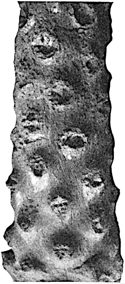
Fig. 161. Halonia tortuosa L. and H. From a specimen in Dr Kidston’s Collection, from the Lower Coal-measures of Ayrshire (No. 1561); ⅔ nat. size.
In 1883 Williamson[308] described a specimen, now in the Leeds Museum, which convinced him that Halonia is merely a special form of Lepidodendron concerned with the production of fertile shoots or strobili. Feistmantel[309] also recognised that Halonia regularis is identical in the form of the cushions with the type known as Lepidophloios laricinus. It is worthy of note that under the name Halonia, Feistmantel[310] figured a piece of decorticated axis characterised by two rows instead of the usual spiral series of large cup-shaped scars. Recent researches have, however, tended to break down the distinction between Ulodendron and Halonia founded respectively on the biseriate and spiral arrangement of the scars or tubercles.
The interpretation of Halonial branches as cone-bearing members of Lepidodendroid plants has passed into a generally accepted statement of fact, but, so far as I know, only one specimen has been figured in which strobili are seen attached to an Halonia axis. This specimen, described by Grand’Eury[311] from the coal-field of Gard, is hardly sufficiently well-preserved to constitute a demonstration of the correctness of the generally received view, which, as is not unusual, has been repeated by one writer after another without due regard being paid to the nature of the evidence on which the statement is based. It may, indeed, be correct to describe Halonial branches as cone-bearing, but there are certain considerations which make one pause before unhesitatingly accepting this explanation. The vascular strand which passes from the central cylinder of the shoot to the tubercle or scar is composed of a solid rod of xylem distinguished from the main stele by the absence of a pith. In such petrified peduncles as have been discovered the stele is of the medullated type. The common occurrence of strobili terminating slender branches of lepidodendroid plants, though not a fatal objection to their attachment to Halonial shoots, shows that in many cases the cones were borne at the tip of leafy shoots. It may be that some of the Halonial scars are in origin like those of the Ulodendron axes of Bothrodendron and mark the position of deciduous vegetative branches.
The first account of the anatomy of Halonia we owe to Dawes[312]; this was followed by a fuller description by Binney[313]. The history of our knowledge of this type of branch has been given by Carruthers[314], who expressed the opinion that Halonia is merely a fertile condition of Lepidophloios and possibly of other lepidodendroid plants. He was also inclined to regard the Halonial tubercles as younger stages of the larger scars characteristic of the genus Ulodendron. Williamson’s contributions to our knowledge of Halonia are of primary importance; he supplied further proof of the Lepidodendroid nature of these branches and advanced our knowledge of their anatomy. In an early paper[315] he expressed the view that the differences on which Halonia and Ulodendron are separated are such as result from a difference in age and are not of generic importance. In the last memoir, of which he was sole author, published by the Royal Society[316], Williamson brought forward further evidence in support of this well-founded opinion.
That the fossils known as Halonia are branches of a lepidodendroid plant is at least certain, and it is probable that the lateral branches which they bore were fertile, though satisfactory proof of this is lacking. We know also that Halonia branches are characterised by the Lepidophloios form of leaf-cushion; there is, however, no sufficient reason to assume that such branches were never attached to stems with the cushions of the Lepidodendron form. The further question, namely whether Williamson was correct in his contention as to the absence of any essential distinction between Ulodendron and Halonia, does not admit of an unchallenged answer. In 1903 Weiss[317] described the anatomy of a specimen of a biseriate Halonia branch of Lepidophloios. The form of the leaf-cushions is unfortunately not very well preserved, but Weiss figures other specimens with two rows of tubercles on which the leaf-cushions are sufficiently distinct to justify a comparison with those of Lepidophloios. He believes with Williamson that it is the presence of tubercles in place of scars which distinguishes Halonia from Ulodendron, and that the arrangement of the tubercles or scars is a matter of little importance. He expresses the opinion justified by the evidence available that the absence or presence of tubercles is merely due to accidents of preservation or, one may add, to difference in age. Kidston[318] dissents from Weiss’s description of his specimen as a biseriate Halonia; he regards it as a Ulodendron branch of Sigillaria discophora (König). Until specimens with more clearly preserved external features are forthcoming it is impossible to settle the point in dispute, but on the facts before us there would seem to be a prima facie case in favour of Weiss’s contention.
The designation Halonia may be retained as a descriptive term for Lepidodendroid shoots characterised by spirally disposed scars or tubercles and bearing leaf-cushions of the Lepidophloios type. In the case of specimens showing prominent tubercles, the superficial tissues are usually absent and, as in the fossil represented in fig. 161, the name Halonia does not necessarily imply the presence of leaf-cushions of a particular type.
vii. Anatomical characters of Vegetative Lepidodendron shoots (Lepidodendron and Lepidophloios).
The type already described under the name Lepidodendron vasculare differs from those dealt with in the following pages chiefly in the anatomy of the stele. The simplest and probably most primitive type of Lepidodendron stem is that in which the xylem forms a solid rod; the type of stele most frequently represented is that of L. Harcourtii, L. fuliginosum, and other species in which the diameter of the stele is greater and a cylinder of primary xylem encloses a comparatively large parenchymatous pith.
1. Lepidodendron esnostense, Renault[319].
This species was founded by Renault on petrified specimens from the Culm beds of Esnost in France. The surface of a young twig bears prominent leaf-cushions of elongated rhomboidal form similar to those of Lepidodendron obovatum (fig. 173) and other species. In older branches the primary cortex is replaced by a considerable thickness of radially disposed secondary cortical tissue which, as shown in tangential section, consists of a reticulum of elongated pointed elements with comparatively thick walls enclosing meshes filled with large-celled parenchyma. It is worthy of note that if such a branch were exposed to decay, the earlier destruction of the more delicate tissue in the meshes of the secondary cortex would produce a series of oval depressions, corresponding to the parenchymatous areas, separated by a projecting reticulum of the more resistant elements: a cast of this partially decayed surface would be indistinguishable from that of some types of Sigillaria or of a Lyginodendron. The inner regions of the cortex of the type-specimens have not been preserved. The xylem, which is the only part of the stele represented, has the form of a protostele or solid cylinder of scalariform tracheids with peripheral groups of narrower protoxylem elements which mark the points of exit of the leaf-traces: in a branch 1–2 cm. wide the xylem column has a diameter of 3 mm. The small leaves (fig. 143, B, C), similar to those of a Sigillaria, are sub-rhomboidal in section near the base and approximately circular near the apex[320]. The mesophyll consists of palisade cells having the appearance of typical chlorophyll-tissue. The heterosporous strobili attributed to this species bore microsporangia on the upper and megasporangia on the lower sporophylls; the megaspores, of which a considerable number occur in each megasporangium, are identical in size with those of another Culm form, Lepidodendron rhodumnense. Some of these have retained traces of prothallus tissue, and in one spore Renault figures what he regards as an archegonium: the drawing is by no means convincing.
2. Lepidodendron rhodumnense, Renault[321].
The species from the Culm of Combres (Loire) agrees in its solid xylem cylinder and in the differentiation of the secondary cortex, as also in the association of two kinds of spore, with Lepidodendron esnostense. A comparison of the leaves of the two types reveals certain differences which may be of specific rank, but, apart from minor differences, these Culm species may be classed under one anatomical type.
3. Lepidodendron saalfeldense, Solms-Laubach[322].
This Devonian species was founded on a specimen 3 × 2·5 cm. broad at the base, which shows the stumps of four branches recalling the dichotomously branched arms of Stigmaria and Pleuromeia. If these are in reality the remains of Stigmaria-like horizontal branches the species affords an interesting example of a Lepidodendron axis with a subterranean rhizome of the type which has been found in several Sigillarian stems. In the upper end of the axis the stele consists of a solid strand of xylem which is not sufficiently well preserved to show the position of the protoxylem groups. A transverse section taken near the base reveals a type of stele differing from that at the upper end in being composed of radially disposed tracheids and in its resemblance to the stele of Stigmaria.
4. Lepidodendron fuliginosum, Williamson. Figs. 162–172, 179, E.
1871. Lepidodendron Harcourtii, Binney, Palæont. Soc., p. 48, Pl. VII. fig. 6.
1872. Halonia regularis, Binney, Palæont. Soc., p. 89, Pl. XV.
1881. Lepidodendron Harcourtii, Williamson, Phil. Trans. Roy. Soc., Vol. 172, p. 288, Pls. XLIX–LII.
1887. Lepidodendron fuliginosum, Williamson, Proc. Roy. Soc., Vol. XLII. p. 6.
1891. Lepidodendron Williamsoni, Solms-Laubach, Fossil Botany, p. 226.
1893. Lepidophloios fuliginosus, Kidston, Trans. Roy. Soc. Edinburgh, Vol. XXXVIII. p. 548.
The name Lepidodendron fuliginosum was proposed by Williamson in 1887 for petrified stems previously included by him in Witham’s species L. Harcourtii, but subsequently recognised as a distinct type characterised by “the greater uniformity in the composition of the entire cortex” and by other features some of which do not constitute distinctive characters. The species agrees with L. Harcourtii and with L. Veltheimianum in having a medullated stele; it is distinguished not only by the more frequent preservation of the middle cortex, a fact due to a difference in minute structure, but chiefly by the peculiar structure of the secondary tissue added to the stele; this is in part composed of radial series of parenchymatous cells and of a varying amount of tracheal tissue the elements of which are narrower than in other species and are characterised also by their sinuous vertical course. As is pointed out in the sequel, the anatomical features of L. fuliginosum, as at present understood, are not confined to one type of Lepidodendron stem. Specimens have been described with leaf-cushions of the form characteristic of L. aculeatum, L. obovatum and Lepidophloios combined with the anatomical features of Williamson’s species: it is possible that the two species L. obovatum and L. aculeatum are not really distinct[323], but it is certain that shoots with both the Lepidodendron and Lepidophloios cushions may have the same type of anatomical structure.
A more detailed knowledge of the structural features of Lepidodendron shoots may enable us to define anatomical species with more exactness than is possible at present. There can, however, be little doubt that well-marked anatomical features may be associated with more than one specific form of shoot as defined by the form of the leaf-cushions.
Solms-Laubach proposed the name Lepidodendron Williamsoni for the anatomical type L. fuliginosum of Williamson, but the latter name has been generally adopted.
In the following account special attention is directed to the nature and origin of the secondary stelar tissue and to the secretory zone, as difference of opinion exists as to the interpretation of these features. Among the best examples of shoots of Lepidodendron fuliginosum without secondary tissue or in which it is feebly developed are those originally described by Binney. The stele includes a large parenchymatous pith, the cells of which frequently show signs of recent division, a feature observed also in the pith of the large stem of L. Wünschianum, represented in figs. 181, 182. The primary xylem cylinder has an irregularly crenulate outer edge like that of L. Wünschianum and L. Harcourtii and the protoxylem elements occupy an exarch position. Isodiametric reticulately-pitted elements are met with both on the inner and outer edge of the xylem.
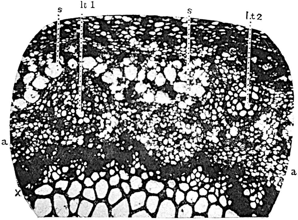
Fig. 162. Lepidodendron fuliginosum. Part of the stele in transverse section. (Binney Collection, Sedgwick Museum, Cambridge.)
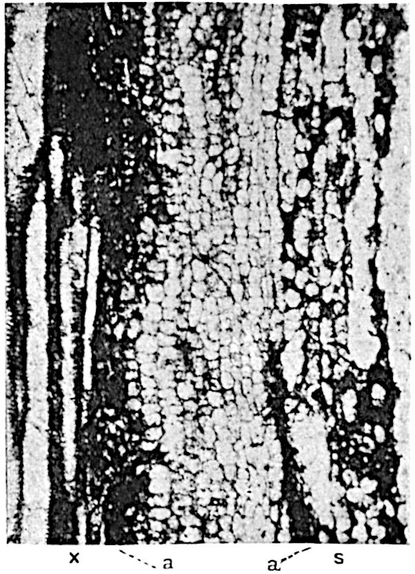
Fig. 163. Lepidodendron fuliginosum. Longitudinal section. (Binney Collection, Cambridge.)
Figs. 162 and 163 illustrate the structure of the outer portion of the xylem and adjacent tissues in a section of a shoot 3·8 cm. × 2·5 cm. in diameter, which is in the act of branching, as shown by the occurrence of two steles of equal size. A figure of the complete section will be found in Binney’s memoir[324], and additional illustrations were published in 1899[325].
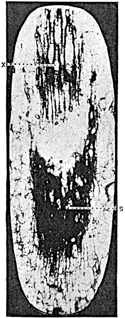
Fig. 164. Lepidodendron fuliginosum. Leaf-trace. (Binney Collection, Cambridge.)
The primary xylem (figs. 162, 163, x) is succeeded by 2–3 rows of polygonal cells with dark contents and associated with isodiametric tracheae: these pass into clearer parenchymatous tissue, a, characterised by the arrangement of the cells in vertical series, to which the term meristematic zone has been applied. The secretory zone, s, abutting on the meristematic zone, consists of more or less disorganised parenchymatous cells and broader and more elongated spaces; it is interrupted here and there by an outgoing leaf-trace, as at lt 1 and lt 2 in fig. 162. The secretory zone is succeeded by a homogeneous inner cortex like that described in L. vasculare; part of this region is seen at the upper edge of fig. 162. The broad middle cortex, which is separated from the inner cortex by a sharply defined boundary, is composed of rather small lacunar parenchymatous tissue consisting of sinuous tubular elements interspersed among isodiametric cells of various sizes (fig. 166, p). In the middle cortical region the leaf-traces pursue an almost horizontal course; one is shown in fig. 164, in oblique longitudinal section, in a reversed position; the xylem, x, should be on the inner side of the secretory tissue, s. The clear space between the two parts of the vascular bundle was originally occupied by a few layers of parenchymatous cells, as seen in the transverse sections, figs. 165 and 166. In some specimens the leaf-traces pass through the middle cortex in a much more vertical course, as shown by the section represented in fig. 165. This section illustrates the structure of a typical leaf-trace with unusual clearness; it shows the tangentially elongated group of xylem, the strand of tissue which occupies the position of phloem, s (to which the term secretory zone is applied), the compact parenchyma between the two parts of the bundle, and surrounding the whole a narrow sheath sharply contrasted by the smaller and more uniform size of the cells from the middle cortex, a few cells of which are seen in the photograph. The middle cortex shows a well-defined junction with the more compact outer cortical region, which consists of primary parenchyma passing externally into a zone of phelloderm composed of thick-walled and more elongated cells. A noticeable feature in many Lepidodendron shoots is the occurrence of a circle of strands of secretory cells often surrounding fairly large ducts just internal to the edge of phelloderm: similar strands form irregularly concentric circles, as was pointed out in the case of L. vasculare, in the phelloderm itself.
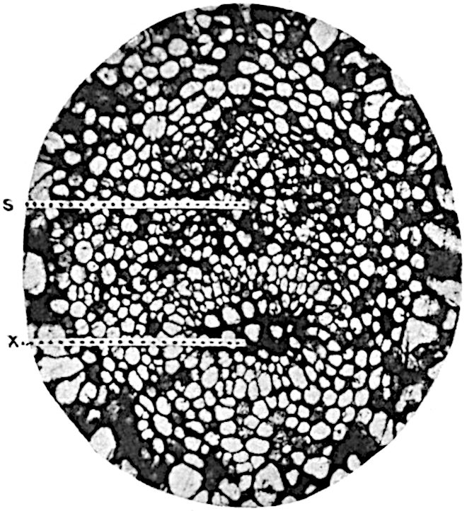
Fig. 165. Lepidodendron fuliginosum. Leaf-trace: x, xylem; s, secretory zone. (Binney Collection, Cambridge.)
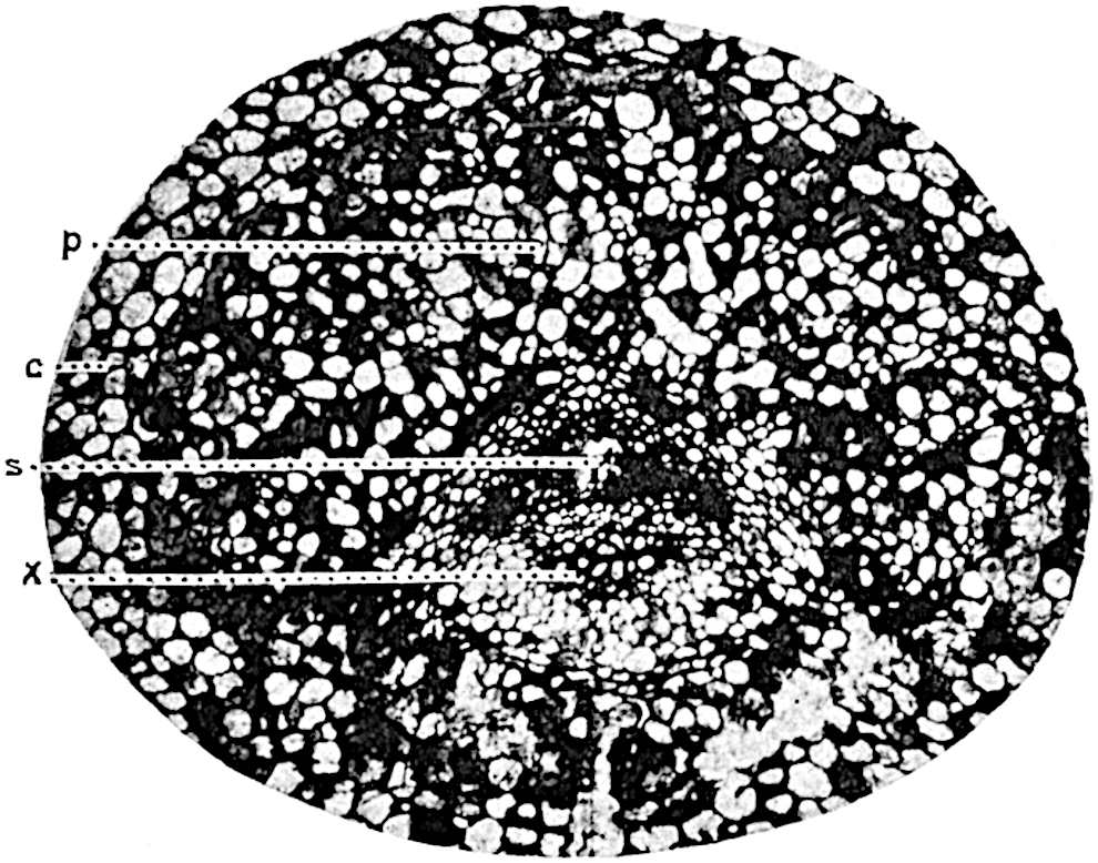
Fig. 166. Lepidodendron fuliginosum. Leaf-trace: p, parichnos. (Binney Collection, Cambridge.)
Fig. 166 shows a leaf-trace in the outer cortex accompanied by its crescent-shaped parichnos, p, derived from the middle cortex and by means of which the outer cortex and the lamina of the leaves are connected with the inner region of the shoot. This lacunar middle cortex and parichnos doubtless constitute an aerating tissue-system which after leaf-fall is exposed directly to the air at the ends of the parichnos arms on the leaf-scars.
Some of the sections in the Binney Collection (Sedgwick Museum, Cambridge) show early stages in the production of secondary xylem: in the section represented in fig. 167 the secretory zone is succeeded on its inner face by a zone of radially elongated cells, m, which are clearly in a meristematic condition. The same section shows also the more radially extended form of the xylem of a leaf-trace with its internal protoxylem, px, in contrast to the tangentially elongated form which is assumed during its passage through the cortex (cf. figs. 165, 166).
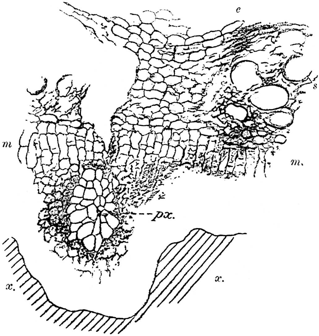
Fig. 167. Lepidodendron fuliginosum. (Binney Collection, Cambridge.)
Some sections of Lepidodendron fuliginosum in the Manchester University Collection are of special interest from the point of view of the method of secondary thickening. In the section reproduced in fig. 168, B, the meristematic zone is seen to consist in part of radially elongated elements, m, with parallel cross-walls evidently of recent origin. The same tissue is shown also in fig. 168, C, a, D, a, and in fig. 169, A, a This band of meristem, which we may speak of as the cambium, occurs in the outer region of the meristematic zone immediately internal to the secretory zone, sc.
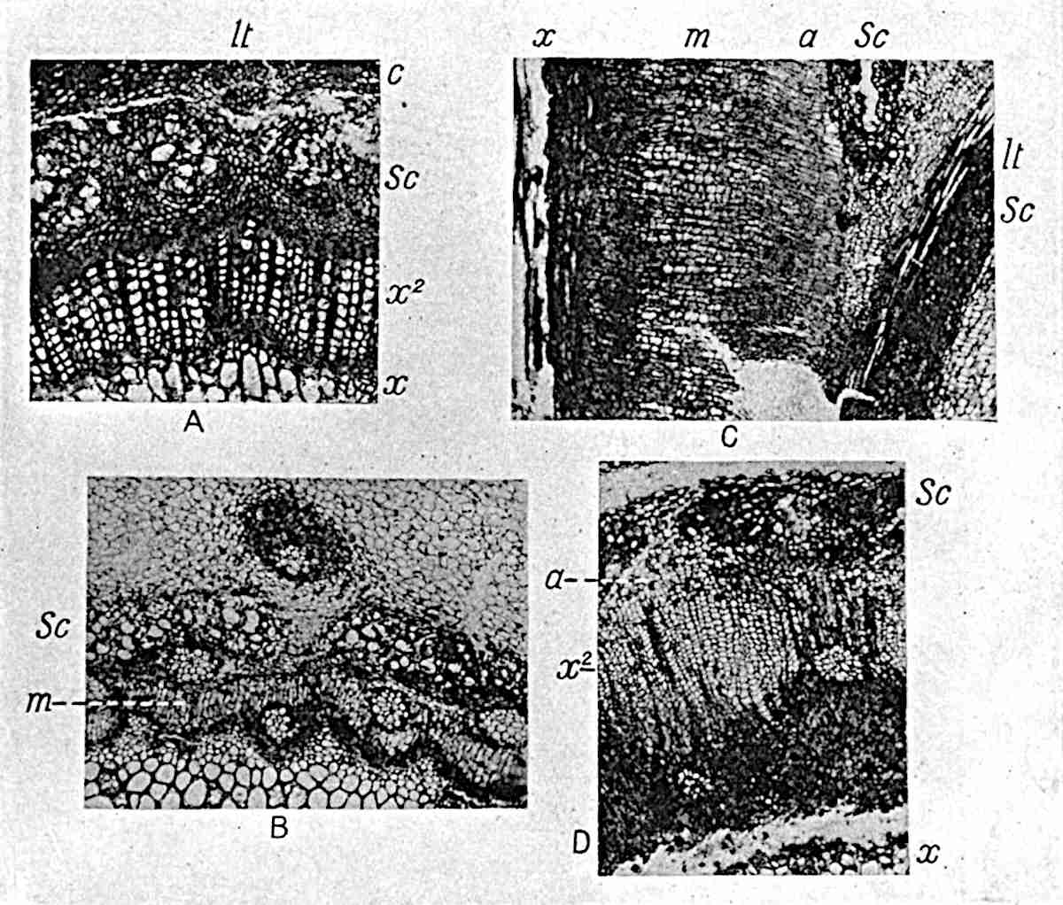
Fig.
168.
Lepidodendron vasculare. (Botany School, Cambridge.)
Lepidodendron fuliginosum. (From a specimen from Shore, Lancashire, in the Cambridge Botany School Collection).
L. fuliginosum. (“Biseriate Halonia” of Weiss No. 257, Manchester University Museum.)
L. fuliginosum. (Manchester Univ. Museum.)
The result of the activity of this cambium band is the production of secondary parenchyma and tracheal tissue. In fig. 179, E, drawn from a portion of the section represented in fig. 168, B, a projecting arm of primary xylem is seen at x; this is followed by 2–3 layers of parenchymatous cells, some of which have dark contents, and beyond this is seen a group of secondary elements, tr, cut across somewhat obliquely, which are evidently products of the cambial cells on the inner margin of the secretory zone, sc. The longitudinal section (fig. 169, D) shows the cambial cells, a, next the secretory zone, sc, passing internally into crushed and imperfectly preserved elongated elements which are presumably miniature tracheae, and these are succeeded by older and more completely lignified xylem elements, x. In larger shoots the amount of secondary tissue is considerably greater; it may consist almost entirely of short-celled parenchyma (fig. 168, C, from x to sc), or it may include a large proportion of radially disposed and vertically elongated tracheae (fig. 168, D, x2, and fig. 170, A, x2), or it may consist of parenchyma containing scattered groups of tracheae (fig. 169, A, x2)[326].
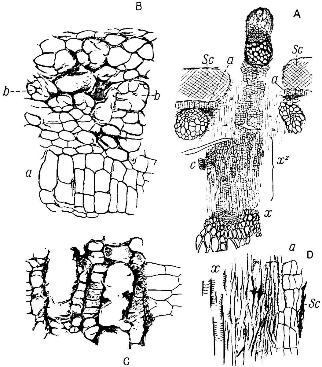
Fig.
169.
Lepidodendron fuliginosum.
A, B. (Manchester University Collection. No. Q. 645 A.)
B, C. (Manchester. No. 257.)
D. (Manchester. No. 6.)
Fig. 169, A, is a diagrammatic sketch of the tissues—1 mm. wide—between the primary xylem, x, and the inner cortex. The primary xylem is succeeded by short parenchymatous cells followed by a zone of radially elongated elements passing occasionally into rows of narrow scalariform tracheae, some of which, owing to their sinuous longitudinal course (fig. 171, C), are seen in oblique section, as at C, fig. 169, A. At its outer edge this secondary tissue, x2, consisting of parenchyma and tracheae, passes into the cambial band (fig. 169, B, a).
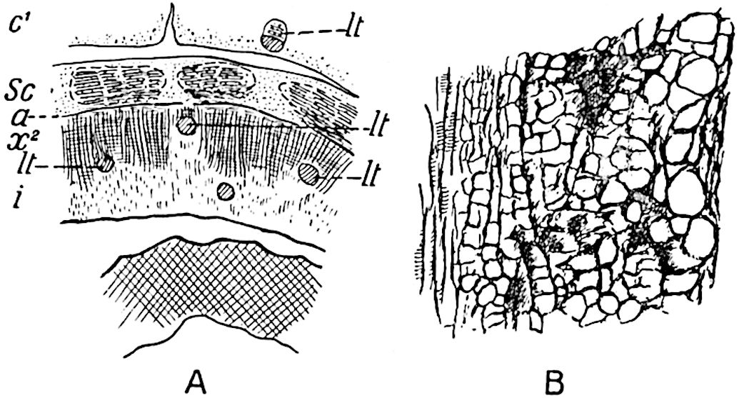
Fig. 170. Lepidodendron fuliginosum. (From sections in the Manchester Museum.)
The radial longitudinal section represented in fig. 168, C, is taken from the fossil described by Weiss as a biseriate Halonia; it agrees sufficiently closely in structure with others referred to Lepidodendron fuliginosum to be classed as an example of this anatomical type. A complete transverse section of the stem measures 9 × 6·3 cm.; the breadth of the tissues between the edge of the primary xylem and the outer edge of the secretory zone is 2·5 mm. The middle cortical region, characterised by the sooty appearance, which led Williamson to choose the specific name fuliginosum, is traversed by the leaf-traces and is sharply differentiated from both the inner and outer cortex. The longitudinal section (fig. 168, C) shows the outer edge of the primary xylem, x, abutting on a band of dark and small-celled parenchyma which passes into the broad zone of secondary tissue, m, the inner region of which consists of fairly thick-walled elements in radial series passing externally into the thin-walled cells of the cambial region, a, on the inner edge of the secretory zone, sc. This section shows also the interruption of the secretory zone by an outgoing leaf-trace, lt, the lower part of which, sc, is continued downwards into the secretory zone. The exit of a leaf-trace produces a gap in the secretory zone of the stem, but not in the xylem. If we applied the term phloem to the secretory zone—a course adopted by Prof. F. E. Weiss and some other authors, but which I do not propose to follow—we should speak of a phloem foliar-gap as a characteristic feature of a Lepidodendron shoot. This applies to other species of the genus as well as to L. fuliginosum.
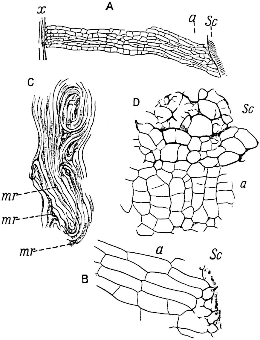
Fig. 171. Lepidodendron fuliginosum. (From sections in the Manchester Museum.)
Fig. 171, A, shows more clearly the broad zone of secondary parenchyma with the thinner-walled cambial region, a; the latter is represented on a larger scale in fig. 171, B. The section shown in fig. 168, D, and in fig. 170, A, affords an example of a stem in which the secondary tissue consists largely of narrow scalariform tracheae, x2; the primary stele has a diameter of 1 cm.; the secondary xylem, x2, forms a fairly broad zone of parenchyma and tracheal elements through which leaf-traces pass vertically, a fact of some interest in comparison with the horizontal course which they pursue through the medullary rays in the normal secondary wood of L. vasculare and L. Wünschianum. The secondary tracheae pass gradually into thin-walled cambial cells (a, fig. 168, D; 170, A) with parallel tangential walls. Fig. 171, C, shows the sinuous course of the secondary tracheae as seen in longitudinal section, and a few small groups of parenchymatous cells, mr, which may be of the nature of medullary rays, enclosed between the winding scalariform tracheae.
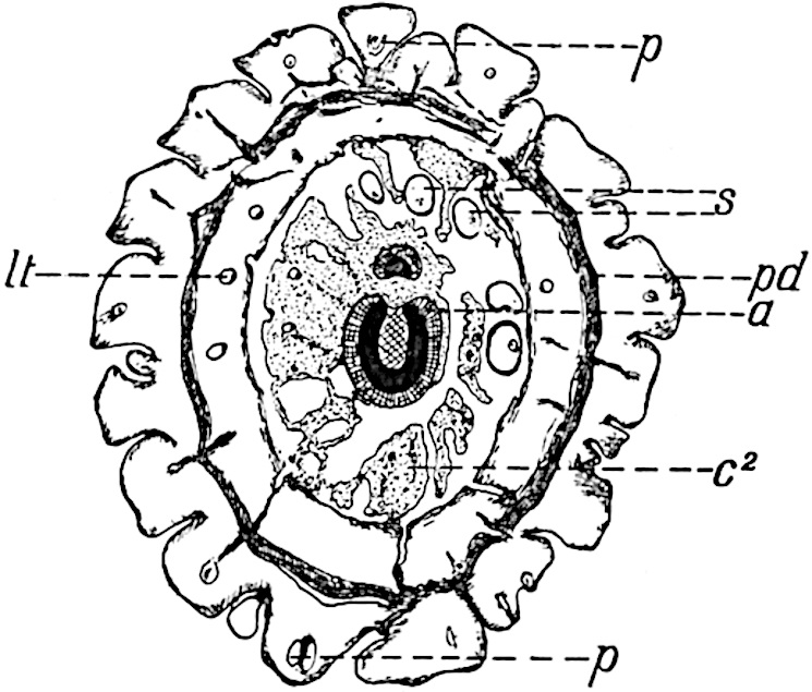
Fig. 172. Lepidodendron fuliginosum. From a section (4 × 3·4 cm.) in the Williamson Collection, British Museum (No. 379), figured by Williamson, Phil. Trans. R. Soc. 1881, Pl. 52.
The secretory zone of Lepidodendron fuliginosum agrees essentially with that of other species; it usually presents the appearance shown in fig. 168, B, sc; fig. 169, B and C; fig. 170, B (longitudinal section); fig. 171, D, sc. The comparatively large clear spaces which characterise this tissue, as seen in fig. 168, B, appear to owe their origin to groups of small cells which gradually break down and give rise to spaces containing remnants of the disorganised elements, as in fig. 171, D, and fig. 169, B, b. The secretory tissue seen in fig. 170, B, consists of large and small parenchymatous cells without any of the broad sacs or spaces such as are shown in fig. 169, C.
Fig. 172 represents a diagrammatic sketch of a transverse section (4 × 3·4 cm. in diameter) of a young shoot from the Lower Coal-Measures of Lancashire figured by Williamson[327] in 1881 as Lepidodendron Harcourtii. It shows the features characteristic of L. fuliginosum and is of importance as affording an example of a shoot giving off a branch from the stele to supply a lateral axis of the type characteristic of Halonia. The exit of the branch-stele forms a gap in the main stele; a ramular gap as distinguished from a foliar gap. The outgoing vascular strand is at first crescentic, but becomes gradually converted into a solid stele. The primary xylem of the main stele (black in the figure) consists of a ring six tracheae in breadth; this is succeeded by a few layers of dark parenchymatous cells and a band of radially elongated elements, a, which abuts on the secretory zone. The middle lacunar cortex, c2, with Stigmaria rootlets, s, is fairly well preserved. In the outer cortex occur several leaf-traces, lt, accompanied by spaces originally occupied by the parichnos strand, p. A band of secondary cortex, consisting chiefly of phelloderm, is seen at pd. The prominent leaf-cushions, some of which show the parichnos, p, appear to be of the Lepidophloios type.
•••••
It remains to consider the external characters of Lepidodendroid shoots possessing the anatomical features represented by the comprehensive species Lepidodendron fuliginosum.
Certain sections exhibiting this type of structure were described by Binney in 1872 as Halonia regularis[328] on evidence supplied by Mr Dawes, who stated that they were cut from a specimen bearing Halonia tubercles. The section represented in fig. 172 is no doubt from an Halonia axis. In 1890 Cash and Lomax[329] stated that they had in their possession a stem of the L. fuliginosum type with the external features of Lepidophloios; this identification has been confirmed by Kidston[330] and Weiss[331]. It is, however, equally clear that certain species with the elongated leaf-cushions of Lepidodendron must be included among examples of shoots with the anatomical characters of L. fuliginosum.
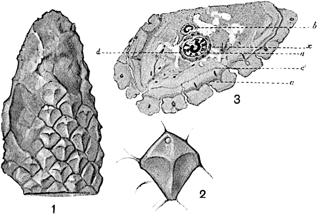
Fig. 173. Lepidodendron obovatum. (From a specimen lent by Dr D. H. Scott.)
Dr Scott[332] published in 1906 a short account of the structure of a specimen from the Lower Coal-Measures of Lancashire, the external features of which were identified by Kidston with those of Lepidodendron obovatum Sternb. Dr Scott generously allowed me to have drawings made from his specimen; these are reproduced in fig. 173. The form of the leaf-cushion is by no means perfect; there is a well-marked median ridge, and the small circular scar near the upper end of some of the cushions may represent the ligular cavity. At the base of the leaf-cushions a cortical meristem has produced a zone of secondary cortex; at c a second meristem is seen in the outer cortex: the dark dots in the cortex mark the positions of leaf-trace bundles. The inner cortex, d, is a more compact tissue surrounding the imperfectly preserved secretory zone. From the medullated stele a lateral branch, b, is being given off; its crescentic form becoming changed to circular as it passes nearer to the surface.
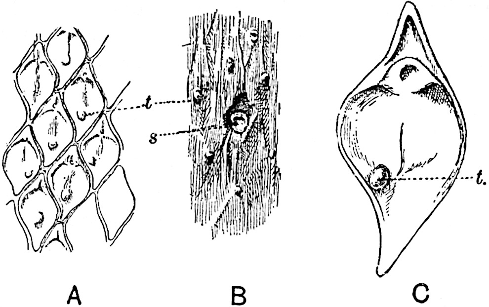
Fig. 174. Lepidodendron aculeatum. (Cambridge Botany School.)
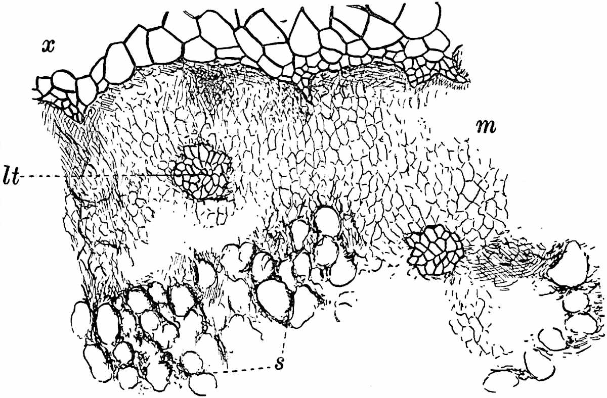
Fig. 175. Lepidodendron aculeatum. (Cambridge Botany School.)
A type of Lepidodendron, L. Hickii, founded on anatomical characters by Mr Watson[333], is believed by him to possess leaf-cushions like those of L. obovatum; if this is so, it is interesting, as he points out, to find two distinct anatomical types associated with one species. Watson thinks it probable that the “species” L. obovatum includes at least two widely different species. This merely emphasizes the importance of correlating structure and external characters as far as available data permit.
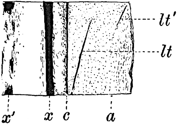
Fig. 176. Lepidodendron aculeatum. (Cambridge Botany School.)
The specimen, of which part of the surface is shown in fig. 174, is in all probability L. aculeatum Sternb. This was described by me in detail in The Annals of Botany (1906) as another example of the co-existence of the Lepidodendron fuliginosum type of anatomy with a true Lepidodendron. The locality of the specimen is not known. The leaf-cushions are 1·5 cm. long with tapered upper and lower ends; a ligular cavity may be recognised on some parts of the fossil, also faint indications of leaf-trace scars. The tubercles (fig. 174, A–C, t) probably represent leaf-traces which the shrinkage of the superficial tissues has rendered visible in the lower part of their course. The circular scar, s (fig. B), on the partially decorticated surface is apparently a wound. The stele is sufficiently well preserved to justify its reference to L. fuliginosum. The irregularly crenulated edge of the primary xylem, x (fig. 175), is succeeded by a broad band of parenchyma (the meristematic zone), m, and beyond this are remnants of the secretory zone, s. The structure of the leaf-traces corresponds with that of other specimens of the type, but the much steeper course of these vascular strands, lt, lt′ (fig. 176), is a feature in which this example differs from most of those referred to L. fuliginosum. Such evidence as is available would seem to point to the absence of trustworthy criteria enabling us to separate, on anatomical grounds, Lepidophloios and Lepidodendron[334].