Volume 1 — CHAPTER XV. (continued 2)
Reading edition
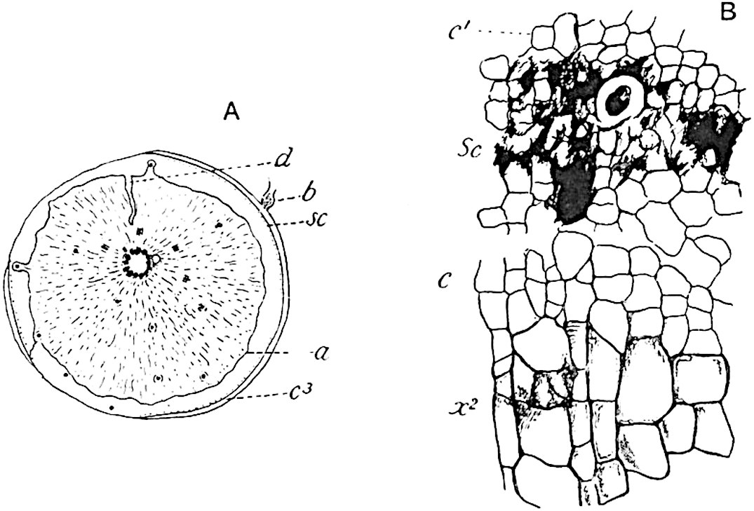
Fig. 177. Stigmaria radiculosa (Hick). (From sections in the Manchester University Collection.)
Stigmaria radiculosa (Hick).
We have no proof of the nature of the subterranean organs of Lepidodendron fuliginosum, though it is not improbable that the specimens described below may be correctly assigned by Weiss to that species. Prof. Weiss[335] has made an interesting contribution to our knowledge of a type first described by Hick[336] under the name Tylophora radiculosa, a designation which he afterwards altered to Xenophyton radiculosum[337] and for which we may now substitute Stigmaria radiculosa (Hick). Prof. Williamson expressed the opinion that Xenophyton exhibited considerable affinity with Stigmaria ficoides and Weiss’s further study of the species leads him to regard Hick’s plant as probably the Stigmarian organ of Lepidodendron fuliginosum. The diagrammatic transverse section represented in fig. 177, A (4·5 cm. in diameter), shows an outer cortex of parenchyma, c3, consisting in part of radial rows of secondary tissue and of a band of compact parenchyma bounded by the wavy line a; at sc is a series of secretory strands exactly like those in a corresponding position in Lepidodendron fuliginosum and other species of the genus. The greater part of the organ is occupied by a lacunar and hyphal middle cortex identical in structure with that shown in fig. 178, B, drawn from a rootlet. At d, fig. 177, A, the middle cortex has been invaded by a narrow tongue of outer cortical tissue. The stele is characterised by a large pith filled with parenchyma; in Stigmaria ficoides[338] the general absence of pith-tissue has led to the inference that the stele was hollow. The xylem is represented by a ring of bundles separated by broad medullary rays; each bundle contains a few small, apparently primary, elements on its inner edge but is mainly composed of radial rows of secondary tracheae x2, fig. 177, B. On the outer face of the secondary xylem occur a few smaller and thinner walled cells, c, having the appearance of meristematic tissue; from these additional tracheae were added to the xylem. This meristematic zone occurs, as in the stems of Lepidodendron, immediately internal to the secretory tissue, sc; at c1, fig. 177, B, is seen the inner cortical tissue.
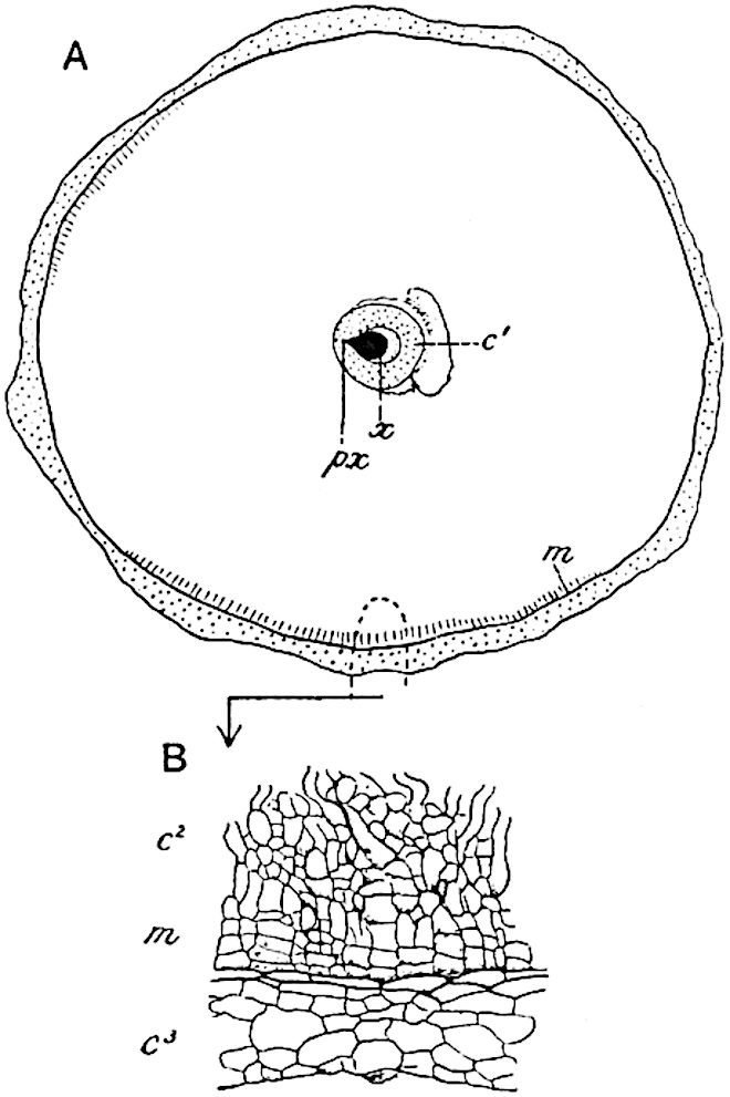
Fig. 178. Rootlet of Stigmaria. (From a section in the Manchester Collection.)
In surface-view a specimen figured by Hick[339] shows a number of circular scars agreeing in shape and arrangement with the rootlet scars of Stigmaria ficoides. At b in fig. 177, A, the basal portion of a rootlet is shown in organic connexion with the outer cortex. The rootlet-bundles are given off from the stele as in other examples of Stigmaria; each bundle consists of a triangular strand of xylem with an endarch protoxylem at the narrow end accompanied by a portion of the secretory tissue as in the leaf-traces. As in Stigmaria ficoides the rootlets are attached to the outer cortex above a cushion of small cells. It is interesting to find that rootlet-bundles, as seen in tangential section of the main axis, are associated with a parichnos strand, but this is on the xylem side of the vascular strand, whereas in the case of leaf-traces the parichnos is on the other side of the bundle.
Fig. 178, A, represents a transverse section of a rootlet (6 mm. in diameter) associated with Stigmaria radiculosa and probably belonging to this species. The xylem strand x is composed of a group of tracheae with a single protoxylem strand, px, at the pointed end and with small metaxylem elements at the broad end next the space originally occupied by the so-called phloem. A parenchymatous sheath, c′, surrounds the bundle, and beyond this is the broad middle cortex, a small portion of which is shown on a larger scale in fig. 178, B; as Weiss points out, some of the outermost cells of the lacunar cortex (m) are clearly in a state of meristematic activity.
The preservation of the middle cortex and the small quantity of secondary xylem are characters which this Stigmaria shares with Lepidodendron fuliginosum, and although decisive evidence is still to seek, we may express the opinion that Weiss’s surmise of a connexion between Stigmaria radiculosa and Lepidodendron fuliginosum is probably correct.
5. Lepidodendron Harcourtii. Fig. 179, A–D.
In 1831 Mr Witham[340] published an anatomical description of a fragment of a Lepidodendron which he named Lepidodendron Harcourtii after Mr C. G. V. Vernon Harcourt from whom the specimen was originally obtained. The fossil was found in rocks belonging to the Calciferous series in Northumberland. Witham reproduced the account of this species in his classic work on Fossil Vegetables[341], and Lindley and Hutton[342], who examined Mr Harcourt’s material, published a description of it in their Fossil Flora in which they expressed the view that Lepidodendron is intermediate between Conifers and Lycopods. Adolphe Brongniart[343] included in his memoir on Sigillaria elegans an account of Witham’s species based on material presented to the Paris Museum by Mr Hutton and Robert Brown. Dr Kidston[344] has shown that the actual transverse section figured by Witham is now in the York Museum; a piece of stem in the same Museum, which is not the specimen from which Witham’s section was cut, supplied the transverse section figured by Brongniart. The figures given by Lindley and Hutton do not appear to have been made from the York specimens. In 1887 Williamson[345] published a note in which he pointed out that some of the specimens described by him as L. Harcourtii should be transferred to a distinct species, which he named L. fuliginosum. Subsequently in 1893 he gave a fuller account of Witham’s species; it has, however, been shown by Dr Kidston and by Mr Watson[346] that certain specimens identified by Williamson as L. Harcourtii differ sufficiently from that type to be placed in another species, for which Watson proposes the name L. Hickii.
A paper on L. Harcourtii published by Bertrand[347] in 1891 extends our knowledge of this type in regard to several anatomical details. It was recognised by Williamson that the absence of secondary wood in shoots possessing the anatomical characters of L. Harcourtii is a feature to which no great importance should be attached. It is possible that the large stems from the Isle of Arran described by Williamson[348] as Lepidodendron Wünschianum, in which the secondary wood is well developed, may be specifically identical with the smaller specimens from Northumberland and elsewhere which are recognised as examples of Witham’s type.
The diagrammatic sketch shown in fig. 179, A, was made from a section figured by Williamson in 1893[349]; it has a diameter of 9 × 8·5 cm. The stele is of the medullated type like that of L. Wünschianum, and the outer edge of the primary xylem is characterised by sharp and prominent projecting ridges similar to those of L. fuliginosum but rather more prominent. Parenchymatous cells succeed the xylem, as in other species, but in this case there is no indication of meristematic activity; beyond this region occur occasional patches of a partially destroyed secretory zone. Remains of a lacunar tissue are seen in the middle cortical region; also numerous leaf-traces, lt, consisting of a tangentially elongated xylem strand accompanied by a strand of secretory zone tissue enclosed in a sheath of delicate parenchyma. In the inner part of the outer cortex, c3, the leaf-traces lie in a space originally occupied by the parichnos; in the outer portion of the same region a band of secondary cortex, pd, has been formed; immediately internal to this occur numerous patches of secretory tissue, represented by small dots in the drawing close to pd; one is shown on a larger scale in fig. B.
The position of the phellogen is seen at a; external to this are radial rows of rather large cells with dark contents.
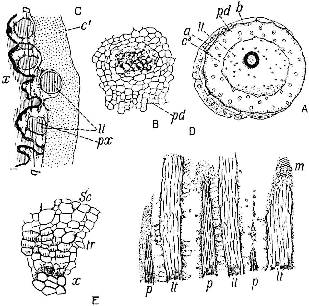
Fig.
179.
A–D. Lepidodendron Harcourtii, Witham.
E. Lepidodendron fuliginosum, Shore, Lancashire.
A, B. From a specimen in the Williamson Collection, British Museum (No. 380), from Airdrie, Scotland.
C, D. From sections in the Collection of Dr Kidston, from Shore, Lancashire.
Fig. 179, C, x, shows the characteristic form of the primary xylem edge, beyond which are seen oval or circular leaf-traces with a mesarch protoxylem, lt, px. It is possible that this specimen may not be specifically identical with Witham’s species, but it represents a very similar if not identical type; it may on the other hand be referable to L. fuliginosum. The importance of the specimen, apart from its precise specific position, is that it serves to illustrate the general appearance of the xylem surface met with in both species, L. Harcourtii and L. fuliginosum. A tangential longitudinal section, taken through the line ab in fig. C, is represented in fig. 179, D. The xylem of the leaf-traces lt, consisting chiefly of scalariform tracheae, alternates with patches of crushed and delicate parenchyma which immediately abut on the primary xylem; at p, p, the section passes through some of the projecting arms of the xylem cylinder; at m is seen a patch of meristematic zone tissue. This section together with the similar section of Lepidodendron vasculare described on a previous page demonstrates that the projecting ridges of the primary xylem form apparently vertical bands: they are not characterised by a lattice-work arrangement as described by Bertrand and by other authors who have accepted his conclusions. If a reticulum of intersecting ridges were present on the face of the xylem cylinder its existence would be revealed by such a section as that represented in fig. 179, D.
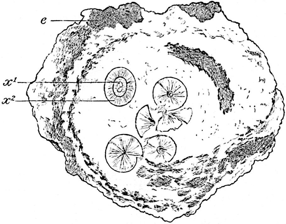
Fig. 180. Lepidodendron Wünschianum. From Arran. (⅕ nat. size.) (Sedgwick Museum, Cambridge.)
6. Lepidodendron Wünschianum (Williamson). Figs. 180–184.
Reference was made in Volume I. to the occurrence of large stems of a Lepidodendron in volcanic beds of Calciferous sandstone age in the island of Arran[350]. These were discovered and briefly described by Mr Wünsch in 1867[351] and afterwards named by Carruthers Lomatophloyos Wünschianus[352]. Mr Carruthers visited the locality and published an account of the peculiar method of preservation of the plant remains[353]. It is, however, to Williamson[354] that we owe the more complete description of these Arran stems. Portions of large stems from the Arran beds are preserved in the British Museum, the Sedgwick Museum, Cambridge, and in the Manchester Museum. The section of one of these is shown in fig. 180; an outer shell of bark encloses a mass of volcanic ash in which are embedded several woody cylinders originally described as “internal piths[355],” and by Carruthers as young stems produced from spores which had germinated in the hollow trunk of a large tree. The true interpretation was supplied by Williamson who showed that a stem of the dimensions of that represented by the outer cortex, e, fig. 180, must have possessed a single stele of the size of those seen in the interior of the hollow trunk. The additional woody cylinders, or steles, were derived from other stems, and carried, probably by water, into the partially decayed trunk. In addition to large Lepidodendron stems Williamson described smaller shoots as well as an Halonial branch and made brief reference to some cones described by Binney[356] in 1871 from the same locality.
The following account of Lepidodendron Wünschianum is based on an exceptionally fine specimen discovered by Mr T. Kerr of Edinburgh in Calciferous sandstone volcanic ashes at Dalmeny in Linlithgowshire. The material from this locality described by Mr Hill and myself[357] was generously placed in my hands by Dr Kidston of Stirling. Fig. 181, A, shows a transverse section, 33 cm. in diameter, consisting of a shell of outer cortical tissue enclosing a core of light-coloured volcanic ash; on the decay of the more delicate middle cortex the cylindrical stele dropped to one side of the hollow trunk. The stele, fig. 182, has a diameter of 6·5 cm.; the centre is occupied by concentric layers of silica, s, surrounded externally by the remains of a parenchymatous pith, p, made up of isodiametric and sinuous hypha-like elements like those in the middle cortex of Lepidodendron shoots. On the inner edge of the primary xylem, x′, occur several isodiametric tracheae with fine scalariform and reticulate thickening bands like those in the central region of the stele of Lepidodendron vasculare: it is probable that these elements are vestiges of conducting tissue which in ancestral forms formed a solid and not a medullated stele.
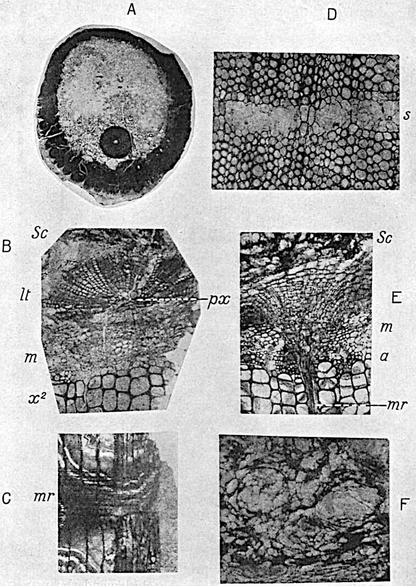
Fig. 181. Lepidodendron Wünschianum. Calciferous Sandstone, Dalmeny. (A, Sedgwick Museum, Cambridge. B–F, Botany School, Cambridge.)
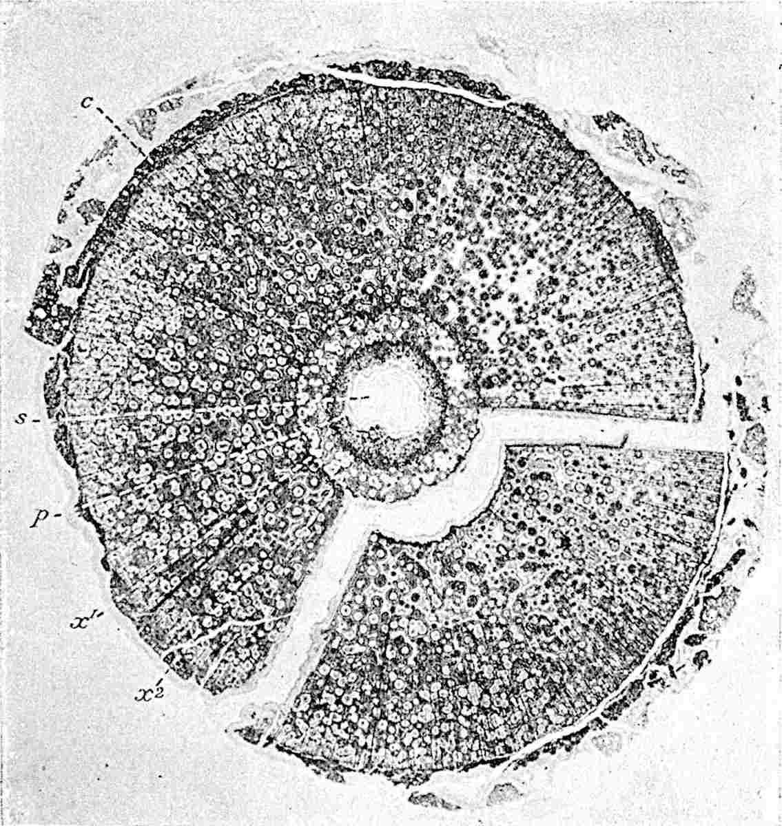
Fig. 182. Lepidodendron Wünschianum. The stele of the stem shown in fig. 181, A. (Cambridge Botany School.)
The primary xylem is limited externally by an unequally fluted surface with exarch protoxylem elements; it is, however, noteworthy that there is not always a very clearly defined difference between the small protoxylem and the large centripetally developed tracheae. Immediately beyond the primary xylem occur numerous thin-walled parenchymatous cells with spiral and reticulate pitting; beyond these is the broad zone of secondary xylem, x2, composed of scalariform tracheae and numerous medullary rays consisting of one, two, or several rows of radially elongated elements with spiral and reticulate pitting. In tangential sections the rays are seen to vary considerably in size, some being made up of a single row of cells while others are longer and broader; through the latter leaf-traces pass horizontally. Portions of medullary rays are seen at mr in fig. 181, C and E.
The leaf-traces given off from projecting ridges on the outer edge of the primary xylem pass upwards for a short distance and then bend outwards through a broad medullary ray; on reaching the limit of the secondary xylem they again bend sharply upwards, appearing in transverse section at lt fig. 181, B. Each leaf-trace consists at first of long tracheae accompanied by numerous thin-walled spiral and reticulate parenchymatous cells derived from the tissue in contact with the outer edge of the primary wood. Fig. 181, B, shows a leaf-trace near the edge of the secondary xylem; it consists of a group of primary tracheae, with narrower protoxylem elements, px, near the outer margin, almost completely enclosed by radially disposed series of smaller and more delicate tracheae. These secondary elements of the leaf-trace are apparently added during its passage through the medullary ray, but additions are also made to this tissue by the meristematic zone, m, fig. 181, B and E. In contact with the outermost tracheae of normal size at the edge of the secondary xylem there are some smaller lignified elements, as at a, fig. 181, E, and at T, fig. 183; this juxtaposition of large and small tracheae has been referred to in the description of L. vasculare.
Prof. Williamson[358], in his account of the Arran specimens of this species, expressed the opinion that the trees probably perished “in consequence of the mephitic vapours which filled the atmosphere”; it maybe that in the striking difference in the diameter of the conducting elements on the margin of the wood we have evidence of approaching death.
Beyond the most recently formed tracheae we have a band of delicate parenchymatous cells (m, figs. B and E, 181; C, figs. 183, 184) forming the meristematic zone[359]. The longitudinal section represented in fig. 184 shows some recently formed narrow tracheae, T, and beyond these the meristematic zone composed of thin-walled short cells, C, arranged in horizontal rows. It is this small-celled tissue to which the name phloem has been applied by some authors[360], a term which seems to me to be misleading and inappropriate. In passing through this zone of dividing cells the leaf-traces become surrounded by an arc of meristem from which elements are added to the radially placed rows of secondary tracheae. Beyond the meristematic region portions of the secretory zone are preserved, consisting of large sacs or spaces and small dark cells as seen in figs. 181, B, E, sc, F; 183, 184. This tissue has the same structure as in L. vasculare and in L. fuliginosum: it is a striking fact that there are no indications of any additions to the secretory zone even in stems with such a large amount of secondary xylem as in the Dalmeny specimen (fig. 182, x2). If the secretory zone were of the nature of phloem we should expect to see signs of additions made to it in the course of growth. In this connexion it is worth mentioning that in the recent fern Botrychium (Ophioglossaceae) secondary xylem is formed in the stem, but apparently no additions are made to the phloem. The structure of the secretory zone tissue as seen in the longitudinal section fig. 184, S, is also a serious difficulty in the way of accepting the designation phloem as employed by Scott and Weiss. Between the secretory zone and the outer cortical region, no tissues have been preserved. The shell of bark consists chiefly of radial rows of elongated cells with rather thick walls characterised by the occurrence of small intercellular spaces and by tangentially placed bands of secretory cells and sacs (fig. 181, D, s). Immediately internal to the secondary cortex or phelloderm occur groups of secretory tissue as shown in the section of L. Harcourtii (fig. 179, B).
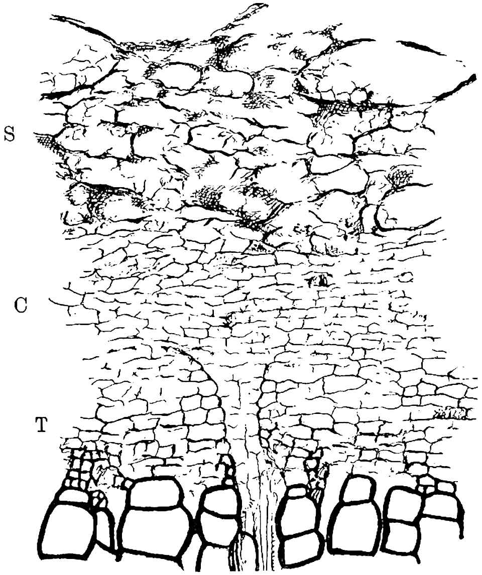
Fig. 183. Lepidodendron Wünschianum. From the specimen shown in fig. 181, S, secretory zone; C, meristem; T, immature tracheae.
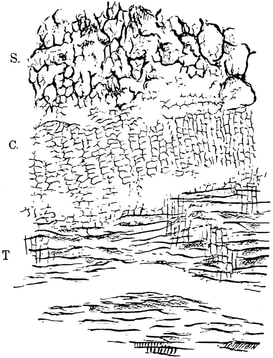
Fig. 184. Lepidodendron Wünschianum. Longitudinal section of the specimen represented in transverse section in fig. 183.
The large tree shown in transverse section in fig. 181, A, has lost its leaf-cushions; the bark, as seen in the lower part of the photograph, presents a fissured appearance like that with which we are familiar on an old Oak or Elm stem. A radial longitudinal section through the phelloderm revealed the existence of a crushed leaf-trace passing outwards in an approximately horizontal course accompanied by a strand of parenchymatous tissue[361] having the characteristic structure of a parichnos. It is probable that the surface of this partially decorticated stem differed in appearance from that of an old Sigillaria (cf. fig. 198) in the much smaller and less conspicuous parichnos strands.
In addition to the large stems of L. Wünschianum from Arran and Dalmeny numerous examples of smaller axes from the former locality are represented in the Williamson collection (British Museum). Some of the twigs are characterised by a solid stele (protostele) giving off numerous leaf-traces accompanied by short spirally thickened tracheids like those which occur at the outer edge of the primary xylem in the larger stem: these extend into the leaf where they are arranged round the vascular bundle like the transfusion tracheids[362] in many recent conifers. The surface of these smaller shoots bears large leaf-cushions which are seen in longitudinal section to have the form characteristic of Lepidophloios. It is worthy of note that a section of a bifurcating axis of this species from the Calciferous Sandstone of Craigleith (British Museum Collection[363]), although its diameter is 19 × 14 cm., shows no signs of secondary wood. This late appearance of secondary xylem and other anatomical features suggest the possibility of the specific identity of L. Wünschianum and L. Harcourtii[364].
In 1871 Binney[365] described a specimen of a heterosporous cone, Lepidostrobus Wünschianus, from Arran exhibiting the ordinary features of lepidodendroid strobili; this was probably borne by Lepidodendron Wünschianum.
7. Lepidodendron macrophyllum (Williamson). Fig. 186, C.
The diagrammatic sketch reproduced in fig. 186, C, was made from the transverse section of a small twig, slightly less than 2 cm. in its longest diameter, originally figured by Williamson[366] in 1872. Earlier in the same year Carruthers[367] published a short account of the same form based on specimens collected by Mr Butterworth from the Coal-Measures of Lancashire near Oldham, but both authors refrained from instituting a new specific name. In a later publication Williamson spoke of the type as Lepidodendron macrophyllum[368]. Williamson’s species has nothing to do with Lycopodites macrophyllus of Goldenberg[369]. The most striking feature of this rare form is the large size of the leaf-cushions, which are of the Lepidophloios type, in proportion to the diameter of the shoot. The stele consists of a ring of xylem, all of which is primary in the sections so far described, enclosing a parenchymatous pith: a Stigmarian rootlet is shown at s.
8. Lepidodendron Veltheimianum Sternb. (General account). Figs. 157, 185, 186, A, B.
1820. “Schuppenpflanze,” Rhode, Beit. zur Pflanzenkunde der Vorwelt, Pl. III. fig. 1.
1825. Lepidodendron Veltheimianum, Sternberg, Flora der Vorwelt, Pl. LII. fig. 5.
1836. Pachyphloeus tetragonus, Goeppert, Die fossilen Farnkräuter, Pl. xLIII. fig. 5.
1852. Sagenaria Veltheimiana, Goeppert, Foss. Flora des Übergangsgebirges, Pls. XVII–XXIV.
1875. Lepidodendron Veltheimianum, Stur, Culm Flora, p. 269, Pls. XVIII–XXII.
1886. Lepidodendron Veltheimianum, Kidston, Catalogue of Palaeozoic plants, British Museum, p. 160.
1901. Lepidodendron Veltheimianum, Potonié, Silur und Culm Flora, p. 116, figs. 72–76.
1904. Lepidodendron Veltheimianum, Zalessky, Mém. Com. Géol. Russie, Pl. IV. figs. 4, 5.
1906. Lepidodendron Veltheimi, Potonié, Königl. Preuss. geol. Landesanstalt, Lief. III.
The above list may serve to call attention to a few synonyms[370] of this plant, and to a selection of sources from which full information may be obtained as to the history of our knowledge of this characteristic and widely spread Lower Carboniferous type.
Lepidodendron Veltheimianum is represented by casts of stems, the largest of which hitherto described reaches a length of 5·22 metres with a maximum diameter of 63 cm.; this specimen, figured by Stur[371], consists of a tapered main axis giving off smaller lateral shoots, some of which exhibit dichotomous branching. Fig. 185, C and D, represent the external features of a well-preserved cast and impression respectively. Oblique rows of prominent cushions wind round the surface of the stem and branches: each cushion is prolonged upwards and downwards in the form of a narrow ridge with sloping sides which connects adjacent cushions by an ogee curve. At the upper limit of the broader kite-shaped portion of the cushion the ligular pit forms a conspicuous feature; immediately below this is the leaf-scar with its three small scars,—the lateral parichnos strands and the central leaf-trace. The two oval areas shown in fig. 185, D, just below the lower edge of the leaf-scars, represent the parichnos arms which impinge on the surface of the cushions on their way to the leaves, as explained on a previous page. It is possible that these areas were visible on the living stem as strands of loose parenchyma comparable with the lenticel-like pits on the stipules of Angiopteris[372] and the leaf-bases of Cyatheaceous ferns, or it may be that their prominence in the specimen before us is the result of the decay of a thin layer of superficial cortex which hid them on the living tree. Fig. 185, B, illustrates the appearance of a stem in a partially decorticated condition (Bergeria state). A further degree of decortication is seen in fig. 185, A, which represents the Knorria condition.
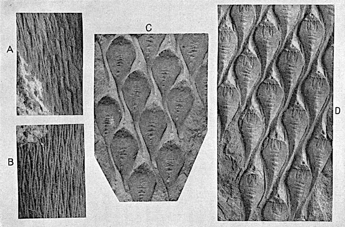
Fig. 185. Lepidodendron Veltheimianum. From specimens in Dr Kidston’s Collection. (Approximately nat. size.)
Fig. 157 shows a Ulodendron axis of this species; in the lower part the specimen illustrates the partial obliteration of the surface features as the result of the splitting of the outer bark consequent on growth in thickness of the tree. By an extension of the cracks, shown in an early stage in fig. 157, the leaf-cushions would be entirely destroyed and the surface of the bark would be characterised by longitudinal fissures simulating the vertical grooves and ridges of a Sigillarian stem. The large stumps of trees shown in the frontispiece to Volume I. are probably, as Kidston[373] suggests, trunks of L. Veltheimianum in which the leaf-cushions have been replaced by irregular longitudinal fissures. In old stems of Sigillaria the enlarged parichnos areas constitute a characteristic feature (p. 205), but it does not follow that the absence of large parichnos scars is a distinguishing feature of all Lepidodendra.
In this species, as in others, the form of the leaf-cushion exhibits a considerable range of variation dependent on the thickness of the shoot; the contiguous cushions of young branches become stretched apart as the result of increasing girth of the whole organ, and casts of still older branches may exhibit very different surface-features[374]. The leaves as seen on impressions of slender branches are comparatively short, reaching a length of 1–2 cm. It is important to notice that leafy twigs of this species may bear terminal cones[375] resembling in form those of Picea excelsa and other recent conifers, though differing essentially in their morphological features.
The fossil stumps of trees represented in the frontispiece to Volume I. bear horizontally spreading and dichotomously branched root-like organs having the characters of Stigmaria ficoides[376]. Geinitz has suggested that Stigmaria inaequalis Göpp. may be the underground portion of Lepidodendron Veltheimianum.
It is unfortunately seldom possible to connect petrified Lepidodendron cones with particular species of the genus based on purely vegetative characters, but it is practically certain that we are justified in recognising certain strobili described by Williamson[377] from the Calciferous Sandstone series of Burntisland on the Firth of Forth as those of Lepidodendron Veltheimianum. Williamson believed that the cone which he described belonged to the plant with shoots characterised by the anatomical features of his species Lepidodendron brevifolium (= L. Veltheimianum), a conclusion which is confirmed by Kidston[378]. The cone of L. Veltheimianum, which reached a diameter of at least 1 cm. and a length of 4 cm., agrees in essentials with other species of Lepidostrobus; the axis has a single medullated stele of the same general type as that of the vegetative shoots of Lepidodendron fuliginosum and L. Harcourtii. The sporophylls are described by Williamson as spirally disposed, and Scott notices that in some specimens they are arranged in alternate whorls; as in recent Lycopods both forms of phyllotaxis may occur in the same species. The heterosporous nature of this strobilus, to which Scott first applied the name Lepidostrobus Veltheimianus, is clearly demonstrated by the two longitudinal sections contributed by Mr Carruthers and figured by Williamson in 1893[379].
Each sporophyll, attached almost at right angles to the cone-axis, bears a radially elongated sporangium seated on the median line of its upper face; its margins are laterally expanded as a thin lamina; from the middle of the lower face a narrow keel extends downwards between two sporangia belonging to a lower series. From the base of a sporangium a mass of sterile tissue penetrates into the spore-producing region as in the large sporangia of Isoetes (cf. fig. 191, H, a, and fig. 133, H). The distal and free portion of the sporophylls is bent upwards as a protecting bract. Some of the sporangia in the upper part of the cone produced numerous microspores, while 8–16 megaspores occur in the lower sporangia. The megaspores, having a mean diameter of 0·8 mm. “quite 40 times the size of the microspores[380],” are characterised by tubular capitate appendages, and by a conspicuous three-lobed projection (fig. 191, E)[381] which, as Scott suggests, may represent the outer spore-wall which has split as the result of germination. It is not improbable, as shown in fig. 191, I, that this cap was present before germination. The megaspores represented in fig. 191, I, illustrate their characteristic form as seen in a section of a megasporangium, Sm; the open beak-like portion of the larger spore is probably the apical region which has split along the three-rayed lines. These lines form a characteristic feature of both recent and extinct spores and denote their origin in tetrads. The spore shown in fig. 191, E[382], illustrates the external features. The apical region of the prothallus of a megaspore of Lepidodendron Veltheimianum described by Mr Gordon[383] consists of smaller cells than those occupying the greater part of the spore-cavity, a differentiation which he compares with that of the prothallus of Selaginella.
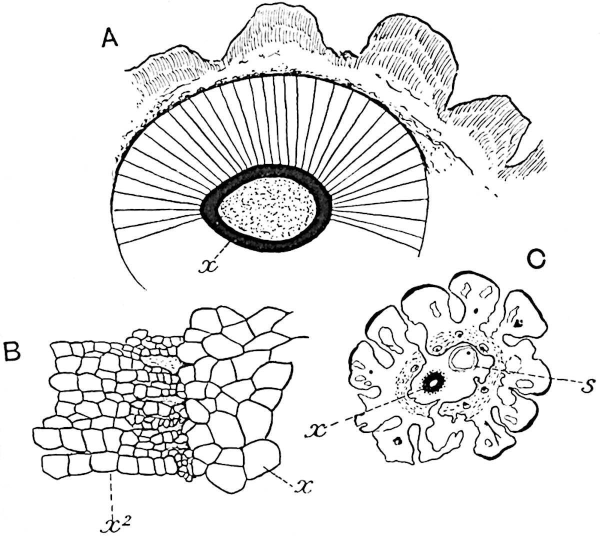
Fig.
186.
A, B. Lepidodendron Veltheimianum. (Botany School, Cambridge.)
C. Lepidodendron macrophyllum. (British Museum. No. 377.)
x, Primary xylem; x2, secondary xylem; s, Stigmarian rootlet.
There can be little doubt that the petrified shoots described by Williamson[384] from the Calciferous Sandstone beds of Burntisland as Lepidodendron brevifolium are identical with specimens possessing the external features of L. Veltheimianum. In 1872 Dawson expressed the opinion that Williamson’s species should be referred to L. Veltheimianum, and evidence subsequently obtained confirms this view. The stele of this species is of the medullated type, differing from that of L. fuliginosum and L. Harcourtii in the absence of prominent ridges on the external surface of the primary xylem, and from L. vasculare in the possession of a parenchymatous pith. In younger twigs the cortex consists of fairly homogeneous tissue, but in older branches there is a greater distinction between a delicate middle cortex and a stronger outer cortex. Fig. 186, A, represents a stem in which the vascular cylinder is composed of a primary xylem ring, x, 1·5 mm. broad, succeeded by a zone of secondary wood 1·2 cm. in breadth. The junction between the primary and secondary xylem is shown on a larger scale in fig. 186, B. The tissues abutting on the secondary xylem have not been preserved; the outer cortex, which consists chiefly of secondary elements, is divided superficially into unequal ridges corresponding to the leaf-cushions which have been more or less obliterated as the result of growth in thickness of the stem.
9. Lepidodendron Pedroanum (Carruthers).
In 1869 Mr Carruthers described some specimens of vegetative stems and isolated sporangia, collected by Mr Plant in Brazil, as Flemingites Pedroanus[385]. From a more recent account published by Zeiller[386] it is clear that Carruthers’ species is a true Lepidodendron; an examination of the type-specimens in the British Museum confirms this determination. The contiguous leaf-cushions have rounded angles similar in form to those of Lepidodendron Veltheimianum and L. dichotomum, but it is not unlikely that the Brazilian plant is specifically distinct from European species. A figure of one of the specimens on which Carruthers founded the species is given by Arber[387] in his Glossopteris Flora. The Brazilian plant is chiefly interesting as affording proof of the existence of Lepidodendron in the southern hemisphere; the species has also been recognised in South Africa from material collected by Mr Leslie at Vereeniging[388].
As Zeiller[389] has suggested, it is not improbable that the fossils described by Renault[390] from Brazil as Lycopodiopsis Derbyi may be the petrified stems of Lepidodendron Pedroanum. The structure of the central cylinder of Renault’s species is of the type represented by L. Harcourtii; the xylem forms a continuous ring and does not consist of separate strands of tracheae as Renault believed.
10. Lepidodendron australe (M’Coy). Figs. 187, A–C.
Specimens described under this name are interesting rather on account of their extended geographical range and geological antiquity than on botanical grounds. The drawings reproduced in fig. 187 illustrate the characteristic appearance of this Lower Carboniferous and Upper Devonian type, as represented by a specimen recently described[391] from the Lower Karroo (Dwyka) series, which is probably of Carboniferous age, near Orange River Station, South Africa. The surface is divided into polygonal or rhomboidal areas (figs. A and B) 8–9 mm. long and 7–8 mm. broad, arranged in regular series and representing leaf-scars, comparable with those of Sigillaria Brardi and other species, or possibly partially decorticated leaf-cushions. A short distance below the apex of each area there is a more or less circular prominence or depression (fig. 187, B) and on a few of the areas there are indications of a groove (fig. A, g) extending from the raised scar to the pointed base, as at g, g.
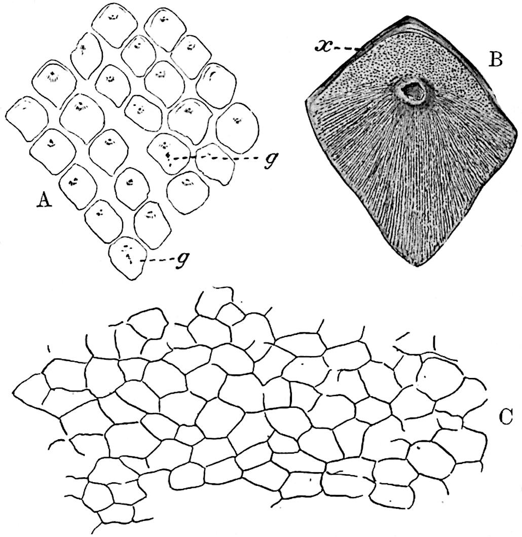
Fig. 187. Lepidodendron australe. Fig. A, nat. size.
In examining the graphitic layer on the surface of the South African specimen shown in fig. 187, A, use was made of a method recently described by Professor Nathorst[392]. A few drops of collodion were placed on the surface, and after a short interval the film was removed and mounted on a slide. The addition of a stain facilitated the microscopic examination and the drawing of the collodion film. The cell-outlines (fig. 187, C) on the surface of the polygonal areas may be those of the epidermis, but they were more probably formed by a subepidermal tissue; the scar, which interrupts the continuity of the flat surface, may mark the position of a leaf-base, or, assuming a partial decortication to have occurred prior to fossilisation, it may represent a gap in the cortical tissue caused by the decay of delicate tissue which surrounded the vascular bundle of each leaf in its course through the cortex of the stem. If the impression were that of the actual surface of a Lepidodendron or a Sigillaria, we should expect to find traces of the parichnos appearing on the leaf-scar as two small scars, one on each side of the leaf-bundle. In specimens from Vereeniging described in 1897[393] as Sigillaria Brardi, which bear a superficial resemblance to that shown in fig. A, the parichnos is clearly shown. On the other hand, an impression of a partially decorticated Lepidodendroid stem need not necessarily show the parichnos as a distinct feature: owing to its close association with the leaf-trace in the outer cortex, before its separation in the form of two diverging arms, it would not appear as a distinct gap apart from that representing the leaf-bundle. The absence of the parichnos may be regarded as a point in favour of the view that the impression is that of a partially decorticated stem. Similarly, the absence of any demarcation between a leaf-cushion and a true leaf-scar such as characterises the stems of Lepidodendra and many Sigillariae is also favourable to the same interpretation.
In 1872 Mr Carruthers[394] described some fossils from Queensland, some of which appear to be identical with that shown in fig. 187 under the name Lepidodendron nothum, Unger[395], a species founded on Upper Devonian specimens from Thuringia. The Queensland plant is probably identical with Dawson’s Canadian species, Leptophloeum rhombicum[396]. In 1874 M’Coy[397] instituted the name Lepidodendron australe for some Lower Carboniferous specimens from Victoria, Australia: these are in all probability identical with the Queensland fossils referred by Carruthers to Unger’s species, but as the identity of the German and Australian plants is very doubtful[398] it is better to adopt M’Coy’s specific designation.
Krasser[399] has described a similar, but probably not specifically identical, type from China; from Devonian rocks of Spitzbergen Nathorst[400] has figured, under the name Bergeria, an example of this form of stem, and Szajnocha[401] has described other specimens from Lower Carboniferous strata in the Argentine.
Lepidodendron australe has been recorded from several Australian localities[402] from strata below those containing the genus Glossopteris and other members of the Glossopteris, or, as it has recently been re-christened, the Gangamopteris[403] Flora.
viii. Fertile shoots of Lepidodendron.
A. Lepidostrobus.
The generic name Lepidostrobus was first used by Brongniart[404] for the cones of Lepidodendron, the type-species of the genus being Lepidostrobus ornatus, the designation given by the author of the genus to a Lepidostrobus previously figured by Parkinson[405] in his Organic Remains of a Former World. The generic name Flemingites proposed by Carruthers[406] in 1865, under a misapprehension as to the nature of spores which he identified as sporangia, was applied to specimens of true Lepidostrobi. Brongniart also instituted the generic name Lepidophyllum for detached leaves of Lepidodendron, both vegetative and fertile; the specimen figured by him in 1822 as Filicites (Glossopteris) dubius[407], and which was afterwards made the type-species of the genus, was recognised as being a portion of the lanceolate limb of a large single-veined sporophyll belonging to a species of Lepidostrobus.
In an unusually large Lepidophyllum, or detached sporophyll of Lepidostrobus, in the Manchester University Museum, the free laminar portion reaches a length of 8 cm.
It is not uncommon to find Lepidodendron preserved in the form of a shell of outer cortex, which has become separated along the phellogen from the rest of the stem; as the result of compression the cylinder of bark may assume the appearance of a flattened stem covered with leaf-cushions. A specimen preserved in this way was described by E. Weiss as a cone of Lomatophloios macrolepidotus Gold., and is quoted by Solms-Laubach and other authors[408] as an example of an unusually large Lepidostrobus. An examination of the type-specimen in the Bergakademie of Berlin convinced me that Weiss had mistaken the partially destroyed leaf-cushions for sporophylls, and Stigmarian rootlets, which had invaded the empty space, for sporangia[409].
In external appearance some species of Lepidostrobus bear a superficial resemblance to the cone of a Spruce Fir (Picea excelsa), but the surface of a lycopodiaceous strobilus is usually covered by the overlapping and upturned laminae which terminate the more or less horizontal sporangium-bearing portion of the sporophyll.
Fig. 188 affords a good example of a long and narrow Lepidostrobus. This specimen from the Middle Coal-Measures of Lancashire has a length of 23 cm.; like other Lepidostrobi it is borne at the tip of a slender shoot. The fossil is sufficiently well preserved to show the characteristic radially elongated form of the large sporangia and the long and upturned distal portions of the sporophylls.
We may briefly describe Lepidostrobus as follows:—Cylindrical strobili consisting of an axis containing a single cylindrical stele which agrees generally with that of the vegetative shoots of L. Harcourtii and other species. The amount of parenchymatous pith varies in different forms; in some the primary xylem is almost solid. The middle cortical region, which has usually been destroyed before fossilisation, possesses the loose lacunar structure characteristic of this region in the vegetative branches. The thicker walled outer cortex is continued at the periphery into crowded, usually spirally disposed sporophylls, each of which consists of a more or less horizontal pedicel, which may be characterised by a keel-like median ridge on its lower surface, while to the central region of the upper face is attached a large radially elongated sporangium. One of the chief differences between a Lepidodendron cone and those of the recent genus Lycopodium is the greater radial elongation of the sporangia in the former. Some species of Lepidostrobus may have been homosporous; some are known to be heterosporous. In the latter the megasporangia borne on the lower sporophylls usually contain several megaspores as in Isoetes (cf. fig. 133, E). Beyond the distal end of the sporangium the sporophyll becomes broader in a horizontal plane and is bent upwards as a lanceolate limb; it may also be prolonged a short distance downwards as a bluntly triangular expansion.
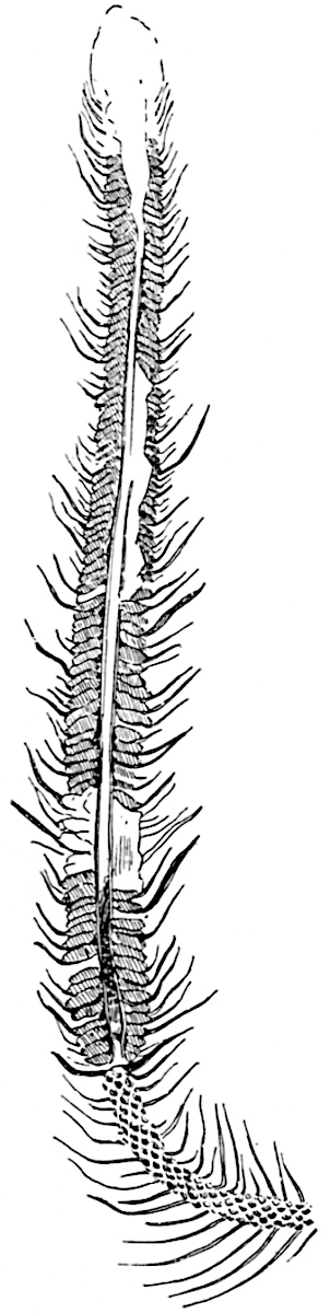
Fig. 188. Lepidostrobus. Middle Coal-Measures, Bardsley, Lancashire. From a specimen in the Manchester Museum. (½ nat. size.)
There can be little doubt that the Palaeozoic Lepidodendra, like Lycopodium cernuum (fig. 123) and other recent Lycopods, usually bore their cones at the tips of slender shoots. The fertile shoot of Lepidophloios scoticus shown in fig. 160, B, affords one of several instances supporting this statement; similar examples are figured by Brongniart[410], Morris[411], and by more recent writers. The apparently sessile cone figured by Williamson[412] from a specimen in the Manchester Museum is certainly not in situ, but is accidentally associated with the stem.
The general absence of secondary wood in the steles of Lepidostrobi is, as Dr Kidston[413] points out, consistent with the view that the cones were shed on maturity and that fertilisation probably took place on the ground, or perhaps on the surface of the water where the slender hairs of the megaspores (fig. 191, F, I) may have served to catch the microspores.
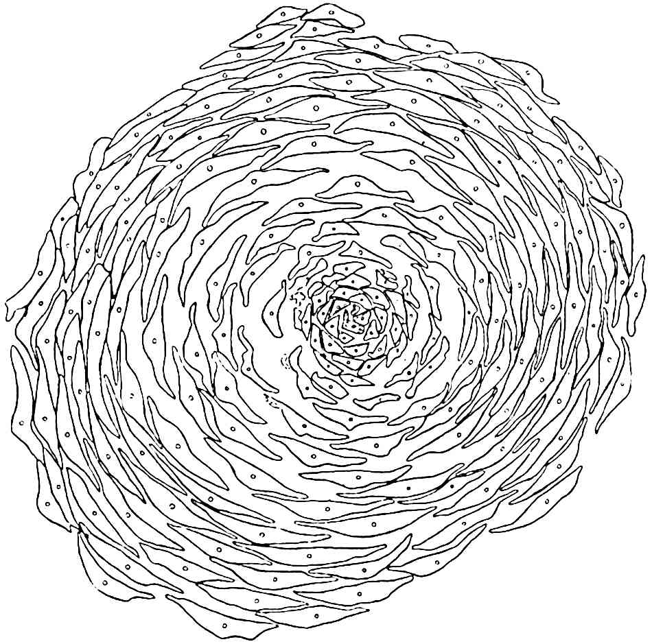
Fig. 189. Lepidostrobus. Section through the apical region of a cone above the axis. (Manchester University Collection.)
Fig. 189 is an accurate representation of a transverse section, 6 mm. in diameter, of what is no doubt the apical portion of a Lepidostrobus from the Coal-Measures of Shore, Lancashire. The section cuts across the upturned free laminae above the level of the apex of the cone-axis. Each lamina contains a small vascular bundle composed of a few tracheae and some thin-walled cells surrounded by delicate mesophyll tissue. Immediately in front of the distal end of a sporangium a small ligule is borne on the upper face of the sporophyll (fig. 191, A, B, l) occupying the same position as in Selaginella (cf. fig. 131, F). Strands of vascular tissue pass in a steeply ascending course from the xylem to the pedicels of sporophylls, finally curving upwards and ending in the upper limb. Each vascular bundle consists of a strand of xylem, apparently of mesarch structure, accompanied by a few layers of parenchyma on its outer face and by a group of cambiform elements, the whole being enclosed in a sheath of parenchyma continuous with the inner cortex of the cone axis. The vascular bundle is accompanied by a parichnos in the outer cortex and in the sporophyll.
Reference has already been made to the belief on the part of some palaeobotanists that the large scars of Ulodendron represent attachment-surfaces of sessile cones, and reasons have been given against the acceptance of this view.
There is considerable range in the size of Lepidostrobi. An incomplete specimen, 33 cm. long and 6 cm. broad, which may have been 50 cm. in length, is described by Renault and Zeiller[414] from the Commentry Coal-field. The larger cones afford a striking demonstration of the enormous spore-output of some species of Lepidodendron.
Among the earliest accounts of the anatomy of Lepidostrobus are those by Hooker[415] and Binney[416]. One of the specimens described by the former author (fig. 190) affords an interesting example of an unusual manner of fossilisation; a hollow stem or Lepidodendron is filled with sedimentary material containing several pieces of Lepidostrobi in an approximately vertical position.
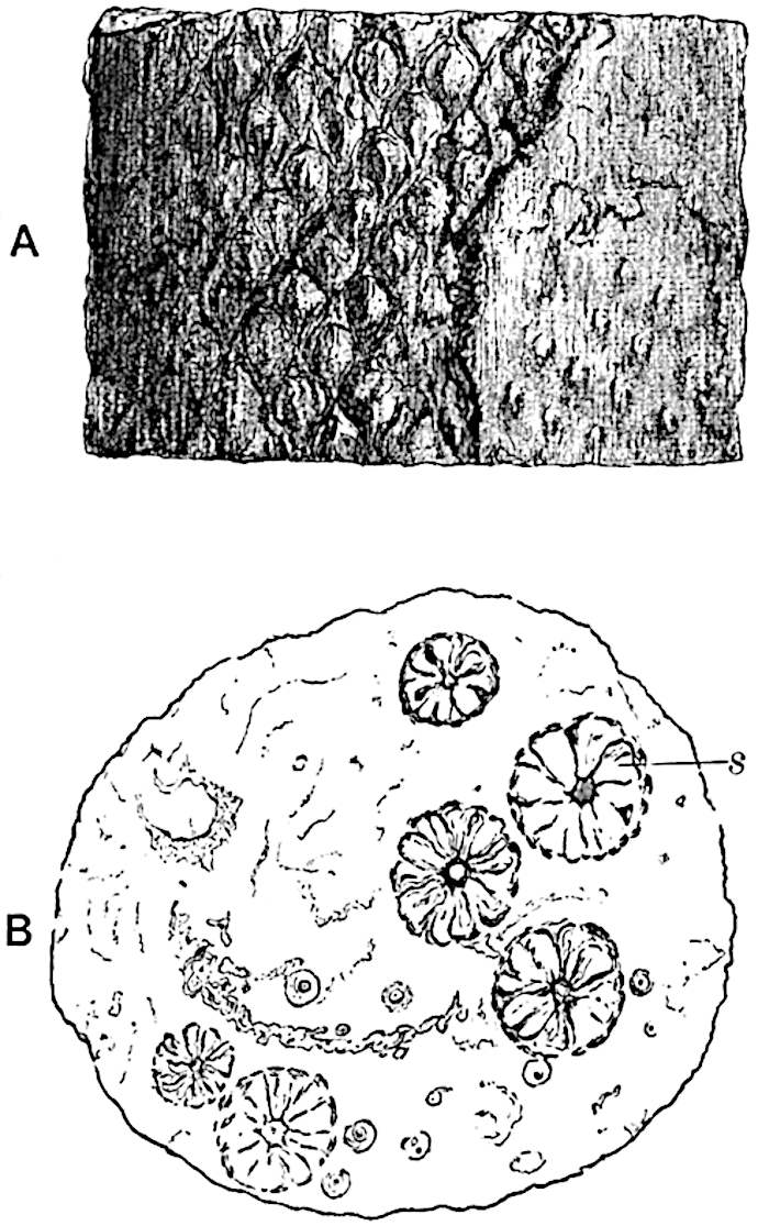
Fig.
190.
Lepidodendron
stem with
Lepidostrobi
. (After Hooker.)
Side-view showing leaf-cushions on the left-hand side and the Knorria condition on the right.
View of transverse section; s, sections of Lepidostrobi.
The fact that Lepidostrobi usually occur as isolated specimens renders it impossible in most cases to refer them to particular species of Lepidodendron. Neither external features nor anatomical characters afford satisfactory criteria by which to correlate vegetative and fertile shoots; in some measure this is due to the imperfection of our knowledge as regards the range of structure within the limits of species; it is also due to lack of information as to the extent to which the transition from sterile to fertile portions of a shoot is accompanied by anatomical differences. Prof. Williamson wrote: “I have for many years endeavoured to discover some specific characters by which different Lepidostrobi can be distinguished and identified, but thus far my efforts have been unsuccessful[417].” In a few cases, such as those mentioned in the description of Lepidodendron Veltheimianum and L. Wünschianum, it has been possible to correlate cones and vegetative shoots.
The most complete account we possess of the anatomy of Lepidodendron cones is that by Mr Maslen[418], who first demonstrated the occurrence of a ligule on the sporophylls, and thus supplied a missing piece of evidence in support of the generally accepted view as to the homology of the sporangium-bearing members and foliage leaves.
i. Lepidostrobus variabilis (Lindley and Hutton).
1811. “Strobilus,” Parkinson, Organic Remains, Vol. I. p. 428, Pl. IX. fig. 1.
1828. Lepidostrobus ornatus, Brongniart, Prodrome, p. 87.
1831. L. variabilis, Lindley and Hutton, Foss. Flora, Pls. X. XI.
1831. L. ornatus, Lindley and Hutton, Foss. Flora, Pl. XXVI.
1837. L. ornatus var. didymus, Ibid. Pl. CLXIII.
1850. Arancarites Cordai, Unger, Genera et Spec. Plant. foss. p. 382.
1875. Lepidostrobus variabilis, Feistmantel, Palaeontographica, Vol. LXIII. Pl. XLIV.
1886. L. variabilis, Kidston, Cat. Palaeozoic Plants, p. 197.
1890. L. ornatus, Zeiller, Flor. Valenciennes, p. 497, Pl. LXXVI. figs. 5, 6.
—— L. variabilis, Zeiller, Flor. Valenciennes, p. 499, Pl. LXXVI. figs. 3, 4.
Under this specific name are included strobili from Upper Carboniferous rocks which, in spite of minor differences, may be considered as one type. The cylindrical cones vary considerably in size, some reaching a length of 50 cm. or more. The sporophylls are attached by a pedicel, 4–8 mm. long, at right angles to the axis, while the distal portion forms an oval lanceolate limb 10–20 mm. in length. The sporangia are 4–8 mm. long.
The branched example figured by Lindley and Hutton[419] as a variety (L. ornatus var. didymus) illustrates a phenomenon not uncommon in both Palaeozoic and recent lycopodiaceous strobili.
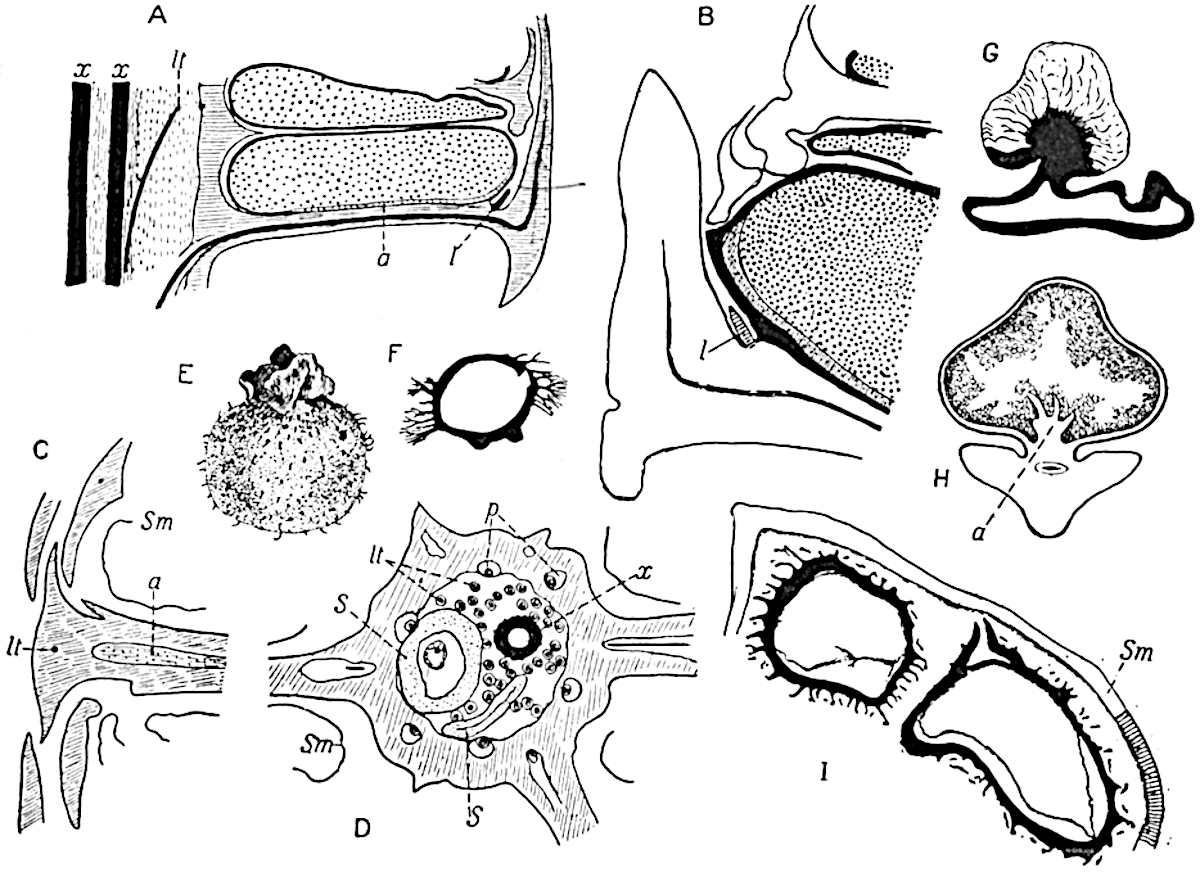
Fig.
191.
Lepidostrobus.
A–D. L. oldhamius.
B, C, D. From sections in the Binney Collection, Cambridge.
E. Megaspore. (After Kidston.)
F. Megaspore (Coal-Measures, Halifax). (After Williamson.)
G. Megaspore of Lepidostrobus foliaceus. (After Mrs Scott.)
H. Tangential section of sporangium. (After Bower.)
I. Part of sporangium wall, Sm, of the cone of Lepidodendron Veltheimianum, enclosing two megaspores. (Cambridge Botany School.)
ii. Lepidostrobus oldhamius Williamson[420]. Fig. 191, A–D.
Williamson[421] instituted this term for strobili previously described by Binney[422], without adequate evidence, as the cones of Lepidodendron Harcourtii. In shape and in the main morphological features this type resembles L. variabilis, which is however known only in the form of casts and impressions. A cone of L. oldhamius, 2–3 cm. in diameter, possesses a medullated stele consisting of a ring of primary xylem (fig. 191, D, x) with exarch protoxylem and no secondary elements. Maslen found several short tracheae at the periphery of the xylem and states that these led him to compare the cone with the vegetative shoots of Lepidodendron vasculare, but the common occurrence of such elements in different types of shoot renders them of little or no specific value. The inner cortex is like that of vegetative shoots of Lepidodendron and the middle cortex, which was no doubt of the type described in Lepidostrobus Brownii, is represented by a gap in the sections, beyond which is the stronger outer cortex (fig. 191, D) passing into the horizontal pedicels of the sporophylls. The section of the axis reproduced in fig. 191, D, was figured by Binney[423] as Lepidodendron vasculare. The leaf-traces, several of which are seen in the middle cortical region in fig. D, lt, consist of a strand of scalariform tracheae, with a mesarch protoxylem, succeeded by a few parenchymatous cells; beyond these there is usually a small gap which was originally occupied by a strand of thin-walled cells. It is important to note that in one sporophyll-trace figured by Maslen[424] there is a strand of thin-walled elongated elements abutting on the xylem, which he describes as phloem. This tissue is certainly more like true phloem than any which has hitherto been described in the leaf-traces of vegetative shoots. The state of preservation is not, however, sufficiently good to enable us to recognise undoubted phloem features.
In such cones as I have examined no tissue has been seen which shows the histological features characteristic of the secretory zone of vegetative shoots: the “phloem” (Maslen) occupies the position in the sporophyll bundle which in the vascular bundles of foliage leaves is occupied by a dark-celled and partially disorganised tissue in continuity with the secretory zone of the main stele. It may be that in the strobili this tissue occurred in a modified form, but even assuming that the section figured by Maslen shows true phloem, an assumption based on slender evidence, this is not sufficient justification for the application of the term phloem to a tissue occupying a corresponding position in vegetative shoots and distinguished by well-marked histological features.
The sporophyll-traces, as seen in the outer cortex in fig. 191, D, are partially surrounded by a large crescentic space, p, which was originally occupied by the parichnos. The sporangia are attached along the middle line of the sporophyll and, as in Lepidostrobus Brownii, a cushion of parenchyma projects into the lower part of the sporangial cavity (fig. 191, A, a; C, a).
The diagrammatic sketch of part of a section in the Binney Collection reproduced in fig. 191, B, shows the position of the ligule, l. No megaspores have been discovered in any specimens of this type; the microspores, which occur both singly and in tetrads, have a length of 0·02–0·03 mm.
The drawing shown in fig. 191, A, based on a section in the Binney Collection, illustrates the general arrangement of the parts of a typical Lepidostrobus. I have made use of this sketch instead of that given by Maslen, as his figure conveys the idea that the sporophylls are superposed, whereas, whether they are verticillate or spiral, a radial longitudinal section would not cut successive sporangia in the same plane.
iii. Lepidostrobus Brownii (Brongn.).
In 1843 a specimen of a portion of a petrified cone was purchased by the British Museum, assisted by the Marquis of Northampton and Robert Brown, for £30 from a French dealer. This fossil, from an unknown locality, was briefly described by Brown in 1851[425] and named by him Triplosporites, but in a note added to his paper he expressed the opinion that the generic designation Lepidostrobus would be more appropriate. Brongniart afterwards named the cone Triplosporites Brownii[426], and Schimper[427] described it in his Traité as Lepidostrobus Brownii. The type-specimen is preserved in the British Museum and the Paris Museum possesses a piece of the same fossil.
The central axis of the cone has a stele of the type characteristic of Lepidodendron fuliginosum and L. Harcourtii, and the xylem is surrounded by a thin-walled tissue described by Bower[428] as possibly phloem; but in the absence of longitudinal sections it is impossible to say how far the tissue external to the xylem agrees with that in Lepidodendron stems. The sporophylls consist of a horizontal portion, to the upper face of which the radially elongated sporangia are attached, one to each sporophyll; beyond the distal end of the sporangium the sporophyll bends sharply upwards as a fairly stout lamina. The wall of the sporangium is composed of several layers of cells, as shown in a drawing published by Bower[429]; in the interior occur groups of microspores, and from a ridge of tissue which extends along the whole length of the sporangium irregular trabeculae of sterile tissue project into the sporangial cavity, as in Isoetes (fig. 191, H: cf. fig. 133, H).
Further information in regard to Lepidostrobus Brownii has recently been supplied by Prof. Zeiller[430], who recognises the existence of a ligule, and draws attention to some interesting histological features in the tissue of the sporophylls[431].
Spores of Palaeozoic Lycopodiales.
The calcareous nodules from the Coal seams of Yorkshire and Lancashire are rich in isolated spores, many of which are undoubtedly those of Lepidostrobi. Examples of spores were figured by Morris[432] in 1840, and their occurrence in coal has been described by several authors, one of the earliest accounts being by Balfour[433]. The drawings of Palaeozoic and recent spores published by Kidston and Bennie[434] demonstrate a striking similarity between the megaspores of existing and extinct Lycopods, the chief difference being the larger size of the fossils.
The general generic name Triletes, originally used by Reinsch[435], is a convenient term by which to designate Pteridophytic spores which cannot be referred to definite types.
It is usual to find more than four megaspores in each megasporangium in Palaeozoic and not infrequently, as we have seen, in Mesozoic lycopodiaceous strobili, but in some Palaeozoic cones, e.g. Bothrostrobus (fig. 216) and Lepidostrobus foliaceus[436], a single tetrad only appears to have reached maturity.
The occurrence of long simple or branched and sometimes capitate hairs is a common feature of Carboniferous megaspores (fig. 191, E, F, I). It is possible that these appendages served to catch the microspores, thus facilitating fertilisation. A peculiar form of megaspore has been described by Mrs Scott[437], and assigned by her to Lepidostrobus foliaceus, the megasporangium of which apparently contained only four spores. As shown in fig. 191, G, a large bladder-like appendage characterised by radiating veins is attached to the thick spore-coat; it is suggested that this excrescence may be compared with the “swimming” apparatus of the recent water-fern Azolla. The epithet swimming which it is customary to apply to the appendages of Azolla megaspores would seem to be inappropriate if Campbell[438] is correct in stating that spores of Azolla are incapable of floating.
B. Spencerites.
Spencerites insignis (Williamson). Fig. 192.
1878. Lepidostrobus sp., Williamson, Phil. Trans. R. Soc., p. 340, Pl. XXII.
1880. Lepidostrobus insignis, Williamson, Phil. Trans. R. Soc., p. 502, Pl. XV. figs. 8–12.
1889. Lepidodendron Spenceri, Williamson, Phil. Trans. R. Soc., p. 199, Pl. VII. figs. 20–22; Pl. VIII. fig. 19.
1897. Spencerites insignis, Scott, Phil. Trans. p. 83, Pls. XII–XV.
Another type of lycopodiaceous strobilus, differing sufficiently from Lepidostrobus to deserve a special generic designation, is that originally described by Williamson[439], from the Lower Coal-Measures of Yorkshire, as a type of Lepidostrobus, L. insignis, but afterwards[440] more fully investigated and assigned to a new genus by Scott[441]. It should be pointed out that in a later publication Williamson spoke of the lycopodiaceous axis, which he suspected might belong to his L. insignis, as possibly worthy of recognition as a distinct generic type.
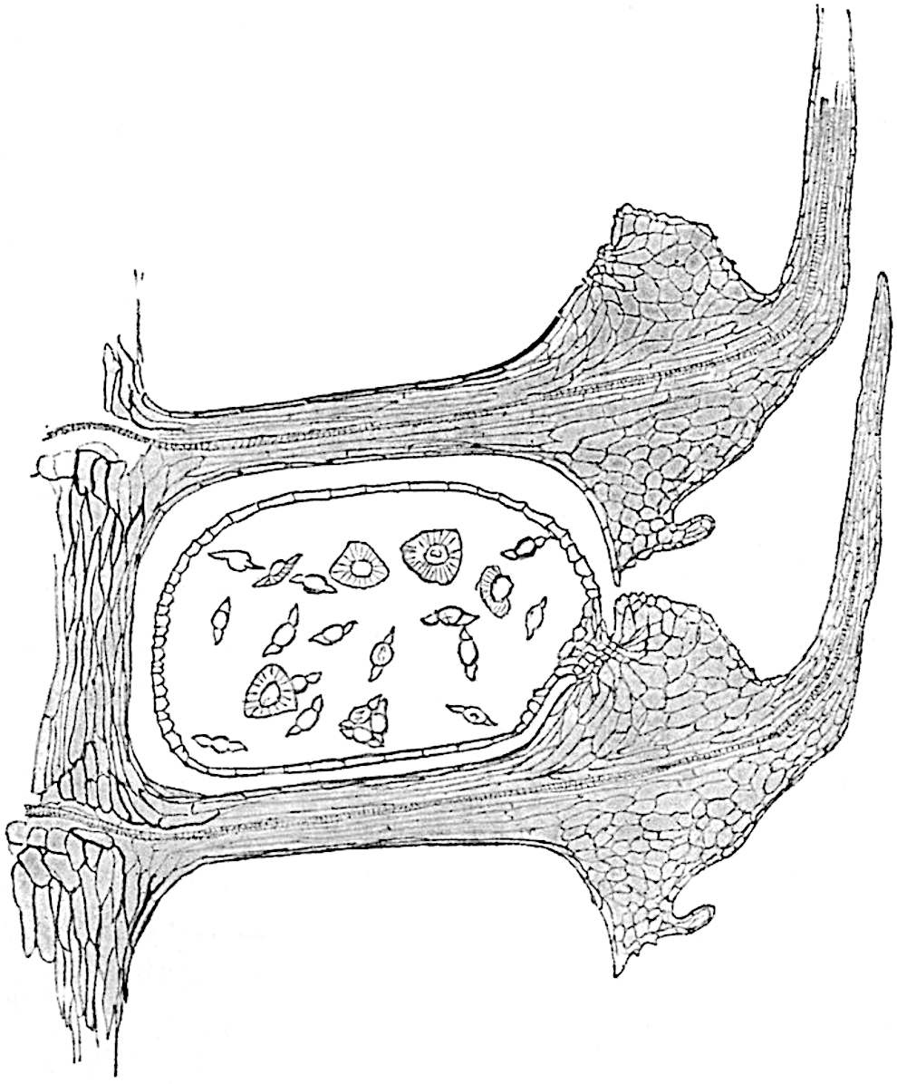
Fig. 192. Spencerites insignis (Williamson). (After Miss Berridge.)
Of the two species included by Scott in his genus Spencerites only one, S. insignis, need be considered. Since the publication of Scott’s paper our knowledge of this type has been extended by Miss Berridge[442] and by Prof. Lang[443].
The axis of the strobilus has a stele characterised by a pith of elongated elements, most of which have thin walls; the xylem cylinder possesses about twenty protoxylem strands forming more or less prominent exarch ridges. The cortex exhibits a differentiation comparable with that in the shoots of Lepidodendron. The sporophylls are arranged in alternating verticils, each whorl consisting of ten members: the narrow horizontal pedicel of a sporophyll, containing a single vascular bundle, as shown in fig. 192, is expanded distally into a prominent upper lobe bearing a cushion of small and delicate cells, to which the sporangium is attached, and prolonged obliquely upwards as a free leaf-like lamina. The lower blunt prolongation of the sporophylls appears to form a thick dorsal lobe, but, as Lang has pointed out, it is highly probable that the present form of the dorsal lobe is of secondary origin, and is “due to the disappearance of a mucilage cavity from a large sporophyll base[444].” As Miss Berridge remarks, the vascular bundle of the sporophyll does not give off a branch to the ventral lobe and sporangium. In attachment, in shape, and in the structure of the wall the sporangia differ markedly from those of Lepidostrobi. The spores, which also constitute a characteristic feature of the genus, have a maximum diameter of 0·14 mm.; they are described as oblate spheroids with a broad hollow wing running round the equator (fig. 192) comparable with the air-sacs of the pollen of Pinus. Scott points out that the spores of Spencerites are intermediate in size between the microspores of Lepidodendron and the megaspores of Lycopodium; it is difficult therefore to decide to which category they should be referred. Spencerites is clearly distinct from Lepidostrobus; the absence of a ligule, the manner of attachment of the sporangia, and the form and size of the spores, are characteristic features.
A comparison of Spencerites with the strobili of Lycopodium cernuum (figs. 123, 126–129) has recently been made by Lang, who draws attention to the striking agreement as regards general plan and even detailed structural features between the Palaeozoic and the recent type of strobilus. It is interesting to find, as Lang points out, that in the original account of the fossil cone by Williamson, the view is expressed that the sporangiophores were confluent. An examination of the section figured by Williamson[445] led Lang to confirm this opinion. It would be out of place to enter here into a detailed comparison of Spencerites insignis and the cone of Lycopodium, but the resemblances are considered by Lang to be sufficiently close to suggest that the striking similarity may be indicative of relationship[446].
It is worthy of notice that the radial section of Spencerites (fig. 192) presents a fairly close resemblance to a corresponding section through a cone-scale of Agathis (Kauri Pine)[447]. In each case the megasporangium is attached by a narrow pedicel to the sporophyll and the latter has a similar form in the two plants, though the extent of the resemblance is somewhat lessened by Lang’s more complete account of the Palaeozoic type. If the Spencerites sporangia possessed an integument the similarity with the Agathis ovule would of course be much closer: recent palaeobotanical investigations have shown that ovules and sporangia are not separated by impassable barriers.
[Since this Chapter was set up in type a paper has appeared by Dr Bruno Kubart on a new species of Spencerites spore, S. membranaceus, from the Ostrau-Karwiner Coal-basin (Austria). The spores are larger than those of S. insignis and in some the cells of a prothallus are preserved. Kubart figures a section of a spore containing a group of seven cells, a central cell, which he regards as an antheridial mother-cell, surrounded by six wall-cells. Kubart (90).]