Front matter (continued 2)
Reading edition
Plate III.
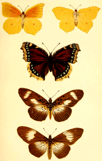
BUTTERFLIES.
The family of Pieridæ, or whites, again afford us admirable examples of the development of spots. The prevailing colours are white, black and yellow: green appears to occur in the Orange-tips (Anthocaris), but it is only the optical effect of a mixture of yellow and grey or black scales. The species are very variable, as a rule, and hence of importance to us; and there are many intermediate species on the continent and elsewhere which render the group a most interesting study.
The wood white (Leucophasia sinapis) Fig. 1, Plate IV., is a pure white species with an almost square dusky tip to the fore-wings of the male. In the female this tip is very indistinct or wanting, Fig. 4, Plate IV. In the variety Diniensis, Fig. 2, Plate IV., this square tip appears as a round spot.
The Orange-tips, of which we have only one species in Britain (Anthocaris cardamines) belongs to a closely allied genus, as does also the continental genus Zegris. The male Orange-tip (A. cardamines) is white with a dark grey or black tip, and a black discoidal spot. A patch of brilliant orange extends from the dark tip to just beyond the discoidal spots. In the female this is wanting, but the dark tip and spot are larger than in the male.
Let us first study the dark tip. In L. sinapis we have seen that it extends right to the margin of the wing in the male, but in the female is reduced to a dusky spot away from the margin. In A. cardamines the margin is not coloured quite up to the edge, but a row of tiny white spots, like a fringe of seed pearls, occupies the inter-spaces of the veins. On the underside these white spots are prolonged into short bars, see Plate IV. In the continental species A. belemia we see the dark tip to be in a very elementary condition, being little more than an irregular band formed of united spots, there being as much white as black in the tip, Fig. 5, Plate IV. In A. belia, Fig. 6, Plate IV., the black tip is more developed, and in the variety simplonia still more so, Fig. 7, Plate IV. We here see pretty clearly that this dark tip has been developed by the confluence of irregular spots.
Turning now to the discoidal spot we shall observe a similar development. Thus in:—
A. cardamines, male, it is small and perfect.
Do. female, "larger"
A. belemia "large"
A. belia "large with white centre.
Do. v. simplonia "small and perfect.
[15] A. eupheno, female, "nearly perfect.
Do. male, "a band.
We here find two distinct types of variation. In A. belia we have a tendency to form an ocellus, and in A. eupheno the spot of the female is expanded into a band in the male.
The orange flush again offers us a similar case; and with regard to this colour we may remark that it seems to be itself a development from the white ground-colour of the family in the direction of the red end of the spectrum. Thus in the Black-veined white (Aporia cratægi) we have both the upper and under surfaces of the typical cream-white, for there is no pure white in the family. In the true whites the under surface of the hind-wings is lemon-yellow, in the female of A. eupheno the ground of the upper surface is faint lemon-yellow, and in the male this colour is well-developed. The rich orange, confined to a spot in G. rhamni becomes a flush in G. Cleopatra, and a vivid tip in A. cardamines. These changes are all developments from the cream white, and may be imitated accurately by adding more and more red to the primitive yellow, as the artist actually did in drawing the plate.
In A. cardamines the orange flush has overflowed the discoidal spot, as it were, in the male, and is absent in the female. But in A. eupheno we have an intermediate state, for as the figures show, in the female, Fig. 8, the orange tip only extends half-way to the discoidal spot, and in the male it reaches it. Moreover it is to be noticed that the flow of colour, to continue the simile, is unchecked by the spot in cardamines, but where the spot has expanded to a bar in eupheno it has dammed the colour up and ponded it between bar and tip. An exactly intermediate case between these two species is seen in A. euphemoides, Fig. 10, Plate IV., in which the spot is elongated, and dribbles off into an irregular band, into which the orange has trickled, as water trickles through imperfect fascines. This series of illustrations might be repeated in almost any group of butterflies, but sufficient has been said to show how spots can spread into patches, either by the spreading of one or by the coalescence of several.
Plate IV.
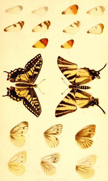
SPOTS and STRIPES.
We will now take an illustration of the formation of stripes or bars from spots, and in doing so must call attention to the rarity of true stripes in butterflies. By a true stripe I mean one that has even edges, that is, whose sides are uninfluenced by structure. In all our British species such as P. machaon, M. artemis, M. athalia, V. atalanta, L. sibilla, A. iris, and some of the Browns, Frittilaries and Hair-streaks, which can alone be said to be striped, the bands are clearly nothing more than spots which have spread up to the costæ, and still retain traces of their origin either in the different hue of the costæ which intersect them, or in curved edges corresponding with the interspaces of the costæ. This in itself is sufficient to indicate their origin. But in many foreign species true bands are found, though they are by no means common. Illustrations are given in Plate IV., of two Swallow-tails, Papilio machaon, Fig. 11, and P. podalirius, Fig. 12, in which the development of a stripe can readily be seen.
In machaon the dark band inside the marginal semi-lunar spots of the fore-wings retain traces of their spot-origin in the speckled character of the costal interspaces, and in the curved outlines of those parts. In podalirius the semi-lunar spots have coalesced into a stripe, only showing its spot-origin in the black markings of the intersecting costæ; and the black band has become a true stripe, with plain edges. Had only such forms as this been preserved, the origin of the spots would have been lost to view.
It may, however, be said, though I think not with justice, that we ought not to take two species, however closely allied, to illustrate such a point. But very good examples can be found in the same species. A common German butterfly, Araschnia Levana, has two distinct varieties, Levana being the winter, and prorsa the summer form; and between these an intermediate form, porima, can be bred from the summer form by keeping the pupæ cold. Dr. Weismann, who has largely experimented on this insect, has given accurate illustrations of the varieties. Plate V. is taken from specimens in our possession. In the males of both Levana, Fig. 4, and prorsa, Fig. 1, the hind-wing has a distinct row of spots, and a less distinct one inside it, and in the females of both these are represented by dark stripes. In porima we get every intermediate form of spots and stripes, both in the male and female, and as these were hatched from the same batch of eggs, or, are brothers and sisters, it is quite impossible to doubt that here, at least, we have an actual proof of the change of spots into stripes.
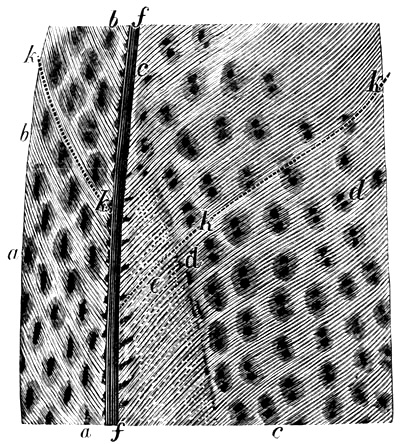
Fig. 1. Part of secondary feather of Argus Pheasant.
a.a. Elongated spots, incipient ocelli. b. Interspaces. c.c. Axial line. d.d. Double spots, incipient ocelli. e. Minute dottings. f.f. Shaft. k.k. Line of feathering.
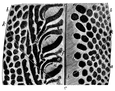
Fig. 2. Part of secondary wing feather of Argus Pheasant.
a. Oval. Axis at right angles. b. Round. c.c. Shaft. d. Imperfect ocellus. e. Expansion of stripe. f. Interspace. g. Stalk. h. Edge of feather.
k. Line of feathering.
Plate V.
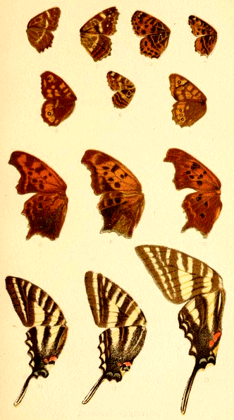
SEASONAL VARIETIES.
The change of spots more or less irregular into eye-spots, or ocelli, is equally clear; and Darwin's drawing of the wings of Cyllo leda[16] illustrates the point well. "In some specimens," he remarks, "large spaces on the upper surfaces of the wings are coloured black, and include irregular white marks; and from this state a complete gradation can be traced into a tolerably perfect ocellus, and this results from the contraction of the irregular blotches of colour. In another series of specimens a gradation can be followed from excessively minute white dots, surrounded by a scarcely visible black line, into perfectly symmetrical and larger ocelli." In the words we have put in italics Darwin seems to admit these ocelli to be formed from blotches; and we think those of the Argus pheasant can be equally shown to arise from spots.
Darwin's beautiful drawings show, almost as well as if made for the purpose, that the bars are developed from spots.[17] In Fig. 1 is shown part of a secondary wing feather, in which the lines k. k. mark the direction of the axis, along which the spots are arranged, perfectly on the right, less so on the left. The lengthening out of the spots towards the shaft is well seen on the right, and the coalescence into lines on the left. In Fig. 2 we have part of another feather from the same bird, showing on the left elongated spots, with a dark shading round them, and on the right double spots, like twin stars, with one atmosphere around them. Increase the elongation of these latter, and you have the former, and both are nascent ocelli. We here, then, have a regular gradation between spots, bands, and ocelli, just as we can see in insects.
In some larvæ, those of the Sphingidæ especially, ocelli occur, and these may be actually watched as they grow from dots to perfect eye-spots, with the maturity of the larva.
Even in some mammals the change from spots to stripes can be seen. Thus, the young tiger is spotted, and so is the young lion; but, whereas in the former case the spots change into the well-known stripes (which are really loops), in the latter they die away. The horse, as Darwin long ago showed, was probably descended from a striped animal, as shown by the bars on a foal's leg. But before this the animal must have been spotted; and the dappled horses are an example of this; and, moreover, almost every horse shows a tendency to spottiness, especially on the haunches. In the museum at Leiden a fine series of the Java pig (Sus vittatus) is preserved. Very young animals are banded, but have spots over the shoulders and thighs; these run into stripes as the animal grows older; then the stripes expand, and, at last meeting, the mature animal is a uniform dark brown. Enough has now, I trust, been said upon this point to show that from spots have been developed the other markings with which we are familiar in the animal kingdom.
The vegetable kingdom illustrates this fact almost as well. Thus, the beautiful leaves of the Crotons are at first green, with few or no coloured spots; the spots then grow more in number, coalesce, form irregular bands, further develop, and finally cover the whole, or almost the whole, of the leaf with a glow of rich colour. Some of the pretty spring-flowering orchid callitriche have sulphur-yellow petals, with dark rich sepia spots; these often develop to such an extent as to overspread nearly all the original yellow. Many other examples might be given.
Hitherto we have started with a spot, and traced its development. But a spot is itself a developed thing, inasmuch as it is an aggregation of similarly coloured cells. How they come about may, perhaps, be partly seen by the following considerations. Definite colour-pattern has a definite function—that of being seen. We may, therefore, infer that the more definite colour is of newer origin than the less definite. Hence, when we find the two sexes differently coloured, we may generally assume that the more homely tinted form is the more ancient. For example, some butterflies, like the gorgeous Purple Emperor (Apatura iris), have very sombre mates; and it seems fair to assume that the emperor's robes have been donned since his consort's dress was originally fashioned.
That the object of brilliant colour is display is shown partly by the fact that in those parts of the wings of butterflies which overlap the brilliant colour is missing, and partly by the generally brighter hues of day-flying butterflies and moths than of the night-flying species. Now, the sombre hues of nocturnal moths are not so much protective (like the sober tints of female butterflies and birds), because night and darkness is their great defender, as the necessary result of the darkness: bright colours are not produced, because they could not be seen and appreciated. In these cases it is very noticeable how frequently the colour is irregularly dotted about—irrorated or peppered over the wings, as it were. This irregular distribution of the pigment cells, if it were quite free from any arrangement, might be looked upon as primitive colouring, undifferentiated either into distinct colour or distinct pattern. If we suppose a few of the pigment cells here and there to become coloured, we should have irregular brilliant dottings, just as we actually see in many butterflies, along the costa. The grouping together of these colour dots would give rise to a spot, from which point all is clear.
That some such grouping or gathering together, allied to segregation, does take place, a study of spots, and especially of eye-spots, renders probable. What the nature of the process is we do not know, nor is it easy to imagine. But let us suppose a surface uniformly tinted brown. Then, if we gather some of the colouring matter into a dark spot we shall naturally leave a lighter area around it, just as we see in all our Browns and Ringlets. In this way we can see how a ring-spot can be formed. To make it a true eye-spot, with a light centre, we must also suppose a pushing away of the colour from that centre. A study of ocelli naturally suggests such a process, which is analogous to the banding of agates, and all concentric nodules. Darwin, struck with this, seems to adopt it as a fact, for he says, "Appearances strongly favour the belief that, on the one hand, a dark spot is often formed by the colouring matter being drawn towards a central point from a surrounding zone, which is thus rendered lighter. And, on the other hand, a white spot is often formed by the colour being driven away from a central point, so that it accumulates in a surrounding darker zone."[18] The analogy between ocelli and concretions may be a real one. At any rate beautiful ocelli of all sizes can be seen forming in many iron-stained sand-stones. The growth of ocelli may thus be a mechanical process adapted by the creature for decorative purposes, but the artistic colouring of many eye-spots implies greater effort.
There is, however, one set of colour lines in birds and insects that do not seem to arise from spots in the ordinary way. These are the coloured feather-shafts of birds, and the coloured nerves or veins in a butterfly's wing, In these the colour has a tendency to flow all along the structure in lines.
Conclusion. The results arrived at in this chapter may be thus summarised:—
Spots, ocelli, stripes, loops, and patches may be, and nearly always are, developed from more or less irregular spots.
This is shown both by the study of normal colouring, or by abnormal colouring, or decolouring in disease.
Even the celebrated case of the Argus Pheasant shows that the bands from which the ocelli are developed arose from spots.
CHAPTER VII. Colouration in the Invertebrata.
I
IF the principle of the dependence of colour-pattern upon structure, enunciated in the preceding pages be sound, we ought to find certain great schemes of colouration corresponding to the great structural subdivisions of the animal kingdom. This is just what we do find; and before tracing the details, it will be as well to group the great colour-schemes together, so that a general view of the question can be obtained at a glance.
The animal kingdom falls naturally into two divisions, but the dividing line can be drawn in two ways. If we take the most simple classification, we have:—
1. Protozoa, animals with no special organs.
2. Organozoa, animals possessing organs.
Practically this classification is not used, but we shall see that from our point of view it is a useful one. In the most general scheme the divisions are:—
1. Invertebrata, animals without backbones.
2. Vertebrata, animals with backbones.
The invertebrata are divided into sub-kingdoms, of which the protozoa form one. These protozoa possess, as it were, only negative properties. In their simplest form they are mere masses of protoplasm, even lacking an investing membrane or coat, and never, even in the highest forms, possessing distinct organs. It is this simplicity which at once separates them entirely from all other animals.
The other sub-kingdoms are:—
Cœlenterata, of which the jelly-fishes are a type; animals possessing an alimentary canal, fully communicating with the general cavity of the body, but without distinct circulatory or nervous systems.
Annuloida, of which the star-fishes are a type; animals having the alimentary canal shut off from the body-cavity, and possessing a nervous system, and in some a true circulatory system.
Annulosa, of which worms, lobsters, and insects are types; animals composed of definite segments, arranged serially, always possessing true circulatory and nervous systems.
Mollusca, of which oysters and whelks are types; animals which are soft-bodied, often bearing a shell, always possessing a distinct nervous system and mostly with a distinct heart.
In old systems of classification, the Cœlenterata and Annuloida were united into one sub-kingdom, the Radiata, in consequence of their radiate or star-like structures.
As colouration, according to the views here set forth, depends upon structure, we may classify the Invertebrata thus:—
Protozoa Structureless.
Cœlenterata Annuloida Radiata. Radiate structure.

Annulosa Segmented "
Mollusca Marginate "
The mollusca are said to be marginate in structure because, in those possessing shells—the mollusca proper—the shell is formed by successive additions to the margin or edge of the shell, by means of the margin of the mantle, or shell-secreting organ.
Now we shall proceed to show that the schemes of colouration follow out these structure-plans, and thus give additional force to the truth of the classification, as well as showing that, viewed on a broad scale, the present theory is a true one.
We can, in fact, throw the whole scheme into a table, as follows:—
SYSTEMS OF COLOURATION.
System of Colouring. Structure. Sub-kingdoms.
A. No Axial Decoration. A. No Axial Structure. A. Invertebrata.
1. No definite system. No definite organs. Protozoa.
2. Radiate system. Radiate structure. Cœlenterata, Annuloida.
3. Segmental system. Segmental structure. Annulosa.
4. Marginate system. Marginate growth. Mollusca.
5. B. Axial Decoration. B. Axial Structure. B. Vertebrata.
Axial system. Axial structure. Vertebrata.
Protozoa. The protozoa are generally very minute, and always composed of structureless protoplasm. Their peculiarities are rather negative than positive, there being neither body segments, muscular, circulatory, nor nervous systems. Even the denser exterior portion (ectosarc) possessed by some of them seems to be rather a temporary coagulation of the protoplasm than a real differentiation of that material.
Here, then, we have to deal with the simplest forms of life, and if colouration depends upon structure, these structureless transparent creatures should lack all colour-pattern, and such is really the case. Possessing no organs, they have no colouration, and are generally either colourless or a faint uniform brown colour, and through their colourless bodies the food particles show, often giving a fictitious appearance of colouring.
To this general statement there is a curious and most telling exception. In a great many protozoa there exists a curious pulsating cell-like body, called the contractile vesicle, which seems to be a rudimentary organ, whose function is unknown. Here, then, if anywhere, traces of colouring should be found, and here it is accordingly found, for, though generally clear and colourless, it sometimes assumes a pale roseate hue. This may be deemed the first attempt at decoration in the animal kingdom, and it is directly applied to the only part which can be said to possess structure. Beautiful examples are plentiful in Leidy's magnificent volume on Freshwater Rhizopods.
Cœlenterata. These animals fall into two groups, the Hydrozoa, of which the hydra and jelly-fishes are types, and the Actinozoa, of which the sea-anemonies and corals are types. Most of the cœlenterata are transparent animals, but it is amongst them we first come across opaque colouring.
Of the lowest forms, the hydras, nothing need be said here, as they are so much like the protozoa in their simplicity of structure.
The Corynida, familiar to many of our sea-side visitors by their horny brown tubes (Tubularia), attached to shells and stones, are next in point of complexity. Within the tube is found a semi-fluid mass of protoplasm, giving rise at the orifice to the polypite, which possesses a double series of tentacles. These important organs are generally of a vivid red colour, thus emphasizing their importance in the strongest manner. Other members of the order are white, with pink stripes.
In the larval stage many of the animals belonging to the above and allied orders, are very like the true jelly-fishes. These free swimming larvæ, or gonophores, possess four radiating canals, passing from the digestive sac to the margins of the bell, and these are often the seat of colour. In these creatures, too, we find the earliest trace of sense organs, and consequently, the first highly differentiated organs, and they appear as richly coloured spots on the margins of the bell. The true oceanic Hydrozoa again afford us fine examples of structural colouration. The beautiful translucent blue-purple Velella, which is sometimes driven on to our shores, is a case in point; and its delicate structure lines are all emphasized in deeper hues. The true jelly-fishes (Medusidæ) with their crystal bells and radiating canals, frequently show brilliant colour, and it is applied to the canals, and also to the rudimentary eye-specks, which are frequently richly tinted, and in all cases strongly marked. In the so-called "hidden-eyed" Medusæ we find the same arrangement of colour, the same emphasized eye-specks, and the reproductive organs generally appear as a vivid coloured cross, showing through the translucent bell.
Turning now to the Actinozoa, of which the sea anemonies and corals are types, we are brought first into contact with general decorative, more or less opaque colour, applied to the surface of the animal. In the preceding cases the animals have been almost universally transparent or translucent, and the colouration is often applied to the internal organs, and shows through. In the sea-anemonies we find a nearer approach to opacity, in the dense muscular body, though even this is often translucent, and the tentacles generally so, often looking like clouded chalcedony. The wealth of colour to be found in these animals gives us a very important opportunity of studying decoration, where it first appears in profusion.
One of the first points that strikes even a casual observer is that amongst the sea-anemonies the colouration is extremely variable, even in the same species and in the same locality. This is in strong contrast to what we generally find amongst the higher organisms, such as insects and birds; for though considerable variation is found in them, it does not run riot as in the anemonies. It would almost appear as if the actual colour itself was of minor importance, and only the pattern essential; the precise hue is not fixed, is not important, but the necessity of colour of some sort properly arranged is the object to be attained. Whether this idea has a germ of truth in it or not, it is hard to say, but when we take the fact in connection with its occurrence just where opacity begins, connecting this with the transparency of the lower organisms, and the application of vivid colour to their internal organs, one seems to associate the instability of the anemony's colouring with the transference of colour from the interior to the exterior. Certain it is, that vivid colour never exists in the interior of opaque animals; it is always developed under the influence of light. The white bones, nerves and cartilages, and the uniform red of mammalian muscles, are not cases of true decorative colouring in our sense of the term, for all bodies must have some colour. All bone is practically white, all mammalian muscle red, but for these colours to be truly decorative, it would be necessary for muscles of apparently the same character often to be differently tinted, just as the apparently similar hairs on a mammal, and scales on an insect, are variously painted. This we do not find, for the shaft-bones and plate-bones, and even such odd bones as the hyoid are all one colour; and no one would undertake to tell, by its hue, a piece of striped from a piece of unstriped muscle. Decorative colouring must be external in an opaque animal; it may be internal in a transparent one.
The connection thus shown between decoration and transparency seems to suggest that hypodermal colour is the original, and epidermal the newer scheme: that the latter was derived from the former. This agrees with Haagen's shrewd hint that all mimetic colour was originally hypodermal. Certain it is that the protective colour that is still under personal control, as in the chameleon, &c., is always hypodermal.
The common crass (Bunodes crassicornis) is so extremely variable, that all one can say of it is, that it is coloured red and green. But this colour is distributed in accordance with structure. The base, or crawling surface, not being exposed to the light, is uncoloured. The column, or stem, is irregularly spotted, and striped in accordance with the somewhat undifferentiated character of its tissue, but the important organs, the tentacles, are most definitely ornamented, the colour varying, but the pattern being constant. This pattern is heart-shaped, with the apex towards the point of the tentacle; that is to say, the narrow part of the pattern points to the narrow part of the tentacle.
In the common Actinea mesembryanthemum, which is often blood red, the marginal bodies, probably sense-organs, are of the most exquisite turquoise blue colour, and the ruby disc thus beaded is as perfect an example of simple structural decoration as could be desired. A zone of similar blue runs round the base of the body.
Turning now to the corals, which are simply like colonies of single anemonies with a stony skeleton, we have quite a different arrangement of hues. No sight is more fascinating than that of a living-coral reef, as seen through the clear waters of a lagoon. The tropical gardens ashore cannot excel these sea-gardens in brilliancy or variety of colour. Reds, yellows, purples, browns of every shade, almost bewilder the eye with their profusion; and here again we find structural decoration carried out to perfection. The growing points of white branching corals (Madrepores) are frequently tipped with vivid purple, and the tiny polyps themselves are glowing gem-stars. In the white brain-corals, the polyps are vivid red, green, yellow, purple and so on; but in almost every case vividly contrasting with the surrounding parts, the colour changing as the function changes.
The Alcyonariæ, which include the sea-fans, sea-pens, and the red coral of commerce, practically bring us to the end of the Cœlenterata, and afford us fresh proof of the dependence of colour upon structure and function. The well-known organ-pipe coral (Tubipora musica) is of a deep crimson colour, and the polyps themselves are of the most vivid emerald green, a contrast that cannot be excelled. Almost equally beautiful is the commercial coral (Corallium rubrum) whose vivid red has given a name to a certain tint. In this coral the polyps are of a milk-white colour.
It must be remembered that in these cases the colour seems actually to be intentional, so as to form a real and not merely an accidental contrast between the stony polypidom and the polyp, for the connecting tissue (cœnosarc) is itself as colourless as it is structureless.
Gathering together the facts detailed in this chapter we find:—
1. That the Protozoa are practically colourless and structureless.
2. That in those species which possess a rudimentary organ (contractile vesicle) a slight decoration is applied to that organ.
3. That in the Cœlenterata the colouration is directly dependent upon the structure.
4. That in transparent animals the colouration is applied directly to the organ whether it be internal as in the canals or ovaries, or external, as in the eye-specks.
5. That in opaque animals, as in the sea-anemonies, the colouring is entirely external.
6. That it is very variable in hue, but not in pattern.
7. That the most highly differentiated parts (tentacles, eye-specks), are the most strongly coloured.
8. That in the corals an emphatic difference occurs between the colour of the polypidom (or "coral") and the polyp.
CHAPTER VIII. Details of Protozoa.
T
THE Protozoa are divided into three orders.
I.—Gregarinidæ. II.—Rhizopoda. III.—Infusoria.
I. The Gregarinidæ consist of minute protozoa, parasitic in the interior of insects, &c., and like other internal parasites are colourless, as we should expect.
II. The Rhizopoda may, for our purpose, be divided into the naked forms like Amœba, and those which possess a skeleton, such as the Radiolaria, the Foraminifera and the Spongia.
Of these the naked forms are colourless, or uniformly tinted, excepting the flush already described as emphasizing the contractile vesicle.
The Foraminifera are the earliest animals that possess a skeleton or shell, and though generally very small, this shell is often complex, and of extreme beauty, though their bodies retain the general simplicity of the protozoa, indeed, they are said to possess no contractile vesicle. Still the complexity of their shells places them on a higher level than the naked rhizopoda.
In these animals we find the first definite colour, not as a pattern, but as simple tinting of the protoplasm. The general hue is yellowish-brown (as in Amœba), but deep red is not uncommon. The deepest colour is found in the oldest central chambers, becoming fainter towards the periphery, where it is often almost unrecognisable.[19]
The Radiolaria are minute organisms with still more complex skeletons, and are considered by Haeckel[20] to be more highly organized than the preceding order. They consist of a central portion containing masses of minute cells, and an external portion containing yellow cells. Here we have the first differentiation of parts in the external coating and internal capsule, and side by side with this differentiation we find colour more pronounced, and even taking regional tints in certain forms.
We may notice the following genera as exhibiting fine colour:—
Red. Eucecryphalus, Arachnocorys, Eucrytidium, Dictyoceras. Yellow. Carpocanium, Dictyophimus, Amphilonche. Purple. Eucrytidium, Acanthostratus. Blue. Cyrtidosphæra, Cœlodendrum. Green. Cladococeus, Amphilonche. Brown. Acanthometra, Amphilonche.
Examples of these may be seen in the plates of Haeckel's fine work, and as an illustration of regional decoration we cite Acanthostratus purpuraceus, in which the central capsule is seen to run from red to orange, and the external parts to be colourless, with red markings in looped chains.
Spongocyclia also exhibits this regional distinction of colour very clearly, the central capsule being red and the external portion yellow.
The Spongida, or sponges, are, broadly speaking, assemblages or colonies of amœba-like individuals, united into a common society. Individually the component animals are low, very low, in type, but their union into colonies, and the necessity for a uniform or common government has given rise to peculiarities that in a certain sense raise them even above the complex radiolaria. Some, it is true, are naked, and do not possess the skeleton that supports the colony, which skeleton forms what we usually call the sponge; but even amongst these naked sponges the necessity for communal purposes over and above the mere wants of the individual, raises them a step higher in the animal series. A multitude of individuals united by a common membrane, living in the open sea, it must have happened that some in more immediate contact with the food-producing waters, would have thriven at the expense of those in the interior who could only obtain the nutriment that had passed unheeded by the peripheral animals. But just as in higher communities we have an inflowing system of water and an out-flowing system of effete sewerage quite uncontrolled, and, alas, generally quite unheeded by the individuals whose wants are so supplied; so in the sponges we have a system of inflowing food-bearing water and an out-flowing sewage, or exhausted-water system. This is brought about by a peculiar system of cilia-lined cells which, as it were, by their motion suck the water in, bringing with it the food, and an efferent system by which the exhausted liquid escapes. These cilia-lined cells are the first true organs that are to be found in the animal kingdom, and according to the views we hold, they ought to be emphasized with colour, even though their internal position renders the colouration less likely. This we find actually to be the case, and these flagellated cells, as they are called, are often the seat of vividest colour.
The animal matter, or sarcode, or protoplasm of sponges falls into three layers, just as we find the primitive embryo of the highest animals; and just as the middle membrane of a mammalian ovum develops into bone, muscle and nerve, so the middle membrane (mesosarc) of the sponges develops the hard skeleton, and in this most important part we find the colour cells prevail. Sollas, one of our best English authorities upon sponges, writes, "The colours of sponges, which are very various, are usually due to the presence of pigment granules, interbedded either in the endosarc of the flagellated cells, or in the mesodermic cells, usually of the skin only, but sometimes of the whole body."[21]
We can, then, appeal most confidently to the protozoa as illustrating the morphological character of colouration.
CHAPTER IX. Details of Cœlenterata.
I. Hydrozoa.
A. Hydrida.
T
THE Hydras, as a rule, are not coloured in our sense of the term; that is to say, they are of a general uniform brown colour. But in one species, H. viridis, the endoderm contains granules of a green colour, which is said to be identical with the green colouring matter of leaves (chlorophyll). This does not occur in all the cells, though it is present in most. The green matter occurs in the form of definite spherical corpuscles, and these colour-cells define the inner layer of the integument (the endoderm), and render it distinct.[22] That portion of the endoderm which forms the boundary of the body-cavity has fewer green corpuscles, but contains irregular brown granules, thus roughly mapping out a structural region.
We thus see that even in so simple a body as the Hydra the colouring matter is distributed strictly according to morphological tracts.
B. Tubularida. The Tubularian Hydroids are the subject of an exhaustive and admirably illustrated monograph by Prof. J. Allman, from which the following details are culled. These animals are with few exceptions marine, and consist either of a single polypite or of a number connected together by a common flesh, or cœnosarc. Some are quite naked, others have horny tubes, into which, however, the polypites cannot retreat. The polypites consist essentially of a sac surrounded with tentacles; and one of their most striking characters is their mode of reproduction. Little buds (gonophores) grow from the cœnosarc, and gradually assume a form exactly like that of a jelly-fish. These drop off, and swim freely about; and are so like jelly-fishes that they have been classed among them as separate organisms.
The Tubulariæ are all transparent; and in them we find structural colouration finely shown, the colour, as is usual in transparent animals, being applied directly to the different organs.
Writing of the colour, Prof. Allman says: "That distinct secretions are found among the Hydroida, and that even special structures are set aside for their elaboration, there cannot now be any doubt.
"One of the most marked of these secretions consists of a coloured granular matter; which is contained at first in the interior of certain spherical cells, and may afterwards become discharged into the somatic fluid. These cells, as already mentioned, are developed in the endoderm;[23] in which they are frequently so abundant as to form a continuous layer upon the free surface of this membrane. It is in the proper gastric cavity of the hydranth and medusa, in the spadix of the sporosac, and in the bulbous dilatations which generally occur at the bases of the marginal tentacles of the medusæ, that they are developed in greatest abundance and perfection; but they are also found, more or less abundantly, in the walls of probably the whole somatic cavity, if we except that portion of the gastrovascular canals of the medusa which is not included within the bulbous dilatations.
"In the parts just mentioned as affording the most abundant supply of these cells, they are chiefly borne on the prominent ridges into which the endoderm is thrown in these situations; when they occur in the intervals between the ridges they are smaller, and less numerous.
"The granular matter contained in the interior of these cells varies in its colour in different hydroids. In many it presents various shades of brown; in others it is a reddish-brown, or light pink, or deeper carmine, or vermilion, or orange, or, occasionally, a fine lemon-yellow, as in the hydranth of Coppinia arcta, or even a bright emerald green, as in the bulbous bases of the marginal tentacles of certain medusæ. No definite structure can be detected in it; it is entirely composed of irregular granules, irregular in form, and usually aggregated into irregularly shaped masses in the interior of the cells. It is to this matter that the colours of the Hydroida, varying, as they do, in different species, are almost entirely due.
"The coloured granular matter is undoubtedly a product of true secretion; and the cells in which it is found must be regarded as true secreting cells. These cells are themselves frequently to be seen as secondary cells in the interior of parent cells, from which they escape by rupture, and then, falling into the somatic fluid, are carried along by its currents, until, ultimately, by their own rupture, they discharge into it their contents.
"We have no facts which enable us to form a decided opinion as to the purpose served by this secretion. Its being always more or less deeply coloured, and the fact of its being abundantly produced in the digestive cavity, might suggest that it represented the biliary secretion of higher animals. This may be its true nature, but as yet we can assert nothing approaching to certainty on the subject; indeed, considering how widely the cells destined for the secretion of coloured granules are distributed over the walls of the somatic cavity, it would seem not improbable that the import of the coloured matter may be different in different situations; that while some of it may be a product destined for some further use in the hydroid, more of it may be simply excretive, taking no further part in the vital phenomena, and intended solely for elimination from the system." [24]
Here we have very definite statements by a highly trained observer of the distribution of colour in the whole of these animals, and of the conclusions he draws from them.
Firstly as to the colour itself. We find it true colour—brown, pink, carmine, vermilion, orange, lemon-yellow, and even emerald green; a set of hues as vivid as any to be found in the animal kingdom. It is difficult to conceive these granules to be merely excrementitious matter; for in such simple creatures, feeding upon such similar bodies, one would hardly expect the excretive matter to be so diversified in tint. Moreover, excrementitious matter is not, as a rule, highly coloured, but brown. Thus, we see in the Rhizopods the green vegetable matter which has been taken in as food becomes brown as the process of assimilation goes on; and, indeed, colour seems almost always to be destroyed by the act of digestion.
Still, it by no means follows that this colour, even if it is produced for the sake of decoration, as we suggest, may not owe its direct origin to the process of digestion. The digestive apparatus is the earliest developed in the animal kingdom, and in these creatures is by far the most important; the cœlenterata being, in fact, little more than living stomachs. If, then, colouration be structural, what is more likely than that the digestive organs should be the seat of decoration in such transparent creatures?
Secondly, as to the distribution of the colour. We find it "frequently forming a continuous layer upon the free surface of" the endoderm, in the "spadix of the sporosac," and in the "bulbous terminations" of the canals, that colour is best developed. In other words, the colour is distributed structurally, and is most strongly marked where the function is most important.
Prof. Allman gives no hint that the colour may be purely decorative, and is naturally perplexed at the display of hues in such vigour; but if this be one of the results of the differentiation of parts, of the specialization of function, then we can, at least, understand why we find such brilliant colour in these creatures, and why it is so distributed.
As an illustration of the Tubularia we have selected Syncoryne pulchella, Fig. 2, Pl. VI., and its medusa, Fig. 1. The endoderm of the spadix of the hydranths is of a rich orange colour, which becomes paler as it descends towards the less highly organized stem. Medusæ are seen in various stages of development, and one, mature and free, is shown. In these the manubrium, and the bulbous terminations of the canals are also seen to be coloured orange.
In these medusæ we find the first appearance of sensory organs. They consist of pigment-cells enclosed in the ectoderm, or outside covering; and are singular as presenting the first true examples of opaque colouring in the animal kingdom. They are associated with nerve cells attached to a ring of filamentous nerve matter, surrounding the base of the bell. In some important respects the pigment differs from that in other parts of the animal. It is more definite in structure; and the whole ocellus is "aggregation of very minute cells, each filled with a homogeneous coloured matter." [25] These ocelli, and similar sense organs, called lithocysts, are always situated over the bulbous termination of the canals. The pigment is black (as in this case), vermilion, or deep carmine.
Plate VI.
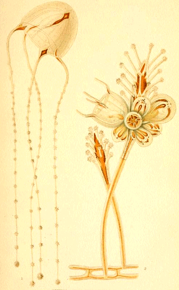
SYNCORYNE PULCHELLA.
The dependence of colour upon structure is thus shown to hold good throughout these animals in a most remarkable manner, and the acceptance of the views here set forth gives us an insight into the reasons for this colouration which, as we have seen, did not arise from the study of the question from the ordinary point of view.
C. Sertularida. These animals are very similar to the last, but they are all compound, and the polypites can be entirely withdrawn within the leathery investment or polypary. Their mode of reproduction is also similar, and their colouration follows the same general plan. Being so like the preceding order, it is unnecessary to describe them.
B. Siphonophora.
The Siphonophora are all free-swimming, and are frequently called Oceanic Hydrozoa. They are divided into three orders, viz.:—
a. Calycophoridæ. b. Physophoridæ. c. Medusidæ.
a. Calycophoridæ. These animals have a thread-like cœnosarc, or common protoplasm, which is unbranched, cylindrical, and contractile. They are mostly quite transparent, but where colour exists it is always placed structurally. Thus, in Diphyes the sacculi of the tentacles are reddish, in Sphæronectes they are deep red, and in Abyla the edges of the larger specimens are deep blue. [26]
b. Physophoridæ. These creatures are distinguished by the presence of a peculiar organ, the float, or pneumatophore, which is a sac enclosing a smaller sac. The float is formed by a reflexion of both the ectoderm and endoderm, and serves to buoy up the animal at the surface of the sea. The best known species is the Physalia, or Portuguese Man-o'-War.
Prof. Huxley, in his monograph on the Oceanic Hydrozoa, gives many details of the colouration; and, not having had much opportunity of studying them, the following observations are taken from his work. It will be seen that the Physophoridæ illustrate the structural distribution of colour in a remarkable manner.
Stephanomia amphitridis, the hydrophyllia, colourless, and so transparent as to be almost imperceptible in water, cœnosarc whitish, enlarged portions of polypites, pink or scarlet, sacs of tentacles scarlet.
The enlarged portion of the polypites is marked with red striæ, "which are simply elevations of the endoderm, containing thread-cells and coloured granules." The small polypites do not possess these elevations, and are colourless.
Agalma breve, like a prismatic mass of crystal, with pink float and polypites.
Athorybia rosacea, float pink, with radiating dark-brown striæ, made up of dots; polypites lightish red, shading to pink at their apices; tentacles yellowish or colourless, with dark-brown sacculi; thread-cells dark brown.
Rhizophysa filiformis, pink, with deep red patch surrounding the aperture of the pneumatocyst.
Physalia caravilla, bright purplish-red, with dark extremities, and blue lines in the folds of the crest; polypites violet, with whitish points, larger tentacles red, with dark purple acetabula, smaller tentacles blue, bundles of buds reddish.
P. pelagica, in young individuals pale blue, in adult both ends green, with highest part of crest purple, tentacles blue, with dark acetabula; polypites dark blue, with yellow points.
P. utriculus. Prof. Huxley describes a specimen doubtfully referred to this species very fully, as follows:—
"The general colour of the hydrosoma is a pale, delicate green, passing gradually into a dark, indigo blue, on the under surface.
"The ridge of the crest is tipped with lake, and the pointed end is stained deep bluish-green about the aperture of the pneumatocyst.
"The bases of the tentacles are deep blue; the polypites deep blue at their bases, and frequently bright yellow at their apices; the velvetty masses of reproductive organs and buds on the under surface are light green."
He further remarks that the tentacles have reniform thickenings at regular intervals, and "the substance of each thickening has a dark blue colour, and imbedded within it are myriads of close-set, colourless, spherical thread-cells."
It would not be possible to find a more perfect example of regional colouration. Not only is each organ differently coloured, but the important parts of each organ, like the ridge of the crest, the bases of the tentacles, and the thread-cell bearing ridges of the tentacles, are also emphasized with deep colour.
Velella. This beautiful creature, which sometimes finds its way to our shores, is like a crystal raft fringed with tentacles, and having an upright oblique crest, or sail. The margins of the disk and crest are often of a beautiful blue colour, and the canals of the disk become deep blue as they approach the crest. The polypites may be blue, purple, green, or brown.
C. Medusidæ. The structure and colouration of the true Medusæ are so like that of the medusiform larvæ of the other Hydrozoa, that they need not be particularly described.
D. Lucernarida. Of this sub-class we need only cite the Lucernaria themselves; which are pretty bell-shaped animals, having the power of attaching themselves to seaweeds, etc., and also of swimming freely about. Round the margin are eight tufts of tentacles, opposite eight lobes, the membrane between the lobes being festooned. In L. auricula, a British species, the membrane is colourless and transparent, the lobes bright red, or green, and the tentacles blue.
As a group the Hydrozoa display regional colouration in a very perfect manner.
II. Actinozoa.
It is not necessary to trace the colouration through all the members of this group, but we will trace the variation of colour through two species of anemonies, which have been admirably studied by Dr. A. Andres. [27] The first column shows the general hue, the second the tints of that hue which are sufficiently marked to form varieties as cochineal red, chocolate, bright red, rufous, liver-coloured, brown, olive, green and glaucous. The third column gives the spotted varieties, from which it will be seen that the chocolate, liver, and green coloured forms have each coloured varieties. It will be seen that the range of colour is very great, passing from pale pink, through yellowish-brown to blue-green.
Prevailing colour. Uniform varieties. Spotted varieties. Allied species.
White. ? A.candida.
" coccinea.
" chiocca. tigrina.
Red. rubra.
" rufosa.
Yellow. hepatica. fragacea.
" umbra.
" olivacea.
" viridis opora.
" glaucus
Blue. ?
Varieties of Actinea Cari.
The following brief descriptions illustrate the distribution of the colour:—
Actinea Cari.
Uniform varieties (Homochroma).
Column. Tentacles. Gonidia. Zone.
α. Hepatica red brown. azure. azure. azure.
β. Rubra crimson. violet.
γ. Chiocca scarlet. white. wanting, or flesh coloured.
δ. Coccinea cochineal. yellowish.
ε. Olivacca olive-brown green. azure. azure.
ζ. Viridis green. azure. azure. azure.
Spotted varieties (Heterochroma).
η. Tigrina red, spotted yellow.
θ. Fragacea liver, spotted clear green. azure or white. indistinct.
ι. Opora green spotted, and striped yellow. azure.
In this table the varieties above mentioned are further particularized. The column is the stalk or body, the tentacles are the arms, the gonidia the eye spots, and the zone the line round the base. It will be noticed that these regions are often finely contrasted in colour.
Bunodes gemmaceus is another variable form, and the following varieties are recognised.
Heterochroma.
α. Ocracea (type), peristome ochre yellow, zone black, tentacles grey, with blue and white spots.
β. Pallida, peristome whitish grey unbanded, tentacles with white spots.
γ. Viridescens, peristome greenish white unbanded, tentacles with white spots and rosy shades.
δ. Aurata, column at base golden, peristome intenser yellow with crimson flush, tentacles grey with ochreous and white spots.
ε. Carnea, column at base flesh coloured, peristome rosy, tentacles rosy, with white spots.
Homochroma.
ζ. Rosea, like ε, but with rosy tubercles.
η. Nigricans, peristome blackish, with blue and green reflexions (riflessi).
A few other examples may be given, all of which can be studied in Dr. André's magnificently coloured plates.
Aiptasia mutabilis is yellow brown, the tentacles spotted in longitudinal rows, the spots growing smaller towards the tip, thus affording a perfect example of the adaptation of colour to structure.
Anemonia sulcata has normally long light yellow pendulous tentacles tipped with rose, but a variety has the column still yellow but the tentacles pale green, tipped with rose.
Bunodes rigidus has the column green, with rows of crimson tubercles, the tentacles are flesh-coloured, except the outer row which are pearly; the peristome is green, with brown lips.
CHAPTER X. The Colouration of Insects.
I
IN the decoration of insects and birds, nature has exerted all her power; and amongst the wealth of beauty here displayed we ought to find crucial tests of the views herein advocated. It will be necessary, therefore, to enter somewhat into detail, and we shall take butterflies as our chief illustration, because in them we find the richest display of colouring. The decoration of caterpillars will also be treated at some length, partly because of their beauty, and partly because amongst them sexual selection cannot possibly have had any influence.
Butterflies are so delicate in structure, so fragile in constitution, so directly affected by changes of environment, that upon their wings we have a record of the changes they have experienced, which gives to them a value of the highest character in the study of biology. In them we can study every variation that geographical distribution can effect; for some species, like the Swallow-tail (Papilio machaon) and the Painted Lady (Cynthia cardui), are almost universal, and others, like our now extinct Large Copper (Lycæna dispar), are excessively local, being confined to a very few square miles. From the arctic regions to the tropics, from the mountain tops to the plains, on the arid deserts and amidst luxuriant vegetation, butterflies are everywhere to be found.
Before entering into details, it will be as well to sketch some of the broad features of butterfly decoration. In the first place they are all day-fliers, and light having so strong an influence upon colour, there is a marked difference in beauty between them and the night-flying moths. A collection of butterflies viewed side by side with a collection of moths brings out this fact more strongly than words can describe, especially when the apparent exceptions are considered; for many moths are as brightly coloured as butterflies. These will be found to belong either to day-flying species, like the various Burnets (Zygæna), Tiger Moths (Arctia), or evening flyers like the Hawk Moths (Sphyngidæ.) The true night-flying, darkness-loving moths cannot in any way compare with the insects that delight in sunshine. We see the same thing in birds, for very few nocturnal species, so far as we are aware, are brilliantly decorated.
Another salient feature is the difference that generally exists between the upper and lower surfaces of the wings. As a rule, the upper surface is the seat of the brightest colour. Most butterflies, perhaps all, close their wings when at rest, and the upper wing is generally dropped behind the under wing, so that only the tip is visible. The under surface is very frequently so mottled and coloured as to resemble the insect's natural surroundings, and so afford protection. It does not follow that this protective colouring need be dull, and only when we know the habit of the insect can we pronounce upon the value of such colouring. The pretty Orange-tip has its under wings veined with green, and is most conspicuous in a cabinet, but when at rest upon some umbelliferous plant, with its orange tip hidden, these markings so resemble the environment as to render the insect very inconspicuous. The brilliant Argynnis Lathonia, with its underside adorned with plates of metallic silver, is in the cabinet a most vivid and strongly-marked species; but we have watched this insect alight among brown leaves, or on brown stones, outside Florence, where it is very common, and find that these very marks are a sure protection, for the insect at rest is most difficult to see, even when it is marked down to its resting-place.
But some butterflies have parts of the under surface as gaily decorated as the upper; and this not for protection. This may be seen to some extent in our own species, for instance in the orange-tip of the Orange-tip, and the red bar in the upper wing of the Red Admiral (V. atalanta). If we watch these insects, the conviction that these are true ornaments is soon forced upon us. The insect alights, perhaps alarmed, closes its wings, and becomes practically invisible. With returning confidence it will gradually open its wings and slowly vibrate them, then close them again, and lift the upper wing to disclose the colour. This it will do many times running, and the effect of the sudden appearance and disappearance of the bright hues is as beautiful as it is convincing. None can doubt the love of display exhibited in such actions.
The delicacy of their organization renders butterflies peculiarly susceptible to any change, and hence they exhibit strong tendencies to variation, which make them most valuable studies. Not only do the individuals vary, but the sexes are often differently coloured. Where two broods occur in a season they are sometimes quite differently decorated, and finally a species inhabiting widely different localities may have local peculiarities.
We can thus study varieties of decoration in many ways, and we shall treat of them as follows:—
1. Simple Variation, in which the different individuals of a species vary in the same locality.
2. Local Variation, in which the species has marked peculiarities in different localities.
3. Sexual Dimorphism, in which the sexes vary.
4. Seasonal Dimorphism, in which the successive broods differ.
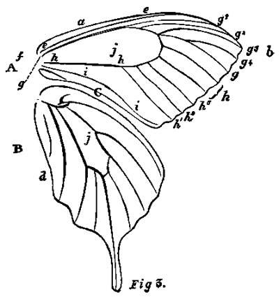
Fig. 3. Diagram of Butterfly's Wing.
A. Upper Wing. B. Lower Wing. a. Costal Margin. b. Hind Margin. c. Inner " d. Anal Angle. e. Costa. f. Costal nervure. g. Sub-costal do. g1-4. Branches of do. h. Median nervure. i. Sub-median do. j. Discoidal Cell. k. Discoidal Veins.
In order fully to understand the bearing of the following remarks it is necessary to know something of the anatomy and nomenclature of butterflies. Fig. 3 is an ideal butterfly. The wing margins are described as the Costal, which is the upper strong edge of the wing, the Hind margin, forming the outside, and the Inner margin, forming the base. The nervures consist of four principal veins; the Costal, a simple nervure under the costa, the Sub-costal, which runs parallel to the costal and about halfway to the tip emits branches, generally four in number; the Median occupying the centre of the wing and sending off branches, usually three in number, and the Sub-median below which is always simple. There are thus two simple nervures, one near the costal the other near the inner margin, and between them are two others which emit branches. Between these two latter is a wide plain space known as the discoidal cell. Small veins called the discoidal pass from the hind margin towards the cell, and little transverse nervures, known as sub-discoidal, often close the cell. By these nervures the wing is mapped out into a series of spaces of which one, the discoidal cell, is the most important.
The nervures have two functions, they support and strengthen the wing, and being hollow serve to convey nutritive fluid and afterwards air to the wing.
The wings are moved by powerful muscles attached to the base of the wings close to the body and to the inside of the thorax, all the muscles being necessarily internal. "There are two sets which depress the wings; firstly a double dorsal muscle, running longitudinally upwards in the meso-thorax;[28] and, secondly, the dorso-ventral muscles of the meso- and meta-thorax,[29] which are attached to the articulations of the wings above, and to the inside of the thorax beneath. Between these lie the muscles which raise the wings and which run from the inner side of the back of the thorax to the legs."[30] When we consider the immense extent of wing as compared with the rest of the body, the small area of attachment, and the great leverage that has to be worked in moving the wings, it is clear that the area of articulation of the wing to the body is one in which the most violent movement takes place. It is here that the waste and repair of tissue must go on with greatest vigour, and we should, on our theory, expect it to be the seat of strong emphasis. Accordingly we commonly find it adorned with hairs, and in a vast number of cases the general hue is darker than that of the rest of the wing, and so far as we have been able to observe, never lighter than the body of the wing. Even in the so-called whites (Pieris) this part of the wing is dusky, and instances are numerous on Plate IV.
The scales, which give the colour to the wings, deserve more than a passing notice. They are inserted by means of little stalks into corresponding pits in the wing-membrane, and overlap like tiles on a roof; occasionally the attachment is a ball and socket (Morphinæ), in which case it is possible the insect has the power of erecting and moving its scales. The shapes are very numerous, but as a rule they are short. To this there is a remarkable exception on the wings of the males of certain butterflies, consisting of elongated tufted prominences which appear to be connected with sense-organs. They are probably scent-glands, and thus we find, even in such minute parts as scales, a difference of function emphasized by difference of ornamentation, here showing itself in variety of forms; but, as we have said, ornamentation in form is often closely allied to ornamentation in colours. In some butterflies, indeed, these scales are aggregated into spots, as in Danais, and have a different hue from the surrounding area.
The scales are not simple structures, but consist of two or more plates, which are finely striated. The colouring matter consists of granules, placed in rows between the striæ, and may exist upon the upper surface of the upper membrane (epidermal), or the upper surface of the under or middle plate (hypodermal), or the colour may be simple diffraction colour, arising from the interference of the lightwaves by fine striæ.
Dr. Haagen, in the admirable paper before mentioned, has examined this question thoroughly, and gives the results set forth in the following table:—
Epidermal Colours.
Metallic blues and greens Bronze Gold Silver Black Brown Red (rarely) Persistent after death.

Hypodermal Colours.
Blue Green Yellow Milk-white Orange and shades between Red Fading after death.
The hypodermal colours are usually lighter than the epidermal, and are sometimes changed by a voluntary act. Hypodermal and epidermal colours are, of course, not peculiar to insects; and, as regards the former, it is owing to their presence that the changing hues of fishes, like the sole and plaice, and of the chameleon are due.
The great order Lepidoptera, including butterflies and moths, seems to the non-scientific mind to be composed of members which are pretty much alike, the differences being of slight importance; but this is not in reality the case, for the lepidoptera might, with some accuracy, be compared to the mammalia, with its two divisions of the placental and non-placental animals. Comparing the butterflies (Rhopalocera) to the placental mammals, we may look upon the different families as similar to the orders of the mammalia. Were we as accustomed to notice the differences of butterflies as we are to remark the various forms of familiar animals, we should no longer consider them as slight, but accord to them their true value. When in the mammalia we find animals whose toes differ in number, like the three-toed rhinoceros and the four-toed tapir, we admit the distinction to be great, even apart from other outward forms. So, too, the seal and lion, though both belonging to the carnivora, are readily recognized as distinct, but the seals may easily be confounded by the casual observer with the manatees, which belong to quite a different order.
Thus it is with the Lepidoptera, for from six-legged insects, whose pupæ lie buried beneath the soil, like most moths, we pass to the highest butterflies, whose fore-legs are atrophied, and whose pupæ hang suspended in the open air; and this by easy intermediate stages. Surely, if six-legged mammals were the rule, we should look upon four-legged ones as very distinct; and this is the case with the butterflies. It is necessary to make this clear at starting, in order that we may appreciate to its full value the changes that have taken place in the insects under study.
Butterflies (Rhopalocera) are grouped into four sub-families, as under:—
1. Nymphalidæ, having the fore-legs rudimentary, and the pupæ suspended from the base of the abdomen.
2. Erycinidæ, in which the males only have rudimentary fore-legs.
3. Lycænidæ, in which the fore-legs of the males are smaller than those of the females, and terminate in a simple hook.
4. Papilionidæ, which have six perfect pairs of legs, and in which the pupæ assume an upright posture, with a cincture round the middle.
It may, at first sight, appear curious that the imperfect-legged Nymphalidæ should be placed at the head of the list, but this is based upon sound reasoning. The larva consists of thirteen segments, and, in passing to the mature stage, the second segment alone diminishes in size, and it is to this segment that the first pair of legs is attached. Looking now to the aerial habits of butterflies, we can understand how, in the process of evolution towards perfect aerial structure, the legs, used only for walking, would first become modified; and, naturally, those attached to the segment which decreases with development would be the first affected. When this is found to be combined with an almost aerial position of the pupæ, we see at once how such insects approach nearest to an ideal flying insect. It is a general law that suppression of parts takes place as organisms become specialized. Thus, in the mammalia, the greatest number of toes and teeth are found in the lowest forms and in the oldest, simplest fossil species.
A butterfly is, indeed, little more than a beautiful flying machine; for the expanse of wing, compared with the size of the body, is enormous.
CHAPTER XI. The Colouration of Insects.
(Continued.)
General Scheme of Colouring. So various are the patterns displayed upon the wings of butterflies, that amidst the lines, stripes, bars, dots, spots, ocelli, scalloppings, etc., it seems at first hopeless to detect any general underlying principle of decoration; and this is the opinion that has been, and is still, held by many who have made these insects a special study. Nevertheless, we will try to show that beneath this almost confused complexity lie certain broad principles, or laws, and that these are expressed by the statement that decoration is primarily dependent upon structure, dependent upon the laws of emphasis and repetition, and modified by the necessity for protection or distinction.
To render this subject as plain as possible, British species will be selected, as far as possible, and foreign ones only used when native forms do not suffice.
The body of by far the greater number of species is either darker or of the same tint as the mass of the wings; and only in rare cases lighter. When the body has different tints, it is generally found that the thorax and abdomen differ in colour, and in many cases the base of the thorax is emphasized by a dark or light band.
On the wings the functional importance of the parts attached to the body is generally darker, perhaps never lighter, than the ground of the wing, and is frequently further emphasized by silky hairs. This has already been sufficiently pointed out.
The wing area may be divided into the strong costal margin, the hind margin, the nervules, and the spaces; and, however complex the pattern may be, it is always based upon these structure lines.
In the majority of insects the costal margin is marked with strong colour. This may be noticed in Papilio Machaon, P. merope, Vanessa antiopa, and the whites in Plate IV. The extreme tip of the fore-wings is nearly always marked with colour, though this may run into the border pattern. This colour is dark or vividly bright, and we know no butterfly, not even dark ones, that has a light tip to the wings. Sometimes, it is true, the light bead-border spots run to the tip, but these are not cases in point. The development of tips has been traced in Chapter VI., and need not be repeated.
The hind margin of both wings is very commonly emphasized by a border, of which V. Antiopa, Pl. III. Fig. 3, is a very perfect example.
The border pattern may consist of one or more rows of spots, lines, bands, or scallops;[31] and there is frequently a fine fringe, which in many cases is white, with black marks, and to which the term bead-pattern may be applied.
A definite relation subsists in most cases between the shape of the hind margin and the character of the border-pattern. The plain or simple bordered wings have plain border patterns, and the scalloped wings have scalloped borders; or rather scalloped borders are almost exclusively confined to scalloped wings. In our English butterflies, for instance, out of the 62 species:—
33 have plain margins to the wings. In all the border is plain, or wanting.
20 have the fore-wings plain, and the hind-wings scalloped, and in all the hind-wings are scalloped and the fore-wings plain, or with slightly scalloped border-patterns.
9 have scalloped margins and scalloped border-patterns.
Another relation between structure and pattern is found in those insects which have tailed hind-wings, for the tail is very frequently emphasized by a spot, often of a different colour from the rest of the wing as in the Swallow-Tails, Plates IV. and V.
Yet another point may be noticed. In each wing there is a space, the discoidal cell, j Fig. 3, at the apex of which several nervures join, forming knots. These are points at which obstacles exist to the flow of the contents, and they are almost always marked by a distinct pattern. We thus have a discoidal spot in very many butterflies, in nearly all moths; and in the other orders of winged insects the decoration is even more pronounced, as any one may see who looks at our dragon-flies, wasps, bees, or even beetles.
In some insects the decoration of the body is very marked, as in our small dragon-flies, the Agrions. In one species, for example, A. Puella, the male is pale blue banded with black, and the female bronze black, with a blue band on the segment, bearing the sexual organ; the ovipositors are also separately decorated. The male generative organs are peculiar, in that the fertilizing fluid is conveyed from one segment to a reservoir at the other end of the abdomen. Both the segments bearing these organs are marked by special decoration. The peculiar arrangement of the sexual organs in dragon-flies is very variable, and certain segments are modified or suppressed in some forms, as was shown by J. W. Fuller.[32] In every case the decoration follows the modification. In the thorax of dragon-flies, too, the principal muscular bands are marked out in black lines. This distinct representation of the internal structure is beautifully shown in Æschna and Gomphina, and in the thorax of Cicada, as shown by Dr. Haagen in the paper quoted in the last chapter.
We may, then, safely pronounce that the decoration of insects is eminently structural.
Simple Variation. Cases of simple variation have been already cited in our description of spots and stripes, and it only remains to show that in this, as in all other cases, the variation is due to a modification of original structural decoration.
To take familiar examples. Newman, in his British Butterflies, figures the varieties of the very common Small Tortoiseshell (Vanessa urticæ). In the normal form there is a conspicuous white spot on the disc of the fore-wings, which is absent in the first variety, owing to the spreading of the red-brown ground colour. This variety is permanent on the Mediterranean shores. In variety two, the second black band, running from the costa across the cell, is continued across the wing. The third variety, Mr. Newman remarks, is "altogether abnormal, the form and colouring being entirely altered." Still, when we examine the insect closely, we find it is only a modification of the original form. The first striking difference is in the margin of the wings, which in the normal form is scalloped with scallop-markings, whereas, in the variety the margins are much simpler, and the border pattern closely corresponds with it, having lost its scalloping. In the fore-wing some of the black bands and spots are suppressed or extended, and the extensions end rigidly at nervules. The dark colouring of the hind-wings has spread over the whole wing. We thus see that the decoration, even in varieties called abnormal, still holds to structural lines, and is a development of pre-existing patterns.
No one can have examined large series of any species without being impressed with the modification of patterns in almost every possible way. For instance, we have reared quantities of Papilio Machaon, and find great differences, not only in the pattern, but in the colour itself. A number of pupæ from Wicken Fen, Cambridgeshire, were placed in cages, into which only coloured light could fall, and though these experiments are not sufficiently extended to allow us to form any sound conclusions as to the effect of the coloured light, we got more varieties than could be expected from a batch of pupæ from the same locality. The tone of the yellow, the quantity of red, the proportion of the yellow to the blue scales in the clouds, varied considerably, but always along the known and established lines.
The variations in the colour of Lepidoptera has been most admirably treated by Mr. J. Jenner Weir in a paper, only too short, read before the West Kent Natural History Society.[33] He divides variations into two sections, Aberrations or Heteromorphism, and constant variations or Orthopæcilism, and subdivides each into six classes, as under:—
Heteromorphism.
Albinism white varieties.
Melanism black do.
Xanthism pallid do.
Sports or occasional variations not included in the above.
Gynandrochomism females coloured as males.
Hermaphroditism sexes united.
Orthopæcilism.
Polymorphism variable species.
Topomorphism local varieties.
Atavism reversion to older forms.
Dimorphism two constant forms.
Trimorphism three do. do.
Horeomorphism seasonal variation.
In some cases, he remarks, variations are met with which may with equal propriety be classed in either section.
Albinism he finds to be very rare in British species, the only locality known to him being the Outer Hebrides. This reminds us of Wallace's remark upon the tendency to albinism in islands. Xanthism, he finds to be more plentiful, and quotes the common Small Heath (Cænonympha pamphilus) as an illustration. In these varieties we have simply a bleaching of the colouring matter of the wings, and therefore no departure from structural lines. Melanism arises from the spreading of large black spots or bars, or, as in Biston betularia, a white moth peppered with black, dots by the confluence of small spots; for this insect in the north is sometimes entirely black. It is singular that insects have a tendency to become melanic in northern and alpine places, and this is especially the case with white or light coloured species. (See Plate IV., Fig. 17) It has recently been suggested that this darkening of these delicate membranous beings in cold regions is for the purpose of absorbing heat, and this seems very probable. [34]
Of ordinary spots it is merely necessary to remark, that they are all cases in our favour. Thus, in Satyrus hyperanthus we have "the ordinary round spots ... changed into lanceolate markings"; this takes place also in C. davus. The other cases of aberration do not concern us.
When, however, we come to the cases in which a species has two or more permanent forms, it is necessary to show that they are in all cases founded on structure lines. The patterns, as shown in Plate V., Figs. 1-13, are always arranged structurally, and the fact that not only are intermediate forms known, as in Araschnia porima, Plate V., Fig. 6, but that the various forms are convertible into one another, would in itself be sufficient to show that in these cases there is no departure from the general law. In Grapta interrogationis, Plate V., Figs. 8-10, we see in the central figure one large spot above the median nervure, in the left-hand form this is surmounted by another spot above the lowest sub-costal branch, and in the right-hand figure this latter spot is very indistinct. We have here a perfect gradation, and the same may be said of the colouration of the lower wings. Take again the three forms of Papilio Ajax in the same plate, Figs. 11-13, and we have again only modifications of the same type.
In local varieties, as in seasonal forms, we have again nothing more than developments of a given type, as is well shown in Plates IV. & V., Figs. 13-18 & 1-13.
When, however, we come to mimetic forms, whether they mimic plants, as in Plate I., or other species, as in Plates II. & III., a difficulty does seem to arise.
The leaf butterfly (Kallima inachus), Plate I., offers no trouble when we view the upper surface only with its orange bands, but its under surface, so marvellously like a dead leaf that even holes and microscopic fungi are suggested, does seem very like a case in which structure lines are ignored. Take, for instance, the mark which corresponds to the mid-ribs, running from the tail to the apex of the upper wing; it does not correspond to any structure line of the insect. But if we take allied and even very different species and genera of Indian and Malayan butterflies, we shall find every possible intermediate form between this perfect mimicry and a total lack of such characters. To cite the most recent authority, the various species of the Genera Discophora, Amathusia, Zeuxidia, Thaumantis, Precis, &c., figured so accurately in Distant's Rhopalocera Malayana, will give all the steps.
In the cases of true mimicry, as in Figs. 1-3, Plates II. & III., where insects as different as sheep from cats copy one another, we find that of course structure lines are followed, though the pattern is vastly changed. The Papilio merope, Fig. 1, Plate II., which mimics Danais niavius, Fig. 3, does so by suppressing the tail appendage, changing the creamy yellow to white—a very easy change, constantly seen in our own Pieridæ—and diffusing the black. A similar case is seen in Figs. 4-5, Plate III., where a normally white butterfly (Panopœa hirta) mimics a normally dark one of quite a different section. Here again the change is not beyond our power of explanation. Where a Papilio like merope mimics a brown species like Danais niavius, we have a still greater change in colour, but not in structural pattern.
If we ascribe to these insects the small dose of intelligence we believe them to possess, we can readily see how the sense of need has developed such forms.
Local varieties present no difficulty under such explanation. The paramount necessity for protection has given the Hebridran species the grey colour of the rocks, and the desert species their sandy hue.
Plate VII.
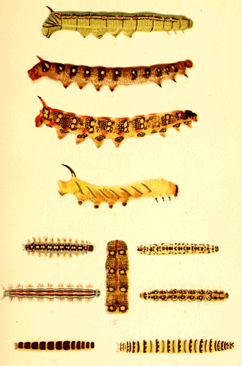
CATERPILLARS.
Finally, to take the case of caterpillars, Weismann has admirably worked out the life history of many forms, and shows how the complex markings have arisen by development. Broadly, a caterpillar consists of 13 segments, the head being one. The head is often marked with darker colour, and the last segment with its clasping feet is also very frequently emphasized, as in Figs. 1 & 3, Plate VII. The spiracles are generally marked by a series of spots, and often connected by a line. Here the tendency to repetition shows itself strongly, for not only the spiracles themselves, but the corresponding points in the segments without spiracles are frequently spotted, and, moreover, these spots are frequently repeated in rows above the spiracular line. Of this, Deilephila galii and D. Euphorbiæ, Figs. 1-5, Plate VII., are good examples.
The segmentation is also generally emphasized, as shown in all the examples on the plate, but in its simplicity in Fig. 10.
Running down the centre of the back a more or less distinct line is often seen, as shown in the figures. This corresponds with the great dorsal alimentary canal lying just below the skin, and Weismann has shown that in young larvæ this line is transparent, and the green food can be seen through the skin. We have here, perhaps, a relic of the direct colouration noticed in the transparent cœlenterata.
Where larvæ possess horns either upon the head, as in Apatura iris and Papilio machaon, or on the tail, as in many of the sphyngidæ, like Figs. 1-5, Plate VII., these appendages are always emphasized in colour. As they are frequently oblique, we often find that this obliquity is continued as a slanting spot, as in D. galii and euphorbiæ, and sometimes repeated as a series of oblique stripes, as in Fig. 4.
It must be admitted that in insects we have strong evidence of structural decoration.
CHAPTER XII. Arachnida.
T
THE Arachnida include the scorpions and spiders, and as the former are tolerably uniform in colour, our remarks will be confined to the latter.
The thorax is covered with a horny plate, while the abdomen only possesses a soft skin, and neither show any traces of segmentation. From the thorax spring four pairs of legs, and a pair of palpi, or feelers. Immediately beneath the skin of the abdomen lies the great dorsal vessel, which serves as a heart. This vessel is divided into three chambers, the general aspect of which is shown in Fig. 9, Plate VIII., taken from Gegenbaur's Comparative Anatomy.[35]
From this heart the blood passes by vessels to each of the limbs, the palpi, etc., as offsets from the double-branched aorta. The shape of this dorsal vessel is peculiar, and its importance in respect to colouration will be immediately apparent.
The primary scheme of colouration in the Arachnida seems to be the distinguishing of the cephalothorax from the abdomen by a different colour. Thus, of the 272 species of British spiders represented in Blackwell's work,[36] no less than 203 have these parts differently coloured, and only 69 are of the same hue, and even in these there is often a difference of tint. So marked is this in certain cases that the two parts form vivid contrasts. Of this cases are given in the following list.
Cephalothorax. Abdomen.
Eresus cinnabarinus, Black, Bright Red.
Thomisus floricolens, Green, Brown.
—— cinereus, Brown, Blue.
—— trux, Red, Brown.
Sparassus smaragdulus, Green, Red and yellow.
As a rule the abdomen is darker than the cephalothorax, and many species have the former red-brown and the latter black.
The legs, usually, take the colour of the cephalothorax, and are, hence, generally lighter than the abdomen, but to this there are exceptions. Where the individual legs differ in colour, the two first pairs are the darkest, and the dark hue corresponds in tint with the dark markings on the cephalothorax. The joints of the legs are in many species emphasized with dark colour, which is often repeated in bands along the limb.
The most remarkable point is, however, the pattern on the abdomen, which, though varied in all possible ways, always preserves a general character, so that we might speak with propriety of a spider-back pattern. This pattern is fairly well illustrated in the genus Lycosa, but is seen to perfection, and in its simplest form in Segestria senoculata, Plate VIII., Fig. 1, and in Sparassus smaragdulus, Plate VIII., Fig. 2.
This peculiar pattern is so like the dorsal-vessel that lies just beneath, that it is difficult to avoid the conclusion that we have here an actual case of the influence of internal organs on the integument, and this we believe to be the case. No matter how curious the abdominal markings may seem to be, they never so far depart from this fundamental pattern as to appear independent of it.
Thus, in the genus Lycosa, which is by no means the best for the purpose, but is chosen as illustrating Gegenbaur's diagram, Plate VIII., we have the dorsal-vessel well marked in L. piscatoria, Plate VIII., Fig. 3, from which may be developed the other forms. In L. andrenivora, Plate VIII., Fig. 4, the male shows the vessel-mark attenuated posteriorly; and in the female, Fig. 5, the hinder part has become broken up into detached marks, still preserving the original shape, while the upper part remains practically unchanged. In L. allodroma the disintegration of the mark has further advanced, for in the male, Fig. 6, the upper portion has lost something of its shape, and the lower part is a series of isolated segments. This process is carried still further in the female, Fig. 8, where the upper portion is simplified, and the lower almost gone. In L. campestris, Fig. 10, the mark is reduced to a stripe, corresponding with the upper part of the vessel-mark only: and, lastly, in the male L. agretyca, Fig. 7, this upper part is represented by two spots, though even here traces of the original form can be seen.
A simplification of marking of another sort is seen in L. rapax, Fig. 13, where the chamber-markings are almost obliterated, and merely an irregular stripe left. The stages by which this modification is arrived at are too obvious to need illustration.
In some species the lower portion of the vessel-mark is reduced to small dots, as in L. cambrica, fluviatilis, piratica, and others; and the stages are very clear. Starting with the isolated chamber-marks, as in L. allodroma, Fig. 5, we get, firstly, a set of spots, as in L. picta, which, in the female, Fig. 16, are still connected with the chamber-marks, but in the male, Fig. 17, are isolated. This leads us, by easy steps, to such forms as L. latitans, Fig. 14, which consists of a double row of spots upon dark stripes.
The intimate connection thus shown to subsist between the characteristic decoration of the abdomen of spiders, and the shape of the important dorsal organ beneath, seems to be strong evidence of effect that internal structure may have upon external decoration. [37]
The cephalothorax of spiders, being covered with a hardened membrane, does not show such evidence clearly, for it appears to be a law that the harder the covering tissue, the less does it reflect, as it were, the internal organs. The hard plates of the armadillo are thus in strong contrast to the softer skins of other animals.
Nevertheless, there does appear, occasionally, to be some trace of this kind of decoration in the cephalothorax of certain spiders, though it would be hard to prove. The blood vessels of this part (see Fig. 9), though large, are not nearly so prominent as the great dorsal vessel. The chief artery enters the cephalothorax as a straight tube, forks, and sends branches to the limbs, palpi, and eyes. In many species, notably in the genus Thomisus, a furcate mark seems to shadow the forked aorta. This is best shown in T. luctuosus, Plate VIII., Fig. 11. Moreover, in this and other genera, lines frequently run to the outer pair of eyes, which alone are supplied with large arteries, see Fig. 9.
However this may be, it is certain that the entire decoration of spiders follows structural lines, and that the great dorsal vessel has been emphasized by the peculiar pattern of the abdomen.
Plate VIII.
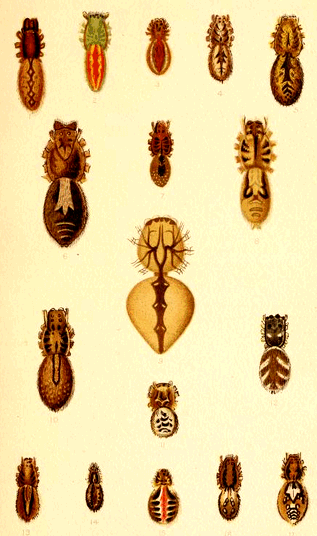
SPIDERS.
CHAPTER XIII. Colouration of Invertebrata (Continued).
O
OF the Arthropoda, including the lobsters, crabs, shrimps, etc., little can be said here, as we have not yet been able to study them with anything like completeness. Still, we find the same laws to hold good. The animals are segmented, and we find their system of colouration segmental also. Thus, in the lobsters and crabs there is no dorsal line, but the segments are separately and definitely decorated. The various organs, such as the antennæ and eyes, are picked out in colour, as may be beautifully seen in some prawns.
When we come to the Mollusca, we meet with two distinct types, so far as our subject is concerned; the naked and the shelled. In the naked molluscs, like the slugs, we have decoration applied regionally, as is shown to perfection in the Nudibranchs, whose feathery gills are often the seat of some of the most vivid hues in nature.
The shell-bearing mollusca are proverbial for their beauty, but it is essential to bear in mind that the shell does not bear the same relation to the mollusc that the "shell" of a lobster does to that animal. The lobster's shell is part of its living body; it is a true exo-skeleton, whereas the shell of a mollusc is a more extraneous structure—a house built by the creature. We ought, on our view, to find no more relation between the decoration of a shell and the structure of its occupant, than we do in the decoration of a human dwelling-house to the tenant.
The shell consists of carbonate of lime, under one or both of the forms known to mineralogists as calcite and aragonite. This mineral matter is secreted by an organ called the mantle, and the edge, or lip, of the mantle is the part applied to this purpose. The edge of the mantle is the builder's hand, which lays the calcareous stones of the edifice. The shell is built up from the edge, and the action is not continuous but seasonal, hence arise the markings known as lines of growth. In some cases the mantle is expanded at times into wing-like processes, which are turned back over the shell, and deposit additional layers, thus thickening the shell.
In all the forms of life hitherto considered the colouring matter is deposited, or formed, in the substance of the organ, or epidermal covering, but in the mollusca this is not the case. The colouring matter is entirely upon the surface, and is, as it were, stencilled on to the colourless shell. This is precisely analogous to the colouring of the shells of birds' eggs. They, too, are calcareous envelopes, and the colouring matter is applied to the outside, as anyone can see by rubbing a coloured egg. In some eggs several layers of colouring matter are superimposed.
In no case does the external decoration of molluscan shells follow the structure lines of the animal, but it does follow the shape of the mantle. The secreting edge may be smooth, as in Mactra, regularly puckered, as in most Pectens, puckered at certain points, as in Trigonia, or thrown into long folds, as in Spondylus. In each of these cases the shell naturally takes the form of the mantle. It is smooth in Mactra, regularly ribbed in Pecten, tubercled in Trigonia, and spined in Spondylus. Where the inside of the shell is coloured as in some Pectens, regional decoration at once appears and the paleal lines, and muscular impressions are bounded or mapped out with colour.
It is a significant fact that smooth bivalves are not so ornate as rugose ones, and that the ridges, spines, and tubercles of the latter are the seats of the most prominent colour.
Similar remarks apply to univalve shells, which are wound on an imaginary vertical axis. They may be smooth, as in Conus and Oliva, rugose, as in Cerithium, or spined, as in Murex. The structure of these shells being more complex than that of bivalves, we find, as a rule, they are more lavishly ornamented, and the prominent parts of the shell, and especially the borders, are the seat of strongest colour. In some cases, as in adult Cowries (Cypræa), the mantle is reflexed so as to meet along the median line, where we see the darkest colour.
The rule amongst spiral shells is to possess spiral and marginal decoration, and this is what we should expect. The Nautilus repeats in the red-brown markings of its shell, the shape of the septa which divide the chambers, though, as is often the case, they are generally more numerous than the septa.
The naked Cephalopoda, or cuttle-fishes, often possess a distinct dorsal stripe, and when our views were first brought before the Zoological Society, this fact was cited as an objection. To us it seems one of the strongest of favourable cases, for these animals possess a sort of backbone—the well-known cuttle-bone—and hence they have a dorsal line.
Some shells, as Margarita catenata, have a chain-pattern, and in this case the action of the pigment cells takes place at regular and short intervals. Others, as Mactra stultorum, the stencilling forms a series of lines and spots, generally enlarging into rays.
The whole subject of the decoration of shells deserves much more time than we have been able to give to it as yet.
CHAPTER XIV. Colouration of Vertebrata.
T
THE vertebrata, as their name implies, are distinguished by the possession of an internal skeleton, of which the backbone is the most essential part, and the general, but not universal, possession of limbs or appendages.
Consequently we find that the dorsal and ventral surfaces are almost invariably coloured differently, and the dorsal is the darker in the great majority of instances. Generally the spine is marked by a more or less defined central line, and hence this system of colouration may be termed axial, because it is in the direction of the axes, or applied about the axes.
Fishes. Where fishes have not been modified out of their original form, as are the soles, plaice, and other flat fish, we find the dorsal region darker than the ventral, and even here the under surfaces are the lightest. Even in cases like the Char, Fig. 1, Plate IX., where vivid colour is applied to the abdomen, the dorsum is the darker. The dorsum is often marked by a more or less well-defined dark band, as in the mackerel and perch, Fig. 2, Plate IX. There are sometimes parallel bands at right angles to the above, as in the perch and mackerel; and this is a common feature, and apparently a very old one, as we find it in the young of fishes whose adults are without these rib-like marks, such as the trout and pike.
It is only necessary to inspect any drawings of fishes to see that their colouration is on a definite principle, although rather erratic. Important functional parts, like the gills, fins, and tail, are generally marked in colour more or less distinctly, as may be seen, for instance, in our common fresh-water fishes, like the roach and perch. The line of mucus-secreting glands running along the sides is usually marked by a dark line. These facts point distinctly to structural decoration.
Plate IX.
CHAR and PERCH.
There are in some fishes, like the John Dory, curious eye-like dark spots, which we cannot refer to a structural origin, though a better acquaintance with the class might reveal such significance.
The Amphibia have not been well studied by us, and we must leave them with the remark that they seem to bear out the view of structural decoration, as is seen in our English newts. Some are, however, modified out of all easy recognition.
Reptiles. Among the reptiles, the snakes, Fig. 4, may be selected for illustration. Snakes are practically little more than elongated backbones, and are peculiar from the absence of limbs. The colouring matter does not reside so much in the scales as in the skin beneath, so that the sloughs do not illustrate the decoration. Hence, we might expect to find here a direct effect of morphological emphasis.
The ornamentation of snakes is very similar throughout the class, both in water and land snakes; as may be seen by Sir W. Fayrer's work on Venomous Snakes. This ornamentation is of a vertebral pattern, placed along the dorsal surface, with cross lines, which may represent ribs.
Where the ribs are wanting, as in the neck, the pattern changes, and we get merely longitudinal markings.
In the Python, Fig. 4, there are, near the central line, numerous round spots, which apparently emphasize the neural processes. There are diagonal markings on some species which illustrate the development of colour-spots already alluded to.