PLATE NO. XIb Slightly later stage of acute burning.
Reading edition
The swelling in the conjunctiva above and below has subsided, but the vascular injection remains, and the solid white oedema in the palpebral aperture is still well marked. The cornea is grey in the exposed area.
[For History of the case see page facing Plate XII.
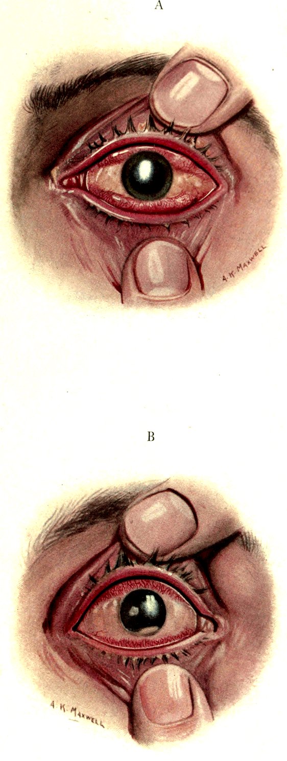
Plate XI.
History of the case. The casualty was caused by the bursting of a Yellow Cross shell close to the man when he was riding a restive mule, and his box respirator was momentarily displaced. A fine spray of the liquid must have splashed lightly over his right side, for cutaneous blisters developed on the neck, the cheek, and the forehead on this side only. The right eye showed serious burning with the central white band, while the left eye was only in the state of general red conjunctivitis.
With the lowering of the nutrition of the corneal epithelium, secondary infection is liable to take place. In this case an infiltrated corneal ulcer is seen associated with a hypopyon. It is therefore important when there is conjunctival discharge, which indicates secondary infection, that in addition to the use of atropine the conjunctival sac should be cleansed by frequent bland irrigations and by the instillation of antiseptic drops so as to check infection of any corneal ulceration which may develop. Otherwise the infective progress which has led to hypopyon may progress till panophthalmitis supervenes.