PLATE NO. IX Ulceration of trachea by mustard gas.
Reading edition
The characteristic feature is the sloughing of the tracheal mucous membrane. The reddening of the base of the tongue and of the pharynx, with a sharp delimitation where the oesophagus has refused ingress to the toxic vapour, is seen also with chlorine and other irritant gasses. But the pharyngeal inflammation with mustard gas may proceed further to a local ulceration that will cause dysphagia for many days.
The mucous membrane of the trachea and bronchi is affected by di-chlor-ethyl-sulphide in much the same way as is the skin. It reacts with an intense inflammation, and death of the surface layers soon results. The mass of necrotic tissue, exuded fibrin, and pus cells may form a yellowish-grey slough in which all manner of organisms flourish. Subsequently this false membrane comes away in patches or in entire casts from the raw surface of the bronchial wall.
Meantime the infected débris and secretions tend to accumulate in the bronchial ramifications at the bases of the lungs, and infection may spread from them into the lung tissues and alveoli. Septic broncho-pneumonia, localised abscesses, superficial pleurisy, and even empyema or pyopneumothorax then develop and cause death.
The drawing is of a trachea at the twelfth day after gassing. The base of the tongue and the pharynx show characteristic inflammation. Yellow necrotic sloughs lie on the larynx and at the bifurcation of the trachea. Between these the trachea is red and glistening, because it is now completely denuded of both mucous membrane and of slough. The dotted line points to a little group of ulcers on the posterior wall from which bleeding has occurred. The trachea and bronchi contained an abundance of thin yellow pus.
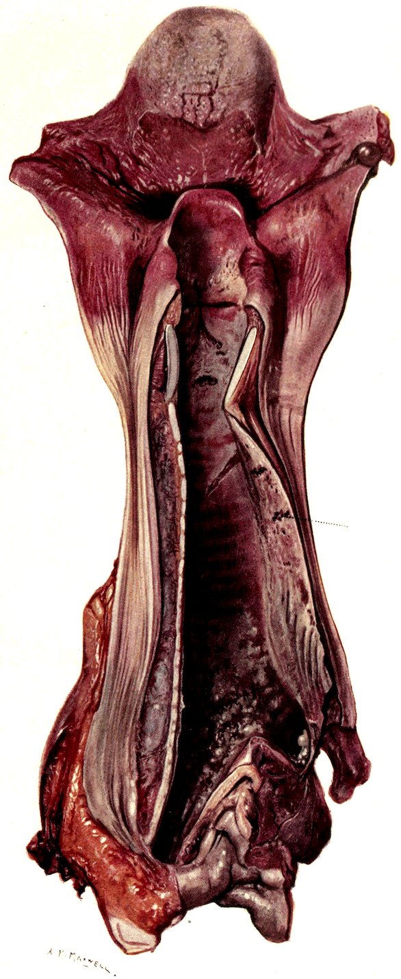
Plate IX.