Chapter III · (continued 2)
Reading edition
A. ANEMIA
This is a deficiency of hemoglobin, or red corpuscles, or both. It is either primary or secondary. The distinction is based chiefly upon etiology, although each type presents a more or less distinctive blood-picture. Secondary anemia is that which is symptomatic of some other pathologic condition. Primary anemia is that which progresses without apparent cause.
1. Secondary Anemia.—The more important conditions which produce secondary or symptomatic anemia are:
(a) Poor nutrition, which usually accompanies unsanitary conditions, poor and insufficient food, etc.
(b) Acute infectious diseases, especially rheumatism and typhoid fever. The anemia is more conspicuous during convalescence.
(c) Chronic infectious diseases: tuberculosis, malaria, syphilis, leprosy.
(d) Chronic exhausting diseases, as heart disease, chronic nephritis, cirrhosis of the liver, and gastro-intestinal diseases, especially when associated with atrophy of gastric and duodenal glands. The last may give an extreme anemia, indistinguishable from pernicious anemia.
(e) Chronic poisoning, as from lead, arsenic, and phosphorus.
(f) Hemorrhage—either repeated small hemorrhages, as from gastric cancer and ulcer, uterine fibroids, etc., or a single large one.
(g) Malignant tumors: these affect the blood partly through repeated small hemorrhages, partly through toxic products, and partly through interference with nutrition.
(h) Animal Parasites.—Some cause no appreciable change in the blood; others, like the Uncinaria and Bothriocephalus latus, may produce a very severe anemia, almost identical with pernicious anemia. Anemia in these cases is probably due both to toxins and to abstraction of blood.
The blood-picture varies with the grade of anemia. Diminution of hemoglobin is the most characteristic feature. In mild cases it is slight, and is the only blood change to be noted. In very severe cases hemoglobin may fall to 15 per cent. Red corpuscles are diminished in all but very mild cases, while in the severest cases the red corpuscle count is sometimes below 2,000,000. The color index is usually decreased.
Although the number of leukocytes bears no relation to the anemia, leukocytosis is common, being due to the same cause.
Stained films show no changes in very mild cases. In moderate cases variations in size and shape of the red cells and polychromatophilia occur. Very severe cases show the same changes to greater degree, with addition of basophilic degeneration and the presence of normoblasts in small or moderate numbers. Megaloblasts in very small numbers have been encountered in extremely severe cases. Blood-plaques are usually increased.
2. Primary Anemia.—The commonly described varieties of primary anemia are pernicious anemia and chlorosis, but splenic anemia may also be mentioned under this head.
(1) Progressive Pernicious Anemia.—It is frequently impossible to diagnose this disease from the blood examination alone. Severe secondary anemia sometimes gives an identical picture. Remissions, in which the blood approaches the normal, are common. All the clinical data must, therefore, be considered.
Hemoglobin and red corpuscles are always greatly diminished. In none of Cabot's 139 cases did the count exceed 2,500,000, the average being about 1,200,000. In more than two-thirds of the cases hemoglobin was reduced to less extent than the red corpuscles: the color-index was, therefore, high. A low color-index probably indicates a mild type of the disease.
The leukocyte count may be normal, but is commonly diminished to about 3000. The decrease affects chiefly the polymorphonuclear cells, so that the lymphocytes are relatively increased. In some cases a decided absolute increase of lymphocytes occurs. Polymorphonuclear leukocytosis, when present, is due to some complication.
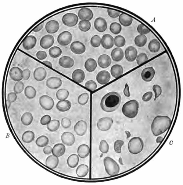
FIG. 84.—A, Normal blood; B, chlorosis; C, pernicious anemia. The plate shows the sharp contrast between cells rich in hemoglobin and the pale cell of chlorosis, and also the poikilocytosis and marked variations in size noted in pernicious anemia. A normoblast and megaloblast also appear. Stained smears (from Greene's "Medical Diagnosis").
PLATE VIII
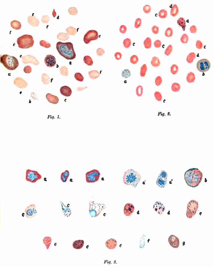
EXPLANATION OF PLATE VIII.
Fig. 1.—Preparation from an advanced case of progressive pernicious anemia from unknown cause: a, Megaloblasts or gigantoblasts; the protoplasm shows marked polychromasia; b, stained granules in erythrocytes with normally stained protoplasm; c and d, polychromatophilic degeneration; e, megalocytes; f, normocytes. Fig. 2.—Preparation from the same case taken some time later while the patient was subjectively and objectively in perfect health: a, Punctate erythrocytes with normal and anemic degenerated protoplasm; b, polynuclear leukocyte; c, normal red blood-corpuscles; d, somewhat enlarged erythrocytes. Fig. 3.—Series of cells from a case of severe progressive pernicious anemia of unknown etiology; preparation made two days antemortem: a, Nucleated red blood-corpuscles characterized as normoblasts by the intense staining of the nuclei; a' and a", karyokinetic figures in erythrocytes; the protoplasm finely punctate; b, beginning karyolysis in a megaloblast; c, erythroblasts with coarse granulation of the protoplasm; d, nuclear remains (?) and fine granulation of the protoplasm; e and f, finely punctate red blood-corpuscles; g, megalocyte with two blue nuclei; nuclear remains (?) in the polychrome protoplasm.
(Nothnagel-Lazarus.)
The red corpuscles show marked variation in size and shape (Plate VIII and Fig. 84). There is a decided tendency to large oval forms, and despite the abundance of microcytes, the average size of the corpuscles is generally strikingly increased. Polychromatophilia and basophilic degeneration are common. Nucleated red cells are always present, although in many instances careful search is required to find them. In the great majority of cases megaloblasts exceed normoblasts in number. This ratio constitutes one of the most important points in diagnosis, since it is practically unknown in other diseases. Blood-plaques are diminished.
The rare and rapidly fatal anemia which has been described under the name of aplastic anemia is probably a variety of pernicious anemia. Absence of any attempt at blood regeneration explains the marked difference in the blood picture. Red corpuscles and hemoglobin are rapidly diminished to an extreme degree. The color index is normal or low. The leukocyte count is normal or low, with relative increase of lymphocytes. Stained smears show only slight variations in size, shape, and staining properties of the red cells. There are no megaloblasts, and few or no normoblasts.
(2) Chlorosis.—The clinical symptoms furnish the most important data for diagnosis. The blood resembles that of secondary anemia in many respects.
The most conspicuous feature is a decided decrease of hemoglobin (down to 30 or 40 per cent. in marked cases), accompanied by a slight decrease in number of red corpuscles. The color-index is thus almost invariably low, the average being about 0.5.
As in pernicious anemia, the leukocytes are normal or decreased in number, with a relative increase of lymphocytes.
In contrast to pernicious anemia (and in some degree also to secondary anemia) the red cells are of nearly uniform size, are uniformly pale (Fig. 84), and their average diameter is somewhat less than normal. Changes in size, shape, and staining reactions occur only in severe cases. Erythroblasts are rarely present. The number of plaques is generally decreased.
(3) Splenic Anemia.—This is an obscure form of anemia associated with great enlargement of the spleen. It is probably a distinct entity. There is decided decrease of hemoglobin and red corpuscles, with moderate leukopenia and relative lymphocytosis. Osler's fifteen cases averaged 47 per cent. hemoglobin and 3,336,357 red cells. Stained films show notable irregularities in size, shape, and staining properties only in advanced cases. Erythroblasts are uncommon.
B. LEUKEMIA
Except in rare instances, diagnosis is easily made from the blood alone. Two types of the disease are commonly distinguished: the myelogenous and the lymphatic. Atypical and intermediate forms are not uncommon. Pseudoleukemia, because of its clinical similarity to lymphatic leukemia, is generally described along with leukemia.
1. Myelogenous Leukemia.—This is usually a chronic disease, although acute cases have been described.
Hemoglobin and red corpuscles show decided decrease. The color-index is moderately low.
Most striking is the immense increase in number of leukocytes. The count in ordinary cases varies between 100,000 and 300,000. Counts over 1,000,000 have been met. During remissions, the leukocyte count may fall to normal.
While these enormous leukocyte counts are equaled in no other disease, and approached only in lymphatic leukemia and extremely high-grade leukocytosis, the diagnosis, particularly during remissions, depends more upon qualitative than quantitative changes. Although all varieties are increased, the characteristic and conspicuous cell is the myelocyte. This cell never appears in normal blood; extremely rarely in leukocytosis; and never abundantly in lymphatic leukemia. In myelogenous leukemia myelocytes usually constitute more than 20 per cent. of all leukocytes. Da Costa's lowest case gave 7 per cent. The neutrophilic form is generally much more abundant than the eosinophilic. Both show considerable variations in size. Very constant also is a marked absolute, and often a relative, increase of eosinophiles and basophiles. Polymorphonuclear neutrophiles and lymphocytes are relatively decreased.
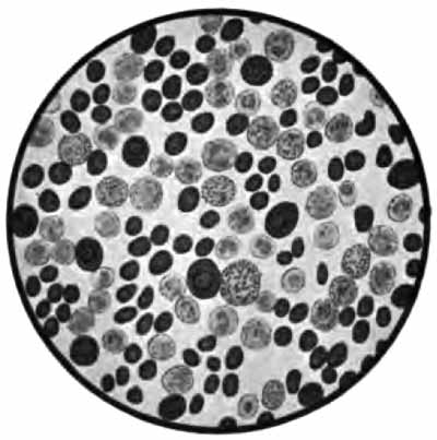
FIG. 85.—Blood from a case of splenomedullary leukemia, stained with Ehrlich's triple stain (Jakob).
The red cells show the changes characteristic of a severe secondary anemia, except that nucleated reds are commonly abundant; in fact, no other disease gives so many. They are chiefly of the normoblastic type. Megaloblasts are uncommon. Blood-plaques are generally increased.
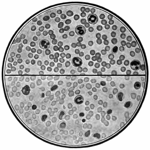
FIG. 86.—Blood: Lymphatic leukemia (lower section); marked leukocytosis (upper section). Stained smears. Compare types of leukocytes with Fig. 85 (from Greene's "Medical Diagnosis").
2. Lymphatic Leukemia.—This form may be either acute or chronic. There is marked loss of hemoglobin and red corpuscles. The color-index is usually moderately low.
The leukocyte count is high, but lower than in the myelogenous type. Counts of 100,000 are about the average, but in many cases are much lower. This high count is referable almost wholly to increase of lymphocytes. They generally exceed 90 per cent. of the total number. In chronic cases they are chiefly of the small variety; in acute cases, of the large form. Myelocytes are rare.
The red corpuscles show the changes usual in severe secondary anemia. Erythroblasts are seldom abundant. Blood-plaques are decreased.
3. Pseudoleukemia (Hodgkin's disease) resembles lymphatic leukemia in that there is marked and progressive enlargement of the lymph-nodes. There is, however, no distinctive blood-picture. The changes in hemoglobin and red cells resemble those of a moderate symptomatic anemia, with rather low color-index. The leukocytes are commonly normal in number and relative proportions.
4. Anæmia Infantum Pseudoleukæmica.—Under this name von Jaksch described a rare disease of infancy, the proper classification of which is uncertain. There is enlargement of liver and spleen, and sometimes of lymph-nodes, together with the following blood-changes: grave anemia with deformed and degenerated red cells and many erythroblasts of both normoblastic and megaloblastic types; great increase in number of leukocytes (20,000 to 100,000), and great variations in size, shape, and staining of leukocytes, with many atypical forms and a few myelocytes.
The table on the following page contrasts the distinctive blood-changes in the more common conditions.
DIFFERENTIAL DIAGNOSIS OF BLOOD DISEASES
SECONDARY ANEMIA. PERNICIOUS ANEMIA. CHLOROSIS. MYELOGENOUS LEUKEMIA. LYMPHATIC LEUKEMIA.
Hemoglobin. Diminished according to degree of anemia. Diminished. Greatly diminished. Decidedly diminished. Markedly diminished.
Red Corpuscle Count. Normal in mild cases; diminished in all others. Greatly diminished. Slightly diminished. Decidedly diminished. Markedly diminished.
Color-index. Normal or slightly diminished. High. Low. Usually slightly diminished. Usually slightly diminished.
Leukocyte Count. Not necessarily affected; leukocytosis common. Normal or diminished. Normal or diminished. Extremely high. Very high.
Red Corpuscles on Stained Films. Variations in size and shape in moderate cases; variations in staining reactions and normoblasts in severe cases. Marked variations in size, shape, and staining reactions. Average size increased. Tendency to large oval forms. Erythroblasts always present; megaloblasts exceed normoblasts. Nearly uniform size and shape; average size decreased; pale centers. Erythroblasts very rare. Similar to secondary anemia, except normoblasts generally very numerous. Similar to secondary anemia. Erythroblasts not numerous.
Leukocytes on Stained Films. Normal proportions or increase of polynuclears. Lymphocytes relatively, sometimes absolutely, increased. Lymphocytes apt to be relatively increased. Large numbers of myelocytes (average, 20 per cent.). Absolute increase of eosinophiles and basophiles. Relative decrease of polynuclears and lymphocytes. Lymphocytes exceed 90 per cent. Other varieties relatively decreased.