Chapter II · (continued 2)
Reading edition
PLATE IV
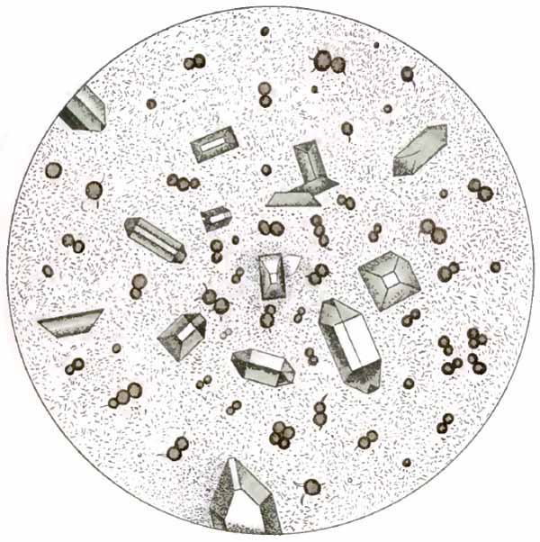
Sediment of alkaline fermentation (after Hofmann and Ultzmann).
These crystals occur only when ammonia is present in excess. They are generally found along with the phosphates in decomposing urine and have no clinical significance.
B. ORGANIZED SEDIMENTS
The principal organized structures in urinary sediments are: tube-casts; epithelial cells; pus-corpuscles; red blood-corpuscles; spermatozoa; bacteria; and animal parasites. They are much more important than the unorganized sediments just considered.
1. Tube-casts.—These interesting structures are albuminous molds of the uriniferous tubules. Their presence in the urine probably always indicates some pathologic change in the kidney, although this change may be very slight or transitory. Large numbers may be present in temporary irritations and congestions. They do not in themselves, therefore, imply organic disease of the kidney. They probably occur only in urine which contains, or has recently contained, albumin.
While it is not possible to draw a sharp dividing-line between the different varieties, casts may be classified as: (1) Hyaline casts; (2) waxy casts; (3) granular casts; (4) fatty casts; (5) casts containing organized structures—(a) epithelial casts; (b) blood-casts; (c) pus-casts; (d) bacterial casts. As will be seen, practically all varieties are modifications of the hyaline.
The significance of the different varieties is more readily understood if one considers their mode of formation. Albuminous material, the source and nature of which are not definitely known, probably enters the lumen of a uriniferous tubule in a fluid or plastic state. It there hardens into a mold, which, when washed out by the urine, retains the shape of the tubule, and contains within its substance whatever structures and débris were lying free within the tubule or were loosely attached to its wall. If the tubule be small and have its usual lining of epithelium, the cast will be narrow; if it be large or entirely denuded of epithelium, the cast will be broad. A cast, therefore, indicates the condition of the tubule in which it is formed.
The search for casts must be carefully made. The urine must be fresh, since hyaline casts soon dissolve when it becomes alkaline. It should be thoroughly centrifugalized. When the sediment is abundant, casts, being light structures, will be found near the top. In cystitis, where casts may be entirely hidden by the pus, the bladder should be irrigated to remove as much of the pus as possible and the next urine examined. In order to prevent solution of the casts the urine, if alkaline, must be rendered acid by previous administration of boric acid or other drugs.
(1) Hyaline Casts.—Typically, these are colorless, homogeneous, semitransparent, cylindric structures, with parallel sides and usually rounded ends. Not infrequently they are more opaque or show a few granules or an occasional cell, either adhering to them or contained within their substance. Generally they are straight or curved; less commonly, convoluted. Their length and breadth vary greatly: they are sometimes so long as to extend across several fields of a medium-power objective, but are usually much shorter; in breadth, they vary from one to seven or eight times the diameter of a red blood-corpuscle. (See Figs. 2, 41, 42, and 46.)
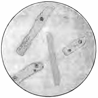
FIG. 41.—Hyaline casts showing fat-droplets and leukocytes (obj. one-sixth) (Boston).
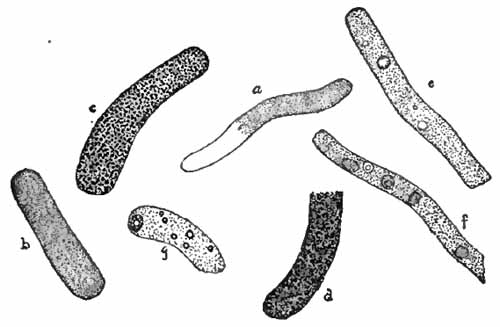
FIG. 42.—Various kinds of casts: a, Hyaline and finely granular cast; b, finely granular cast; c, coarsely granular cast; d, brown granular cast; e, granular cast with normal and abnormal blood adherent; f, granular cast with renal cells adherent; g, granular cast with fat and a fatty renal cell adherent (Ogden).
Hyaline casts are the least significant of all the casts, and occur in many slight and transitory conditions. Small numbers are common following ether anesthesia, in fevers, after excessive exercise, and in congestions and irritations of the kidney. They are always present, and are usually stained yellow when the urine contains much bile. While they are found in all organic diseases of the kidney, they are most important in chronic interstitial nephritis. Here they are seldom abundant, but their constant presence is the most reliable urinary sign of the disease. Small areas of chronic interstitial change are probably responsible for the few hyaline casts so frequently found in the urine of elderly persons.
Very broad hyaline casts commonly indicate complete desquamation of the tubular epithelium, such as occurs in the late stages of nephritis.
(2) Waxy Casts.—Like hyaline casts, these are homogeneous when typical, but frequently contain a few granules or an occasional cell. They are much more opaque than the hyaline variety, and are usually shorter and broader, with irregular, broken ends, and sometimes appear to be segmented. They are grayish or colorless, and have a dull waxy look, as if cut from paraffin (Figs. 43 and 61). They are sometimes composed of material which gives the amyloid reactions. Waxy casts are found in most advanced cases of nephritis, where they are an unfavorable sign.
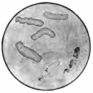
FIG. 43.—Waxy casts (upper part of figure). Fatty and fat-bearing casts (lower part of figure) (from Greene's "Medical Diagnosis").
Casts which resemble waxy casts but have a distinctly yellow color, as if cut from beeswax (so-called "fibrinous casts"), are often seen in acute nephritis. They have less serious significance than the true waxy variety.
(3) Granular Casts.—These are merely hyaline casts in which numerous granules are embedded (Figs. 42, 44, 46, and 61).
Finely granular casts contain many fine granules, are usually shorter, broader, and more opaque than the hyaline variety, and are more conspicuous. Their color is grayish or pale yellow.
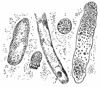
FIG. 44.—Granular and fatty casts and two compound granular cells (Stengel).
Coarsely granular casts contain larger granules and are darker in color than the finely granular, being often dark brown owing to presence of altered blood-pigment. They are usually shorter and more irregular in outline, and more frequently have irregularly broken ends.
(4) Fatty Casts.—Small droplets of fat may at times be seen in any variety of cast. Those in which the droplets are numerous are called fatty casts (Figs. 43 and 44). The fat-globules are not difficult to recognize. Staining with osmic acid or Sudan (p. 109) will remove any doubt as to their nature.
The granules and fat-droplets seen in casts are products of epithelial degeneration. Granular and fatty casts, therefore, always indicate partial or complete disintegration of the renal epithelium. The finely granular variety is the least significant, and is found when the epithelium is only moderately affected. Coarsely granular, and especially fatty casts, indicate a serious parenchymatous nephritis.
(5) Casts Containing Organized Structures.—Cells and other structures are frequently seen adherent to a cast or embedded within it. (See Figs. 41 and 42). When numerous, they give name to the cast.
(a) Epithelial casts contain epithelial cells from the renal tubules. They always imply desquamation of epithelium, which rarely occurs except in parenchymatous inflammations (Figs. 60 and 61).
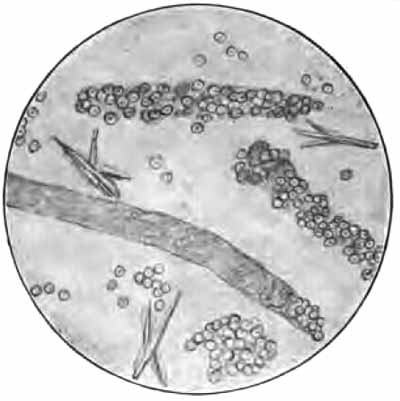
FIG. 45.—Red blood-corpuscles and blood-casts (courtesy of Dr. A. Scott) (obj. one-sixth) (Boston).
(b) Blood-casts contain red blood-corpuscles, usually much degenerated (Figs. 45 and 60). They always indicate hemorrhage into the tubules, which is most common in acute nephritis or an acute exacerbation of a chronic nephritis.
(c) Pus-casts (see Fig. 62), composed almost wholly of pus-corpuscles, are uncommon, and point to a chronic suppurative process in the kidney.
(d) True bacterial casts are rare. They indicate a septic condition in the kidney. Bacteria may permeate a cast after the urine is voided.
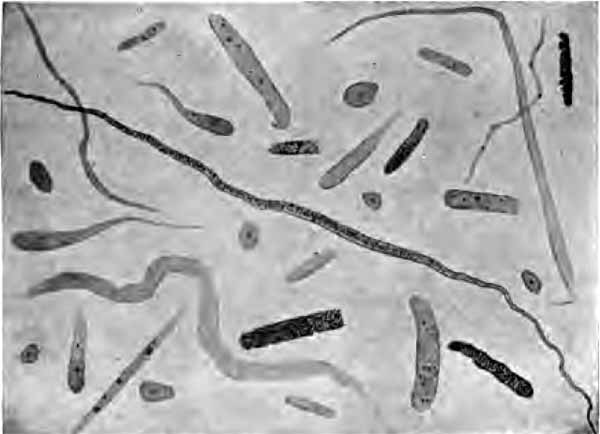
FIG. 46.—Hyaline and granular casts, mucous threads, and cylindroids. There are also a few epithelial cells from the bladder (Wood).
The structures most likely to be mistaken for casts are:
(1) Mucous Threads.—Mucus frequently appears in the form of long strands which slightly resemble hyaline casts (Fig. 46). They are, however, more ribbon-like, have less well-defined edges, and usually show faint longitudinal striations. Their ends taper to a point or are split or curled upon themselves, and are never evenly rounded, as is commonly the case with hyaline casts.
(2) Cylindroids.—This name is sometimes given to the mucous threads just described, but is more properly applied to certain peculiar structures more nearly allied to casts. They resemble hyaline casts in structure, but differ in being broader at one end and tapering to a slender tail, which is often twisted or curled upon itself (Fig. 46). They frequently occur in the urine along with hyaline casts, and have no definite pathologic significance.
(3) Masses of amorphous urates or phosphates or very small crystals (Fig. 47), which accidentally take a cylindric form, or shreds of mucus covered with granules, closely resemble granular casts. Application of gentle heat or appropriate chemicals will serve to differentiate them. When urine contains both mucus and granules, large numbers of these "pseudo-casts," all lying in the same direction, can be produced by slightly moving the cover-glass from side to side. It is possible—as in urate infarcts of infants—for urates to be molded into cylindric bodies within the renal tubules.
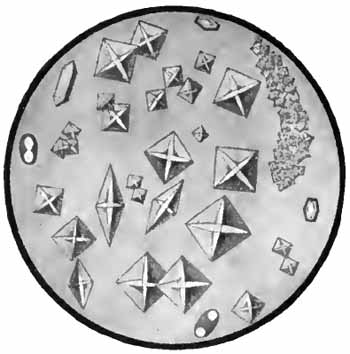
FIG. 47.—Calcium-oxalate crystals, showing a pseudo-cast of small crystals (Jakob).
(4) Hairs and fibers of wool, cotton, etc. These could be mistaken for casts only by beginners. One can easily become familiar with their appearance by suspending them in water and examining with the microscope (Fig. 57).
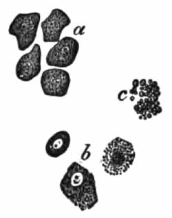
FIG. 48.—Renal epithelium from nephritic urine: a, Polyhedral epithelium in nephritis of scarlet fever; b and c, different grades of fatty degeneration in renal epithelium in chronic nephritis (X400) (after Bizzozero).
(5) Hyphæ of molds are not infrequently mistaken for hyaline casts. Their higher degree of refraction, their jointed or branching structure, and the accompanying spores will differentiate them (Fig. 58).
2. Epithelial Cells.—A few cells from various parts of the urinary tract occur in every urine. A marked increase indicates some pathologic condition at the site of their origin. It is sometimes, but by no means always, possible to locate their source from their form. Any epithelial cell may be so granular from degenerative changes that the nucleus is obscured. They are usually divided into three groups:
(1) Small, round, or polyhedral cells are about the size of pus-corpuscles, or a little larger, with a single round nucleus. Such cells may come from the deeper layers of any part of the urinary tract. They are uncommon in normal urine. When they are dark in color, very granular, and contain a comparatively large nucleus, they probably come from the renal tubules, but their origin in the kidney is not proved unless they are found embedded in casts. Renal cells are abundant in parenchymatous nephritis, especially the acute form. They are nearly always fatty—most markedly so in chronic parenchymatous nephritis, where their substance is sometimes wholly replaced by fat-droplets ("compound granule cells") (see Figs. 44, 48, 60, and 61).
(2) Irregular cells are considerably larger than the preceding. They are round, pear-shaped, or spindle-shaped, or may have tail-like processes, and are hence named large round, pyriform, spindle, or caudate cells respectively. Each contains a round or oval distinct nucleus. Their usual source is the deeper layers of the urinary tract, especially of the bladder. Caudate forms come most commonly from the pelvis of the kidney (see Figs. 49, b, 50, 62, and 63).
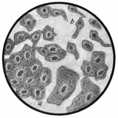
FIG. 49.—Epithelial cells from urethra and bladder: a, Squamous cells from superficial layers; b, irregular cells from deeper layers (Jakob).
(3) Squamous or pavement cells are large flat cells, each with a small, distinct, round or oval nucleus (Fig. 49, a). They are derived from the superficial layers of the ureters, bladder, urethra, or vagina. Those from the bladder are generally rounded, while those from the vagina are larger, thinner, and more angular. Great numbers of these vaginal cells, together with pus-corpuscles, may be present when leukorrhea exists.
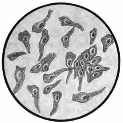
FIG. 50.—Caudate epithelial cells from pelvis of kidney (Jakob).
3. Pus-corpuscles.—A very few leukocytes are present in normal urine. They are more abundant when mucus is present. An excess of leukocytes, mainly of the polymorphonuclear variety, with albumin, constitutes pyuria—pus in the urine.
When at all abundant, pus forms a white sediment resembling amorphous phosphates macroscopically. Under the microscope the corpuscles appear as very granular cells, about twice the diameter of a red blood-corpuscle (Figs. 51 and 63). In freshly voided urine many exhibit ameboid motion, assuming irregular outlines. Each contains one irregular nucleus or several small, rounded nuclei. The nuclei are obscured or entirely hidden by the granules, but may be brought clearly into view by running a little acetic acid under the cover-glass. This enables one to easily distinguish pus-corpuscles from small round epithelial cells, which resemble them in size, but have a single, rather large, round nucleus.
Pyuria indicates suppuration in some part of the urinary tract—urethritis, cystitis, pyelitis, etc.—or may be due to contamination from the vagina, in which case many vaginal epithelial cells will also be present. In general, the source of the pus can be determined only by the accompanying structures (epithelia, casts) or by the clinical signs.
A fairly accurate idea of the quantity of pus from day to day may be had by shaking the urine thoroughly and counting the number of corpuscles per cubic millimeter upon the Thoma-Zeiss blood-counting slide.
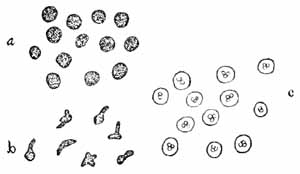
FIG. 51.—Pus-corpuscles: a, As ordinarily seen; b, ameboid corpuscles; c, showing the action of acetic acid (Ogden).
4. Red Blood-corpuscles.—Urine which contains blood is always albuminous. Very small amounts do not alter its macroscopic appearance. Larger amounts alter it considerably. Blood from the kidneys is generally intimately mixed with the urine and gives it a hazy reddish or brown color. When from the lower urinary tract, it is not so intimately mixed, and settles more quickly to the bottom, the color is brighter, and small clots are often present.
Red blood-corpuscles are not usually difficult to recognize with the microscope. When very fresh, they have a normal appearance, being yellowish discs of uniform size (normal blood). When they have been in the urine any considerable time, their hemoglobin may be dissolved out, and they then appear as faint colorless circles or "shadow cells" (abnormal blood), and are more difficult to see (Fig. 52; see also Figs. 45 and 60). They are apt to be swollen in dilute and crenated in concentrated urines. The microscopic findings may be corroborated by chemic tests for hemoglobin, although the microscope may show a few red corpuscles when the chemic tests are negative.
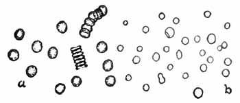
FIG. 52.—Blood-corpuscles: a, Normal; b, abnormal (Ogden).
When not due to contamination from menstrual discharge, blood in the urine, or hematuria, is always pathologic. Blood comes from the kidney tubules in severe hyperemia, in some forms of nephritis, and in renal tuberculosis and malignant disease. The finding of blood-casts is the only certain means of diagnosing the kidney as its source. Blood comes from the pelvis of the kidney in renal calculus (Fig. 62), and is then usually intermittent, small in amount, and accompanied by a little pus and perhaps crystals of the substance forming the stone. Considerable hemorrhages from the bladder may occur in vesical calculus, tuberculosis, and newgrowths. Small amounts of blood generally accompany acute cystitis.
5. Spermatozoa are generally present in the urine of men after nocturnal emissions, after epileptic convulsions, and in spermatorrhea. They may be found in the urine of both sexes following coitus. They are easily recognized from their characteristic structure (Fig. 53). The one-sixth objective should be used, with subdued light and careful focusing.
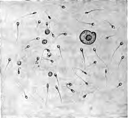
FIG. 53.—Spermatozoa in urine (Ogden).
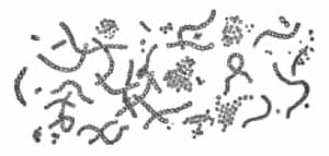
FIG. 54.—Micrococcus ureæ (after von Jaksch).
6. Bacteria.—Normal urine is free from bacteria in the bladder, but becomes contaminated in passing through the urethra. Various non-pathogenic bacteria, notably Micrococcus ureæ (Fig. 54), are always present in decomposing urine. In suppurations of the urinary tract pus-producing organisms may be found. In many infectious diseases the specific germs may be eliminated in the urine without producing any local lesion. Typhoid bacilli have been known to persist for months and even years after the attack.
Bacteria produce a cloudiness which will not clear upon filtration. They are easily seen with the one-sixth objective in the routine microscopic examination. Ordinarily, no attempt is made to identify any but the tubercle bacillus and the gonococcus.
Tubercle bacilli are nearly always present in the urine when tuberculosis exists in any part of the urinary tract, but are often difficult to find, especially when the urine contains little or no pus.
Detection of Tubercle Bacilli in Urine.—The urine should be obtained by catheter after careful cleansing of the parts.
(1) Centrifugalize thoroughly, after dissolving any sediment of urates or phosphates by gentle heat or acetic acid. Pour off the supernatant fluid, add water, and centrifugalize again. Addition of one or two volumes of alcohol will favor centrifugalization by lowering the specific gravity.
(2) Make thin smears of the sediment, adding a little egg-albumen if necessary to make the smear adhere to the glass; dry, and fix in the usual way.
(3) Stain with carbol-fuchsin, steaming, for at least three minutes.
(4) Wash in water, and then in 20 per cent. nitric acid until only a faint pink color remains.
(5) Wash in water.
(6) Soak in alcohol fifteen minutes or longer. This decolorizes the smegma bacillus (p. 35), which is often present in the urine, and might easily be mistaken for the tubercle bacillus. It is unlikely, however, to be present in catheterized specimens. It is always safest to soak the smear in alcohol for several hours or over night, since some strains of the smegma bacillus are very resistant.
(7) Wash in water.
(8) Apply Löffler's methylene-blue solution one-half minute.
(9) Rinse in water, dry between filter-papers, and examine with the one-twelfth objective.
When the bacilli are scarce, the following method may be tried. It is applicable also to other fluids. If the fluid is not albuminous, add a little egg-albumen. Coagulate the albumen by gentle heat and centrifugalize. The bacilli will be carried down with the albumen. Separate the albumen, mix with artificial gastric juice (for preparation of which see test for pepsin, p. 222), and set in an incubator or warm place until digested. Finally, centrifugalize and stain as described above. The bacilli do not stain so well as in the ordinary methods.
A careful search of many smears may be necessary to find the bacilli. They usually lie in clusters (see Plate V). Failure to find them in suspicious cases should be followed by inoculation of guinea-pigs; this is the court of last appeal, and must also be sometimes resorted to in order to exclude the smegma bacillus.
In gonorrhoea gonococci are sometimes found in the sediment, but more commonly in the "gonorrheal threads," or "floaters." In themselves, these threads are by no means diagnostic of gonorrhea. Detection of the gonococcus is described later (p. 264).
PLATE V
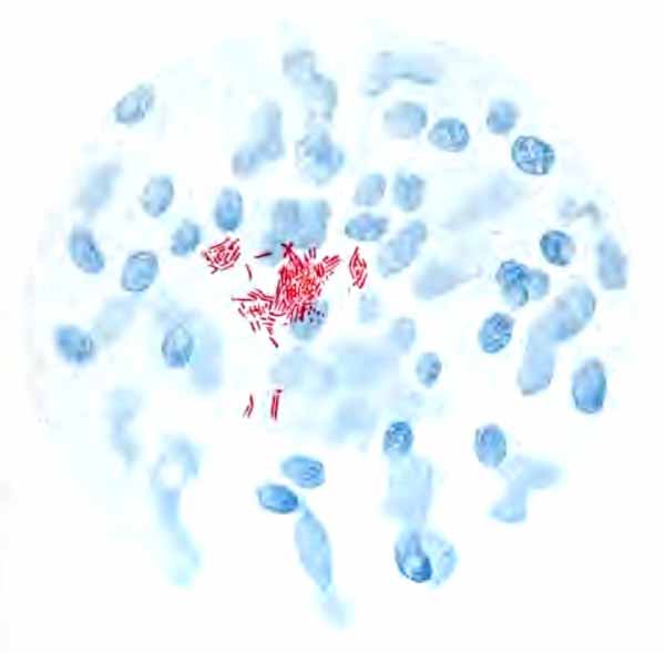
Tubercle bacilli in urinary sediment; X800 (Ogden).
7. Animal parasites are rare in the urine. Hooklets and scolices of Tænia echinococcus (Fig. 55) and embryos of Filaria sanguinis hominis have been met. In Africa the ova, and even adults, of Distoma hæmatobium are common, accompanying "Egyptian hematuria."
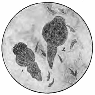
FIG. 55.—1, Scolex of tænia echinococcus, showing crown of hooklets; 2, scolex and detached hooklets (obj. one-sixth) (Boston).
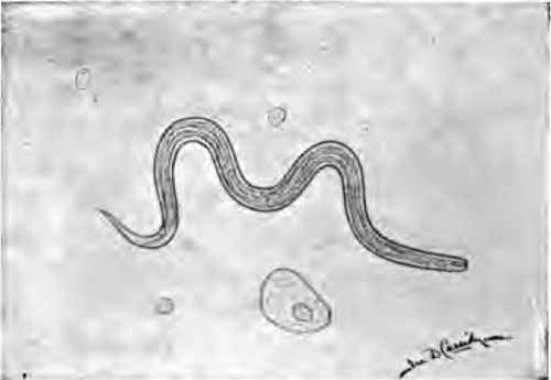
FIG. 56.—Embryo of "vinegar eel" in urine, from contamination; length, 340 µ; width, 15 µ. An epithelial cell from bladder and three leukocytes are also shown (studied with Dr. J. A. Wilder).
Other parasites, most of which are described in Chapter VI, may be present from contaminations. A worm which is especially interesting is Anguillula aceti, the "vinegar eel." This is generally present in the sediment of table vinegar, and may reach the urine through use of vinegar in vaginal douches, or through contamination of the bottle in which the urine is contained. It has been mistaken for Strongyloides intestinalis and for Filaria sanguinis hominis. It closely resembles the former in both adult and embryo stages. The young embryos have about the same length as filaria embryos, but are nearly twice as broad and the intestinal canal is easily seen (compare Figs. 56 and 107).
C. EXTRANEOUS STRUCTURES
The laboratory worker must familiarize himself with the microscopic appearance of the more common of the numerous structures which may be present from accidental contamination (Fig. 57).
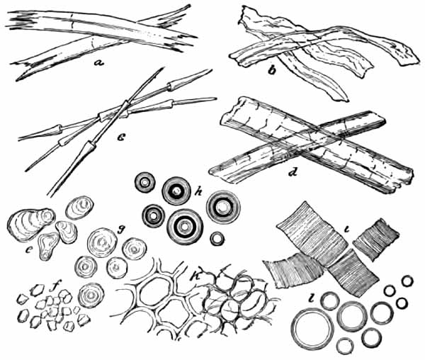
FIG. 57.—Extraneous matters found in urine: a, Flax-fibers; b, cotton-fibers; c, feathers; d, hairs; e, potato-starch; f, rice-starch granules; g, wheat-starch; h, air-bubbles; i, muscular tissue; k, vegetable tissue; l, oil-globules.
Yeast-cells are smooth, colorless, highly refractive, spheric or ovoid cells. They sometimes reach the size of a leukocyte, but are generally smaller (see Fig. 88, l). They might be mistaken by the inexperienced for red blood-corpuscles, fat-droplets, or the spheric crystals of calcium oxalate, but are distinguished by the facts that they are not of uniform size; that they tend to adhere in short chains; that small buds may often be seen adhering to the larger cells; and that they do not give the hemoglobin test, are not stained by osmic acid or Sudan but are colored brown by Lugol's solution, and are insoluble in acids and alkalis. Yeast-cells multiply rapidly in diabetic urine, and may reach the bladder and multiply there.
Mold fungi (Fig. 58) are characterized by refractive, jointed, or branched rods (hyphæ), often arranged in a network, and by highly refractive, spheric or ovoid spores. They are common in urine which has stood exposed to the air.
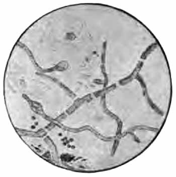
FIG. 58.—Aspergillus from urine (Boston).
Fibers of wool, cotton, linen, or silk, derived from towels, the clothing of the patient, or the dust in the air are present in almost every urine. Fat-droplets are most frequently derived from unclean bottles or oiled catheters. Starch-granules may reach the urine from towels, the clothing, or dusting-powders. They are recognized by their concentric striations and their blue color with iodin solution. Lycopodium granules (Fig. 59) may also reach the urine from dusting-powders. They might be mistaken for the ova of parasites. Bubbles of air are often confusing to beginners, but are easily recognized after once being seen. Scratches and flaws in the glass of slide or cover are likewise a common source of confusion to beginners.
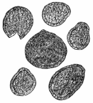
FIG. 59.—Granules of lycopodium (Saxe).
IV. THE URINE IN DISEASE
In this section the characteristics of the urine in those diseases which produce distinctive urinary changes will be briefly reviewed.
1. Renal Hyperemia.—Active hyperemia is usually an early stage of acute nephritis, but may occur independently as a result of temporary irritation. The urine is generally decreased in quantity, highly colored, and strongly acid. Albumin is always present—usually in traces only, but sometimes in considerable amount for a day or two. The sediment contains a few hyaline and finely granular casts and an occasional red blood-cell. In very severe hyperemia the urine approaches that of acute nephritis.
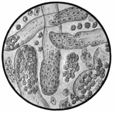
FIG. 60.—Sediment from acute hemorrhagic nephritis: Red blood-corpuscles; leukocytes; renal cells not fattily degenerated; epithelial and blood-casts (Jakob).
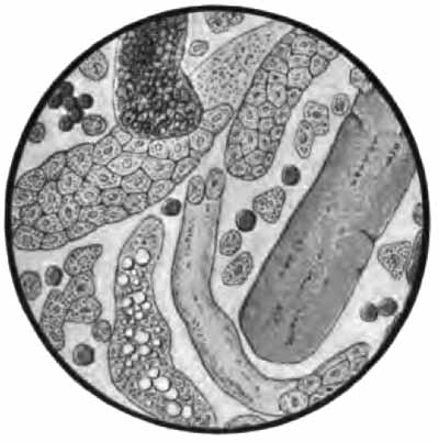
FIG. 61.—Sediment from chronic parenchymatous nephritis: hyaline (with cells attached), waxy, brown granular, fatty, and epithelial casts; fattily degenerated renal cells; and a few white and red blood-corpuscles (Jakob).
Passive hyperemia occurs most commonly in diseases of the heart and liver and in pregnancy. The quantity of urine is somewhat low and the color high, except in pregnancy. Albumin is present in small amount only. The sediment contains a very few hyaline or finely granular casts. In pregnancy the amount of albumin should be carefully watched, as any considerable quantity, and especially a rapid increase, strongly suggests approaching eclampsia.
2. Nephritis.—The various degenerative and inflammatory conditions grouped under the name of nephritis have certain features in common. The urine in all cases contains albumin and tube-casts, and in all well-marked cases shows a decrease of normal solids, especially of urea and the chlorids. The characteristics of the different forms are well shown in the table on opposite page, modified from Hill.
THE URINE IN NEPHRITIS
PHYSICAL. CHEMIC. MICROSCOPIC.
Acute nephritis. Quantity diminished, often very greatly. Color dark; may be red or smoky. Specific gravity, 1.020 to 1.030. Urea and chlorids low. Much albumin: up to 1.5 per cent. Reaction acid. Sediment abundant, red or brown. Many casts, chiefly granular, blood and epithelial varieties. Red blood-cells abundant. Numerous renal epithelial cells and leukocytes.
Chronic parenchymatous nephritis. Quantity usually diminished. Color variable, often pale and hazy. Specific gravity, 1.010 to 1.020. Urea and chlorids low. Largest amounts of albumin: up to 3 per cent. Reaction acid. Sediment rather abundant. Many casts of all varieties: fatty casts and casts of degenerated epithelium most characteristic. Blood present in traces: abundant only in acute exacerbations. Numerous fattily degenerated renal epithelial cells, often free globules of fat, and a few leukocytes.
Chronic interstitial nephritis. Quantity markedly increased, especially at night. Color pale, clear. Specific gravity, 1.005 to 1.015. Urea and chlorids low in well-marked cases. Albumin present in traces (often overlooked), increasing in late stages. Reaction acid. Sediment very slight. Few narrow hyaline and finely granular casts. No blood except in acute exacerbations. Very few renal cells. Uric acid and calcium-oxalate crystals common.
Amyloid degeneration of kidney. Quantity moderately increased. Color pale, clear. Specific gravity, 1.012 to 1.018. Slight decrease of urea and chlorids. Variable amounts of albumin and globulin. Sediment slight. Moderate number of hyaline, finely granular, and sometimes waxy casts.
3. Renal Tuberculosis.—The urine is pale, usually cloudy. The quantity may not be affected, but is apt to be increased. In early cases the reaction is faintly acid and there are traces of albumin and a few renal cells. In advanced cases the urine is alkaline, has an offensive odor, and is irritating to the bladder. Albumin in varying amounts is always present. Pus is nearly always present, though frequently not abundant. It is generally intimately mixed with the urine, and does not settle so quickly as the pus of cystitis. Casts, though present, are rarely abundant, and are obscured by the pus. Small amounts of blood are common. Tubercle bacilli are nearly always present, although animal inoculation may be necessary to detect them.
4. Renal Calculus.—The urine is usually somewhat concentrated, with high color and strongly acid reaction. Small amounts of albumin and a few casts may be present as a result of kidney irritation. Blood is frequently present, especially in the daytime and after severe exercise. Crystals of the substance composing the calculus—uric acid, calcium oxalate, cystin—may often be found. The presence of a calculus generally produces pyelitis, and variable amounts of pus then appear, the urine remaining acid in reaction.
5. Pyelitis.—In pyelitis the urine is slightly acid, and contains a small or moderate amount of pus, together with many spindle and caudate epithelial cells. Pus-casts may appear if the process extends up into the kidney tubules (see Fig. 62). Albumin is always present, and its amount, in proportion to the amount of pus, is decidedly greater than is found in cystitis.
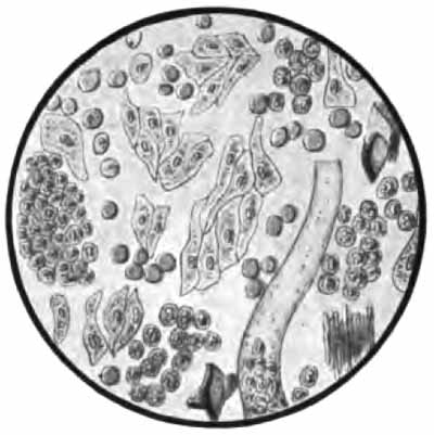
FIG. 62.—Sediment from calculous pyelitis: numerous pus-corpuscles, red blood-corpuscles, and caudate and irregular epithelial cells; a combination of hyaline and pus-cast; and a few uric-acid crystals (Jakob).
6. Cystitis.—-In acute and subacute cases the urine is acid and contains a variable amount of pus, with many epithelial cells from the bladder—chiefly large round, pyriform, and rounded squamous cells. Red blood-corpuscles are often numerous.
In chronic cases the urine is generally alkaline. It is pale and cloudy from the presence of pus, which is abundant and settles readily into a viscid sediment. The sediment usually contains abundant amorphous phosphates and crystals of triple phosphate and ammonium urate. Vesical epithelium is common. Numerous bacteria are always present (see Fig. 63).
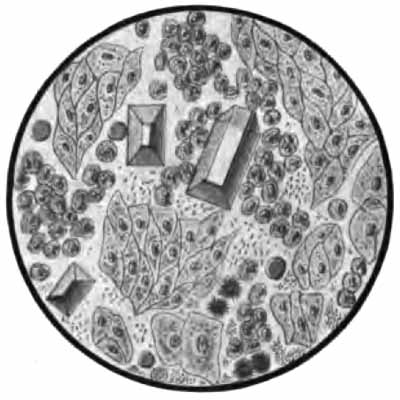
FIG. 63.—Sediment from cystitis (chronic): numerous pus-corpuscles, epithelial cells from the bladder, and bacteria; a few red blood-corpuscles and triple phosphate and ammonium urate crystals (Jakob).
7. Vesical Calculus, Tumors, and Tuberculosis.—These conditions produce a chronic cystitis, with its characteristic urine. Blood, however, is more frequently present and more abundant than in ordinary cystitis. With neoplasms, especially, considerable hemorrhages are apt to occur. Particles of the tumor are sometimes passed with the urine. No diagnosis can be made from the presence of isolated tumor cells. In tuberculosis tubercle bacilli can generally be detected.
8. Diabetes Insipidus.—Characteristic of this disease is the continued excretion of very large quantities of pale, watery urine, containing neither albumin nor sugar. The specific gravity varies between 1.001 and 1.005. The daily output of solids, especially urea, is increased.
9. Diabetes Mellitus.—The quantity of urine is very large. The color is generally pale, while the specific gravity is nearly always high—1.030 to 1.050, very rarely below 1.020. The presence of glucose is the essential feature of the disease. The amount of glucose is often very great, sometimes exceeding 8 per cent., while the total elimination may exceed 500 gm. in twenty-four hours. It may be absent temporarily. Acetone is generally present in advanced cases. Diacetic acid may be present, and usually warrants an unfavorable prognosis.