TUNICATA.
Reading edition
Wall Case to left of main entrance to Shell Gallery.
The Tunicata are marine animals, the majority of which live, in their adult stage, a stationary life, fixed to the rocks or sea-bottom, but a comparatively small number are free-swimming.
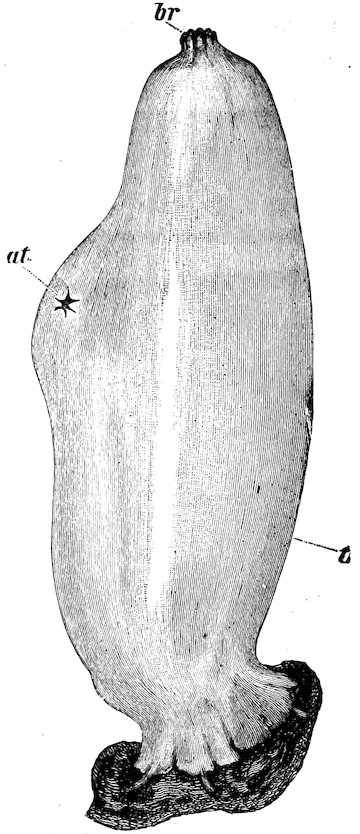
Fig. 1. Ascidia mentula from the right side. at, atrial aperture; br, branchial aperture; t, test. [After Herdman: Tunicata, Encyc. Britannica.]
They occur in the form of cartilaginous or leathery sacs, fleshy incrustations, solid fleshy masses, free-swimming, barrel-shaped animals, solitary or united into chains or hollow cylinders; or, lastly, of minute free-swimming tadpole-shaped organisms. To explain briefly the structure of a Tunicate, Ascidia mentula (Fig. 1), is selected. The 84animal, which lives on a muddy bottom, in from five to twenty fathoms, resembles a conical sac fixed by the broader end, of grayish green colour and about 4 inches in height. At the narrower end are two orifices, one terminal—the branchial orifice or mouth, and the other a little lower—the atrial orifice: the former has eight lobes and the latter six.
When the Ascidian is undisturbed, the orifices are wide open, and currents enter by the branchial and leave by the atrial orifice. On the least alarm, the orifices close, jets of water being at the same time squirted out; hence the popular name “Sea-squirts” given to these animals.
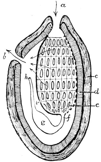
Fig. 2. Diagrammatic section of Ascidia representing the three sacs, and the branchial sac as the pharynx or throat. a, branchial; and b, atrial orifice; c, tunic or test; d, mantle; e, branchial sac; f, gullet; g, stomach; h, anal orifice; i, dorsal lamina; dotted line indicates the endostyle.
The Ascidian is orientated as follows: hold the animal with the branchial orifice pointing forwards and the atrial upwards; the branchial orifice will be anterior and the opposite end posterior; the atrial orifice will lie on the upper or dorsal aspect, the opposite aspect being lower or ventral, and the sides right and left. The aspects, in fact, correspond with those of a vertebrate animal. A 85vertical section roughly shows the animal to be formed of three concentric sacs (Figs. 2, 3). The outermost, which is tough and membranous, is called the Test or Tunic, the whole group owing its name to the presence of this protective covering.
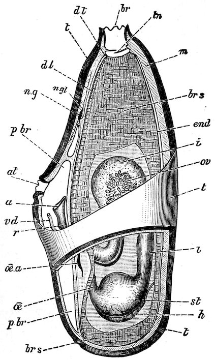
Fig. 3. Diagrammatic dissection of A. mentula. at, atrial orifice; br, branchial orifice; a, anal orifice; brs, branchial sac; dl, dorsal lamina; end, endostyle; m, mantle; ng, nerve ganglion; oea, orifice of gullet; pbr, peribranchial cavity; st, stomach; t, test; tn, tentacles. (After Herdman: Tunicata, Encyc. Britannica.)
The middle sac, termed the Mantle, which almost corresponds in shape to the outer, is composed of connective tissue, muscle-fibres, blood-vessels, etc.; in spirit specimens, the mantle is shrunk away from the test except at the orifices and at a point behind, where vessels enter the test.
The innermost or Branchial Sac is attached behind the branchial orifice and along the ventral edge, but otherwise hangs free in the 86interior, the space around and outside of the sac being termed the atrial or peribranchial cavity.
The delicate walls of the branchial sac, which resemble fine muslin, are perforated by innumerable vertical slits, termed stigmata, arranged in transverse rows (Fig. 4).
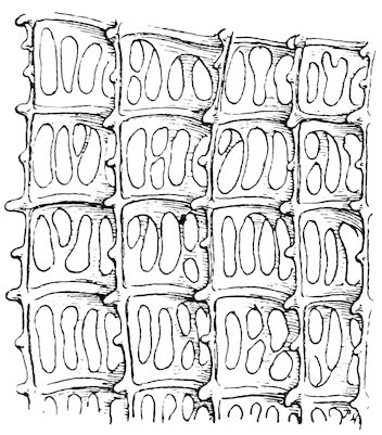
Fig. 4. Ascidia mentula. Part of wall of branchial sac showing stigmata. Magnified.
The margins of the stigmata are lined with cilia which set up currents; and the water which enters by the branchial orifice, passes through the stigmata into the atrial cavity, and thence out through the atrial orifice. The walls of the branchial sac are chiefly composed of a sieve-like meshwork of fine blood-vessels arranged in transverse and longitudinal rows. The currents of water passing through the stigmata aërate the blood in the vessels. Besides the stigmata, the branchial sac has two relatively large orifices, viz., the branchial orifice or mouth, and, at the opposite end, the opening into the gullet. The branchial sac is, in fact, a capacious throat or pharynx (Diagram Fig. 2 and Fig. 14). Inside the branchial orifice is a circle of fine tentacles, which guard the entrance to the branchial sac. The food of the animal consists of minute animal and vegetable organisms.
It may be wondered how this food is secured, seeing that the currents of water are continually passing through the sieve-like walls of the branchial sac to the exterior again. Within the branchial orifice and above the branchial sac are two circular ciliated ridges with a groove between, which is full of viscid secretion; the cilia on the ridges direct particles into the groove where they are retained by the mucus.
87Passing backwards along the ventral edge of the branchial sac is a thick-lipped furrow, which appears like a rod in the thin-walled sac, and hence is called the endostyle. This organ secretes the mucus which is carried up by ciliary action to the circular groove in front of the branchial sac, and thence to the gullet along a fold or crest, termed the dorsal lamina, situated along the dorsal edge of the branchial sac.
The gullet opens into a large stomach situated posteriorly on the left side of the branchial sac. The stomach opens into the intestine, which, after forming a loop, terminates in the anal orifice or vent opening into the atrial cavity.
The tubular heart lies below the stomach, a remarkable feature in the circulation consisting in the periodic reversal of the blood current. An elongated nerve ganglion is situated between the branchial and atrial orifices.
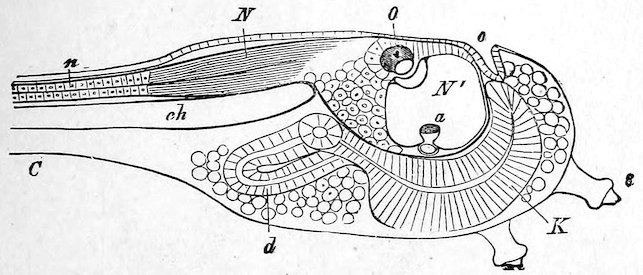
Fig. 5. Ascidian Tadpole with part only of the tail C. Magnified section. N, nervous system with enlarged brain in front and narrow spinal cord behind n; N′, cavity of brain; O, the single cerebral eye lying in the brain; a, auditory organ; K, pharynx; d, intestines; o, rudiment of mouth; ch, notochord or primitive backbone. (From Gegenbaur’s ‘Elements of Comparative Anatomy.’)
Ascidia mentula is hermaphrodite. The egg develops into a minute tadpole-like larva which swims about by means of its tail. Water entering by the mouth passes out through the gill-slits. A nerve-tube extending along the back and tail is swollen in front into a brain-vesicle; and underneath the long nerve-tube behind the brain is a stiff skeletal rod or axis—the notochord—which constitutes the rudiment of a backbone. Inside the brain are two unpaired sense organs, an eye and an organ of hearing (Fig. 5). After swimming freely for a few hours, the larva settles down head foremost and 88fixes itself by papillæ on the anterior end (Figs. 6, 7). Presently the tail becomes absorbed, and the posterior end of the nerve-tube, and the brain with its eye and hearing organ, undergo atrophy, the nerve ganglion of the adult alone representing the cerebrospinal axis of the larva. The branchial sac and intestines develop greatly, and growth proceeds in such a manner that the mouth is pushed round to a position opposite to the fixed area, and gradually the animal becomes the adult ascidian.
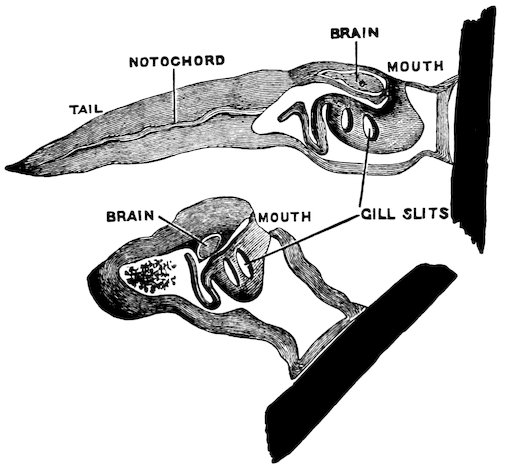
Fig. 6. Degeneration of Ascidian Tadpole to form the adult. The black pieces represent the rock or stone to which the Tadpole has fixed its head.
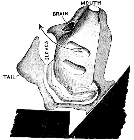
Fig. 7. Very young Ascidian with only two gill-slits. (Figs. 6, 7, from Lankester’s ‘Degeneration.’)
89This wonderful metamorphosis presents a striking example of DEGENERATION resulting from the adoption of a fixed mode of life. The active free-swimming larva with its brain, eye, hearing organ, and muscular tail becomes transformed into a comparatively inert sac.
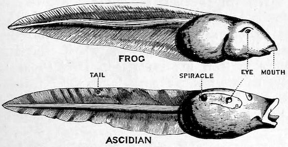
Fig. 8. Tadpole of Frog and Ascidian. Surface view. (Lankester’s ‘Degeneration.’)
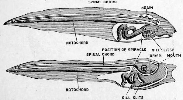
Fig. 9. Tadpole of Frog and Ascidian. Diagram representing the chief internal organs. (Lankester’s ‘Degeneration. A chapter in Darwinism.’)
The tadpole of an Ascidian resembles that of a frog (Figs. 8, 9), not merely superficially, but also in its general structure and mode of development. The Tunicata are now generally regarded as a degenerate offshoot from the ancestral stock of the Vertebrata, in that the larva possesses a skeletal rod (rudimentary backbone) separating the dorsally situated nerve-tube (cerebrospinal axis) from the ventrally situated intestinal tube, the existence of the cerebral 90eye in the Ascidian tadpole further tending to confirm the truth of this theory. Apart from a knowledge of the course of their development, Tunicata would have been classed among the Invertebrata, but the structure of the larva clearly reveals the affinities of the group to the backboned animals.
Ascidia mentula belongs to the group of Simple Ascidians which are all fixed, and are either solitary or joined into colonies in which each individual or ascidiozooid has a distinct test of its own. In the Compound Ascidians, which form colonies by budding, the ascidiozooids are buried in a common investing mass and have no separate tests. In a third group, the Salpa-like Ascidians, the ascidiozooids are united to form free-swimming colonies shaped like hollow cylinders open at one end. The above three groups belong to one great Order—the Ascidiacea. A second Order, Thaliacea, includes the free-swimming Salpa and Doliolum, which exhibit alternation of generations in their life history. A third Order Larvacea, includes very minute free-swimming forms which possess a tail in the adult stage. There are sixteen families of Tunicata.
The following is a tabular view of Prof. Herdman’s classifications:—
Order I. Ascidiacea. Sub-order 1. Ascidiæ Simplices, 4 Families.
Sub-order 2. „ Compositæ, 7 Families.
Sub-order 3. „ Salpiformes, 1 Family.
Order II. Thaliacea 3 Families.
Order III. Larvacea 1 Family.
Order I.—Ascidiacea.
The Ascidiacea include the great majority of species. With the exception of the one genus Pyrosoma, they lead a fixed or stationary life.
Sub-order 1.—Ascidiæ Simplices.
The Simple Ascidians are mostly solitary; in a few forms, however, colonies arise by budding from stolons, but each individual has a distinct test. The four families into which the sub-order is divided are chiefly characterised by the nature of the test, the number of lobes round the branchial and atrial orifice, and the character of the branchial sac.
In the family Molgulidæ the tough membranous test is often coated with sand; the branchial aperture is six-lobed, the atrial four-lobed, the branchial sac has long folds or pleats, and the stigmata are curved or arranged in spirals.
91Molgula gigantea, which is one of the largest of the Ascidians, and which attains a length of over thirteen inches, forms a tough conical sac; the branchial and atrial orifices at the upper end have six and four lobes respectively. The test is leathery, smooth above, but coated with sand below. The exhibited specimen, which comes from the Straits of Magellan, has several specimens of the stalked Boltenia legumen attached to the lower part of the test.
The curious Molgula oculata (Fig. 10) has a soft oval or rounded body coated with sand. The branchial and atrial orifices have respectively six and four lobes. Specimens grow attached to the rocks and also live free in the sand. The surface of the test is provided with hairs, which adhere to the rocks and collect particles of sand. The adhesion not being very firm, specimens are easily detached by currents and collected into heaps by the eddies; when living in the sand only the two dark orifices are visible. The sand coating has been supposed to confer protection by mimicry of the environment; but Professor Lacaze Duthiers found, much to his chagrin, that the sandy tests of his specimens were of no avail in securing them from being devoured by crabs who seemed to scent their prey from afar.
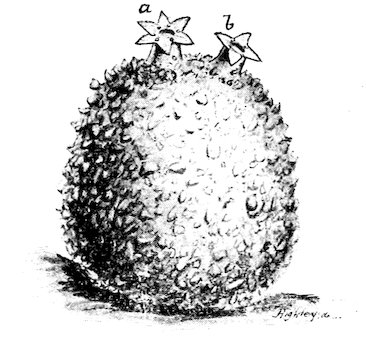
Fig. 10. Mogula oculata. a, branchial; b, atrial orifice.
In the family Cynthiidæ the test is usually leathery, the branchial and atrial apertures four-lobed, and the branchial sac folded into longitudinal pleats.
92The genera Boltenia and Culeolus include species in which the body is attached to a peduncle.
The large exhibited specimen of Boltenia pachydermatina is 28 inches in length, the head being 4 and the stalk 24 inches long. The two four-lobed apertures are along one edge, the branchial being the lower; the body is marked with long deep furrows, and the stalk with transverse wrinkles. Culeolus perlucidus, from 1600 fathoms in the Southern Ocean, is in the form of a small pear-shaped head on a slender stalk, the total length being 4½ inches. The branchial orifice forms a transverse slit with raised lips near the stalk, the slit-like atrial orifice being near the rounded end of the body. Culeolus moseleyi, another slender-stalked form, was obtained from 2425 fathoms in the Central Pacific.
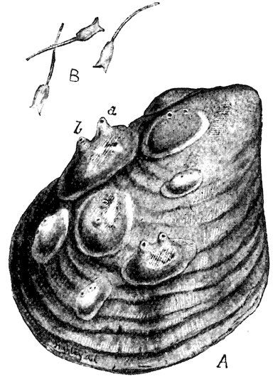
Fig. 11. A. Styelopsis grossularia on shell. B. Tadpoles of same, × 9. a, branchial; b, atrial orifice. (B, after Sir J. Dalyell.)
The little Cynthiid Styelopsis grossularia (Fig. 11), popularly known as the “Currant Squirter,” occurs in the form of bright red hemispherical blobs on stones and shells; when undisturbed, the branchial and atrial orifices expand and project upwards. The eggs are brilliant red in colour. Sir John Dalyell was the first to discover the tadpole form, which is about ⅒ inch long (Fig. 11, B), and to 93observe the tadpoles become fixed and develop into fixed Ascidians. He calls the active little swimming larvæ “Spinulæ,” from their resemblance to small pins.
The family Ascidiidæ includes forms with a gelatinous or cartilaginous test; the branchial and atrial orifices usually have 8 and 6 lobes respectively; the branchial sac is without folds.
Ascidia mentula, described above, belongs to this family.
Chelyosoma is characterised by the test forming tortoise-like horny plates on the upper surface. The exhibited specimen of C. macleayanum (Fig. 12) comes from Greenland; the upper hemispherical part of the test is divided into 8 plates; the branchial and atrial orifices are situated in the joints between the plates.
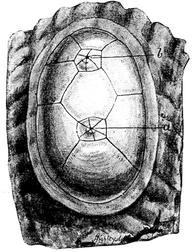
Fig. 12. Chelyosoma macleayanum, slightly enlarged. a, branchial; b, atrial orifice.
The fine specimen of Phallusia mammillata from Naples consists of several individuals partly fused together; the branchial and atrial orifices are wide open, and the mantle can be seen through the thick knobby translucent test.
In Rhodosoma the test is modified so as to form stiff plates recalling the valves of a bivalve shell. One plate is attached to the rocks, the other closing against the first like a lid; the anterior end of the animal with its branchial and atrial orifices is visible only when the 94lid is open. The Mediterranean species R. callense (Fig. 13) grows attached to the rocks. The little exhibited specimen is on a fragment of shell in front of a black patch. The figure shows specimens with the lid open and closed.
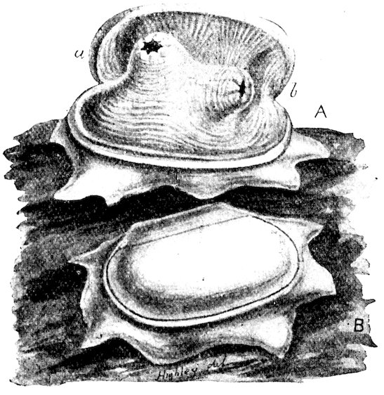
Fig. 13. Rhodosoma callense, × 10. A, “valve” open; B, shut. a, branchial; b, atrial orifice. (After Lacaze Duthiers.)
Family Clavelinidæ. The body is attached to a creeping stolon or mass of stolons, from which new individuals arise by budding. The other three families of Simple Ascidians included solitary forms, but the Clavelinidæ are social, and form colonies wherein each individual has its own test.
Clavelina lepadiformis (Fig. 14) forms graceful crystal vases about an inch in height. The figure shows one individual, but usually the processes at the base extend out as stolons whence other individuals arise.
Diazona violacea, from Cornwall, forms beautiful purple disk-shaped colonies in which the ascidiozooids arise from a basal mass of stolons. Sometimes the ascidiozooids die down, leaving only a smooth violet pad, which in due time produces a new crop of ascidiozooids.
95
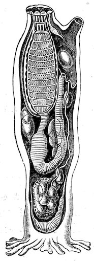
Fig. 14. Clavelina lepadiformis; diagrammatic, showing the anatomy. The oval bodies are the eggs; at lower end lies the tubular heart; the root-like processes at the base grow into stolons, whence other ascidiozooids arise.
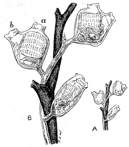
Fig. 15. Perophora listeri; A, slightly, B, further magnified. Ascidiozooids in right, left, and lateral aspects. a, branchial; b, atrial orifice.
96The remarkable Rhopalæa neapolitana, from Naples, may be roughly compared to an hour-glass with a very long constriction. The test is smooth in the upper part, but knobby and encrusted with foreign bodies below. The upper or thoracic end contains the branchial sac, and the lower or abdominal portion the stomach, heart, and reproductive organs, the gullet and intestine traversing the whole length of the narrow central region. Although from its general structure Rhopalæa is a Clavelinid, it is not certainly known to produce buds.
Perophora listeri (Fig. 15) occurs in the form of little jelly-like transparent blobs rising by short stalks from a silvery thread-like stolon. Owing to their small size and transparency, it is possible to examine specimens alive under the microscope, the currents passing through the stigmata in the walls of the branchial sac, and the beating of the heart being distinctly visible. The rapid motion of the cilia surrounding stigmata gives the appearance of dark wheels all rotating in the same direction. The heart beats so as to drive the blood current so many times in one direction, and then after a short pause, in the reverse direction.
The exhibited specimen growing on an oyster shell, is from Plymouth.
Sub-order 2.—Ascidiæ Compositæ.
The Compound Ascidians are fixed forms, which give rise to colonies by budding, the individuals being immersed in a common mass and not possessing separate tests.
Although reduced to an extremely small size each individual or ascidiozooid of a colony possesses the same organs as a large Simple Ascidian, excepting that the former does not possess a separate test. Frequently the individuals of a colony are grouped into systems, in which the atrial orifices open into a common cloaca. The little ascidiozooids vary greatly in shape in the different families. In the Polyclinidæ, for instance, they are long, the organs being so to speak, drawn out, and being arranged in three regions, the thoracic, abdominal and post-abdominal, the first region containing the branchial sac, the second the stomach, and the third the heart and reproductive organs. In the Distomidæ, the body exhibits two regions, thoracic and abdominal, the heart and reproductive organs lying alongside of the stomach. The Botryllidæ comprise only one region, the stomach and the other organs being situated by the side of the branchial sac.
97The Compound Ascidians include seven families which are characterised chiefly by the method of bud formation, and by the arrangement of the organs into one, two, or three regions.
It is only possible, from limits of space, to refer to a few interesting forms.
The species of Botryllus are those most commonly met with. They form richly coloured gelatinous incrustations on rocks and seaweeds. B. violaceus (Figs. 16, 17, and 18 D) is blue with white lines; B. smaragdus, green; B. marionis, brown with white and carmine; B. castaneus, purple, and so on. The individuals are arranged in circular systems with the branchial orifices round the circumference and the atrial orifices opening into a common central cavity (Fig. 17), the whole colony being composed of groups of systems.
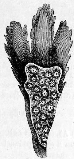
Fig. 16. Botryllus violaceus on seaweed. (After H. Milne-Edwards.)
The exhibited specimen of B. violaceus was grown in the tanks of the Biological Station at Plymouth. The red specimen of B. aurolineatus, from Naples, shows well the branchial and cloacal orifices. In Botrylloides, the individuals form elliptical or elongated systems.
Colella thomsoni was obtained near the Philippines at a depth of 10 fathoms. The specimen, which is about 7 inches in length, 98resembles an elongated head of clover on a thickened stalk. The individuals which compose the head are arranged in spiral lines, the atrial orifice of each ascidiozooid opening separately and not into a common cloaca.
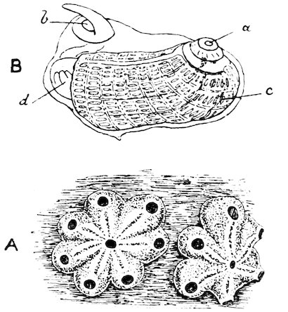
Fig. 17. A. Botryllus violaceus, magnified, showing two systems of 6 and 7 ascidiozooids. B. One ascidiozooid extracted. a, branchial; b, atrial orifices; c, branchial sac; d, stomach. (After H. Milne-Edwards.)
Colella quoyi (Fig. 18 A), from 25 fathoms off Kerguelen Island, forms a rounded head on a short peduncle, the total height being one inch. The ascidiozooids are arranged in vertical lines in the “head,” each line consisting of a double zigzag series.
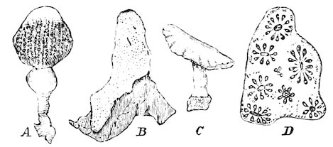
Fig. 18. Colonies of Ascidiæ compositæ, natural size. A. Colella quoyi. B. Leptoclinum neglectum. C. Pharyngodictyon mirabile. D. Botryllus. (After Herdman, Challenger Report and Encyclopædia Britannica.)
Julinia ignota, from the Antarctic regions, forms long narrow 99colonies, which attain a length of nearly three feet. One end is attached, the rest of the colony apparently lying along the sea-bottom.
Amaroucium roseum from Naples forms translucent gelatinous masses; a slice is exhibited, showing the long slender ascidiozooids immersed in the mass.
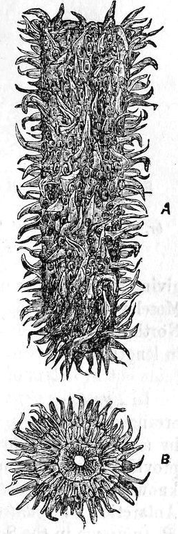
Fig. 19. Pyrosoma elegans, natural size. A. Side view of entire colony. B. End view of open extremity. (Herdman: Tunicata, Encyclopædia Britannica.)
Pharyngodictyon mirabile (Fig. 18 C), from 1600 fathoms in the Southern Indian Ocean, resembles a small mushroom, and is about one inch in height. This species is one of the few deep-sea Compound Ascidians.
Leptoclinum albidum is a common and widely distributed species; it occurs in the form of thin white crusts. The glistening white appearance is due to the common test being densely crowded with minute stellate spicules of carbonate of lime.
The specimen of Leptoclinum neglectum (Fig. 18 B) encrusts a fragment of sponge.
Goodsiria pedunculata from the Straits of Magellan, forms a rounded cartilaginous mass attached by a short peduncle; sometimes several masses are attached to each other. Each of the small dark oval areas on the surface corresponds to the branchial and atrial orifices of one ascidiozooid.
Sub-order 3.—Ascidæ Salpiformes.
The Salpiform Ascidians comprise only one genus, Pyrosoma, which occurs in the form of free-swimming colonies shaped like hollow cylinders closed and rounded at one end and open and truncate at the other (Fig. 19). The wall of the cylinder is formed of a single layer of ascidiozooids (Fig. 20), so arranged that all the atrial orifices open into the interior of the cylinder, and all the branchial orifices on the exterior, the two kinds of orifices being at opposite ends of the body, and not close together, as in most simple and compound Ascidians.
100Specimens vary in size from a few inches to upwards of four feet in length, and, as the name of the genus implies,[22] they are brilliantly phosphorescent. Sometimes they occur in innumerable multitudes, giving rise to a zone of greenish light extending for miles. Professor Moseley records that during the voyage of the Challenger in the North Atlantic a huge specimen of Pyrosoma spinosum, four feet in length, was captured. On tracing his name on its body, the word came out in letters of fire.
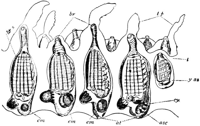
Fig. 20. Section through wall of Pyrosoma, magnified, showing a single layer of ascidiozooids. br, branchial; at, atrial orifice; tp, process of the test; br s, branchial sac. (Herdman: Tunicata, Encyclopædia Britannica.)
In Pyrosoma elegans (exhibited), from Naples, the ascidiozooids are arranged in verticils, and the mouth of the cylinder is surrounded by a movable diaphragm; the outer end of each ascidiozooid is provided with a membranous spine. Six species of Pyrosoma are known. Pyrosoma atlanticum is found in the tropical Atlantic and Antarctic; P. giganteum in the Atlantic, Pacific, and Antarctic; and P. spinosum in the South Atlantic.
Order II.—Thaliacea.
The Thaliacea are free-swimming Tunicates, which exhibit alternation of generations in their life history. There are three families, Salpidæ, Octacnemidæ, and Doliolidæ.
Salpidæ.—The Salpas are transparent barrel-shaped organisms 101which occur in abundance at the ocean surface. They are so transparent that they are rarely seen, except in calm weather from the side of small boats; yet they frequently swarm in countless multitudes. From five to ten bands of muscles partially or entirely surround the body, like hoops. The branchial and atrial openings are at or near the opposite ends of the body. The branchial sac has almost disappeared, the dorsal lamina and ventral gutter (or endostyle) alone remaining, the interval between the two on each side representing an enormous stigma; the dorsal lamina, or “gill” is the transversely striated band passing obliquely across the body and forming the only barrier between the branchial and atrial cavities. Water enters at the mouth, and, by the contraction of the muscle-hoops, is driven out through the atrial aperture at the opposite end, which is then closed by a sphincter muscle. The elastic walls of the body expand, and water again enters through the mouth, the valve-like lips of which prevent its being driven out that way. The Salpa swims along in jerks, and along with each gulp of water takes in Radiolaria, Foraminifera, etc., which are retained by the mucus of the endostyle and carried to the gullet. The Salpa, in fact, lives, as Professor Brooks observes, in a “living broth,” so abundant is the food supply.
The intestines usually form an oval mass termed the “nucleus,” which is a conspicuous object at the posterior end.
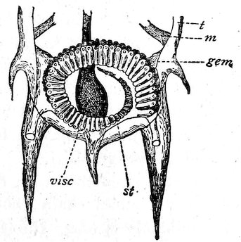
Fig. 21. Posterior part of solitary form of Salpa democratica-mucronata, showing a chain of embryos nearly ready to be set free. gem, young chain of Salpæ; st, stolon; t, test; visc, visceral mass.
The solitary Salpa above described is asexual. In the ventral region of its body it forms a stolon which becomes segmented into a series of buds (Fig. 21). As the stolon grows the end series of buds 102breaks off in the form of a chain and swims away, other chains being detached in succession. A chain is formed of individuals arranged in two rows, the individuals in each row being alternate (not opposite).
Each individual of a chain differs from the solitary individual in shape, arrangement of muscle bands, etc., but especially in having reproductive organs. The chain Salpid is hermaphrodite; the embryo develops into a solitary asexual Salpa which produces the chains by budding. The wonderful life history of Salpa was discovered by the poet Chamisso during a voyage round the world in 1819. He observes: “A Salpa mother is not like its daughter or its own mother, but resembles its sister, its granddaughter, and its grandmother.” Here we have an example of “alternation of generations,” a sexual generation (chain form) giving rise to an asexual generation (solitary form), which latter produces the sexual generation.[23]
Most of the species of Salpa have double names owing to the chain and solitary forms having been regarded as distinct species before they were known to be phases in the life history of one and the same species. Salpa runcinata-fusiformis, solitary form (Fig. 22 B), is barrel-shaped, truncated at each end, with terminal orifices, and with nine muscle-bands on the dorsal surface, some of which converge towards each other. An individual of a chain (Fig. 22 A) is fusiform, with six muscle-bands, and with the orifices not terminal, but at each end of the dorsal surface.
The solitary form of S. africana-maxima is barrel-shaped, with truncated ends and terminal orifices, and with nine broad parallel muscle-bands. The chain form is conical at one end, with six bands, and with orifices on the dorsal surface. The exhibited specimen of the chain form, which is in an early stage of growth, contains 202 individuals. The solitary and chain individuals of Salpa costata-tilesii attain a length of six to eight inches. The solitary form has eighteen muscle-bands and two large spines at the posterior end. The individual of the chain has five muscle-bands. A chain of three individuals is exhibited.
Salpa pinnata produces a circular chain; the exhibited specimen of the solitary form shows a small chain about to be detached; a circular chain of six individuals is also exhibited. Species of Salpa abound in all seas, but specimens from Naples have alone been exhibited on account of their good preservation.
103Family Octacnemidæ includes O. bythius, a deep-sea Salpid, in which the body forms a flattened disk produced into eight radiating lobes.
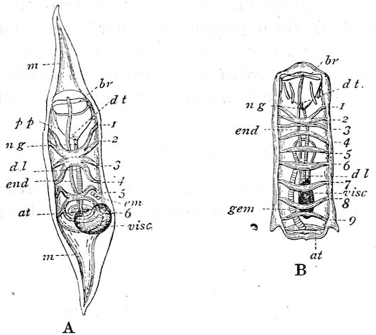
Fig. 22. Salpa runcinata-fusiformis. A. Chain form. B. Solitary form. 1–9, muscle bands; em, embryo; m, mantle; visc, visceral mass or nucleus. (Herdman: Tunicata, Encyclopædia Britannica.)
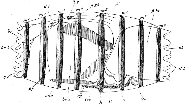
Fig. 23. Doliolum denticulatum, sexual generation, from the left side. m1-m8 muscle bands; at, atrial; br, branchial apertures; br s, branchial sac; sg, stigmata; st, stomach; ng, nerve ganglion; so, sense organs. (After Herdman, Encyclopædia Britannica.)
Family Doliolidæ. The body is cask-shaped and surrounded by circular hoops. The branchial and atrial orifices are at the opposite 104ends. The branchial sac is pierced by two oblique bands of stigmata (Fig. 23 sg). The life history is very complicated. The egg develops into a tailed larva, which develops into a “nurse”; the latter is asexual, and produces three kinds of buds on a stolon, viz. (1) nutritive buds which provide the “nurse” with food, (2) foster forms which are set free as cask-shaped bodies with eight broad muscle-bands, and (3) sexual forms which are attached for a time to the foster forms, but which later become free and give rise to the egg.
Order III.—Larvacea.
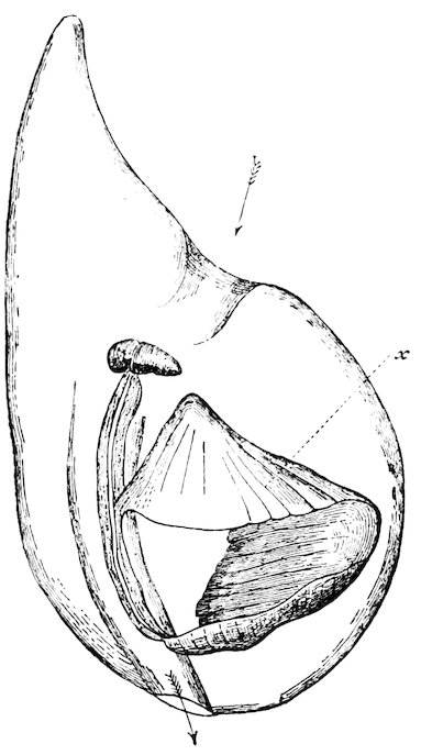
Fig. 24. Oikopleura cophocerca in its “house” (after Fol); seen from right side, × 6. Arrows indicate course of the water; x, lateral reticulated parts of the “house.”
The Larvacea are very minute Tunicata which live at the surface and swim by means of a tail-like appendage, resembling in this and certain other respects the tadpole larva of other Tunicata. They are able to form a temporary test or “house” many times larger than the body (Fig. 24). The organism itself, which is almost lost 105in its large test, is the little hammer-shaped body in the centre of the figure; the streaked areas bound a space in which the tail lashes vigorously. The animal can leave its test and secrete another in a few hours.
The tail is attached to the under or ventral surface of the tiny little barrel-shaped body, and usually points forwards; a skeletal rod, the urochord, runs along its length. The branchial sac has two ciliated openings or gill-clefts leading directly to the exterior, and not opening like the stigmata of the other orders into an atrial cavity.
The order contains one family, the Appendiculariidæ, and four genera, and is represented in all seas.
Oikopleura cophocerca, one of the largest forms, is about half an inch in length. The exhibited specimens came from St. Andrews, Fife. Professor McIntosh reports that occasionally specimens of this species occur in immense quantities, the tow-nets being filled with them.
106