MUSCLES
Reading edition
In the accounts of the muscles the name used by Hudson, et al. (1959) for each muscle is given in parentheses after the name used by me if the two differ.
In the account of each muscle, the description of the condition found in most specimens of the Lesser Prairie Chicken (T. pallidicinctus) is given first. This is hereafter referred to as the typical condition for T. pallidicinctus. Then any individual variations found within this species are given. Under the heading T. cupido any constant differences between this species and typical T. pallidicinctus are given first, and any individual variations found within the species T. cupido (both subspecies considered together) are given second. Under the heading P. p. jamesi any constant differences between this subspecies and the typical condition for T. pallidicinctus (thus these differences are not necessarily constant between the two genera) are given first, and any individual variations found within the subspecies P. p. jamesi are given second.
In the bird embryo, according to the studies of Romer (1927) and Wortham (1948), the muscles within each segment of the leg differentiate from distinct dorsal or ventral mesenchymal masses. Presumably these represent the primitive dorsal extensor and ventral flexor muscle masses. The list below indicates the ontogenetic origin of the avian leg muscles, according to the studies of Romer and Wortham. The individual muscles are discussed in the order in which they are listed below.
Dorsal muscles of thigh
M. extensor iliotibialis lateralis M. extensor iliofibularis
M. extensor iliotibialis anticus M. piriformis
M. ambiens M. gluteus profundus
M. vastus lateralis M. iliacus
M. vastus medialis M. iliotrochantericus medius
M. femoritibialis internus M. psoas
Ventral muscles of thigh
M. flexor cruris lateralis M. adductor superficialis
M. flexor cruris medialis M. adductor profundus
M. caudofemoralis M. obturator
M. flexor ischiofemoralis M. femorocruralis
Ventral muscles of shank
M. gastrocnemius M. flexor perforatus digiti III
M. flexor perforans et perforatus digiti II M. flexor perforatus digiti II
M. flexor perforans et perforatus digiti III M. flexor hallucis longus
M. flexor perforatus digiti IV M. plantaris
M. flexor digitorum longus
M. popliteus
Dorsal muscles of shank
M. peroneus longus M. extensor digitorum longus
M. tibialis anticus M. peroneus brevis
Dorsal muscles of foot
M. extensor hallucis longus M. extensor proprius digiti III
M. abductor digiti II M. extensor brevis digiti IV
M. extensor brevis digiti III
Ventral muscles of foot
M. lumbricalis (M. adductor digiti II—not present)
M. abductor digiti IV (M. adductor digiti IV—not present)
M. flexor hallucis brevis
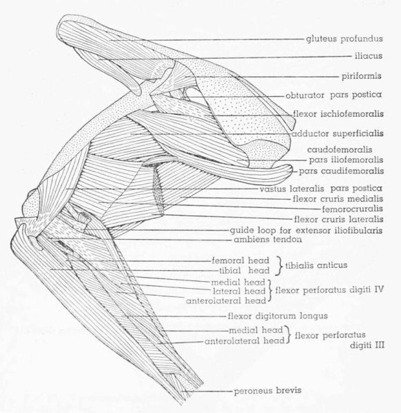
Fig. 12. Tympanuchus pallidicinctus 2L. Lateral view of the superficial muscles of the left leg. × 1.
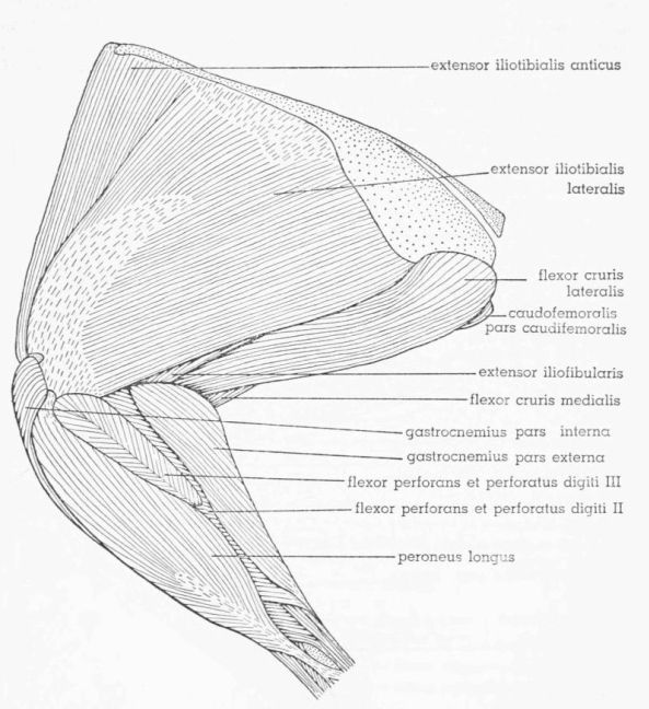
M. Extensor Iliotibialis Lateralis (M. iliotibialis), Figs. 12, 13, 20F, G
T. pallidicinctus
Fig. 13. Tympanuchus pallidicinctus 2L. Medial view of the superficial muscles of the left leg. × 1. Articular capsule shown by concentrically arranged dashes.
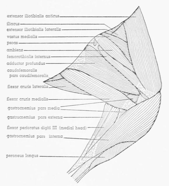
General Description and Relations.—Most superficial muscle on lateral surface of thigh; broad, flat, and triangular; bounded anteriorly by M. extensor iliotibialis anticus and posteriorly by M. flexor cruris lateralis; posterior part considerably thicker than anterior part; anteroproximal and centrodistal parts aponeurotic; extreme posteroproximal corner also aponeurotic (could be considered tough sheet of connective tissue intimately fused with M. extensor iliotibialis lateralis, rather than part of muscle itself; see Fig. 20F); latter aponeurosis, as well as adjacent fleshy fibers, overlapped by M. flexor cruris lateralis; this aponeurosis fused with posterior end of underlying M. caudofemoralis pars iliofemoralis; centrodistal aponeurosis tightly fused to underlying Mm. vastus lateralis and vastus medialis; fleshy fibers posterior to this aponeurosis also fused with M. vastus lateralis, although posterior third of muscle free; fleshy part anterior to this aponeurosis bound by tough connective tissue to underlying M. vastus medialis, although no fusion of fibers; anterior edge tightly bound by strong connective tissue to M. extensor iliotibialis anticus, with some fusion of fibers (proximally); posteroproximal corner bound by tough connective tissue to adjacent muscles; anteroproximal aponeurosis fused with aponeurotic anteroproximal part of underlying M. extensor iliofibularis. Continuous proximal aponeurosis of M. extensor iliotibialis anticus and of M. extensor iliotibialis lateralis underlain by tough fascial sheet overlying M. gluteus profundus; anterior part of this fascia tightly fused to latter muscle but free from overlying aponeurosis; posterior part of this fascia tightly fused to overlying aponeurosis but free from M. gluteus profundus; middle part of fascia fused to both aponeurosis and M. gluteus profundus.
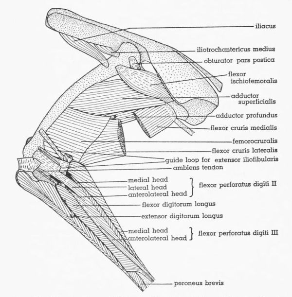
Fig. 14. Tympanuchus pallidicinctus 2L. Lateral view of the muscles of the left leg. The following muscles have been removed: extensor iliotibialis lateralis, extensor iliotibialis anticus, gastrocnemius pars externa and pars interna, and peroneus longus. × 1.
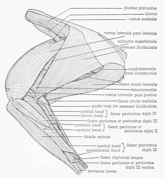
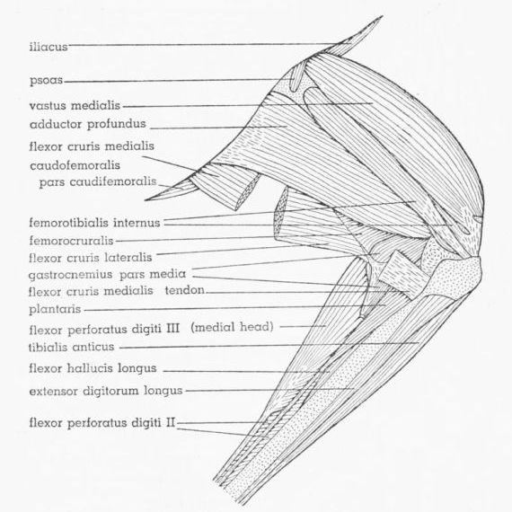
Fig. 15. Tympanuchus pallidicinctus 2L. Medial view of the muscles of the left leg. The following muscles have been removed: extensor iliotibialis lateralis, extensor iliotibialis anticus, ambiens, flexor cruris lateralis (in part), flexor cruris medialis (in part), gastrocnemius pars externa and pars interna, and peroneus longus. × 1.
Origin.—Approximately the anterior half attaches by an extensive aponeurosis, which is continuous anteriorly with that of M. extensor iliotibialis anticus, to the anterior iliac crest, ending posteriorly at the anterior end of the lateral iliac process; the posterior part attaches fleshily to the edge of the entire lateral iliac process and (posterior few mm.) aponeurotically to the entire lateral ischiatic ridge. The proximal part of the belly is much thicker than the fleshy origin. Two accessory aponeuroses associate with the anterior part of the muscle; the proximal one of these comes off the deep surface several mm. distal to the proximal end of the fleshy belly and passes medially between Mm. gluteus profundus and iliacus, fusing to both these muscles, and attaches to the lateral edge of M. iliotrochantericus medius and to the lateral edge of the ilium anterior to the latter; the aponeurosis actually splits into two sheets at the edge of M. iliotrochantericus medius; these sheets fuse to the dorsal and ventral surfaces of the latter muscle, enclosing it; the part of this aponeurosis between Mm. iliacus and iliotrochantericus medius is strongly fused with the underlying body wall. The distal accessory aponeurosis (sometimes weak) comes off the deep surface several mm. distal to the proximal one and passes medially along the ventral surface of M. iliacus, fusing with the latter, then joining the proximal accessory aponeurosis medial to M. iliacus.
Insertion.—The muscle inserts by a broad aponeurosis strongly fused to the underlying Mm. vastus lateralis and vastus medialis; the aponeurosis contributes superficially to the patellar tendon, attaching to the lateral half of the rotular crest.
Fig. 16. Tympanuchus pallidicinctus 2L. Lateral view of the muscles of the left leg. The following muscles, in addition to those listed for Fig. 14, have been removed: ambiens, vastus lateralis pars lateralis, vastus medialis (except for part of patellar tendon), extensor iliofibularis, flexor cruris lateralis (in part), flexor perforans et perforatus digiti II, and flexor perforans et perforatus digiti III. × 1.
Innervation.—A variable number of branches (usually two) of the middle division of the femoral nerve pass ventral to M. iliacus and between Mm. extensor iliotibialis anticus and vastus medialis and enter the deep surface of the anteroproximal part of the muscle. The branch of the middle peroneal division of the sciatic nerve emerges between the proximal ends of Mm. extensor iliofibularis and vastus lateralis and sends twigs into the deep surface of M. extensor iliotibialis lateralis.
Individual Variation.—In two legs, the nerve supplying M. extensor iliotibialis anticus gives twigs into M. extensor iliotibialis lateralis.
T. cupido
Differences From T. pallidicinctus.—The fleshy origin from the lateral iliac process is considerably thicker (reflected in a thicker lateral iliac process).
Individual Variation.—In three legs the nerve supplying M. extensor iliotibialis anticus gives twigs into M. extensor iliotibialis lateralis. In another leg one of the branches to the fused Mm. vastus lateralis and vastus medialis sends a twig into M. extensor iliotibialis lateralis.
Fig. 17. Tympanuchus pallidicinctus 2L. Lateral view of the muscles of the left leg. The following muscles, in addition to those listed for Fig. 16, have been removed: vastus lateralis pars postica, gluteus profundus, flexor cruris medialis (in part), caudofemoralis, flexor perforatus digiti IV, and tibialis anticus. × 1.
P. p. jamesi
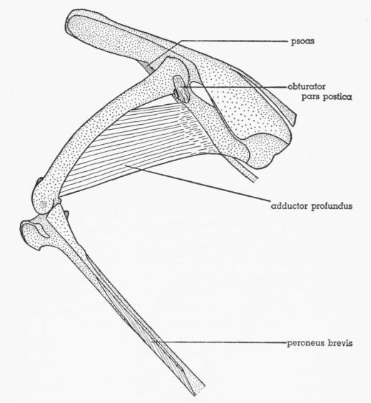
Fig. 18. Tympanuchus pallidicinctus 2L. Lateral view of the muscles of the left leg. The following muscles, in addition to those listed for Fig. 17, have been removed: patellar tendon, iliacus, iliotrochantericus medius, flexor cruris lateralis, flexor cruris medialis, flexor ischiofemoralis, adductor superficialis, femorocruralis, gastrocnemius pars media, flexor perforatus digiti III, flexor perforatus digiti II, flexor hallucis longus, plantaris, flexor digitorum longus, popliteus, and extensor digitorum longus. × 1.
Differences From Typical T. pallidicinctus.—The posteroproximal aponeurosis is more extensive, resulting in a narrower proximal fleshy end (Fig. 20G); the fleshy fibers adjacent to this aponeurosis are not overlapped by M. flexor cruris lateralis. There is a fusion of fibers between the anterodistal fleshy part of M. extensor iliotibialis lateralis and the underlying M. vastus medialis, but there is no fusion of fibers between the anterior edge of M. extensor iliotibialis lateralis and M. extensor iliotibialis anticus. The connective tissue binding the posteroproximal corner to adjacent muscles is stronger. The fleshy part of the origin is narrower, partly tendinous, and much thinner (reflected in a thin lateral iliac process). The proximal border is much more nearly straight, owing to a less pronounced lateral iliac process. The distal accessory aponeurosis is absent.
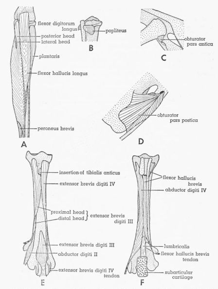
Fig. 19. Tympanuchus pallidicinctus 2L. A. Posterior view of the muscles of the left shank. The following shank muscles, in addition to those listed for Fig. 17, have been removed: gastrocnemius pars media, flexor perforatus digiti III, and flexor perforatus digiti II. × 1. B. Posterior view of the proximal end of the shank, showing the most deeply situated muscle. × 1. C. Lateral view of the head of the left femur and the middle part of the pelvis, showing the deepest part of M. obturator. × 1. D. Medial view of the posteroventral part of the left side of the pelvis, showing the intrapelvic part of M. obturator. × 1. E. Anterior view of the left tarsometatarsus, showing the dorsal intrinsic muscles of the foot. × 1½. F. Posterior view of the left tarsometatarsus, showing the ventral intrinsic muscles of the foot. × 1½.
Individual Variation.—The muscle is usually somewhat fused to the posteroproximal and anteroproximal fleshy corners of the underlying M. extensor iliofibularis.
M. Extensor Iliotibialis Anticus (M. sartorius), Figs. 12, 13
T. pallidicinctus
General Description and Relations.—Anteriormost muscle of thigh; long and strap-shaped; proximal part entirely anterior (adjacent) to M. extensor iliotibialis lateralis; posterior edge of middle part medial to latter muscle; distal part mostly medial to Mm. extensor iliotibialis lateralis and vastus medialis; proximal part aponeurotic, continuous posteriorly with anteroproximal aponeurosis of M. extensor iliotibialis lateralis; anterior edge of M. extensor iliotibialis lateralis bound by strong connective tissue to adjacent part of M. extensor iliotibialis anticus; some fusion of fibers (proximally) between these two muscles; anteroproximal corner of fleshy part of muscle sometimes fused to underlying anterior edge of ilium and fascia covering body wall musculature adjacent (anterior) to ilium.
Origin.—The muscle arises aponeurotically from the anterior part of the anterior iliac crest and (anteroproximal corner) from the anterior end of the median dorsal ridge.
Insertion.—The flat tendon, continuous posteriorly with the superficial tendon of M. femoritibialis internus, fuses to the tendon of M. vastus medialis, contributing superficially to the medial part of the patellar tendon, which attaches to the medial half of the rotular crest; most of the tendon is overlapped by the edge of M. gastrocnemius pars interna.
Innervation.—A branch of the anterior division of the femoral nerve gives twigs into the lateral surface of the posterior part.
Individual Variation.—In two legs, a twig from the anteriormost branch of the middle division of the femoral nerve anastomoses with the typical branch to M. extensor iliotibialis anticus.
T. cupido
Individual Variation.—In several legs, the anterior edge of origin extends forward onto the neural spine of the last free thoracic vertebra. A twig from the middle division of the femoral nerve anastomoses with the typical branch to M. extensor iliotibialis anticus in three legs.
P. p. jamesi
Differences From Typical T. pallidicinctus.—There is no fusion of fibers between M. extensor iliotibialis anticus and M. extensor iliotibialis lateralis.
Individual Variation.—The anterior edge of origin extends forward onto the neural spine of the last free thoracic vertebra in some legs.
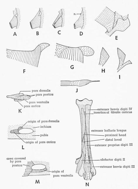
Fig. 20. A-D. Dorsal views of M. iliotrochantericus medius, showing its relationship to femoral notch. × 1. In D, note absence of femoral notch and location of branch of femoral nerve. A. Tympanuchus pallidicinctus 2L. B. T. cupido pinnatus 4L. C. Pedioecetes phasianellus jamesi 1L. D. T. pallidicinctus 3L. E. Medial view of distal end of M. flexor cruris medialis of P. p. jamesi 4L. × 1. Part of insertion is covered by medial collateral ligament. F,G. Lateral views of posteroproximal corner of M. extensor iliotibialis lateralis (removed from specimen). × 1. F. T. pallidicinctus 2L. G. P. p. jamesi 3L. H,I. Dorsolateral views of M. piriformis. × 1. H. P. p. jamesi 1L. I. T. cupido attwateri 1L. J. Lateral view of M. caudofemoralis pars caudifemoralis (removed from specimen) of T. c. pinnatus 4L. × 1. K. Lateral view of extrapelvic part of M. obturator of T. pallidicinctus 3L (bones not shown). × 2. L,M. Region surrounding obturator foramen of T. pallidicinctus 3L, showing points of attachment of three parts of M. obturator (muscles removed). × 3. L. Lateral view. M. Medial view. N. Anterior view of left tarsometatarsus of P. p. jamesi 4L, showing dorsal intrinsic muscles of foot. × 1½. Tendon of M. extensor digitorum longus has been removed.
M. Ambiens, Figs. 13, 16, 17
T. pallidicinctus
General Description and Relations.—Thin and elongate; on medial surface of thigh; broadest above middle of belly; belly narrowed distally, forming long slender tendon passing lateral to distal part of M. extensor iliotibialis anticus; bounded anterolaterally by M. vastus medialis and posterolaterally by Mm. femoritibialis internus and psoas (proximally).
Origin.—The muscle arises by a short flat tendon from the pectineal process.
Insertion.—The long slender tendon enters an elongate channel within the patellar tendon; the point of entrance is at the proximal end of the latter tendon just medial to the patella; the tendon passes distolaterally (within the channel) below the patella and emerges from the distolateral edge of the patellar tendon and then extends distally along the anterolateral surface of the head of the fibula, superficial to the fibular arm of the guide loop for M. extensor iliofibularis, and joins the anterolateral surface of the common tendon of origin of the anterolateral heads of Mm. flexor perforatus digiti III, flexor perforatus digiti IV, and flexor perforatus digiti II; the point of junction is usually immediately proximal to the proximal end of the lateral head of M. flexor digitorum longus.
Innervation.—The branch of the middle division of the femoral nerve that supplies M. femoritibialis internus gives off a tiny twig or twigs that penetrate the lateral surface of the proximal part of M. ambiens.
Individual Variation.—None of significance in T. pallidicinctus or in P. p. jamesi; in T. cupido the origin is partly fleshy in one leg.
M. Vastus Lateralis (M. femoritibialis externus + part of M. femoritibialis medius), Figs. 14, 16
Fisher and Goodman (1955) apply the name femoritibialis externus to the muscle unit that I here term the pars postica of M. vastus lateralis. The reasons for this change are discussed in the section on terminology.
T. pallidicinctus
General Description and Relations.—Thick; on lateral surface of femur deep to M. extensor iliotibialis lateralis; anterior to M. extensor iliofibularis and lateral to M. vastus medialis; much of lateral surface, except proximal part, fused with overlying M. extensor iliotibialis lateralis; deep surface of anterior half fused with M. vastus medialis; proximal part overlapping, but usually not fusing with, insertions of Mm. iliacus and caudofemoralis; partially separable into two parts—pars lateralis and pars postica, former constituting main part of muscle; latter considerably smaller and situated deep to posterodistal part of pars lateralis, except for posterodistal part extending posterior to edge of pars lateralis; proximal part of pars postica strongly fused with pars lateralis; posterodistal tendinous edge of pars lateralis fused or not fused with lateral surface of pars postica; proximal end (narrow) of pars postica tendinous and variable in length.
Origin.—Pars lateralis: This arises fleshily from most of the lateral surface and (distally) from the anterior surface of the femur, extending anteriorly to the anterior intermuscular line, fusing with M. vastus medialis, and extending posteriorly to the posterolateral intermuscular line (proximally) and the origin of pars postica (distally); the proximal end begins at the level of the distal edge of the insertion of M. iliotrochantericus medius, contacting the insertions of Mm. iliotrochantericus medius, piriformis, and flexor ischiofemoralis, and terminates distally at the level of the proximal ends of the femoral condyles.
Pars postica: This arises fleshily and tendinously (proximal end and deep surface) from the posterolateral surface of approximately the distal half of the femur, extends posteromedially to the posterolateral intermuscular line where it contacts the origin of M. femorocruralis, and extends anteriorly to the level of a line drawn diagonally across the femur from the proximal end of the origin (at the posterolateral intermuscular line) to the proximal end of the external condyle; the distal end is anterior (adjacent) to the attachment of the proximal arm of the tendinous guide loop for M. extensor iliofibularis; the origin is adjacent to, but distinct from, the origin of pars lateralis.
Insertion.—Pars lateralis is fused indistinguishably with M. vastus medialis; these two muscles form the main (middle) part of the patellar tendon, which also receives contributions from pars postica and Mm. femoritibialis internus, extensor iliotibialis lateralis, and extensor iliotibialis anticus; the patellar tendon attaches to the entire rotular crest of the tibia; the patella is situated in the proximal part of this tendon; some deep fleshy fibers of M. vastus lateralis pars lateralis and M. vastus medialis attach to the proximal edge of the patella. Pars postica forms a short narrow tendon that fuses to the lateral part of the tendon of pars lateralis, forming the lateralmost part of the patellar tendon. A broad flat vinculum extends from the lateral surface of the femorofibular fascia (defined under M. flexor perforans et perforatus digiti II) to the deep surface of the lateral part of the patellar tendon; a similar vinculum extends from the medial surface of the internal condyle to the deep surface of the medial part of the patellar tendon.
Innervation.—Two or more branches of the middle division of the femoral nerve penetrate the anterior surface of the fused Mm. vastus lateralis (pars lateralis) and vastus medialis; short twigs emerge from the deep surface of pars lateralis and penetrate the superficial surface of the anteroproximal part of pars postica.
Individual Variation.—The proximal ends of M. vastus medialis and M. vastus lateralis are usually separated by a deep notch. In some legs, a small bundle of fibers forming the anteroproximal part of M. vastus lateralis attaches to the lateral surface of M. vastus medialis anterior to this notch.
T. cupido
Individual Variation.—One leg shows the same variation found in T. pallidicinctus (see above). In several legs, pars lateralis does not extend so far proximally as usual, but begins at the level of insertion of M. piriformis (does not contact the insertion of M. iliotrochantericus medius) and may not overlap M. iliacus. In a few legs, no vincula are associated with the patellar tendon.
P. p. jamesi
Individual Variation.—Pars lateralis often begins proximally at the level of the insertion of M. piriformis.
M. Vastus Medialis (Part of M. femoritibialis medius), Figs. 13, 14, 15
T. pallidicinctus
General Description and Relations.—Thick; on anteromedial surface of femur medial to anterior part of M. vastus lateralis pars lateralis; bounded medially by Mm. ambiens and extensor iliotibialis anticus (distally); bounded posteromedially by M. femoritibialis internus; proximal part medial to posterior ends of Mm. iliacus, iliotrochantericus medius, and gluteus profundus; lateral surface, except proximal part, fused with anterior part of M. vastus lateralis pars lateralis; part of lateral surface of M. vastus medialis covered by sheet of fascia attaching to anterior intermuscular line; M. vastus lateralis separable from this fascia, but fascia absent anteriorly and distally and these two muscles indistinguishably fused.
Origin.—The proximal third is attached narrowly by its lateral edge; the distal two thirds is attached broadly by its entire deep surface. The proximal third arises tendinously from the trochanteric ridge and the proximal end of the anterior intermuscular line and fleshily from a narrow area of the femur adjacent (medial) to the latter; the distal part arises tendinously from the anterior intermuscular line and fleshily from a broad adjacent area on the anteromedial surface of the femur, terminating distally at the level of the proximal end of the internal condyle; the posterior edge contacts the origin of M. femoritibialis internus.
Insertion.—Attachment is in common with M. vastus lateralis pars lateralis, which see.
Innervation.—Two or more branches of the middle division of the femoral nerve penetrate the anterior surface of the fused Mm. vastus medialis and vastus lateralis pars lateralis; a variable number of branches of the same division penetrate the medial surface of the proximal part of M. vastus medialis.
Individual Variation.—None of significance in any of the three species studied.
M. Femoritibialis Internus, Figs. 13, 15
T. pallidicinctus
General Description and Relations.—Elongate; on posteromedial surface of femur; bounded anteriorly by M. vastus medialis and posteriorly by M. adductor profundus (overlapping anterior edge of latter); anteroproximal part lateral to M. ambiens; anterodistal corner deep to distal end of M. extensor iliotibialis anticus; distal part of muscle split into superficial and deep layers; superficial layer thin, narrow, and tendinous except for proximal end; deep layer wider, much thicker, and fleshy except for distal end taking form of flat tendon; anterior edge of latter somewhat fused to medial edge of tendon of M. vastus medialis; deep layer widest near distal end of fleshy part; posterior edge of superficial layer fused to underlying deep layer, and anterior edge fused to (continuous with) posterior edge of tendon of M. extensor iliotibialis anticus.
Origin.—The origin is mostly fleshy from the posteromedial surface of the femur between the origin of M. vastus medialis and the posterior intermuscular line, terminating immediately proximal to the internal condyle.
Insertion.—The tendons of both superficial and deep layers attach to the medial part of the rotular crest, forming the medialmost part of the patellar tendon.
Innervation.—The posteriormost branch of the middle division of the femoral nerve penetrates the medial surface of the muscle near the proximal end.
Individual Variation.—None of significance in any of the three species studied.
M. Extensor Iliofibularis (M. biceps femoris), Figs. 12, 14, 16, 17
The term extensor in the name of this muscle does not refer to its function. Howell (1938) used the term extensor to indicate derivation of the muscle from the primitive dorsal extensor muscle mass. (Likewise he used the term flexor to indicate derivation from the primitive ventral flexor muscle mass.)
T. pallidicinctus
General Description and Relations.—Deep to M. extensor iliotibialis lateralis and posterior to femur; broad proximally and narrow distally; posterior to M. vastus lateralis and anterior to proximal part of M. flexor cruris lateralis (superficial to distal part of latter); anteroproximal part aponeurotic, fused to deep surface of aponeurosis of M. extensor iliotibialis lateralis; proximal part of aponeurosis of M. extensor iliofibularis also fused to dorsal edges of underlying Mm. gluteus profundus and piriformis.
Origin.—The posterior part is fleshy from the ventromedial surface of the entire lateral iliac process; the anterior part is aponeurotic from the posterior part of the anterior iliac crest.
Insertion.—The tendon forms along the posterodistal edge of the belly and continues beyond the end of the belly as a cylindrical tendon that passes through the tendinous guide loop (the belly terminates approximately at the level of the guide loop), then extends anterodistally into the shank musculature; the tendon passes between the medial and lateral heads of M. flexor perforatus digiti IV, between the medial and lateral heads of M. flexor perforatus digiti II, lateral to the common tendon of the anterolateral heads of Mm. flexor perforatus digiti IV, flexor perforatus digiti II, and flexor perforatus digiti III, and between the posterior and lateral heads of M. flexor digitorum longus, attaching to the fibular tubercle.
The tendinous guide loop has three arms—proximal femoral, distal femoral, and fibular; the proximal and distal femoral arms unite posterior to the tendon of M. extensor iliofibularis; the proximal arm is medial to, and the distal arm is lateral to, the latter; the fibular arm joins the distal edge of the distal arm lateral to the tendon of M. extensor iliofibularis. The proximal arm extends anteroproximally lateral to the medial head of M. flexor perforatus digiti IV and medial to M. vastus lateralis pars postica, attaching to a narrow line on the anterolateral surface of the femur a short distance proximal to the external condyle and adjacent (posterior) to the origin of M. vastus lateralis pars postica. The distal arm extends anteriorly medial to the posterior head of M. flexor perforans et perforatus digiti II and medial to M. vastus lateralis pars postica, attaching in common with the tendon of origin of M. gastrocnemius pars externa to a small oval area on the posterolateral surface of the femur a short distance proximal to the fibular groove; the arm is also fused to the underlying articular capsule. The fibular arm (broadest of the three) passes deep to, and fused with, the common tendon of origin of the lateral heads of Mm. flexor perforatus digiti IV and flexor perforatus digiti II, superficial to the common tendon of origin of the anterolateral heads of Mm. flexor perforatus digiti IV, flexor perforatus digiti II, and flexor perforatus digiti III, and deep to the tendon of M. ambiens, attaching broadly to a narrow line on the anterolateral surface of the proximal part of the fibula; the arm is also fused to the underlying articular capsule.
Innervation.—A branch of the middle peroneal division of the sciatic nerve sends twigs to the deep surface of the anteroproximal part; the dorsal peroneal division of the sciatic nerve penetrates the deep surface of the proximal end.
Individual Variation.—In some instances a variable number of twigs arises from the peroneal nerve near the middle of the thigh and enters the deep surface of the muscle. They are difficult to expose without breaking and may have been overlooked in some specimens.
T. cupido
Individual Variation.—The same variation is found as in T. pallidicinctus (see above). In one leg, the tendon of insertion bifurcates into proximal and distal arms before attaching.
P. p. jamesi
Differences from Typical T. pallidicinctus.—It arises from the ventral rather than the ventromedial surface of the lateral iliac process (there is no ventromedial surface to this process).
Individual Variation.—In nearly all of the legs, minute twigs to M. extensor iliofibularis come off the peroneal nerve near the middle of the thigh. The insertional tendon tends toward doubleness in two legs.
M. Piriformis (M. gluteus medius et minimus), Figs. 16, 20H, I
T. pallidicinctus
General Description and Relations.—Small, thin, and triangular; lateral to antitrochanter and posterior part of trochanter; deep to M. extensor iliofibularis and posterior (adjacent) to M. gluteus profundus; distal half (or more) tendinous.
Origin.—The muscle arises fleshily from the posterior end of the anterior iliac crest (ventral to the origins of Mm. extensor iliotibialis lateralis and extensor iliofibularis) beginning adjacent to the posterior end of M. gluteus profundus.
Insertion.—The flat tendon narrows, overlaps the anteroproximal corner of insertion of M. flexor ischiofemoralis, and attaches to the lateral surface of the proximal part of the femur immediately anterior to the insertion of M. flexor ischiofemoralis and posterior to the proximal end of M. vastus lateralis; the attachment is posterodistal to the insertion of M. iliotrochantericus medius and posteroproximal to the insertion of M. iliacus.
Innervation.—The small anterior peroneal division of the sciatic nerve turns anteriorly immediately after emerging from the ilio-ischiatic fenestra and passes deep to M. piriformis, giving twigs to the deep surface.
Individual Variation.—In both legs of one specimen, the insertion does not overlap the insertion of M. flexor ischiofemoralis. The posteroproximal corner of the muscle is tendinous in one leg.
T. cupido
Individual Variation.—The anterior border is somewhat fused with the posterior edge of M. gluteus profundus in one leg, while in another there is a slight gap between the origins of M. gluteus profundus and M. piriformis. In one leg, the posterior edge of the origin is aponeurotic. On both sides of one specimen, an accessory tendinous band arises several mm. posterior to the main part of M. piriformis and joins the proximal part of the insertional tendon, thus forming a Y-shaped unit (Fig. 20I); the accessory tendon arises from the anterior end of the lateral iliac process (left side) or from the anterior part of the lateral iliac fossa (right side). The insertion may be proximal (rather than posterior) to the proximal end of M. vastus lateralis. In one leg, the insertional tendon is partly fused to the insertional tendon of M. flexor ischiofemoralis.
P. p. jamesi
Individual Variation.—There is often a gap between the origins of M. gluteus profundus and M. piriformis. In one leg (Fig. 20H), the posteroproximal corner of the muscle is aponeurotic. The insertion is often proximal (rather than posterior) to the proximal end of M. vastus lateralis. In one instance, the insertion does not overlap the insertion of M. flexor ischiofemoralis.
M. Gluteus Profundus (M. iliotrochantericus posterior), Figs. 14, 16
T. pallidicinctus
General Description and Relations.—Large and thick; covering dorsolateral surface of entire preacetabular part of ilium; deep to Mm. extensor iliotibialis lateralis and extensor iliotibialis anticus; bounded posteriorly by M. piriformis and ventrally by M. iliacus; ventral edge fused with anterior part of latter and with proximal accessory aponeurosis of M. extensor iliotibialis lateralis; tough sheet of fascia strongly fused to anterior two thirds of lateral surface; posterior to this, fascia overlying muscle but not attaching to it; posterior half of fascia fused to overlying aponeurosis of M. extensor iliotibialis lateralis; deep surface of muscle somewhat fused to proximal part of M. iliotrochantericus medius.
Origin.—The superficial surface is tendinous from the entire anterior iliac crest except the posterior end and from the crest forming the anterior and anterolateral edges of the ilium; the muscle arises fleshily from the entire dorsolateral surface of the preacetabular ilium as far posteriorly as the level of the pectineal process; the dorsal edge is adjacent (anterior) to the origin of M. piriformis.
Insertion.—The attachment is by a short, wide, thick tendon to a curved line (convex anteriorly) on the lateral surface of the femoral trochanter.
Innervation.—The anterodorsal division of the femoral nerve turns dorsally through the femoral notch of the ilium and penetrates the deep surface of the ventral part of the muscle midway of its length; the anterior peroneal division of the sciatic nerve passes deep to M. piriformis and terminates near the posterodorsal edge of M. gluteus profundus.
Individual Variation.—On both sides of one specimen, the branch from the femoral nerve passes lateral to the extreme anteroproximal corner of M. iliotrochantericus medius instead of through the femoral notch.
T. cupido
Individual Variation.—In one leg, the insertional tendon is strongly fused to the insertional tendon of M. iliotrochantericus medius.
P. p. jamesi
Individual Variation.—None of significance.
M. Iliacus (M. iliotrochantericus anterior), Figs. 13, 14, 15, 16, 17
T. pallidicinctus
General Description and Relations.—Adjacent ventrally to ventrolateral edge of M. gluteus profundus; lateral edge much thicker than medial edge; deep to M. extensor iliotibialis lateralis and anterolateral to M. iliotrochantericus medius; distal (posterior) end passing between proximal ends of Mm. vastus medialis and vastus lateralis pars lateralis; insertion overlapped by latter; dorsal surface of anterior part fused with ventrolateral edge of M. gluteus profundus and with ventral surface of proximal accessory aponeurosis of M. extensor iliotibialis lateralis; ventral surface partly fused with distal accessory aponeurosis of latter muscle.
Origin.—The origin is fleshy and tendinous from the lateral edge of the anterior part of the ilium.
Insertion.—The attachment is by a short flat tendon to the lateral surface of the femur distal to the trochanter and anterodistal to the insertion of M. piriformis and deep to the proximal part of M. vastus lateralis pars lateralis.
Innervation.—The dorsal division of the femoral nerve penetrates the ventral surface.
Individual Variation.—The dorsal division of the femoral nerve may fuse proximally with either the anterior or middle division. In one leg, there are two separate branches to the muscle.
T. cupido
Individual Variation.—The insertion may not be overlapped by M. vastus lateralis. The dorsal division of the femoral nerve is fused proximally with the middle division in one leg.
P. p. jamesi
Differences from Typical T. pallidicinctus.—The fleshy origin is wider.
Individual Variation.—The dorsal division of the femoral nerve may fuse proximally with either the anterior or middle division. In one leg, there are two branches to M. iliacus, one fused with the anterior division and the other with the middle division.
M. Iliotrochantericus Medius, Figs. 17, 20A, B, C, D
T. pallidicinctus
General Description and Relations.—Small and triangular; ventral to posterior half of M. gluteus profundus; all but posteroventral corner deep to latter; posteromedial to M. iliacus, anterior to neck of femur, and dorsolateral (adjacent proximally) to M. psoas; proximal end notched at level of femoral notch for passage of anterodorsal division of femoral nerve; part anterior to femoral notch mainly tendinous; dorsal surface of proximal part somewhat fused to M. gluteus profundus, proximal accessory aponeurosis of M. extensor iliotibialis lateralis split into two sheets enclosing and fusing with M. iliotrochantericus medius, ultimately attaching to lateral edge of ilium in common with origin of latter muscle.
Origin.—The muscle arises from the ventrolateral surface of the ilium anterior to the acetabulum and posterior to the origin of M. iliacus; the anterior part attaches to the ventrolateral edge of the ilium and the posterior part attaches just above the ventral edge. The muscle is not attached to the concavity of the femoral notch (the origin is notched here). The part attaching anterior to the femoral notch is narrow, tendinous, and continuous anteriorly with the accessory aponeurosis of M. extensor iliotibialis lateralis (thus the anterior border of the muscle cannot be exactly delimited). The part attaching posterior to the femoral notch is wider and fleshy (Fig. 20A).
Insertion.—The short flat tendon attaches to the lateral surface of the distal end of the trochanter slightly anterior and immediately distal to the insertion of M. gluteus profundus; the attachment is proximal to the origin of M. vastus lateralis, anteroproximal to the insertion of M. piriformis, and several mm. proximal to the insertion of M. iliacus.
Innervation.—The small posterodorsal division of the femoral nerve penetrates the ventral surface.
Individual Variation.—On both sides of one specimen, the femoral notch is absent and the proximal end of the muscle is not notched; the proximal part is entirely fleshy and the anterior border is well defined (Fig. 20D).
T. cupido
Individual Variation.—The part attaching anterior to the femoral notch has a fleshy origin in one leg (Fig. 20B), but in another, no part attaches anterior to the femoral notch (thus the muscle is not notched). In one leg, the insertional tendon is strongly fused to, and continuous with, the ventral edge of the insertional tendon of M. gluteus profundus.
P. p. jamesi
Individual Variation.—The part attaching anterior to the femoral notch may be mainly or entirely fleshy. In one leg, the part attaching anterior to the femoral notch is entirely separate from, although overlapped by, the main part of the muscle for the entire length of the fleshy belly (Fig. 20C); both parts have a common insertional tendon.
M. Psoas (M. iliacus), Figs. 13, 15, 18
T. pallidicinctus
General Description and Relations.—Small and slender; on medial aspect of proximal end of thigh lateral to proximal end of M. ambiens; ventromedial to M. iliotrochantericus medius; proximal end visible from inside pelvis (medial to inguinal ligament); passes dorsolateral to inguinal ligament.
Origin.—The muscle arises fleshily from the ventrolateral edge of the ilium posterior to the femoral notch and ventral (adjacent) to the origin of M. iliotrochantericus medius.
Insertion.—The attachment is tendinous to the medial surface of the femur a short distance proximal to the origin of M. femoritibialis internus.
Innervation.—The posterior division of the femoral nerve, which spirals completely around M. psoas, gives several twigs into the proximal part.
Individual Variation.—None of significance.
T. cupido
Individual Variation.—In two legs the insertion is partly fleshy.
P. p. jamesi
Individual Variation.—In one leg the insertion is partly fleshy. The posterior division of the femoral nerve perforates the muscle in one instance.
M. Flexor Cruris Lateralis (M. semitendinosus), Figs. 12, 13, 14, 15, 16, 17
This muscle represents only the main head of the muscle for which Fisher and Goodman (1955) used the same name. Their accessory head of M. flexor cruris lateralis is here termed M. femorocruralis.
T. pallidicinctus
General Description and Relations.—Large, thick, and strap-shaped; on posterior surface of thigh; proximal part bounded anteriorly by Mm. extensor iliotibialis lateralis and extensor iliofibularis; anterodistal part deep to latter; bounded medially by Mm. caudofemoralis (proximally) and flexor cruris medialis (distally); proximal end much narrower than remainder and posterior to ilium; fused to underlying tough membrane, which forms body wall posterior to ilium; proximal half of narrow part aponeurotic; distal part of muscle posterior to M. femorocruralis; separated from latter by common raphe to which both attach; caudal muscle (M. transversoanalis) attached aponeurotically to superficial surface of posteroproximal fleshy part of M. flexor cruris lateralis.
Origin.—The origin is tendinous (superficial surface) and fleshy from the entire dorsolateral iliac ridge and fleshy from an area of the ilium below this ridge, also tendinous from the posterior edge of the ilium medial to the dorsolateral iliac ridge, and also tendinous from the transverse processes of the first free caudal vertebra and the vertebra either anterior or posterior to the latter.
Insertion.—M. flexor cruris lateralis and M. femorocruralis insert broadly on opposite sides of a long tendinous raphe that extends parallel to, but some distance posterior to, the distal half of the femur; the distal end of this tendon broadens somewhat and fuses to the medial surface of M. gastrocnemius pars media (continuous with the tendon of the latter); the superficial part of this tendon continues toward the tibiotarsus, soon fusing to the deep surface of the overlying tendon of M. flexor cruris medialis; thus the common tendon of M. flexor cruris lateralis and M. femorocruralis insert in common with both M. flexor cruris medialis and M. gastrocnemius pars media.
Innervation.—A branch of the middle tibial division of the sciatic nerve enters the substance of M. caudofemoralis pars iliofemoralis, and emerges near its ventral edge, then passes lateral to M. caudofemoralis pars caudifemoralis and enters the anterior part of M. flexor cruris lateralis.
Individual Variation.—In three legs, the nerve does not perforate M. caudofemoralis pars iliofemoralis, but passes deep to it.
T. cupido
Individual Variation.—In one leg, a small accessory slip arises from the ventrolateral surface of the caudal musculature and joins the posterior edge of the main part of M. flexor cruris lateralis a short distance dorsal to the pubis. In several legs, the nerve does not perforate M. caudofemoralis pars iliofemoralis, but passes deep to it.
P. p. jamesi
Differences from Typical T. pallidicinctus.—The muscle is wider. The extreme proximal end is fleshy up to its origin, which is fleshy and tendinous from the vertebrae. The common insertional tendon of M. flexor cruris lateralis and M. femorocruralis fuses with the distal end of the fleshy part (instead of tendon) of M. flexor cruris medialis.
Individual Variation.—None of significance.
M. Flexor Cruris Medialis (M. semimembranosus), Figs. 12, 13, 14, 15, 16, 17, 20E
T. pallidicinctus
General Description and Relations.—Most posterior muscle on medial surface of thigh; long and strap-shaped; bounded anteriorly by M. adductor profundus; posteroproximal corner of latter medial to anteroproximal part of M. flexor cruris medialis; bounded laterally by Mm. caudofemoralis (proximally) and flexor cruris lateralis (distally); anteroproximal corner adjacent to posteroventral corner of M. flexor ischiofemoralis and lateral to extreme posteroproximal corner of M. adductor superficialis; distal end tendinous, extending into proximal part of shank; bounded medially by M. gastrocnemius pars interna and laterally by Mm. gastrocnemius pars media and plantaris.
Origin.—The muscle arises by a wide flat tendon from a narrow line on the lateral surface of the ischium dorsal to the ventral ischiatic tubercle.
Insertion.—The wide flat tendon attaches to a narrow line on the medial surface of the proximal part of the tibiotarsus a short distance anterior to the proximal part of M. plantaris and deep to M. gastrocnemius pars interna; the proximal end attaches immediately anterior to the distal end of the medial collateral ligament. Part of the common tendon of Mm. flexor cruris lateralis and femorocruralis fuses with the lateral surface of the tendon of M. flexor cruris medialis, inserting in common with it.
Innervation.—A branch of the middle tibial division of the sciatic nerve passes deep to both heads of M. caudofemoralis and enters the anterior part of M. flexor cruris medialis.
Individual Variation.—In several legs, the anterior edge of the proximal part fits into a deep longitudinal groove in the posterior edge of the proximal part of M. adductor superficialis; the two muscles fuse slightly at this point.
T. cupido
Individual Variation.—In two legs, the extreme posterior end of the origin is from the pubis. In two others, the proximal end is separated by a slight gap from M. adductor superficialis. The nerve arises from the posterior (rather than middle) tibial division in one leg.
P. p. jamesi
Differences from Typical T. pallidicinctus.—The origin is wider; the posterior third to half of the origin is fleshy. The entire origin is from a strongly curved line, the middle part of which attaches to the ventral edge of the ischium posterior to the ventral ischiatic tubercle. The insertion is wider. The insertional tendon attaches posterior (rather than anterior) to the distal end of the medial collateral ligament; the proximal end of the insertion attaches to the articular capsule (Fig. 20E). The insertional tendon is shorter; as a result, the common tendon of Mm. flexor cruris lateralis and femorocruralis fuses with the distal end of the fleshy belly (instead of the tendon) of M. flexor cruris medialis.
Individual Variation.—In two thirds of the legs, the proximal part of the insertion is fleshy rather than tendinous. In one leg, the middle part of the insertional tendon splits into two sheets, one attaching anterior to and one attaching posterior to the distal end of the medial collateral ligament. The nerve may arise from the posterior tibial division (two legs), from the middle tibial division (one leg), or as an independent division of the tibial nerve (three legs). In one leg, the nerve perforates the lateral part of M. flexor ischiofemoralis.
M. Caudofemoralis (M. piriformis), Figs. 12, 13, 14, 15, 16, 20J
T. pallidicinctus
General Description and Relations.—Posterior to proximal part of shaft of femur and deep to M. extensor iliofibularis; posterior part deep to M. flexor cruris lateralis; bounded medially by Mm. flexor ischiofemoralis (dorsally), flexor cruris medialis (posteriorly), and adductor superficialis (anteroventrally); anterior end distal to anterior end of M. flexor ischiofemoralis; two distinct heads—pars iliofemoralis and pars caudifemoralis; pars iliofemoralis dorsal to pars caudifemoralis; posteroventral corner of former overlapped by latter; pars iliofemoralis wider and much shorter than pars caudifemoralis; extreme posterior end of pars iliofemoralis fused to overlying posteroproximal aponeurosis of M. extensor iliotibialis lateralis; small part of ventral edge sometimes fused with underlying tendinous posteroproximal corner of M. flexor cruris medialis; entirely fleshy except for small triangular tendinous area along dorsal margin at point where branch of middle tibial division of sciatic nerve passes deep to muscle; pars caudifemoralis long, thin, narrow, and strap-shaped; overlapping posteroventral corner of ischium; posterior end of fleshy belly narrowed and forming long slender tendon passing into caudal musculature; anterior end forming short narrow tendon fused to deep surface of ventral edge of pars iliofemoralis relatively near insertion; tendon continuous to insertion; fleshy anterodorsal corner of pars caudifemoralis slightly overlapped by ventral edge of pars iliofemoralis; some form of connection usually present between anterior part of M. caudofemoralis pars caudifemoralis and dorsal end of raphe between Mm. flexor cruris lateralis and femorocruralis, most often consisting of narrow weak tendon.
Origin.—Pars iliofemoralis: This arises fleshily from the ventromedial surface of the posterior part of the lateral iliac process, from the entire lateral ischiatic ridge, and from the lateral surface of the ischium anterior to this ridge nearly as far forward as the posterior edge of origin of M. flexor ischiofemoralis; the posteroventral corner reaches the ventral edge of the ischium and usually attaches to the ischiopubic membrane posterior to M. flexor cruris medialis. Pars caudifemoralis: This arises by a narrow tendon from the ventral surface of a broad, thick, tendinous sheet ventral to the pygostyle, which, in turn, attaches to the ventral surface of the pygostyle.
Insertion.—The common belly formed by the union of the two heads narrows (width variable) and attaches to the posterolateral surface of the femur distal to the level of insertion of M. iliacus and in contact with the posterior edge of origin of M. vastus lateralis pars lateralis; the dorsal part is fleshy and the ventral part is tendinous.
Innervation.—A branch of the middle tibial division of the sciatic nerve gives several twigs to the deep surface of pars iliofemoralis; another twig enters the substance of pars iliofemoralis and emerges from the ventral edge of the latter, then enters the dorsal edge of pars caudifemoralis. The latter twig was not found in all legs, but was probably destroyed during dissection.
Individual Variation.—The tendinous area in the dorsal margin of pars iliofemoralis is lacking in one leg and extremely small in some others. In both legs of one specimen, the connection between M. caudofemoralis pars caudifemoralis and the raphe between Mm. flexor cruris lateralis and femorocruralis consists of a small (11 × 2 mm.) but well developed and entirely fleshy muscle slip (Fig. 16). In one leg, the ventral third of this connection is fleshy, the remainder tendinous; in another, this connection is completely lacking.
T. cupido
Individual Variation.—The tendinous area in the dorsal margin of pars iliofemoralis is lacking in one leg. The connection between pars caudifemoralis and the raphe between Mm. flexor cruris lateralis and femorocruralis is lacking in several legs. A conspicuous variation occurring in three legs is the presence of a tendinous area in the belly of pars caudifemoralis, dividing the latter into proximal and distal parts (Fig. 20J). In one leg, the posteroventral corner of pars iliofemoralis arises from the pubis. The origin of pars caudifemoralis in three legs is directly from the anteroventral surface of the pygostyle. In one instance, the insertional tendon of pars caudifemoralis is long and extremely slender and extends for some distance in a groove on the medial surface of pars iliofemoralis before fusing with the latter.
P. p. jamesi
Differences from Typical T. pallidicinctus.—There is no connection at all between pars caudifemoralis and the raphe between Mm. flexor cruris lateralis and femorocruralis. The posteroventral corner of pars iliofemoralis is some distance dorsal to the ventral edge of the ischium and, therefore, does not attach to the ischiopubic membrane.
Individual Variation.—The insertion (narrow) is entirely tendinous in one leg.
M. Flexor Ischiofemoralis (M. ischiofemoralis), Figs. 16, 17
T. pallidicinctus
General Description and Relations.—Thick; on lateral surface of anterior part of ischium; posterior end in lateral iliac fossa; deep to Mm. extensor iliofibularis and caudofemoralis pars iliofemoralis; overlapping ventral extrapelvic part of M. obturator and anteroproximal part of M. adductor superficialis (slightly fused to proximal edge of latter); posteroventral corner contacting anteroproximal corner of M. flexor cruris medialis; extreme anterodorsal corner usually overlapped by tendon of M. piriformis.
Origin.—The muscle arises fleshily from a large area on the lateral surface of the ischium extending ventrally to the origin of M. adductor superficialis, anteriorly to the level of the posterior end of the obturator foramen, dorsally to the ventral border of the ilio-ischiatic fenestra and to the depth of the lateral iliac fossa, and posteriorly approximately to the level of the ventral ischiatic tubercle.
Insertion.—The short flat tendon attaches to the lateral surface of the femur immediately posterior to the insertion of M. piriformis.
Innervation.—The posterior tibial division of the sciatic nerve penetrates the dorsal surface.
Individual Variation.—The ventral part of the insertion may be fleshy.
T. cupido
Individual Variation.—None of significance.
P. p. jamesi
Individual Variation.—In all the legs except one, an additional twig arises from the branch to M. flexor cruris medialis and penetrates the lateral surface of M. flexor ischiofemoralis. The ventral part of the insertion is fleshy in one leg.
M. Adductor Superficialis (M. adductor longus et brevis, pars externa), Figs. 14, 16, 17
T. pallidicinctus
General Description and Relations.—Posterior to femur, lateral to M. adductor profundus, and medial to Mm. flexor ischiofemoralis, caudofemoralis, and femorocruralis; proximal end (fleshy) fused to proximal tendinous end of M. adductor profundus.
Origin.—The origin is fleshy and tendinous from the proximal end of the lateral surface of M. adductor profundus and from a narrow line on the ischium adjacent (dorsal) to the origin of the latter; the posterior part of the origin sometimes extends farther dorsally on the lateral surface of the ischium; the origin does not extend so far anteriorly nor so far posteriorly as the origin of M. adductor profundus; the anterior edge is at the posterior border of the obturator foramen.
Insertion.—The attachment is fleshy and thick (distal end thin) to the posterior surface of the middle part of the femur between the posterior and posterolateral intermuscular lines; the attachment is adjacent (lateral) to the insertion of M. adductor profundus and adjacent (medial) to the origins of Mm. vastus lateralis (proximally) and femorocruralis (distally); the proximal edge is approximately at the level of the distal edge of the insertion of M. caudofemoralis.
Innervation.—A branch of the obturator nerve emerges from the obturator foramen dorsal to the tendon of insertion of M. obturator pars postica, turns ventrally (crossing latter), and passes deep to the anteroproximal corner of M. adductor superficialis, extending posterodistally between the adductor muscles and giving twigs to the medial surface of M. adductor superficialis and to the lateral surface of M. adductor profundus.
Individual Variation.—The anterior edges of the two adductor muscles are so firmly fused together in some cases that the boundaries cannot be identified at this point. In several legs, there is a deep longitudinal groove in the posterior edge of the proximal part of the muscle into which the anterior edge of M. flexor cruris medialis fits.
T. cupido
Individual Variation.—In some cases, the anterior edges of the two adductor muscles are firmly fused together.
P. p. jamesi
Differences from Typical T. pallidicinctus.—The origin is narrower.
Individual Variation.—The anterior edges of the two adductor muscles may be fused together. In one leg, the entire muscle is indistinguishably fused with M. adductor profundus and they appear as a single muscle.
M. Adductor Profundus (M. adductor longus et brevis, pars interna), Figs. 13, 15, 17, 18
T. pallidicinctus
General Description and Relations.—Broad; on medial surface of thigh immediately posterior to femur; bounded posteriorly by M. flexor cruris medialis (medial to anteroproximal corner of latter), anteriorly by M. femoritibialis internus (anterior edge overlapped by latter), and laterally by Mm. adductor superficialis and femorocruralis; proximal end tendinous (except anterior edge), fused to proximal fleshy end of M. adductor superficialis.
Origin.—The muscle arises tendinously from the ventral edge of the ischium extending from the posterior border of the obturator foramen to the ventral ischiatic tubercle and (anterior edge) fleshily from the lateral surface of the pubis ventral to the obturator foramen; the origin is adjacent (ventral) to the origin of M. adductor superficialis.
Insertion.—The attachment is fleshy and tendinous from the posterior intermuscular line and (proximally and distally) from a narrow adjacent area. Proximally there are often two approximately parallel lines a short distance apart, representing points of attachment of the lateral and medial edges of the muscle; if there is only one line proximally, it may represent the attachment of either the lateral or medial edge of the muscle; distally there is usually only one line, representing the lateral edge of the muscle. The distal end extends onto the posterior surface of the proximal part of the internal condyle, and is adjacent (lateral) to the origin of M. femoritibialis internus, adjacent (medial) to Mm. adductor superficialis and femorocruralis, and adjacent (proximal) to M. gastrocnemius pars media.
Innervation.—See M. adductor superficialis.
Individual Variation.—The anterior edges of the two adductor muscles are strongly fused together in some cases.
T. cupido
Individual Variation.—The anterior edge may be fused with that of M. adductor superficialis. The distal end is sometimes slightly fused with M. gastrocnemius pars media. In one leg, the proximal two thirds of the insertion is entirely tendinous, whereas in another the distal end of the insertion is tendinous.
P. p. jamesi
Individual Variation.—The anterior edge (in one leg the entire muscle) in some legs fuses with that of M. adductor superficialis.
M. Obturator (M. obturator externus + M. obturator internus), Figs. 16, 17, 18, 19C, D, 20K, L, M
I am adopting the single name M. obturator for the complex that Fisher (Fisher, 1946; Fisher and Goodman, 1955) subdivides into Mm. obturator externus and obturator internus. The reasons for this change are given in the section on terminology.
For ease of description, it is desirable to apply names to the subdivisions of M. obturator. It has been customary to divide the obturator complex into two parts—an obturator internus and an obturator externus; the latter has often been further subdivided. The evidence given below demonstrates that a primary division of the complex into only two parts is unsatisfactory.
I strongly suspect that comparable parts of the obturator complex have been considered a part of the "internus" in some birds and a part of the "externus" in others. In their work on the Galliformes, Hudson, et al. (1959) subdivide the obturator complex into only two divisions—obturator externus and obturator internus. The extrapelvic part of this complex that arises from the rim of the obturator foramen and inserts in common with the stout tendon of the main intrapelvic part of the obturator internus is considered by them to be a part of the obturator internus. Their obturator externus lies anterior and deep to the extrapelvic part of the obturator internus and inserts separately from the latter. (I also have found this same arrangement in Tympanuchus and Pedioecetes.)
Berger (1952), in his description of the Black-billed Cuckoo (Coccyzus erythrophthalmus), also divides the obturator complex into an obturator internus and an obturator externus; the latter he subdivides into a dorsal and a ventral part. He states (p. 530) that he did not find any measurable differences in myology between C. erythrophthalmus and C. americanus. In order better to compare this arrangement with that in Tympanuchus, I have examined two specimens of C. americanus. My findings in the latter differ from Berger's description (p. 541) in one respect. Whereas Berger states that the dorsal and ventral parts of M. obturator externus are distinct except at their origin, I find them fused for their entire length; the muscle fibers that connect these two parts lie deep to the tendon of M. obturator internus. The origin of all parts of the complex in Coccyzus is similar to that in Tympanuchus. The only notable difference in configuration is that the part in Coccyzus that appears to correspond to the obturator externus of Hudson, et al. (1959) is not separate from the remainder of the extrapelvic part of the muscle. Berger (1952) considers all parts of the muscle having an extrapelvic origin to make up the obturator externus. It appears to me that the dorsal part and a part of the ventral part of the obturator externus of Berger correspond to the extrapelvic fleshy part of the obturator internus of Hudson, et al.
From my limited study, it seems to me to be desirable to recognize four subdivisions of the obturator complex, for which I propose the terms pars antica, pars dorsalis, pars ventralis, and pars postica. These parts exhibit various degrees of fusion in different groups of birds and some parts appear to be absent in certain birds. A study of a wide variety of birds will be required to determine whether or not a subdivision into the four parts proposed here is suitable for birds as a whole.
Applying these terms to Coccyzus, pars postica is equivalent to the entire obturator internus of Berger (1952). Pars dorsalis is apparently equivalent to the dorsal part of Berger's obturator externus. The ventral part of the obturator externus of Berger represents the fused pars antica and pars ventralis.
The main parts of the obturator muscle appear to be pars postica and pars antica. Pars dorsalis and pars ventralis are more variable; in Coccyzus these two parts are closely associated with pars antica whereas in Tympanuchus they are most closely associated with pars postica. Apparently pars dorsalis and pars ventralis may be absent in some birds.
T. pallidicinctus
General Description and Relations.—Deeply situated immediately posterior to head of femur; part extending through obturator foramen and lying inside pelvis; extrapelvic part deep to Mm. flexor ischiofemoralis and piriformis; muscle partially divisible into four parts—pars antica, pars dorsalis, pars ventralis, and pars postica (Fig. 20K); pars postica: mostly inside pelvis; much larger than other parts; broad (narrow anteriorly); on medial surface of ischium; composed of several fascicles; anterior end forming narrow, heavy tendon (with some fleshy fibers on posterior part of deep surface) passing through obturator foramen; anteriormost fleshy fibers of ventralmost fascicle fused with pars ventralis; pars ventralis: essentially extrapelvic (see origin); mostly ventral to tendon of pars postica; superficial to pars antica; fused to anterior fleshy part of pars postica; anterodorsal edge usually adjacent to, and often slightly fused with, ventral edge of pars dorsalis (deep to tendon of pars postica); pars dorsalis: entirely extrapelvic; mostly dorsal to tendon of pars postica; superficial to dorsal part of pars antica; pars antica: extremely short but relatively thick; entirely fleshy; entirely extrapelvic; between obturator foramen and head of femur; anterior surface adjacent to articular capsule; almost completely covered by other parts of muscle; proximal end of posterior surface often slightly fused with adjacent parts of pars ventralis and pars dorsalis.
Origin.—Pars postica: This arises fleshily from the medial surface of the entire ischium except the posterior end, from the dorsomedial and medial surfaces of the anterior half of the pubis as far forward as the obturator foramen, from the internal ilio-ischiatic crest, from the medial surface of the ilium for a short distance posterior to this crest, and from the iliac recess; the posteroventral corner usually arises from the medial surface of the ischiopubic membrane. Pars ventralis: This arises fleshily from the dorsomedial edge of the ventral border of the obturator foramen (Fig. 20M) and (narrowly) from the anterior border of the foramen; this part may or may not arise from the lateral surface of the anteroventral border of the foramen and is usually adjacent along the anterior border of the foramen to pars dorsalis; pars ventralis is continuous along the ventral border of the foramen with the intrapelvic origin of pars postica. Pars dorsalis: This arises fleshily from the lateral surface of the anterodorsal border of the foramen (Fig. 20L) and may extend posteriorly along the dorsal border of the foramen. Pars antica: This arises fleshily from the depresssed area anterior to the obturator foramen (adjacent to pars dorsalis and pars ventralis); the posteroventral corner may arise from the lateral surface of the anteroventral border of the obturator foramen (ventral to the anterior end of pars ventralis; Fig. 20L).
Insertion.—Pars postica: Several tendinous bands (intrapelvic) converge and coalesce, forming a single strong tendon that passes through the obturator foramen and attaches to the lateral surface of the femoral trochanter a short distance posterior to the insertion of M. gluteus profundus and proximal to the insertion of M. flexor ischiofemoralis. Pars ventralis: The attachment is fleshy and tendinous to the ventral edge and the deep surface of the tendon of pars postica. Pars dorsalis: The attachment is fleshy and tendinous to the dorsal edge of the tendon of pars postica. Pars antica: The attachment is fleshy to the posterior surface of the proximal end of the femur several mm. posterior to the insertion of pars postica; the lateral edge attaches to the obturator ridge.
Innervation.—The muscle is supplied by the obturator nerve; several twigs, which do not pass through the obturator foramen, penetrate the anterior part of the medial surface of pars postica; several twigs pass through the obturator foramen and supply pars dorsalis, pars ventralis, and pars antica.
Individual Variation.—In some cases the origin of pars postica does not include the dorsal end of the internal ilio-ischiatic crest nor the ilium posterior to it. Tiny but distinct accessory slips are sometimes present. In one leg a tendinous slip of pars antica extends beyond the remainder of the muscle and inserts independently on the trochanter close to the insertion of pars postica. In another leg, a fleshy and tendinous slip of pars antica attaches to the deep surface of the insertional tendon of pars postica. In still another leg, a fleshy and tendinous slip of pars dorsalis inserts adjacent (anterior) to the dorsal edge of the insertion of pars antica.
T. cupido
Individual Variation.—The variations are similar to those given above for T. pallidicinctus except that there is no slip of pars antica attaching to the tendon of pars postica.
P. p. jamesi.
Individual Variation.—There are variations similar to those given above for T. pallidicinctus except that there is no independent slip of pars antica attaching on the trochanter close to the insertion of pars postica. Pars dorsalis may be quite small. In several legs, pars dorsalis is more closely associated with pars antica than with pars postica; in one of these, pars dorsalis is indistinguishably fused with pars antica (inserting with the latter) except for a few fibers which insert with pars postica.
M. Femorocruralis (M. accessorius semitendinosi), Figs. 14, 15, 16, 17
Fisher (Fisher, 1946; Fisher and Goodman, 1955) considers this muscle as an accessory head of M. flexor cruris lateralis. The reasons for this change in terminology are given in the section on terminology.
T. pallidicinctus
General Description and Relations.—Short and broad; posterior to distal part of femur; deep to Mm. extensor iliofibularis and vastus lateralis pars postica; bounded posteriorly by M. flexor cruris lateralis, medially by Mm. adductor superficialis and adductor profundus, and distally by M. gastrocnemius pars media; fused to a variable degree with the latter (in some cases these two muscles fused firmly together, appearing as single muscle); distal and medial to proximal end of M. flexor perforatus digiti IV.
Origin.—The muscle arises fleshily (thin proximally, thick distally) from the posterior surface of approximately the distal half of the femur between the posterior and posterolateral intermuscular lines. The ventral end is continuous with the origin of M. gastrocnemius pars media, adjacent (medial) to the origin of M. vastus lateralis pars postica, and adjacent (lateral) to the insertions of Mm. adductor superficialis and adductor profundus.
Insertion.—The attachment is to the tendinous raphe in common with M. flexor cruris lateralis (which see).
Innervation.—One or two tiny branches come off the tibial nerve near the distal end of the main trunk of the sciatic nerve, pass anteriorly deep to the peroneal nerve, and penetrate the lateral surface.
Individual Variation.—In two legs, the branch of the medial division of the tibial nerve which supplies M. gastrocnemius pars media sends a twig to the lateral surface of the distal end of M. femorocruralis (in addition to the usual innervation).
T. cupido
Individual Variation.—None of significance.
P. p. jamesi
Differences from Typical T. pallidicinctus.—The muscle is much wider, extending farther proximally on the femur.
Individual Variation.—None of significance.
M. Gastrocnemius, Figs. 12, 13, 15
T. pallidicinctus
General Description and Relations.—Divided into three distinct, widely separated parts—pars externa, pars interna, and pars media; pars externa: large; on posterolateral surface of shank; narrow proximally and distally; bounded anterolaterally by M. flexor perforans et perforatus digiti II and anteromedially by medial head of M. flexor perforatus digiti III; completely separate from pars interna and media except for common tendon of insertion; pars interna: large; on anteromedial surface of shank; narrow distally; bounded anterolaterally by M. peroneus longus and posteromedially by pars media (proximally) and medial head of M. flexor perforatus digiti III; broad sheet of tough connective tissue extending between distal parts of pars externa and pars interna; covering underlying M. flexor perforatus digiti III (medial head), somewhat fused with anteroproximal edge of M. peroneus longus; pars media: small and short; on medial surface of proximal part of shank; deep to tendon of insertion of M. flexor cruris medialis; bounded anteromedially by pars interna, posterolaterally by medial head of M. flexor perforatus digiti III, and proximally by M. femorocruralis; fused to latter, and boundary between the two difficult to locate.
Origin.—Pars externa: The short cylindrical tendon fuses with the anterior half of the distal arm of the tendinous guide loop for M. extensor iliofibularis and attaches in common with the latter to the posterolateral surface of the femur immediately proximal to the fibular condyle; the attachment is proximal (adjacent) to the origin of M. flexor perforans et perforatus digiti II and distal (adjacent) to the origin of M. flexor perforatus digiti IV and is fused to the articular capsule.
Pars interna: The proximal end is partly separable into two layers—a superficial longer one and a deep shorter one. The superficial layer attaches fleshily to the ventral part of the anterior surface of the patella and to the medial half of the superficial surface of the patellar tendon; this layer slightly overlaps the distal fleshy end of M. extensor iliotibialis anticus. The deep layer (overlapped by the superficial layer) attaches to the medial surface of the inner cnemial crest, to the rotular crest medial to the latter, to the medial surface of the proximal part of the tibiotarsus, and (posteroproximal corner) to the distomedial edge of the patellar tendon and to the articular capsule posteromedial to the rotular crest; the entire ventral edge is tendinous, the remainder fleshy.
Pars media: This arises fleshily from an oblique line beginning at the distal end of the origin of M. femorocruralis (continuous with the latter) and extending distomedially across the proximal part of the popliteal area to the proximal edge of the internal condyle, then attaching to the adjacent part of the articular capsule; this part is adjacent (distal) to the insertion of M. adductor profundus and adjacent (proximomedial) to the medial head of M. flexor perforatus digiti IV.
Insertion.—Pars media narrows distally with a narrow tendon along the posterior edge of the fleshy belly; approximately one third of the way down the tibiotarsus the fleshy part terminates and the tendon joins the posterior edge of pars interna, continuing distally in this position. The ossified tendon on the superficial surface of the distal part of pars interna, continuous posteriorly with the tendon of pars media, is joined approximately two thirds of the way down the tibiotarsus by the tendon of pars externa; the fleshy belly of pars interna ends just below the junction. The ossified tendon on the superficial surface of the distal part of pars externa extends beyond the fleshy belly and becomes flexible before joining the tendon of pars interna and media. The common tendon (partly ossified) extends along the posterior surface of the tibiotarsus and widens as it passes posterior to the tibial cartilage, bound to the latter by a thin tough sheet of connective tissue which attaches to the edges of the tibial cartilage, thus forming a sheath for the tendon; the tendon attaches by its edges to the posterior edges of the calcaneal ridges of the hypotarsus, then continues distally (much reduced in thickness) along the posterior surface of the tarsometatarsus, enclosing the flexor tendons; the lateral edge of the tendon attaches to the posterolateral edge of the tarsometatarsus, terminating immediately above the level of the hallux; the medial edge attaches to the edge of the posterior metatarsal crest; the tendon terminates as a thin sheet that attaches to the fascia on the sole of the foot. (Hudson, et al., 1959 consider the posterior metatarsal crest to be an ossified part of the tendon of M. gastrocnemius.)
Innervation.—A branch of the lateral division of the tibial nerve penetrates the proximal part of the medial surface of pars externa. One or two branches of the medial division of the tibial nerve pass deep to M. plantaris and penetrate the deep surface of the posterior part of pars interna. The most proximal branch of the medial division of the tibial nerve penetrates the lateral surface of pars media.
Individual Variation.—None of significance.
T. cupido
Individual Variation.—In one leg, the lateral edge of pars interna overlaps the proximomedial edge of M. peroneus longus; some fibers attach to the lateral surface of the inner cnemial crest.
P. p. jamesi
Differences from Typical T. pallidicinctus.—The proximal end of pars interna does not reach the patella.
Individual Variation.—In one leg, an additional twig to pars media arises from the distal branch to M. femorocruralis.
M. Flexor Perforans et Perforatus Digiti II, Figs. 12, 14
T. pallidicinctus
General Description and Relations.—Long, slender, and Y-shaped; on lateral surface of shank; the two heads enclosing M. flexor perforans et perforatus digiti III; posterior head bounded posteriorly by M. gastrocnemius pars externa; extreme proximal end deep to M. vastus lateralis pars postica; anterior surface fused to posterior surface of M. flexor perforans et perforatus digiti III; deep surface fused to tendinous part of lateral head of M. flexor perforatus digiti IV; anterior head tendinous except for extreme distal end; covered by, and fused to, posterior edge of M. peroneus longus; fused to anterior surface of M. flexor perforans et perforatus digiti III; two heads join above middle of shank; anteroproximal and posterodistal parts of common belly usually tendinous.
Origin.—Anterior head: This arises by a narrow tendon (partly ossified) from the distal tip of the outer cnemial crest. The tendon is so intimately fused with a connective tissue sheet fused to the deep and posterior surfaces of M. peroneus longus and to the anterior surface of M. flexor perforans et perforatus digiti III that M. flexor perforans et perforatus digiti II could be considered to arise from these two muscles. Posterior head: This arises mostly fleshily from the lateral surface of a compound sheet of tough connective tissue formed by the fusion of the tendinous posteroproximal corner of M. flexor perforans et perforatus digiti III, the proximal parts of the tendons of origin of the lateral heads of Mm. flexor perforatus digiti IV and flexor perforatus digiti II, the fibular and distal arms of the guide loop for M. extensor iliofibularis, and the lateral part of the articular capsule; a part of the common tendon of origin of the anterolateral heads of Mm. flexor perforatus digiti III, flexor perforatus digiti IV, and flexor perforatus digiti II also contributes to this sheet, which attaches to the lateral surface of the external condyle of the femur and to the anterolateral surface of the head of the fibula; for convenience in description, this complex connective tissue sheet will hereafter be termed the femorofibular fascia. The anteroproximal corner of the posterior head of M. flexor perforans et perforatus digiti II often attaches to the lateral surface of the vinculum that passes from the femorofibular fascia to the deep surface of the patellar tendon; the extreme proximal end usually attaches fleshily to a small area on the femur immediately proximal to the fibular condyle and adjacent (distal) to the attachment of the distal arm of the guide loop for M. extensor iliofibularis.
Insertion.—The common belly terminates approximately two thirds of the way down the shank; the slender ossified tendon begins along the posteromedial edge of the common belly, continues distally along the posterior surface of the shank, and becomes flexible before passing through the canal in the tibial cartilage that lies posteromedial to the canal for M. flexor digitorum longus. The tendon passes with the tendon of M. flexor perforatus digiti II (medial to the latter) through a canal in the hypotarsus (see M. flexor perforatus digiti II); just below the hypotarsus, the tendon becomes superficial to the tendon of M. flexor perforatus digiti II and farther distally becomes lateral and finally deep to the latter; the tendon is ossified for most of the length of the tarsometatarsus. At the distal end of this bone, the tendon expands before passing onto the ventral surface of digit II between the tendons of Mm. flexor perforatus digiti II and flexor digitorum longus; at the level of the first phalanx, the edges of the tendon extend dorsally around the tendon of M. flexor digitorum longus and fuse, forming a sheath around the latter; the latter emerges from the sheath near the distal end of the first phalanx; the tendon attaches to the proximal end of the subarticular cartilage ventral to the first interphalangeal joint (the strongest attachment is on the medial side).
Innervation.—The lateral division of the tibial nerve sends twigs into the posteromedial edge of the posterior head.
Individual Variation.—In one leg, the fleshy part of the anterior head is unusually long. In another leg, the anterior head is entirely tendinous. In one leg, a bundle of fibers of the posterior head attaches to the deep surface of the distal part of the patellar tendon. In one leg, near the middle of the tarsometatarsus a rather long and narrow but thick and strong vinculum arises from the tendon of M. flexor perforatus digiti II and, farther distally, joins the tendon of M. flexor perforans et perforatus digiti II.
T. cupido
Individual Variation.—In one leg, the posterior head arises in part from the distolateral edge of the patellar tendon and in another, in part from the superficial surface of the distolateral corner of the patellar tendon.
P. p. jamesi
Individual Variation.—None of significance.
M. Flexor Perforans et Perforatus Digiti III, Figs. 12, 14
T. pallidicinctus
General Description and Relations.—Thick, bipinnate; on lateral surface of proximal part of shank between two heads of M. flexor perforans et perforatus digiti II; bounded anteriorly by M. peroneus longus; anterior surface fused with tendinous anterior head of M. flexor perforans et perforatus digiti II; anterolateral edge somewhat fused to posterior edge of M. peroneus longus superficial to latter tendon; posterior surface fused to posterior head of M. flexor perforans et perforatus digiti II; distal part of belly covered by common belly of latter muscle; posteromedial edge fused to underlying lateral head of M. flexor perforatus digiti IV; anteromedial edge usually somewhat fused to underlying M. flexor digitorum longus.
Origin.—The origin is fleshy and tendinous from the edge of the outer cnemial crest and fleshy from the superficial surface of the distolateral part of the patellar tendon; the posteroproximal corner arises tendinously from the femorofibular fascia.
Insertion.—The belly narrows abruptly, terminating approximately at the middle of the shank; the slender ossified tendon extends posterodistally along the shank, becoming flexible before passing posterior to the tibial cartilage deep to the tendon of M. gastrocnemius, medial to the tendon of M. flexor perforatus digiti IV, and superficial to the medial half of the tendon of M. flexor perforatus digiti III; a thin sheet of connective tissue covers the tendon and attaches by its edges to the underlying tendon of M. flexor perforatus digiti III (thus the latter tendon forms a sheath for the tendon of M. flexor perforans et perforatus digiti II); the tendon is ossified for most of the length of the tarsometatarsus; at midlength of the latter, the tendon lies between the tendons of Mm. flexor perforatus digiti IV and flexor perforatus digiti III; near the distal end of the tarsometatarsus, the tendon becomes lateral and then deep to the tendon of M. flexor perforatus digiti III and is connected by a vinculum to the latter (which see). The tendon enters the ventral surface of digiti III between the tendons of Mm. flexor perforatus digiti III and flexor digitorum longus; after sending a dorsal slip (lateral to the tendon of M. flexor digitorum longus) to the subarticular cartilage ventral to the first interphalangeal joint, the tendon divides into two branches, between which emerges the tendon of M. flexor digitorum longus; the lateral branch attaches to the subarticular cartilage of the second interphalangeal joint and to the lateral surface of the distal end of the second phalanx; the medial branch has similar attachments on the medial side of the digit.
Innervation.—A branch of the lateral division of the tibial nerve passes deep to the posterior head of M. flexor perforans et perforatus digiti II and enters the posteromedial edge of M. flexor perforans et perforatus digiti III.
Individual Variation.—In both legs of one specimen, the part arising from the femorofibular fascia appears as a distinct but short accessory head. There is no significant individual variation in T. cupido or P. p. jamesi.
M. Flexor Perforatus Digiti IV, Figs. 14, 16
T. pallidicinctus
General Description and Relations.—On posterolateral aspect of shank deep to M. gastrocnemius pars externa; bounded medially by medial head of M. flexor perforatus digiti III, anterolaterally by posterior head of M. flexor perforans et perforatus digiti II, and anteriorly by M. flexor digitorum longus; divided into three heads—medial (largest), lateral, and anterolateral (smallest); tendon of insertion of M. extensor iliofibularis passing between medial and lateral heads; proximal and anteroproximal parts of lateral head an extremely thin, flat tendon; anterodistal part of tendon fused to lateral surface of fleshy part of underlying lateral head of M. flexor perforatus digiti II; proximal part of tendon fused indistinguishably to tendinous part of underlying lateral head of M. flexor perforatus digiti II; fleshy part of anterolateral head anterodistal to lateral head; proximal part of former a long slender tendon anterior to lateral head; anterior surface of anterolateral head (both fleshy and tendinous parts) fused to tendon of anterolateral head of M. flexor perforatus digiti III; deep surface fused to underlying anterolateral head (fleshy) of M. flexor perforatus digiti II; common tendon of anterolateral heads of M. flexor perforatus digiti IV and M. flexor perforatus digiti III passing medial to tendon of insertion of M. extensor iliofibularis, to peroneal nerve, and to fibular arm of guide loop for M. extensor iliofibularis; tendon of M. ambiens inserting on anterolateral surface of this common tendon; medial head entirely fleshy; medial surface fused to medial head of M. flexor perforatus digiti III; deep surface fused to medial edge of underlying medial head of M. flexor perforatus digiti II; medial and lateral heads joined, forming bipinnate belly (pinnate structure most evident on deep surface); anterolateral head joined to distolateral part of belly.
Origin.—The medial head attaches fleshily to the proximal part of the popliteal area proximal (adjacent) to the origin of M. flexor hallucis longus and distolateral to the distal end of the origin of M. femorocruralis; the attachment extends laterally onto the posterolateral surface of the femur proximal (adjacent) to the common attachment of M. gastrocnemius pars externa and the distal arm of the guide loop for M. extensor iliofibularis; the medial edge of the origin is fused with part of the tendinous origin of the medial head of M. flexor perforatus digiti III.
The broad flat common tendon of the lateral head and the lateral head of M. flexor perforatus digiti II fuses to the superficial surface of the fibular arm of the guide loop for M. extensor iliofibularis and contributes to the femorofibular fascia; consequently the ultimate origin would be the external femoral condyle and the head of the fibula.
The slender common tendon of the anterolateral head and the anterolateral heads of Mm. flexor perforatus digiti II and flexor perforatus digiti III passes deep to the insertional tendon of M. extensor iliofibularis and to the fibular arm of the guide loop for the latter muscle (to which it partly fuses); the tendon attaches to a narrow line on the head of the fibula adjacent to the attachment of the fibular arm of the guide loop and to the deep part of the femorofibular fascia.
Insertion.—The slender ossified tendon becomes flexible before it passes posterior to the tibial cartilage deep to the tendon of M. gastrocnemius, lateral to the tendon of M. flexor perforans et perforatus digiti III, and superficial to the lateral half of the tendon of M. flexor perforatus digiti III; a thin sheet of connective tissue covers the tendon and attaches by its edges to the underlying tendon of M. flexor perforatus digiti III (thus the latter tendon forms a sheath for the tendon of M. flexor perforatus digiti IV; this sheath is separate from a similar sheath surrounding the tendon of M. flexor perforans et perforatus digiti III); the tendon is again ossified where it passes along the posterolateral surface of the tarsometatarsus posterolateral to the tendon of M. flexor perforans et perforatus digiti III; near the distal end of the tarsometatarsus the tendon becomes flexible and expands greatly in width and thickness, and sends a small slip dorsally, medial to the underlying tendons, that attaches to the subarticular cartilage ventral to the trochlea for digit IV; sometimes this slip is continuous with the retinaculum ventral to the tendon at the level of the proximal end of the digit. Several more or less distinct sheets of tough connective tissue lie ventral to all of the flexor tendons at the level of the trochleae and the proximal end of the digits, holding them in place. The tendon narrows as it passes onto the ventral surface of digit IV and soon divides into three branches; the tendon of M. flexor digitorum longus emerges between the medial and middle branches. The lateral branch attaches to the subarticular cartilage ventral to the first interphalangeal joint and is also bound by connective tissue to the ventrolateral surface of the first phalanx. A dorsal slip arises at the point of divergence of the lateral and middle branches and attaches to the subarticular cartilage of the first interphalangeal joint. The middle branch attaches to the subarticular cartilage of the second joint. The medial branch, after sending dorsal slips to each of the first two subarticular cartilages, attaches to the subarticular cartilage of the third interphalangeal joint.
Innervation.—The posterior division of the tibial nerve sends a branch into the posterior edge of the medial head, then passes between the latter and the medial head of M. flexor perforatus digiti III; as it extends distally it gives off twigs to the medial surface of the medial head, to the deep surface of the lateral head, and to the deep surface of the anterolateral head.
Individual Variation.—In one leg, an additional branch arises from the tibial nerve at the level of origin of the posterior division and enters the posterior surface of the medial head; a twig from this branch anastomoses with the first twig of the posterior division to the same head; a branch of the medial division joins the posterior division distal to the origin of the twigs to the medial head but proximal to the origin of the twigs to the other heads.
T. cupido
Individual Variation.—None of significance.
P. p. jamesi
Individual Variation.—In four legs, a tiny vinculum connects with the tendon of M. flexor digitorum longus (which see).
M. Flexor Perforatus Digiti III, Figs. 13, 14, 15, 16, 17
T. pallidicinctus
General Description and Relations.—Divided into two widely separated heads—medial and anterolateral—with completely separate bellies but with common insertional tendon; small anterolateral head on lateral aspect of thigh deep to M. flexor perforans et perforatus digiti II and posterior to M. flexor digitorum longus; fleshy part of head distolateral to belly of M. flexor perforatus digiti IV; fleshy part fused to lateral edge of belly of M. flexor perforatus digiti II; proximal part of head a slender ossified tendon fused to anterior edge of both fleshy and tendinous parts of anterolateral head of M. flexor perforatus digiti IV and to lateral edge of anterolateral head of M. flexor perforatus digiti II; this tendon passing deep to tendon of insertion of M. extensor iliofibularis and to peroneal nerve; large medial head on posteromedial surface of thigh anterior to medial edge of M. gastrocnemius pars externa, lateral to M. gastrocnemius pars media, and medial to M. flexor perforatus digiti IV; fused to medial surface of medial head of latter and to medial edges of Mm. flexor perforatus digiti II and flexor hallucis longus; proximal end of head tendinous.
Origin.—The medial head attaches tendinously to the medial part of the popliteal area in common with the medial head of M. flexor perforatus digiti II and with the medial edges of Mm. flexor perforatus digiti IV (medial head) and flexor hallucis longus; and is also fused to the articular capsule. The anterolateral head arises in common with the anterolateral heads of Mm. flexor perforatus digiti II and flexor perforatus digiti IV (see account of latter).
Insertion.—The short unossified tendon of the anterolateral head and the longer ossified tendon of the medial head join (after the latter becomes flexible) a short distance above the tibial cartilage, forming a broad flat common tendon that passes posterior to the tibial cartilage (in a shallow groove of the latter); the main part of the tendon is deep to the tendons of Mm. flexor perforatus digiti IV and flexor perforans et perforatus digiti III, but forms separate thin sheaths around these two tendons at the level of the tibial cartilage. A thin sheet of connective tissue covers these three tendons and attaches by its edges to the tibial cartilage, forming a sheath for them. These three tendons pass through the superficial groove in the hypotarsus deep to the tendon of M. gastrocnemius; the tendon of M. flexor perforatus digiti III is ossified for most of the length of the tarsometatarsus; a short distance below the hypotarsus, the anterior branch of the tendon of M. peroneus longus attaches broadly to the lateral edge of the tendon of M. flexor perforatus digiti III. In the proximal part of the tarsometatarsus the tendon is deep to the tendon of M. flexor perforans et perforatus digiti III, but farther distally becomes medial and then superficial to the latter and lateral to the tendon of M. flexor perforans et perforatus digiti II; near the distal end of the tarsometatarsus a narrow but strong vinculum extends from the lateral edge of the tendon somewhat distally to the lateral edge of the tendon of M. flexor perforans et perforatus digiti III. At the distal end of the tarsometatarsus the tendon expands before entering the ventral surface of digit III where it soon divides into two branches, between which emerge the tendons of Mm. flexor perforans et perforatus digiti III and flexor digitorum longus; the lateral branch attaches to the subarticular cartilage ventral to the first interphalangeal joint and to the lateral surface of the distal end of the first phalanx; the medial branch has similar attachments on the medial side of the digit.
Innervation.—The posterior division of the tibial nerve passes between the medial heads of M. flexor perforatus digiti III and M. flexor perforatus digiti IV and sends a twig to the lateral surface of the former, then passes deep to the common belly of M. flexor perforatus digiti IV and sends a twig to the posterior surface of the anterolateral head of M. flexor perforatus digiti III.
Individual Variation.—None of significance.
T. cupido
Individual Variation.—In one leg, an extra branch (immediately distal to the branch to M. gastrocnemius pars media) of the medial division of the tibial nerve penetrates the medial surface of the proximal end of the medial head.
P. p. jamesi
Individual Variation.—None of significance.
M. Flexor Perforatus Digiti II,Figs. 15, 17
T. pallidicinctus
General Description and Relations.—Bipinnate; on posterior aspect of shank deep to M. flexor perforatus digiti IV and between two heads of M. flexor perforatus digiti III; bounded anteriorly by Mm. flexor digitorum longus and flexor hallucis longus; proximal part divided into three small heads—medial, lateral, and anterolateral; medial and proximal parts of medial head tendinous and extremely thin except for ossified medial edge; proximal part of lateral head tendinous and lateral to insertional tendon of M. extensor iliofibularis; both tendinous and fleshy parts fused to overlying tendon of M. flexor perforatus digiti IV; narrow anterolateral head fused to overlying anterolateral head of latter muscle and (anterolateral edge) to ossified tendon of anterolateral head of M. flexor perforatus digiti III; lateral edge of common belly fused to latter head; medial edge of muscle fused to medial heads of Mm. flexor perforatus digiti IV and flexor perforatus digiti III and to M. flexor hallucis longus.
Origin.—The medial head attaches by a slender ossified tendon to the medial part of the popliteal area in common with the medial head of M. flexor perforatus digiti III and with the medial edges of Mm. flexor perforatus digiti IV (medial head) and flexor hallucis longus; this head is also fused to the articular capsule. The above-mentioned ossified part of the tendon is situated at the junction of M. flexor perforatus digiti II and M. flexor perforatus digiti III (medial head) and could be considered to be a part of the latter rather than the former. The flat tendon of the lateral head arises in common with the lateral head of M. flexor perforatus digiti IV (which see). The anterolateral head arises in common with the anterolateral heads of Mm. flexor perforatus digiti IV and flexor perforatus digiti III (see former).
Insertion.—The short, slender, ossified tendon becomes flexible and passes through the canal in the tibial cartilage that lies medial to the canal for M. flexor hallucis longus and lateral to the canals for Mm. flexor digitorum longus and flexor perforans et perforatus digiti II. The tendon passes with the tendon of M. flexor perforans et perforatus digiti II (lateral to latter) through the canal in the hypotarsus that is deep to the groove for M. flexor perforatus digiti III and superficial to the canal for M. flexor digitorum longus; the former canal has a bony floor and sides but a fibrous roof; a fibrous partition subdivides the proximal half of this canal, forming a separate channel for each tendon. The tendon is ossified for most of the length of the tarsometatarsus and is situated lateral (adjacent) to the posterior metatarsal crest; immediately below the hypotarsus, the tendon becomes situated deep to the tendon of M. flexor perforans et perforatus digiti II and farther distally becomes situated medial and finally superficial to the latter; at the distal end of the tarsometatarsus the tendon expands greatly and its edges (thick) pass dorsally around the underlying flexor tendons and become continuous with the subarticular cartilage ventral to the trochlea for digit II. The tendon extends onto the ventral surface of digit II and attaches by its edges to the ventromedial and ventrolateral surfaces of the proximal part of the first phalanx (the lateral edge extending farthest distally); the tendons of Mm. flexor perforans et perforatus digiti II and flexor digitorum longus emerge from the distal end of the tendon of M. flexor perforatus digiti II.
Innervation.—The posterior division of the tibial nerve passes between the medial heads of Mm. flexor perforatus digiti III and flexor perforatus digiti IV and gives a twig to the superficial surface of each of the three heads of M. flexor perforatus digiti II and sometimes gives another twig to the superficial surface of the distal part of the common belly.
Individual Variation.—In one leg, a vinculum connects the tendon with that of M. flexor perforans et perforatus digiti II (which see).
T. cupido
Individual Variation.—The canal in the hypotarsus through which the tendon passes has a bony (instead of fibrous) roof in one leg.
P. p. jamesi
Individual Variation.—The variation given above for T. cupido is found in both legs of one specimen.
M. Flexor Hallucis Longus, Figs. 15, 19A
T. pallidicinctus
General Description and Relations.—Elongate and tapering; on posterior aspect of shank deep to M. flexor perforatus digiti II and to proximal end of medial head of M. flexor perforatus digiti IV; bounded anterolaterally by M. flexor digitorum longus and anteromedially by M. plantaris; tendinous anteromedial surface of proximal end fused to common tendon of origin of medial heads of Mm. flexor perforatus digiti III and flexor perforatus digiti II; belly ending approximately halfway down shank.
Origin.—The origin is fleshy and tendinous (anteromedial surface) from the popliteal area immediately distal to the origin of the medial head of M. flexor perforatus digiti IV, extending laterally to the area immediately proximal to the external femoral condyle (medial to the origin of M. gastrocnemius pars externa); the muscle also arises from the proximal end of the posterior part of the articular capsule.
Insertion.—The slender ossified tendon becomes flexible and passes through the canal in the tibial cartilage that lies lateral to the canal for M. flexor perforatus digiti II, then passes through a slight groove in the lateral surface of the hypotarsus and becomes ossified again; midway of the tarsometatarsus, the tendon becomes superficial to the tendon of M. flexor digitorum longus and is connected with the latter by an extensive vinculum, which extends from the deep surface and lateral edge of the tendon of M. flexor hallucis longus distally to the superficial surface of the tendon of M. flexor digitorum longus; the tendon continues, unossified and considerably reduced in size, distally medial to the tendon of M. flexor digitorum longus, and passes through the flexor groove of the first metatarsal anterolateral (adjacent) to the tendon of M. flexor hallucis brevis, then passes deep to the terminal expansion of the latter onto the ventral surface of the hallux; the tendon emerges from under the end of the tendon of M. flexor hallucis brevis and attaches to the ventral surface of the ungual phalanx; a weak dorsal slip attaching to the ventral surface of the distal end of the first phalanx is usually present.
Innervation.—A branch of the medial division of the tibial nerve passes along the medial edge of the muscle, giving several twigs into it.
Individual Variation.—None of significance in any of the three species studied.
M. Plantaris, Figs. 15, 19A
T. pallidicinctus
General Description and Relations.—Elongate and tapering; on posteromedial surface of tibiotarsus; bounded medially by M. gastrocnemius pars interna and tendon of M. flexor cruris medialis, posteriorly by M. gastrocnemius pars media and medial head of M. flexor perforatus digiti III, posterolaterally by M. flexor hallucis longus; medial to M. flexor digitorum longus; anterolateral surface of proximal end often slightly overlapping and fused to posterior surface of medial end of M. popliteus; belly terminating above middle of shank.
Origin.—The origin is fleshy and tendinous (distal edge only) from an elongate area on the posteromedial surface of the proximal end of the tibiotarsus adjacent to the insertion of M. popliteus.