ADAM SEDGWICK, M.A., (continued 2)
Reading edition
Fig. 38. Ovary of a rabbit five days after birth, coloured in the same manner as figs. 35, 36 and 37, but represented on a somewhat smaller scale. Picric acid.
Fig. 38A. Vertical section through a small part of the surface of the same ovary as fig. 38. Zeiss D D, ocul. 2.
Fig. 38B. Small portion of the deeper layer of the germinal epithelium of the same ovary as fig. 38. The figure shews the commencing differentiation of the cells of the germinal epithelium into true ova and follicle cells. Zeiss D D, ocul. 2.
Fig. 39A. Section through a small part of the middle region of the germinal epithelium of a rabbit seven days after birth. Zeiss D D, ocul. 2.
Fig. 39B. Section through a small part of the innermost layer of the germinal epithelium of a rabbit seven days after birth, shewing the formation of Graafian follicles. Zeiss D D, ocul. 2.
Figs. 40A and 40B. Small portions of the middle region of the germinal epithelium of a rabbit four weeks after birth. Zeiss D D, ocul. 2.
Fig. 41. Graafian follicle with two ova, about to divide into two follicles, from a rabbit six weeks after birth. Zeiss D D, ocul. 2.
DESCRIPTION OF PLATES 27 AND 28. (XIII. p. 618.)
Complete List of Reference Letters.
ao. Aorta. cv. Cardinal vein. gl. Glomerulus. gr1. First groove of head-kidney. gr2. Second groove of head-kidney. gr3. Third groove of head-kidney. ge. Germinal epithelium. mrb. Malpighian body. me. Mesentery. md. Müllerian duct. r1. First ridge of head-kidney. r2. Second ridge of head-kidney. r3. Third ridge of head-kidney. Wd. Wolffian duct. x. Fold in germinal epithelium.
Plate 27.
Series A. Sections through the head-kidney at our second stage. Zeiss 2, ocul. 3 (reduced one-third). The second and third grooves are represented with the ridge connecting them, and the rod of cells running backwards for a short distance.
No. 1. Section through the second groove.
No. 2. Section through the ridge connecting the second and third grooves.
No. 3. Section passing through the same ridge at a point nearer the third groove.
Nos. 4, 5, 6. Sections through the third groove.
No. 7. Section through the point where the third groove passes into the solid rod of cells.
No. 8. Section through the rod when quite separated from the germinal epithelium.
No. 9. Section very near the termination of the rod.
No. 10. Last section in which any trace of the rod is seen.
Series B. Sections passing through the head-kidney at our third stage. Zeiss C, ocul. 2. Our figures are representations of the following sections of the series, section 1 being the first which passes through the anterior groove of the head-kidney.
No.1 Section3.
" 2 " 4.
" 3 " 5.
" 4 " 6.
" 5 " 8.
" 6 " 10.
" 7 " 11.
" 8 " 13.
" 9 " 15.
" 10 " 16.
" 11 " 17.
" 12 " 18.
" 13 " 19.
" 14 " 20.
The Müllerian duct extends through eleven more sections.
The first groove (gr1.) extends to No. 3.
The second groove (gr2.) extends from No. 4 to No. 7.
The third groove (gr3.) extends from No. 11 to No. 13.
The first ridge (r1.) extends from No. 2 to No. 5.
The second ridge (r2.) extends from No. 8 to No. 11.
The third ridge (r3.) extends from No. 13 backwards through twelve sections, when it terminates by a pointed extremity.
Fig. C. Section through the ridge connecting the second and third grooves of the head-kidney of an embryo slightly younger than that from which Series B was taken. Zeiss C, ocul. 3 (reduced one-third).
The fold of the germinal epithelium, which gives rise to a deep groove (x.) external to the head-kidney is well marked.
Series G. Sections through the rod of cells constituting the termination of the Müllerian duct at a stage in which the head-kidney is still present. Zeiss C, ocul. 2.
Plate 28.
Series D. Sections chosen at intervals from a complete series traversing the peritoneal opening of the Müllerian duct, the remnant of the head-kidney, and the termination of the Müllerian duct. Zeiss C, ocul. 3 (reduced one-third).
Nos. 1 and 2. Sections through the persistent anterior opening of the head-kidney (abdominal opening of Müllerian duct). The approach of the Wolffian duct to the groove may be seen by a comparison of these two figures. In the sections in front of these (not figured) the two are much more widely separated than in No. 1.
No. 3. Section through the Müllerian duct, just posterior to the persistent opening.
Nos. 4 and 5. Remains of the ridges, which at an earlier stage connected the first and second grooves, are seen passing from the Müllerian duct to the peritoneal epithelium.
No. 6. Rudiment of the second groove (gr2.) of the head-kidney.
Between 6 and 7 is a considerable interval.
No. 7. All traces of this groove (gr2.) have vanished, and the Müllerian duct is quite disconnected from the epithelium.
No. 8. Rudiment of the third groove (gr3.).
No. 9. Müllerian duct quite free in the space between the peritoneal epithelium and the Wolffian duct, in which condition it extends until near its termination. Between Nos. 9 and 10 is an interval of eight sections.
No. 10. The penultimate section, in which the Müllerian duct is seen. A lumen cannot be clearly made out.
No. 11. The last section in which any trace of the Müllerian duct is visible. No line of demarcation can be seen separating the solid end of the Müllerian duct from the ventral wall of the Wolffian duct.
Figs. E. and F. Sections through the glomerulus of the head-kidney from an embryo prior to the appearance of the head-kidney. Zeiss B, ocul. 2. A comparison of the two figures shows the variation in the thickness of the stalk of the glomerulus. E. Section anterior to the foremost Malpighian body. F. Section through both the glomerulus of the head-kidney and that of a Malpighian body. The two are seen to be connected.
Series H. Consecutive sections through the hind end of the Müllerian duct, from an embryo in which the head-kidney was only represented by a rudiment. (The embryo was, perhaps, very slightly older than that from which Series D was taken.) Zeiss C, ocul. 3 (reduced one-third).
No. 1. Müllerian duct is without a lumen, and quite distinct from the Wolffian wall.
No. 2. The solid end of the Müllerian duct is no longer distinct from the internal wall of the Wolffian duct.
No. 3. All trace of the Müllerian duct has vanished.
Series I. Sections through the hinder end of the Müllerian duct from an embryo of about the middle of the sixth day. Zeiss C, ocul. 2 (reduced one-third).
No. 1. The Müllerian duct is distinct and small.
No. 2. Is posterior by twelve sections to No. 1. The Müllerian duct is dilated, and its cells are vacuolated.
No. 3. Penultimate section, in which the Müllerian duct is visible; it is separated by three sections from No. 2.
No. 4. Last section in which any trace of the Müllerian duct is visible; the lumen, which was visible in the previous section, is now absent.
No. 5. No trace of Müllerian duct. Nos. 3, 4, and 5 are consecutive sections.
Fig. K. Section through the hind end of the abdominal opening of the Müllerian duct of a chick of 123 hours. Zeiss C, ocul. 2 (reduced one-third). It illustrates the peculiar cord connecting the Müllerian and Wolffian ducts.
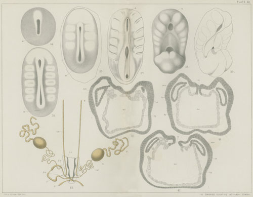
DESCRIPTION OF PLATE 29. (XIV. p. 644.)
Complete List of Reference Letters.
am. Amnion. ch. Notochord. ch´. Notochordal thickening of hypoblast. ep. Epiblast. hy. Hypoblast. m.g. Medullary groove. me.p. Mesoblastic plate. ne. Neurenteric canal (blastopore). pr. Primitive streak.
Series A. Sections through an embryo shortly after the formation of the medullary groove. × 120[4].
Fig. 1. Section through the trunk of the embryo.
Figs. 2-5. Sections through the neurenteric canal.
Fig. B. Surface view of a somewhat older embryo than that from which Series A is taken. × 30.
Series B. Sections through the embryo represented in Fig. B. × 120.
Fig. 1. Section through the trunk of the embryo.
Figs. 2, 3. Sections through the hind end of the medullary groove.
Fig. 4. Section through the neurenteric canal.
Fig. 5. Section through the primitive streak.
Fig. C. Surface view of a somewhat older embryo than that represented in Fig. B. × 30.
EXPLANATION OF PLATES 30, 31, AND 32. (XVII. p. 668.)
Plate 30.
Complete List of Reference Letters.
ch. Cheliceræ. ch.g. Ganglion of cheliceræ. c.l. Caudal lobe. p.c. Primitive cumulus. pd. Pedipalpi. pr.l. Præoral lobe. pp1. pp2. etc. Provisional appendages. sp. Spinnerets. st. Stomodæum.
I-IV. Ambulatory appendages. 1-16. Postoral segments.
Fig. 1. Ovum, with primitive cumulus and streak proceeding from it.
Fig. 2. Somewhat later stage, in which the primitive cumulus is still visible. Near the opposite end of the blastoderm is a white area, which is probably the rudiment of the procephalic lobe.
Fig. 3a and 3b. View of an embryo from the ventral surface and from the side when six segments have become established.
Fig. 4. View of an embryo, ideally unrolled, when the first rudiments of the appendages become visible.
Fig. 5. Embryo ideally unrolled at the stage when all the appendages have become established.
Fig. 6. Somewhat older stage, when the limbs begin to be jointed. Viewed from the side.
Fig. 7. Later stage, viewed from the side.
Fig. 7a. Same embryo as fig. 7, ideally unrolled.
Figs. 8a and 8b. View from the ventral surface and from the side of an embryo, after the ventral flexure has considerably advanced.
Fig. 9. Somewhat older embryo, viewed from the ventral surface.
Plates 31 and 32.
Complete List of Reference Letters.
ao. Aorta. ab.g. Abdominal nerve cord. ch. Cheliceræ. ch.g. Ganglion of cheliceræ. ep. Epiblast. hs. Hemispherical lobe of supra-œsophageal ganglion. ht. Heart. l.l. Lower lip. m. Muscles. me. Mesoblast. mes. Mesenteron. mp.g. Malpighian tube. ms. Mesoblastic somite. œ. Œsophagus. p.c. Pericardium. pd. Pedipalpi. pd.g. Ganglion of pedipalpi. pr. Proctodæum (rectum). pr.c. Primitive cumulus. s. Septum in abdomen. so. Somatopleure. sp. Splanchnopleure. st. Stomodæum. su. Suctorial apparatus. su.g. Supra-œsophageal ganglion. th.g. Thoracic ganglion. v.g. Ventral nerve cord. y.c. Cells derived from yolk. yk. Yolk. y.n. Nuclei of yolk cells.
Ig—IVg. Ganglia of ambulatory limbs. 1—16. Postoral segments.
Fig. 10. Section through an ovum, slightly younger than fig. 1. Shewing the primitive cumulus and the columnar character of the cells of one half of the blastoderm.
Fig. 11. Section through an embryo of the same age as fig. 2. Shewing the median thickening of the blastoderm.
Fig. 12. Transverse section through the ventral plate of a somewhat older embryo. Shewing the division of the ventral plate into epiblast and mesoblast.
Fig. 13. Section through the ventral plate of an embryo of the same age as fig. 3, shewing the division of the mesoblast of the ventral plate into two mesoblastic bands.
Fig. 14. Transverse section through an embryo of the same age as fig. 5, passing through an abdominal segment above and a thoracic segment below.
Fig. 15. Longitudinal section slightly to one side of the middle line through an embryo of the same age.
Fig. 16. Transverse section through the ventral plate in the thoracic region of an embryo of the same age as fig. 7.
Fig. 17. Transverse section through the procephalic lobes of an embryo of the same age. gr. Section of hemicircular groove in procephalic lobe.
Fig. 18. Transverse section through the thoracic region of an embryo of the same age as fig. 8.
Fig. 19. Section through the procephalic lobes of an embryo of the same age.
Fig. 20a, b, c, d, e. Five sections through an embryo of the same age as fig. 9. a and b are sections through the procephalic lobes, c through the front part of the thorax. d cuts transversely the posterior parts of the thorax, and longitudinally and horizontally the ventral surface of the abdomen. e cuts the posterior part of the abdomen longitudinally and horizontally, and shews the commencement of the mesenteron.
Fig. 21. Longitudinal and vertical section of an embryo of the same age. The section passes somewhat to one side of the middle line, and shews the structure of the nervous system.
Fig. 22. Transverse section through the dorsal part of the abdomen of an embryo of the same stage as fig. 9.
EXPLANATION OF PLATE 33. (XX. p. 714.)
Fig. 1. Transverse section through the pelvic fin of an embryo of Scyllium belonging to stage P[5], magnified 50 diameters. bp. basipterygium. br. fin ray. m. muscle. hf. horny fibres supporting the peripheral part of the fin.
Fig. 2. Pelvic fin of a very young female embryo of Scyllium stellare, magnified 16 diameters. bp. basipterygium. pu. pubic process of pelvic girdle (cut across below). il. iliac process of pelvic girdle. fo. foramen.
Fig. 3. Pelvic fin of a young male embryo of Scyllium stellare, magnified 16 diameters. bp. basipterygium. mo. process of basipterygium continued into clasper. il. iliac process of pelvic girdle. pu. pubic section of pelvic girdle.
Fig. 4. Transverse section through the ventral part of the trunk of an embryo Scyllium of stage P, in the region of the pectoral fins, to shew how the fins are attached to the body, magnified 18 diameters. br. cartilaginous fin-ray. bp. basipterygium. m. muscle of fin. mp. muscle-plate.
Fig. 5. Transverse section through the ventral part of the trunk of an embryo Scyllium of stage P, in the region of the pelvic fin, on the same scale as fig. 4. bp. basipterygium. br. cartilaginous fin-rays. m. muscle of the fins. mp. muscle-plate.
Fig. 6. Pectoral fin of an embryo of Scyllium canicula, of a stage between O and P, in longitudinal and horizontal section (the skeleton of the fin was still in the condition of embryonic cartilage), magnified 36 diameters. bp. basipterygium (eventual metapterygium). fr. cartilaginous fin-rays. pg. pectoral girdle in transverse section. fo. foramen in pectoral girdle. pe. epithelium of peritoneal cavity.
Fig. 7. Transverse section through the pectoral fin of a Scyllium embryo of stage P, magnified 50 diameters. bp. basipterygium. br. cartilaginous fin-ray. m. muscle. hf. horny fibres.
Fig. 8. Pectoral fin of an embryo of Scyllium stellare, magnified 16 diameters. mp. metapterygium (basipterygium of earlier stage). me.p. rudiment of future pro- and mesopterygium. sc. cut surface of a scapular process. cr. coracoid process. fr. foramen. hf. horny fibres.
Fig. 9. Skeleton of the pectoral fin and part of pectoral girdle of a nearly ripe embryo of Scyllium stellare, magnified 10 diameters. mp. metapterygium. mes. mesopterygium. pp. propterygium. cr. coracoid process.
EXPLANATION OF PLATES 34-42. (XXII. p. 738.)
List of Reference Letters.
a. Anus. ab. Air-bladder. ab´. Aperture of air-bladder into throat. ac. Anterior commissure. af. Anal fin. al. Alimentary canal. ao. Aorta. ar. Artery. au. Auditory pit. b. Brain. bc. Body-cavity. bd. Bile duct. bd´. Aperture of bile duct into duodenum. bl. Coalesced portion of segmental ducts, forming urinogenital bladder. bra. Branchial arches. brc. Branchial clefts. c. Pyloric caæca. c´. Apertures of caæca into duodenum. cb. Cerebellum. cdv. Cardinal vein. ce. Cerebrum: in figs. 47A and B, anterior lobe of cerebrum. ce´. Posterior lobe of cerebrum. cf. Caudal fin. cn. Centrum. ch. Choroidal fissure. crv. Circular vein of vascular membrane of eye. csh. Cuticular sheath of notochord. cv. Caudal vein. d. Duodenum. dc. Dorsal cartilage of neural arch. df. Dermal fin-rays. dl. Dorsal lobe of caudal fin. dlf. Dorsal fin. e. Eye. ed. Epidermis. ep. Epiblast. fb. Fore-brain. fe. Pyriform bodies surrounding the zona radiata of the ovum, probably the remains of epithelial cells. gb. Gall-bladder. gd. Genital duct. gl. Glomerulus. gr. Genital ridge. h. Heart. ha. Hæmal arch. hb. Hind-brain. hc. Head-cavity. hpd. Hepatic duct. hm. Hyomandibular cleft. hop. Operculum. hy. Hypoblast; in fig. 10, hyoid arch. hyl. Hyaloid membrane. ic. Intercalated cartilaginous elements of the neural arches. in. Infundibulum. ir. Iris. is. Interspinous cartilage or bones. iv. subintestinal vein. ivr. Intervertebral ring of cartilage. k. Kidney. l. Lens. lc. Longitudinal canal, formed by union of the vasa efferentia. lin. Lobi inferiores. ll. Ligamentum longitudinale superius. lr. Liver. lt. Lateral line. ly. Lymphatic body in front of kidney. m. Mouth. mb. Mid-brain. mc. Medullary cord. mel. Membrana elastica externa. mes. Mesorchium. mn. Mandible. md. and mo. Medulla oblongata. ms. Mesoblast. na. Neural arch. na´. Dorsal element of neural arch. nc. Notochord. nve. Network formed by vasa efferentia on inner face of testis. od. Oviduct. od´. Aperture of oviduct into bladder. ol. Nasal pit or aperture. olf. Olfactory lobe. op. Optic vesicle. opch. Optic chiasma. opl. Optic lobes. opth. Optic thalami. orep. Oral epithelium. ov. Ovary. p. Pancreas. pc. Pericardium. pcf. Pectoral fin. pch. Pigmented layer of choroid. pf. Peritoneal funnel of segmental tube of mesonephros. pfp. Peritoneal funnel leading into pronephric chamber. pg. Pectoral girdle. plf. Pelvic fin. pn. Pineal gland. po. Primitive germinal cells. pr. Mesoblastic somite. prc. Pronephric chamber. prn. Pronephros. prn´. Opening of pronephros into pronephric chamber. pt. Pituitary body. py. Pyloric valve. pz. Parietal zone of blastoderm. r. Rostrum. rb. Rib. rc. Rectum. s. Spleen. sc. Seminal vessels passing from the longitudinal canal into the kidney. sd. Suctorial disc. sg. Segmental or archinephric duct. sgt. Segmental tubules. sh. Granular outer portion of the sheath of the notochord in the vertebral regions. smx. Superior maxillary process. snc. subnotochordal rod. so. Somatic mesoblast. sp. Splanchnic mesoblast. spn. Spinal nerve. spv. Spiral valve. st. Stomach. st. Seminal tubes of the testis. sup. Suctorial papillæ. t. Testis. th. Thalamencephalon. thl. Lobes of the roof of the thalamencephalon. tr. Trabeculæ. ug. Urinogenital aperture. v. Ventricle. ve. Vasa efferentia. vh. Vitreous humour. vl. Ventral lobe of the caudal fin. vmt. Ventral mesentery. vn. Vein. vs. Blood-vessel. vsh. Vascular sheath between the hyaloid membrane and the vitreous humour. vth. Vesicle of the thalamencephalon. x. Groove in epiblast, probably formed in process of hardening. y. Yolk. z. Commissure in front of pineal gland. zr. Outer striated portion of investing membrane (zona radiata) of ovum. zr´. Inner non-striated portion of investing membrane of ovum. I. Olfactory nerve. II. Optic nerve. III. Oculomotor nerve. V. Trigeminal nerve. VIII. Facial and auditory nerves.
Plate 34.
Figs. 1-4. Different stages in the segmentation of the ovum.
Fig. 1. Ovum with a single vertical furrow, from above.
Fig. 2. Ovum with two vertical furrows, from above.
Fig. 3. Side view of an ovum with a completely formed blastodermic disc.
Fig. 4. The same ovum as fig. 3, from below, shewing four vertical furrows nearly meeting at the vegetative pole.
Figs. 5-10. External views of embryos up to time of hatching.
Fig. 5. Embryo, 3.5 millims. long, third day after impregnation.
Fig. 6. Embryo on the fifth day after impregnation.
Fig. 7. Posterior part of same embryo as fig. 6, shewing tail swelling.
Fig. 8. Embryo on the sixth day after impregnation.
Fig. 9. Embryo on the seventh day after impregnation.
Fig. 10. Embryo on the eleventh day after impregnation (shortly before hatching).
Fig. 11. Head of embryo about the same age as fig. 10, ventral aspect.
Fig. 12. Side view of a larva about 11 millims. in length, shortly after hatching.
Fig. 13. Head of a larva about the same age as fig. 12, ventral aspect.
Fig. 14. Side view of a larva about 15 millims. long, five days after hatching.
Fig. 15. Head of a larva 23 millims. in length.
Fig. 16. Tail of a larva 11 centims. in length.
Fig. 17. Transverse section through the egg-membranes of a just-laid ovum.
We are indebted to Professor W. K. Parker for figs. 12, 14 and 15.
Plate 35.
Figs. 18-22. Transverse sections of embryo on the third day after impregnation.
Fig. 18. Through head, shewing the medullary keel.
Fig. 19. Through anterior part of trunk.
Fig. 20. Through same region as fig. 19, shewing a groove (x) in the epiblast, probably artificially formed in the process of hardening.
Fig. 21. Through anterior part of tail region, shewing partial fusion of layers.
Fig. 22. Through posterior part of tail region, shewing more complete fusion of layers than fig. 21.
Figs. 23-25. Transverse sections of an embryo on the fifth day after impregnation.
Fig. 23. Through fore-brain and optic vesicles.
Fig. 24. Through hind-brain and auditory pits.
Fig. 25. Through anterior part of trunk.
Figs. 26-27. Transverse[TN 1] sections of the head of an embryo on the sixth day after impregnation.
Fig. 26. Through fore-brain and optic vesicles.
Fig. 27. Through hind-brain and auditory pits.
Plate 36.
Figs. 28-29. Transverse sections of the trunk of an embryo on the sixth day after impregnation.
Fig. 28. Through anterior part of trunk (from a slightly older embryo than the other sections of this stage).
Fig. 29. Slightly posterior to fig. 28, shewing formation of segmental duct as a fold of the somatic mesoblast.
Fig. 30. Longitudinal horizontal section of embryo on the sixth day after impregnation, passing through the mesoblastic somites, notochord, and medullary canal.
Figs. 31-34. Transverse sections through an embryo on the seventh day after impregnation.
Fig. 31. Through anterior part of trunk.
Fig. 32. Through the trunk somewhat behind fig. 31.
Fig. 33. Through tail region.
Fig. 34. Further back than fig. 33, shewing constriction of tail from the yolk.
Figs. 35-37. Transverse sections through an embryo on the eighth day after impregnation.
Fig. 35. Through fore-brain and optic vesicles.
Fig. 36. Through hind-brain, shewing closed auditory pits, &c.
Fig. 37. Through anterior part of trunk.
Fig. 38. Section through tail of an embryo on the ninth day after impregnation.
Plate 37.
Fig. 39. Section through the olfactory involution and part of fore-brain of a larva on the ninth day after impregnation, shewing olfactory nerve.
Fig. 40. Section through the anterior part of the head of the same larva, shewing pituitary involution.
Figs. 41-43. Transverse sections through an embryo on the eleventh day after impregnation.
Fig. 41. Through fore-part of head, shewing the pituitary body still connected with the oral epithelium.
Fig. 42. Slightly further back than fig. 41, shewing the pituitary body constricted off from the oral epithelium.
Fig. 43. Slightly posterior to fig. 42, to shew olfactory involution, eye, and hyomandibular cleft.
Fig. 44. Longitudinal section of the head of an embryo of 15 millims. in length, a few days after hatching, shewing the structure of the brain.
Fig. 45. Longitudinal section of the head of an embryo, about five weeks after hatching, 26 millims. in length, shewing the structure of the brain. In the front part of the brain the section passes slightly to one side of the median line.
Figs. 46A to 46G. Transverse sections through the brain of an embryo 25 millims. in length, about a month after hatching.
Fig. 46A. Through anterior lobes of cerebrum.
Fig. 46B. Through posterior lobes of cerebrum.
Fig. 46C. Through thalamencephalon.
Fig. 46D. Through optic thalami and optic chiasma.
Fig. 46E. Through optic lobes and infundibulum.
Fig. 46F. Through optic lobes and cerebellum.
Fig. 46G. Through optic lobes and cerebellum, slightly in front of fig. 46F.
Plate 38.
Figs. 47A, B, C. Figures of adult brain.
Fig. 47A. From the side.
Fig. 47B. From above.
Fig. 47C. From below.
Fig. 48. Longitudinal vertical section through the eye of an embryo, about a week after hatching, shewing the vascular membrane surrounding the vitreous humour.
Fig. 49. Diagram shewing the arrangement of the vessels in the vascular membrane of the vitreous humour of adult eye.
Fig. 50. Capillaries of the same vascular membrane.
Fig. 51. Transverse section through anterior part of trunk of an embryo on the ninth day after impregnation, shewing the pronephros and pronephric chamber.
Fig. 52. Transverse section through the region of the stomach of an embryo 15 millims. in length, shortly after hatching, to shew the glomerulus and peritoneal funnel of pronephros.
Fig. 53. Transverse section through posterior part of the body of an embryo, about a month after hatching, shewing the structure of the mesonephros, the spiral valve, &c.
Plate 39.
Figs. 54, 55, 56, and 57 are a series of transverse sections through the genital ridge and mesonephros of one side from a larva of 11 centims.
Fig. 54. Section of the lymphatic organ which lies in front of the mesonephros.
Fig. 55. Section near the anterior end of the mesonephros, where the genital sack is completely formed.
Fig. 56. Section somewhat further back, shewing the mode of formation of the genital sack.
Fig. 57. Section posterior to the above, the formation of the genital sack not having commenced, and the genital ridge with primitive germinal cells projecting freely into the body-cavity.
Fig. 58A. View of the testis, mesorchium, and duct of the kidney of the left side of an adult male example of Lepidosteus, 60 centims. in length, shewing the vasa efferentia and the longitudinal canal at the base of the mesorchium. The kidney ducts have been cut open posteriorly to shew the structure of the interior.
Fig. 58B. Inner aspect of the posterior lobe of the testis from the same example, to shew the vasa efferentia forming a network on the face of the testis.
Figs. 59A and B. Two sections shewing the structure and relations of the efferent ducts of the testis in the same example.
Fig. 59A. Section through the inner aspect of a portion of the testis and mesorchium, to shew the network of the vasa efferentia (nve) becoming continuous with the seminal tubes (st). The granular matter nearly filling the vasa efferentia and the seminal tubes represent the spermatozoa.
Fig. 59B. Section through part of the kidney and its duct and the longitudinal canal (lc) at the base of the mesorchium. Canals (sc) are seen passing off from the latter, which enter the kidney and join the uriniferous tubuli. Some of the latter (as well as the seminal tubes) are seen to be filled with granular matter, which we believe to be the remains of spermatozoa.
Fig. 60. Diagram of the urinogenital organs of the left side of an adult female example of Lepidosteus 100 centims. in length. This figure shews the oviduct (od) continuous with the investment of the ovary, opening at od´ into the dilated part of the kidney duct (segmental duct). It also shews the segmental duct and the junction of the latter with its fellow of the right side to form the so-called bladder, this part being represented as cut open. The kidney (k) and lymphatic organ (ly) in front of it are also shewn.
Plate 40.
Fig. 61. Transverse section through the developing pancreas (p) of a larva 11 millims. in length.
Fig. 62. Longitudinal section through portions of the stomach, liver, and duodenum of an embryo about a month after hatching, to shew the relations of the pancreas (p) to the surrounding parts.
Fig. 63. External view of portions of the liver, stomach, duodenum, &c., of a young Fish, 11 centims. in length, to shew the pancreas (p).
Fig. 64. Transverse section through the anterior part of the trunk of an embryo, about a month after hatching, shewing the connection of the air-bladder with the throat (ab´).
Fig. 65. Transverse section through the same embryo as fig. 64 further back, shewing the posterior part of the air-bladder (ab).
Fig. 66. Viscera of an adult female, 100 centims. in length, shewing the alimentary canal with its appended glands in natural position, and the air-bladder with its aperture into the throat (ab´). The proximal part of the duodenum and the terminal part of the intestine are represented as cut open, the former to shew the pyloric valve and the apertures of the pyloric cæca and bile duct, and the latter to shew the spiral valve.
This figure was drawn for us by Professor A. C. Haddon.
Plate 41.
Fig. 67. Transverse section through the tail of an advanced larva, shewing the neural and hæmal processes, the independently developed interneural and interhæmal elements (is), and the commencing dermal fin-rays (df).
Fig. 68. Side view of the tail of a larva, 21 minims. in length, dissected so as to shew the structure of the skeleton.
Fig. 69. Longitudinal horizontal section through the vertebral column of a larva, 5.5 centims. in length, on the level of the hæmal arches, shewing the intervertebral rings of cartilage continuous with the arches, the vertebral constriction of the notochord, &c.
Figs. 70 and 71. Transverse sections through the vertebral column of a larva of 5.5 centims. The red represents bone, and the blue cartilage.
Fig. 70. Through the vertebral region, shewing the neural and hæmal arches, the notochordal sheath, &c.
Fig. 71. Through the intervertebral region, shewing the intervertebral cartilage.
Figs. 72 and 73. Transverse sections through the trunk of a larva of 5.5 centims. to shew the structure of the ribs and hæmal arches.
Fig. 72. Through the anterior part of the trunk.
Fig. 73. Through the posterior part of the trunk.
Plate 42.
Figs. 74-76. Transverse sections through the trunk of the same larva as figs. 72 and 73.
Fig. 74. Through the posterior part of the trunk (rather further back than fig. 73).
Fig. 75. Through the anterior part of the tail.
Fig. 76. Rather further back than fig. 75.
Fig. 77. Longitudinal horizontal section through the vertebral column of a larva of 11 centims., passing through the level of the hæmal arches, and shewing the intervertebral constriction of the notochord, the ossification of the cartilage, &c.
Fig. 78. Transverse section through a vertebral region of the vertebral column of a larva 11 centims. in length.
Fig. 79. Transverse section through an intervertebral region of the same larva as fig. 78.
Fig. 80. Side view of two trunk vertebræ of an adult Lepidosteus.
Fig. 81. Front view of a trunk vertebra of adult.
In figures 80 and 81 the red does not represent bone as in the other figures, but simply the ligamentum longitudinale superius.
EXPLANATION OF PLATES 43-45. (XXIV. p. 854.)
N. B. The series of sections are in all cases numbered from before backwards.
List of Reference Letters.
a.p. Area pellucida. ep. Epiblast. ch. Notochord. gr. Germinal wall. hy. Hypoblast. m. Mesoblast. o.p. Area opaca. pr.g. Primitive groove. pvs. Primitive streak. yk. Yolk of germinal wall.
Plate 43.
Series A, 1 and 2. Sections through the blastoderm before the appearance of primitive streak.
1. Section through anterior part of area pellucida in front of embryonic shield. The hypoblast here forms an imperfect layer. The figure represents about half the section. 2. Section through same blastoderm, in the region of the embryonic shield. Between the epiblast and hypoblast are a number of undifferentiated cells. The figure represents considerably more than half the section.
Series B, 1, 2 and 3. Sections through a blastoderm with a very young primitive streak.
1. Section through the anterior part of the area pellucida in front of the primitive streak. 2. Section through about the middle of the primitive streak. 3. Section through the posterior part of the primitive streak.
Series C, 1 and 2. Sections through a blastoderm with a young primitive streak.
1. Section through the front end of the primitive streak. 2. Section through the primitive streak, somewhat behind 1. Both figures shew very clearly the difference in character between the cells of the epiblastic mesoblast of the primitive streak, and the more granular cells of the mesoblast derived from the hypoblast.
Fig. D. Longitudinal section through the axial line of the primitive streak, and the part of the blastoderm in front of it, of an embryo duck with a well-developed primitive streak.
Plate 44.
Series E, 1, 2, 3 and 4. Sections through blastoderm with a primitive streak, towards the end of the first stage.
1. Section through the anterior part of the area pellucida. 2. Section a little way behind 1 shewing a forward growth of mesoblast from the primitive streak. 3. Section through primitive streak. 4. Section through posterior part of primitive streak, shewing the great widening of primitive streak behind.
Series F, 1 and 2. Sections through a blastoderm with primitive groove.
1. Section shewing a deep pit in front of primitive streak, probably an early indication of the neurenteric canal. 2. Section immediately following 1.
Fig. G. Section through blastoderm with well-developed primitive streak, shewing an exceptionally deep slit-like primitive groove.
Series H, 1 and 2. Sections through a blastoderm with a fully-developed primitive streak.
1. Section through the anterior part of area pellucida, shewing the cubical granular hypoblast cells in this region. 2. Section slightly behind 1, shewing the primitive hypoblast cells differentiated into stellate cells, which can hardly be resolved in the middle line into hypoblast and mesoblast.
Plate 45.
Series I, 1, 2, 3, 4 and 5. Sections through blastoderm somewhat older than Series H.
1. Section through area pellucida well in front of primitive streak. 2. Section through area pellucida just in front of primitive streak. 3. Section through the front end of primitive streak. 4. Section slightly behind 3. 5. Section slightly behind 4.
Series K, 1, 2, 3, 4 and 5. Sections through a blastoderm in which the first traces of notochord and medullary groove have made their appearance. Rather more than half the section is represented in each figure, but the right half is represented in 1 and 3, and the left in 2 and 4.
1. Section through notochord immediately behind the head fold. 2. Section shewing medullary groove a little behind 1. 3. Section just in front of the primitive streak. 4 and 5. Sections through the front end of the primitive streak.
Fig. L. Surface view of blastoderm with a very young primitive streak.
EXPLANATION OF PLATES 46-53[6]. (XXV. p. 871.)
Complete List of Reference Letters.
A. Anus. a. Dorso-lateral horn of white matter in brain. a.g. Accessory gland of male (modified accessory leg gland). at. Antenna. at.n. Antennary nerve. b. Ventro-lateral horn of white matter of brain. b.c. Body-cavity. bl. Blastopore. C. Cutis. c. Postero-dorsal lobe of white matter of brain. c.g. Supra-œsophageal ganglia. cl. Claw. c.m. Circular layer of muscles. co. Commissures between the ventral nerve-cords. co.2. Second commissure between the ventral nerve-cords. co1. 2. Mass of cells developed on second commissure. cor. Cornea. c.s.d. Common duct for the two salivary glands. cu. Cuticle. d. Ventral protuberance of brain. d.l.m. Dorsal longitudinal muscle of pharynx. d.n. Median dorsal nerve to integument from supra-œsophageal ganglia. d.o. Muscular bands passing from the ventro-lateral wall of the pharynx at the region of its opening into the buccal cavity. E. Eye. E. Central lobe of white matter of brain. e.n. Nerves passing outwards from the ventral cords. ep. Epidermis. ep.c. Epidermis cells. F.1, F.2, &c. First and second pair of feet, &c. f. Small accessory glandular tubes of the male generative apparatus. F.g. Ganglionic enlargement on ventral nerve-cord, from which a pair of nerves to foot pass off. f.gl. Accessory foot-gland. F.n. Nerves to feet. g.co. Commissures between the ventral nerve-cords containing ganglion cells. g.o. Generative orifice. H. Heart. h. Cells in lateral division of body-cavity. hy. Hypoblast. i.j. Inner jaw. j. Jaw. j.n. Nerves to jaws. L. Lips. l. Lens. l.b.c. Lateral compartment of body-cavity. le. Jaw lever (cuticular prolongation of inner jaw lying in a backwardly projecting diverticulum of the buccal cavity). l.m. Bands of longitudinal muscles. M. Buccal cavity. M1. Median backward diverticulum of mouth or common salivary duct which receives the salivary ducts. me. Mesenteron. mes. Mesoblastic somite. m.l. Muscles of jaw lever. m.s. Sheets of muscle passing round the side walls of pharynx to dorsal body-wall. od. Oviduct. œ. Œsophagus. œs.co. Œsophageal commissures. o.f.g. Orifice of duct of foot-gland. o.j. Outer jaw. op. Optic ganglion. op.n. Optic nerve. or.g. Ganglionic enlargements for oral papillæ. or.n. Nerves to oral papillæ. or.p. Oral papillæ. o.s. Orifice of duct of segmental organ. ov. Ovary. P. Pads on ventral side of foot. p. Common duct into which the vasa deferentia open. p.c. Posterior lobe of brain. p.d.c. Posterior commissure passing dorsal to rectum. p.f. Internal opening of nephridium into body-cavity. ph. Pharynx. pi. Pigment in outer ends of epidermic cells. pi.r. Retinal pigment. p.n. Nerves to feet. p.p. Primary papilla. pr. Prostate. R. Rectum. re. Retinal rods. R. m. Muscle of claw. s. Vesicle of nephridium. s1. Part of 4th or 5th nephridium which corresponds to vesicle of other nephridia. s.c.1. Region No. 1 of coiled tube of nephridium. s.c.2. Region No. 2 of ditto. s.c.3. Region No. 3 of ditto. s.c.4. Region No. 4 of ditto. s.d. Salivary duct. s.g. Salivary gland. sl.d. Reservoir of slime gland. sl.g. Tubules of slime gland. s.o.1, 2, 3, &c. Nephridia of 1st, 2nd, &c., feet. s.o.f. Terminal portion of nephridium. s.p. Secondary papilla. st. Stomach. st.e. Epithelium of stomach. sy. Sympathetic nerve running in muscles of tongue and pharynx. sy´. Origin of pharyngeal sympathetic nerves. T. Tongue. t. Teeth on tongue. te. Testis. tr. Tracheæ. tr.c. Cells found along the course of the tracheæ. tr.o. Tracheal stigma. tr.p. Tracheal pit. ut. Uterus. v.c. Ventral nerve cord. v.d. Vas deferens. v.g. Imperfect ganglia of ventral cord.
Plate 46.
Fig. 1. Peripatus capensis, × 4; viewed from the dorsal surface. (From a drawing by Miss Balfour.)
Plate 47.
Fig. 2. A left leg of Peripatus capensis, viewed from the ventral surface; × 30. (From a drawing by Miss Balfour.)
Fig. 3. A right leg of Peripatus capensis, viewed from the front side. (From a drawing by Miss Balfour.)
Fig. 4. The last left (17th) leg of a male Peripatus capensis, viewed from the ventral side to shew the papilla at the apex of which the accessory gland of the male, or enlarged crural gland, opens to the exterior. (From a drawing by Miss Balfour.) Prof. Balfour left a rough drawing (not reproduced) shewing the papilla, to which is appended the following note. Figure shewing the accessory genital gland of male, which opens on the last pair of legs by a papilla on the ventral side. The papilla has got a slit-like aperture at its extremity.
Fig. 5. Ventral view of head and oral region of Peripatus capensis. (From a drawing by Miss Balfour.)
Plate 48.
Figs. 6 and 7 are from one drawing.
Fig. 6. Peripatus capensis dissected so as to shew the alimentary canal, slime glands, and salivary glands; × 3. (From a drawing by Miss Balfour.)
Fig. 7. The anterior end of Fig. 6 enlarged; × 6. (From a drawing by Miss Balfour.) The dissection is viewed from the ventral side, and the lips, L., have been cut through in the middle line behind and pulled outwards, so as to expose the jaws, j., which have been turned outwards, and the tongue, T., bearing a median row of chitinous teeth, which branches behind into two. The junction of the salivary ducts, s.d., and the opening of the median duct so formed into the buccal cavity is also shewn. The muscular pharynx, extending back into the space between the 1st and 2nd pairs of legs, is followed by a short tubular œsophagus. The latter opens into the large stomach with plicated walls, extending almost to the hind end of the animal. The stomach at its point of junction with the rectum presents an S-shaped ventro-dorsal curve.
A. Anus. at. Antenna. F.1, F.2. First and second feet. j. Jaws. L. Lips. œ. Œsophagus. or.p. Oral papilla. ph. Pharynx. R. Rectum. s.d. Salivary duct. s.g. Salivary gland. sl.d. Slime reservoir. sl.g. Portion of tubules of slime gland. st. Stomach. T. Tongue in roof of mouth.
Fig. 8. Peripatus capensis, × 4; male. (From a drawing by Miss Balfour.) Dissected so as to shew the nervous system, slime glands, ducts of the latter passing into the oral papilla, accessory glands opening on the last pair of legs (enlarged crural glands), and segmental organs, viewed from dorsal surface. The first three pairs of segmental organs consist only of the vesicle and duct leading to the exterior. The fourth and fifth pairs are larger than the succeeding, and open externally to the crural glands. The ventral nerve-cords unite behind dorsal to the rectum.
A. Anus. a.g. Accessory generative gland, or enlarged crural gland of the 17th leg. at. Antenna. c.g. Supra-œsophageal ganglia with eyes. co. Commissures between the ventral nerve-cords. d.n. Large median nerve to dorsal integument from hinder part of brain. F.1, 2, &c. Feet. g.o. Generative orifice. œ. Œsophagus. œs.co. Œsophageal commissures. or.p. Oral papilla. p.d.c. Posterior dorsal commissure between the ventral nerve-cords. ph. Pharynx. p.n. Nerves to feet, one pair from each ganglionic enlargement. sl.d. Reservoir of slime gland. sl.g. Tubules of slime gland. s.o.1, 2, 3, &c. Segmental organs. v.c. Ventral nerve-cords. v.g. Imperfect ganglia of ventral cords.
Figs. 9 and 10. Left jaw of Peripatus capensis (male), shewing reserve jaws. (From a drawing by Miss Balfour.)
Fig. 9. Inner jaw.
Fig. 10. Outer jaw.
Plate 49.
Figs. 11-16. A series of six transverse sections through the head of Peripatus capensis.
Fig. 11. The section is taken immediately behind the junction of the supra-œsophageal ganglia, c.g., and passes through the buccal cavity, M., and jaws, o.j. and i.j.
Fig. 12. The section is taken through the hinder part of the buccal cavity at the level of the opening of the mouth into the pharynx and behind the jaws. The cuticular rod-like continuation (le.) of the inner jaw lying in a backwardly directed pit of the buccal cavity is shewn; on the right hand side the section passes through the opening of this pit.
Fig. 13. The section passes through the front part of the pharynx, and shews the opening into the latter of the median backward diverticulum of the mouth (M1), which receives the salivary ducts. It also shews the commencement of the ventral nerve-cords, and the backwardly projecting lobes of the brain.
Fig. 14. The section passes through the anterior part of the pharynx at the level of the second commissure (co.2), between the ventral nerve-trunks, and shews the mass of cells developed on this commissure, which is in contact with the epithelium of the backward continuation of the buccal cavity (M1).
Fig. 15. Section through the point of junction of the salivary ducts with the median oral diverticulum.
Fig. 16. Section behind the pharynx through the œsophagus.
b.c. Body-cavity. C. Cutis. c.b.c. Central compartment of body-cavity. c.g. Supra-œsophageal ganglia. c.m. Layer of circular muscles. co. Commissure between ventral nerve-cords. co. 2. Second commissure between the ventral nerve-cords. co1. 2. Mass of cells developed on second commissure (probably sensory). c.s.d. Common duct for the two salivary glands. d.l.m. Dorsal longitudinal muscles of pharynx. d.o. Muscles serving to dilate the opening of the pharynx. Ep. Epidermis. e.n. Nerve passing outwards from ventral nerve-cord. H. Heart. i.j. Inner jaw. j.p. Jaw papillæ. L. Lips of buccal cavity. l.b.c. Lateral compartment of body-cavity. le. Rod-like cuticular continuation of inner jaw, lying in a pit of the buccal cavity. l.m. Bands of longitudinal muscles. M. Buccal cavity. M1. Median backward continuation of buccal cavity. m.l. Muscles of jaw lever. m.s. Muscular sheets passing from side walls of pharynx to dorsal body-wall. œ. Œsophagus. œs.co. Œsophageal commissures. o.j. Outer jaw. ph. Pharynx. s.d. Salivary duct. s.g. Salivary gland. sl.d. Reservoir of slime gland. sy. Sympathetic nerves running in muscles of tongue or pharynx. sy1. Origin of sympathetic nerves to pharynx. T. Tongue. v.c. Ventral nerve-cords.
Figs. 17, 18. Two longitudinal horizontal sections through the head of Peripatus capensis. Fig. 17 is the most ventral. They are both taken ventral to the cerebral ganglia. In Fig. 17 dorsal tracheal pits are shewn with tracheæ passing off from them. (Zeiss a a, Hartnack's camera.) C. Cutis. c.s.d. Common salivary duct. ep. Epidermis. i.j. Inner jaw. M. Buccal cavity. M1. Median backward diverticulum of mouth. o.j. Outer jaw. s.d. Salivary ducts. T. Tongue. t. Teeth on tongue. tr. Tracheæ. tr.p. Tracheal pits.
Plate 50.
Fig. 19. "A, B, C, D, E, F, G." Seven transverse sections illustrating the structure of the supra-œsophageal ganglia. (Zeiss A, Hartnack's camera.) a. Dorso-lateral horn of white matter. b. Ventro-lateral horn of white matter. c. Postero-dorsal lobe of white matter. d. Ventral protuberance of brain. e. Central lobe of white matter. o.p. Optic ganglion.
A. Section through anterior portions of ganglia close to the origin of the antennary nerve. B. Section a little in front of the point where the two ganglia unite. C. Section close to anterior junction of two ganglia. D. Section through origin of optic nerve on the right side. E. Section shewing origin of the optic nerve on the left side. F. Section through the dorso-median lobe of white matter. G. Section near the termination of the dorsal tongue of ganglion cells.
Plate 51.
Fig. 20. Portion of a transverse section through the hinder part of Peripatus capensis (male). The section passes through a leg, and shews the opening of the segmental organ (o.s.), and of a crural gland, o.f.g., and the forward continuation of the enlarged crural gland of the 17th leg (f.gl.). (Zeiss a a, Hartnack's camera.) a.g. accessory gland of male (modified crural gland of last leg). C. Cutis. cl. Claw. cu. Cuticle. ep. Epidermis. f.gl. Crural gland. h. Cells in lateral compartment of body-cavity. o.f.g. Orifice of accessory foot gland. o.s. Opening of segmental organ. P. Three spinous pads on ventral surface of foot. pr. Prostate. R.M. Retractor muscle of claw. s. Vesicle of nephridium. s.c.i. Region No. 1 of coiled part of nephridium. sl.g. Tubule of slime gland. s.o.t. Terminal portion of nephridium. st. Stomach. st.e. Epithelium of stomach. v.c. Ventral nerve-cord. v.d. Vas deferens.
Fig. 21. Longitudinal vertical section through the supra-œsophageal ganglion and œsophageal commissures of Peripatus capensis. (Zeiss a a, Hartnack.) at. Antenna. e. Central lobe of white matter. j. Part of jaw. s.g. Salivary gland.
Fig. 22: drawn by Miss Balfour. Brain and anterior part of the ventral nerve-cords of Peripatus capensis enlarged and viewed from the ventral surface. The paired appendages (d) of the ventral surface of the brain are seen, and the pair of sympathetic nerves (sy1) arising from the ventral surface of the hinder part.
From the commencement of the œsophageal commissures (œs.co.) pass off on each side a pair of nerves to the jaws (j.n.).
The three anterior commissures between the ventral nerve-cords are placed close together; immediately behind them the nerve-cords are swollen, to form the ganglionic enlargements from which pass off to the oral papillæ a pair of large nerves on each side (or.n.).
Behind this the cords present a series of enlargements, one pair for each pair of feet, from which a pair of large nerves pass off on each side to the feet (p.n). at.n. Antennary nerves. co. Commissures between ventral cords. d. Ventral appendages of brain. E. Eye. e.n. Nerves passing outwards from ventral cord. F.g. Ganglionic enlargements from which nerves to feet pass off. j.n. Nerves to jaws. or.g. Ganglionic enlargement from which nerves to oral papillæ pass off. or.n. Nerves to oral papillæ. p.c. Posterior lobe of brain. p.n. Nerves to feet. s.y. Sympathetic nerves.
Fig. 23. Longitudinal horizontal section through the head of Peripatus capensis, shewing the structure of the brain, the antennary and optic nerves, &c. (Zeiss a a, Hartnack's camera.) at. Antenna. at.n. Antennary nerve. cor. Cornea. e. Central mass of white matter. l. Lens. op.n. Optic nerve. ph. Pharynx. p.p. Primary papilla covered with secondary papillæ and terminating in a long spine. sy. Pharyngeal sympathetic nerves.
Fig. 24. Eye of Peripatus capensis, as shewn in a longitudinal horizontal section through the head. The figure is so far diagrammatic that the lens is represented as filling up the whole space between the rods and the cornea. In the actual section there is a considerable space between the parts, but this space is probably artificial, being in part caused by the shrinkage of the lens and in part by the action of the razor. (Zeiss C, Hartnack's camera.) (It appears that the ganglionic region of the eye is covered by a thin capsule, which is omitted in the figure.)
cor. Cornea. l. Lens. op. Optic ganglion. op.n. Optic nerve. pi.r. Pigment. Re. rods. s.p. Secondary papillæ.
Fig. 25. Longitudinal horizontal section through the dorsal skin, shewing the peculiar arrangement of the circular muscular fibres. (Zeiss A, Hartnack's camera.)
Plate 52.
Fig. 26. Portion of ventral cord of Peripatus capensis enlarged, shewing two ganglionic enlargements and the origin of the nerves and commissures. (From a drawing by Miss Balfour.)
co. Commissures. E.n. Nerves passing out from ventral cords. F.n. Nerves to feet. g.co. Commissures between the ventral cords containing ganglion cells. v.g. Ganglionic enlargements.
Fig. 27. Segmental organ from the 5th pair of legs of Peripatus capensis. This nephridium resembles those of the 4th legs, and differs from all the others in its large size and in the absence of any dilatation giving rise to a collecting vesicle on its external portion (enlarged). The terminal portion has the same histological characters as in the case of the hinder segmental organs. (From a drawing by Miss Balfour.)
Fig. 28. Segmental organ or nephridium from the 9th pair of legs of Peripatus capensis, shewing the external opening, the vesicle, the coiled portion and the terminal portion with internal opening (enlarged). (From a drawing by Miss Balfour.)
o.s. External opening of segmental organ. p.f. Internal opening of nephridium into the body-cavity (lateral compartment). s. Vesicle of segmental organ. s1. Portion of segmental organ of 4th and 5th legs, corresponding to vesicle of the other nephridia. s.c.1. First or external portion of coiled tube of nephridium, lined by columnar epithelium with small nuclei; the cells project for very different distances, giving the inner boundary of this region a ragged appearance. s.c.2. Region No. 2 of coiled tube of nephridium, lined by small closely-packed columnar cells. s.c.3. Region No. 3 of coiled tube of segmental organ, lined by large flat cells with large disc-shaped nuclei. s.c.4. Region No. 4 of coiled tube of nephridium; this region is very short and lined by small columnar cells. s.o.t. Terminal portion of nephridium.
Fig. 29. Portion of nephridium of the hindermost leg of Peripatus capensis, seen in longitudinal and vertical section. The figure is given to shew the peritoneal funnel of the nephridium. Portions of the collecting sack (s.) and other parts are also represented. (Zeiss B, Hartnack's camera.)
p.f. Peritoneal funnel. s. Vesicle. s.c.1, s.c.2, s.c.3. Portions of coiled tube.
Fig. 30. Section through a tracheal pit and diverging bundles of tracheal tubes taken transversely to the long axis of the body. (Zeiss E, oc. 2.) (From a rough drawing by Prof. Balfour.)
tr. Tracheæ, shewing rudimentary spiral fibre. tr.c. Cells resembling those lining the tracheal pits, which occur at intervals along the course of the tracheæ. tr.s. Tracheal stigma. tr.p. Tracheal pit.
Fig. 31. Sense organs and nerves attached from antenna of Peripatus capensis (Zeiss, immersion 2, oc. 2.) (From a rough drawing by Prof. Balfour.) The figure shews the arrangement of the epidermis cells round the base of the spine. The spine is seen to be continuous with the inner layer of the cuticle.
Fig. 32. Section through the skin of Peripatus capensis; it shews the secondary papillæ covered with minute spinous tubercles and the relation of the epidermis to them. (The cuticle in the process of cutting has been torn away from the subjacent cells.) The cells of the epidermis are provided with large oval nuclei, and there is a deposit of pigment in the outer ends of the cells. The granules in the protoplasm of the inner ends of the cells are arranged in lines, so as to give a streaked appearance. (Zeiss E, oc. 2.) (From a rough drawing by Prof. Balfour.)
c. Dermis. cu. Cuticle. ep.c. Epidermis cells. pi. Deposit of pigment in outer ends of epidermis cells. s.p. Secondary papillæ.
Fig. 33. Female generative organs of Peripatus capensis, × 5. (From a rough drawing by Prof. Balfour.) The following note was appended to this drawing: Ovary rather to dorsal side, lying in a central compartment of body-cavity and attached to one of the longitudinal septa, dividing this from the lateral compartment between the penultimate pair of legs and that next in front. The oviducts cross before opening to the exterior, the right oviduct passing under the rectum and the left over it. They meet by opening into a common vestibule, which in its turn opens below the anus. On each side of it are a pair of short papillæ (aborted feet?).
F. 16, 17. Last two pairs of legs. od. Oviduct. ov. Ovary. ut. Uterus. v.c. Nerve-cord.
Plate 53.
Figs. 34-39. Five young embryos of Peripatus capensis; ventral view. All, excepting Fig. 37, from drawings by Miss Balfour. In figures 34 to 38a denotes what is probably the anterior extremity.
Fig. 34, Stage A. Youngest embryo found, with slightly elongated blastopore.
Fig. 35, Stage B. Embryo with three mesoblastic somites and elongated blastopore. The external boundaries of the somites are not distinct.
Fig. 36, Stage C. Embryo with five somites. The blastopore is closing in its middle portion.
Fig. 37, Stage D. The blastopore has completely closed in its middle portion, and given rise to two openings, the future mouth and anus. (From a rough drawing left by Professor Balfour.) (Zeiss A, Camera Oberhaus. on level of stage.)
The following note was appended to this drawing in his handwriting: Young larva of Peripatus capensis. I could not tell for certain which was the anterior end. Length, 1.34 mm.
Fig. 38, Stage E. Embryo with about thirteen mesoblastic somites in which the flexure of the hind part of the body has commenced. The remains of the original blastopore are present as the mouth, placed between the second pair of mesoblastic somites, and the anus placed on the concavity of the commencing flexure of the hind part of the body.
Fig. 39. Side view of same embryo.
Figs. 40-42. Drawings by Professor Balfour of three transverse sections through the embryo from which fig. 36 was taken. (Zeiss c, Camera.) Figs. 40 and 42 pass through the region of the blastopore.
bl. Blastopore. ep. Epiblast. hy. Hypoblast. me. Mesenteron. mes. Mesoblastic somite.
Fig. 43. Male generative organs of Peripatus capensis, viewed from the dorsal surface. (From a drawing by Miss Balfour.)
a.g. Enlarged crural glands of last pair of legs. F.16, 17. Last pairs of legs. f. Small accessory glandular tubes. p. Common duct into which vasa deferentia open. p.r. Prostate. te. Testes. v.c. Nerve-cord. v.d. Vas deferens.
PLATE 1
PLATE 2
PLATE 3
PLATE 4
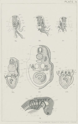
PLATE 5
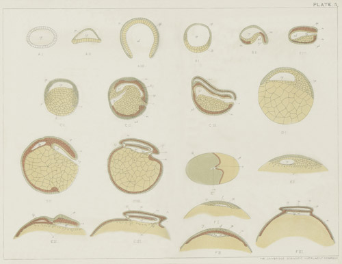
PLATE 6
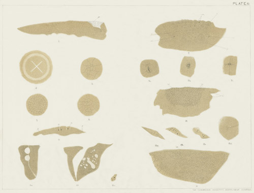
PLATE 7
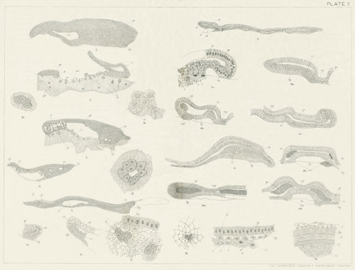
PLATE 8
PLATE 9
PLATE 10
PLATE 11
PLATE 12
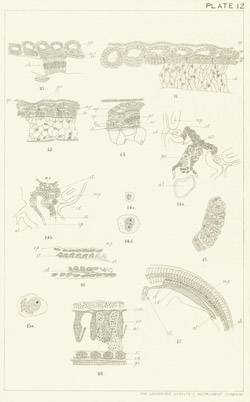
PLATE 13
PLATE 14
PLATE 15
PLATE 16
PLATE 17
PLATE 18
PLATE 19
PLATE 20
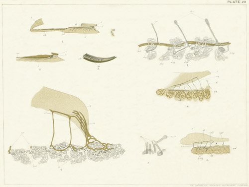
PLATE 21
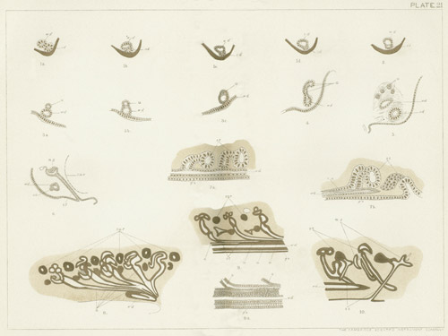
PLATE 22
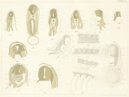
PLATE 23
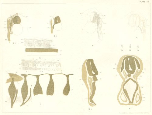
PLATE 24
PLATE 25
PLATE 26
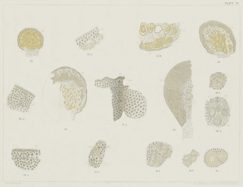
PLATE 27
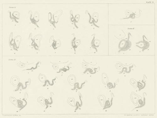
PLATE 28
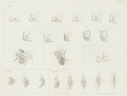
PLATE 29
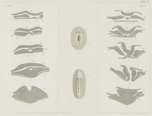
PLATE 30
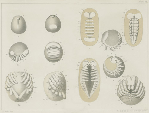
PLATE 31
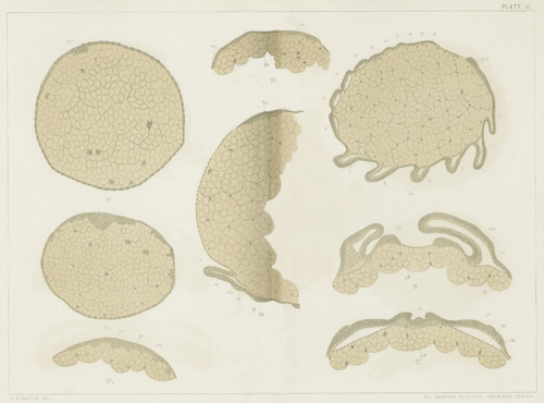
PLATE 32
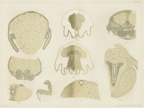
PLATE 33
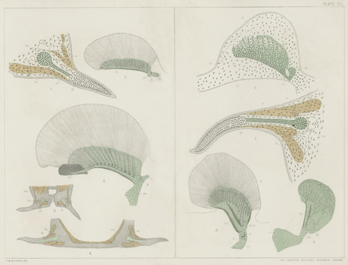
PLATE 34
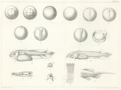
PLATE 35
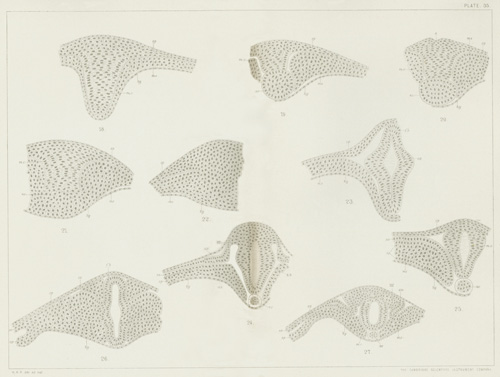
PLATE 36
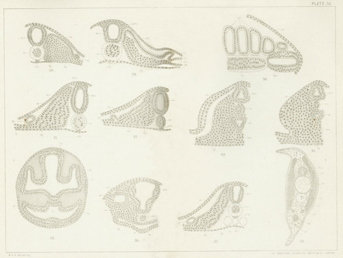
PLATE 37
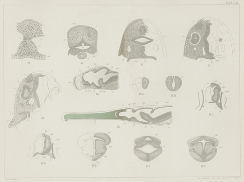
PLATE 38
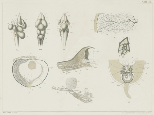
PLATE 39
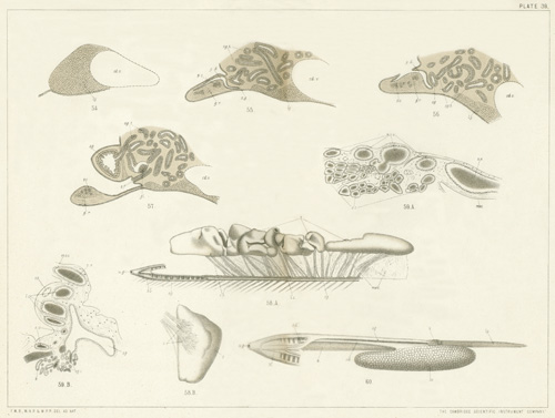
PLATE 40
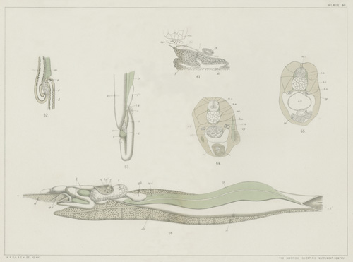
PLATE 41
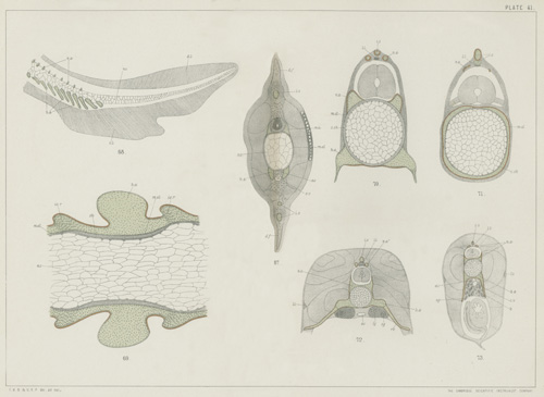
PLATE 42
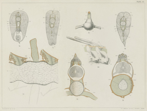
PLATE 43
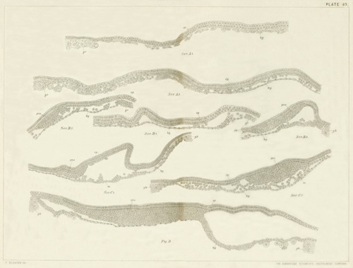
PLATE 44
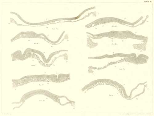
PLATE 45
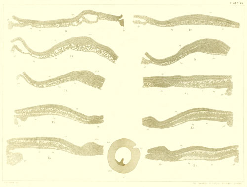
PLATE 46
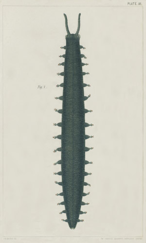
PLATE 47
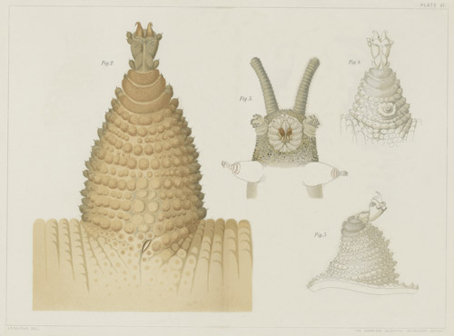
PLATE 48
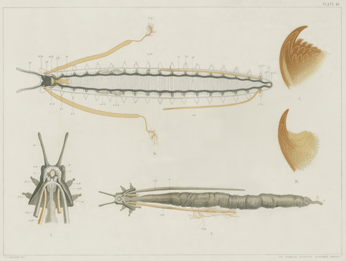
PLATE 49
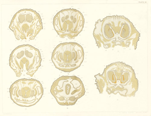
PLATE 50
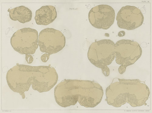
PLATE 51
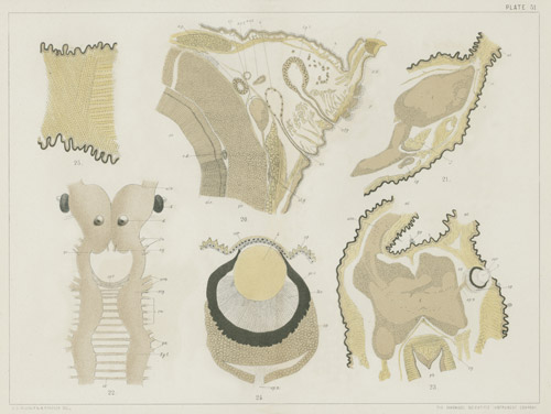
PLATE 52
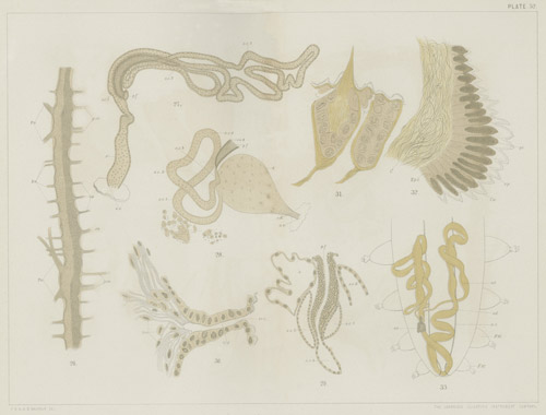
PLATE 53