Volume 2 — CHAPTER X. OF THE FUNCTION OF DIGESTION.
Reading edition
Process of Assimilation in the plant; in the animal—Digestive apparatus in the lower classes of animals; in the higher classes; in man—Digestive processes—Prehension, Mastication, Insalivation, Deglutition, Chymification, Chylification, Absorption, Fecation—Structure and action of the organs by which these operations are performed—Ultimate results—Powers by which those results are accomplished—Two kinds of digestion, a lower and a higher; the former preparatory to the latter.
548. Digestion is the function by which the aliment is converted into nutriment. No food can nourish until it be converted into a fluid analogous in chemical composition to that of the body by which it is assimilated. The conversion of the crude aliment into such a fluid is effected by a vital power peculiar to living beings, by which they subvert the constitution of other organized bodies, and cause them to assume their own. They accomplish this change by the agency of certain secretions which they elaborate in their own organs, and which they add to the substances they receive as aliment. By the action of these secretions, the chemical composition of the aliment is brought into a close affinity to that of the body which it nourishes.
549. This change in the chemical composition of the aliment, by means of fluids secreted by the living bodies which receive it, is manifest in the plant as well as in the animal. The sap, as it issues from the root, is a colourless and limpid fluid; it has a specific gravity a little greater than that of water; it has a sweetish taste; it contains an acid which is sometimes free, and is either the carbonic or the acetic; but more commonly it is combined with lime or potass. To this crude sap, in this the first stage of its formation, vegetable secretions, sugar and mucus, assimilative substances, are superadded, probably by the fibres of the root.
550. As the sap ascends in the stalk, a greater quantity and a greater number of these vegetable secretions are poured into it. In the ratio of its elevation it acquires sugar, mucus, albumen, and an azotized substance analogous to gluten. By the admixture of these assimilative secretions, the crude sap is progressively assimilated nearer and nearer to the chemical composition of the proper nutritive fluid of the plant. Thus prepared, the sap passes to the leaf, in the upper surface of which it undergoes a process analogous to that of digestion in the animal (315), and is converted into proper nutrient matter.
551. The plant can only take up, by absorption, liquid food; it never receives solid substances as aliment: it therefore needs no apparatus for the division, solution, and fluidification of its food; its sole work of assimilation consists in changing the innate affinities of liquid aliment. But animals which live on vegetable and animal substances have to modify, by their digestive juices, the affinities of organic solids: hence assimilation in the animal must necessarily be a more complex operation than it is in the plant.
552. Fixed immovably to the soil by its roots, the nutritive apparatus of the plant is always in contact with its food, which is slowly but unceasingly absorbed according to the wants of its system. But the animal endowed with the faculty of locomotion receives its aliment into the interior of its body, that it may transport its food along with it in all its changes of place; and that, as in the plant, its food may be always in contact with its nutritive apparatus. The interior nutrition of the animal and the convergence of its nutritive apparatus to the centre of its system, and the exterior nutrition of the plant and the divergence of its nutritive apparatus to the peripheral extremity of its body, are differences in their mode of nutrition, connected with essential differences in the mode of life peculiar to the two beings.
553. Plant-like animals have a plant-like mode of nutrition. The transition from the one class to the other is so gradual as to be almost insensible. Fixed to the same spot in the ocean as the tree to the land, the nutritive surface of the poriferous animal is always in contact with the water, as the soil is with the external surface of the plant. The cellular substance of which the bag of the poriferous animal is composed is permeated in all directions by ramifying and anastomosing canals, which, beginning by minute pores placed on the external surface, terminate in larger orifices, termed vents, which are fecal openings. These internal canals are incessantly traversed by streams of water, which enter through the minute, and are discharged through the larger orifices. By these currents the nutrient matter contained in the water is conveyed to every part of the body, and the streams that issue from the fecal orifices abound with minute flocculent particles, the residue of the digested matter. No separate part of the body is appropriated to the function of digestion any more than in the plant; there is merely a general absorbent surface; the water is to this animal what the soil is to the plant; its whole surface is a root; every point of that surface is constantly in contact with its food, and every point is absorbent.
554. In the class above the porifera, the margins of the superficial pores are merely lengthened out into minute sacs, irritable and sentient, surrounded with vibratile cilia (342). These sacs, which are termed polypi, are so many little stomachs, which select, seize, and digest the food brought to them in the currents of water created by the action of the cilia (344).
Fig. CXLVIII.—Hydra Viridis.
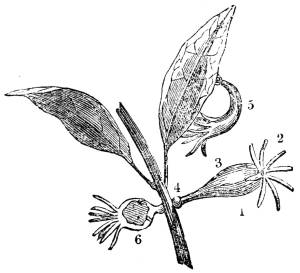
1. The Hydra with its tentacula expanded. 2. The tentacula. 3. The body of the Hydra. 4. Disc for attachment. 5. The Hydra in the act of creeping. 6. The Hydra with an animalcule in its digestive cavity.
555. The fresh-water polype, the little hydra (fig. CXLVIII. 1), is one of these minute sacs detached and endowed with the power of locomotion (fig. CXLVIII. 5), a sentient, self-moving digestive bag. Capable of swallowing animals many times its own size, as the red-blooded worm, this little creature stretches its whole body like a thin elastic membrane over its prey, so as completely to alter its own shape, and the membranous substance of which it is composed becoming transparent by the distention, allows the subsequent process to be distinctly seen. The red fluid of the worm, as the process of digestion advances, is slowly diffused over every part of the internal surface of the polype. The whole internal surface of this minute self-moving bag is digestive; a true and proper stomach (fig. CXLVIII. 6). By dexterous manipulation, this internal surface may be rendered external, and the animal turned completely inside out. Then the external begins to perform the office of the internal surface, carrying on the function of digestion, just as well as that which was primitively formed for it; while the originally digestive becomes the generative surface, for the creature buds from this surface, now the outer one; a striking and instructive illustration of the analogy between the external covering of the animal body or the skin, and its internal lining, or the mucous surface.
Fig. CXLIX.
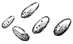
Group of Monades; the dark spots in the interior of their bodies representing their digestive sacs.
556. In the monades (fig. CXLIX.), and in all the lower animalcules, the digestive apparatus, instead of forming the entire internal surface of the body, consists of numerous sacs, which constitute so many separate stomachs, whence the name of the class, polygastrica. When empty, or when filled with water, these digestive sacs cannot be distinguished from the common cellular tissue of the body; but on feeding the animals with coloured organic matter, minutely diffused in water, the coloured particles readily enter the digestive sacs, and render apparent their form and arrangement. In the minutest animal hitherto appreciable, the monas termo, the 2000th part of a line in diameter, four rounded sacs have been seen filled with coloured particles (fig. CXLIX.). Each of these sacs, about the 6000th part of a line in diameter, opens by a narrow neck into a funnel-shaped mouth, surrounded with a single row of long vibratite cilia, by the action of which the floating organic particles are brought within the reach of the mouth. In general, even in this class, an alimentary canal traverses the whole extent of the body, into which all the different stomachs open. Sometimes numerous branches proceed from the main trunks of the alimentary canal, bearing the nutritive matter to the different parts of the body (fig. CL. 2). Often, in order to extend the digestive surface, the alimentary canal is produced, forming rounded enlargements called cœcal appendages, all of which act as so many additional stomachs (fig. CLI. 3). In some individuals, observed under favourable circumstances, nearly 200 of these cœcal stomachs, filled with coloured matter, have been counted, and there may have been many more unseen, because empty and collapsed. In the lowest tribes of this class there is but one orifice to the alimentary canal, the oral; the food entering, and the fecal matter passing out of the system by the same aperture; but in the higher orders there is both an oral and an anal orifice, and the mouth and the anus are placed at opposite extremities of the body, as in the higher animals.
Fig. CL.—Fasciola Hepatica.
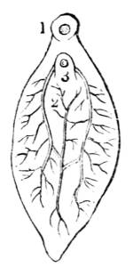
1. Mouth. 2. Alimentary tubes. 3. Sucker.
Fig. CLI.—Aphrodita Aculeata.
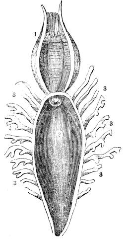
1. Proboscis in a retracted state. 2. Interior of digestive cavity. 3, 3. Cœcal appendages opening into it.
557. Up to this point in the animal series the digestive sacs and the alimentary canal are merely cavities formed in the common cellular tissue of the body, without any lining membrane, without teeth, or without any instruments for dividing and preparing the aliment, and without a single gland, as far as has been ascertained, to assist the digestive process. All the assimilative functions, the respiratory as well as the digestive, appear to be performed by this single surface. But in the ascending scale not only is an apparatus appropriated to digestion, perfectly distinct from that assigned to respiration, but even the stomach and the alimentary canal are separate organs, distinguished from each other, both in structure and function. Still higher in the scale new organs are successively added, as the process becomes more complex and refined, in order to assist the main operations carried on in particular parts of the apparatus; and as that apparatus approaches its highest degree of perfection, not only do the several parts of which it is composed increase in number and complexity, but each part becomes more and more isolated from the rest, a specific office being assigned to each in the division of labour that is made. Viewing, however, the digestive apparatus as a whole, whether simple or complex, whether consisting of a single uninterrupted surface, or divided into many separate portions, its nature is universally and invariably the same, and from the monad to man is endowed with analogous vital energies.
558. Comparative anatomy, which has succeeded in tracing through the different classes, orders, genera, and countless tribes of animals, the modifications in form and structure of the digestive apparatus, has shown that those modifications are invariably in strict adaptation to the kind of food on which the apparatus is destined to act and to the extent of the elaboration requisite to convert crude aliment into proper animal substance. To trace this adaptation through the rising and ever-varying series, is a most interesting and instructive study, not only exhibiting, in the very organs that elaborate its food, the physical and even the mental qualities assigned by the hand of nature to each individual, but oftentimes shedding a clear and bright light on the complex structures of the highest and most perfect organization. Striking and beautiful illustrations are afforded by these investigations of the principle formerly insisted on (vol. i. chap. i. p. 28, 3), that the communication of the higher faculties exalts the apparatus even of the very lowest processes, that the latter may work in harmony with the former. In conformity with this principle, as the nobler endowments exalt the animal in the scale of organization, so even its very digestive apparatus becomes extended, isolated, complex and refined.
559. The highest and most perfect form of the digestive apparatus is that which is disposed in a series of chambers in free communication with each other. In these chambers the food undergoes a succession of changes, by which it is progressively assimilated to the nature of animal substance. This assimilation, however, is never effected by the sole agency of the chambers themselves; it is accomplished, to a great extent, by the influence of special organs placed in the neighbourhood of the digestive chambers. In the lowest animal there is but one substance and one surface for every function; in the highest, even for the performance of the lowest function, there is the combination of many substances which are arranged in complex modes.
560. In man, the digestive chambers are five; the auxiliary organs are many.
The first of these chambers is the cavity called the mouth; the second is the bag termed the pharynx; the pharynx communicates by the esophagus with the third chamber, the stomach; the fourth chamber consists of the convoluted tubes named the small intestines, and the fifth consists of the larger tubes, denominated the large intestines. The assistant organs are, first, numerous appendages to the mouth, namely, the tongue, the teeth, the salivary glands, and the muscles that work the jaws; and, secondly, certain appendages to the small intestines, namely, the pancreas, the liver, the mesenteric glands, and the lacteal vessels.
561. By the mouth the food is softened and reduced to a pulp; by the tongue, materially aided by the soft palate, this pulp, when duly prepared, is transmitted to the pharynx; received by the pharynx, it is sent on to the esophagus; by the esophagus, it is conveyed to the stomach; in the stomach, it is converted into a peculiar substance called chyme; the chyme, passing from the stomach into the first portion of the small intestines, is there converted into the substance called chyle; the chyle, carried slowly along the remaining portion of the small intestines, is successively absorbed by the lacteals; by the lacteals, it is conveyed through the mesenteric glands to the thoracic duct, and by the thoracic duct it is poured into the venous blood close to the heart. By the large intestines the refuse matter is conveyed out of the system.
562. The function of digestion consists, then, of the following processes:—
1. Prehension. 2. Mastication. 3. Insalivation. 4. Deglutition. 5. Chymification. 6. Chylification. 7. Absorption. 8. Fecation.
563. Prehension is the reception of the aliment; mastication is the mechanical comminution of it; insalivation is the admixture of it with certain juices poured into the mouth; deglutition is the transmission of it, when duly moistened and divided, into the stomach; chymification is the conversion of it into chyme; chylification is the conversion of the chyme into chyle; absorption is the assumption of the chyle by the lacteals and the transmission of it into the blood, and fecation is the separation and discharge of the refuse matter. Each part of this extended apparatus is modified in structure so as specially to fit it for the performance of the office which is appropriated to it.
564. The mouth is not merely the opening between the two lips, but consists of an oval chamber, bounded above by the upper jaw and the palate; below by the tongue and the lower jaw; laterally by the cheeks; behind by the soft palate; and before by the lips.
565. The upper and lower jaw, the palate bones, and the teeth, constitute the hard or the bony parts of the mouth. The soft parts consist of the lips, the cheeks, the soft palate, the tongue, and the mucous membrane which lines the whole.
566. The lips and cheeks are composed principally of muscles, covered on the outside by the skin, and lined on the inside by the mucous membrane of the mouth. In the interspaces between the muscles is disposed a quantity of fat, which gives form to the face, facilitates the movements of the muscles, and protects the glands, blood-vessels, and nerves, with which all these organs are most abundantly supplied.
567. The roof of the mouth, called the palate, consists partly of bony and partly of membranous substance. The bony part of the palate forms an arch in the upper jaw, the position of which in the erect posture is horizontal: the membranous part of the palate consists of the mucous membrane of the mouth, which affords a covering to the bony part of the palate.
Fig. CLII.—View of the Mouth, showing particularly the Soft Palate, Tonsils, and Tongue.
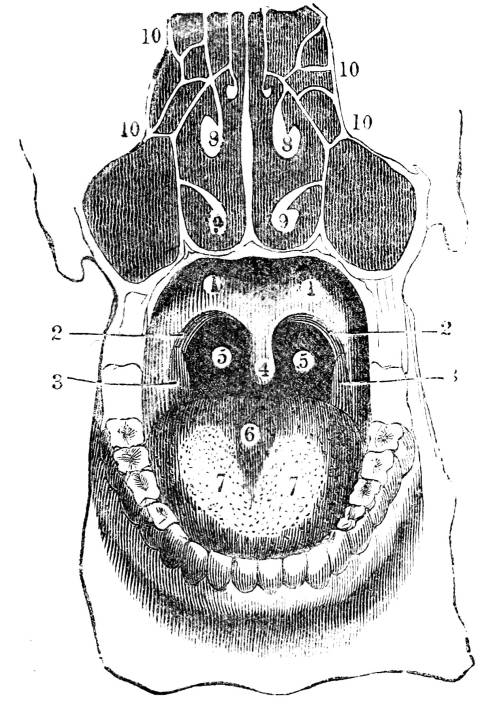
1. Anterior arch of the soft palate. 2. Posterior arch. 3. Tonsils or amygdalæ. 4. Uvula. 5. Communication between the mouth and pharynx. 6. The tongue. 7. Anterior or nervous papillæ. 8 and 9. The upper and lower turbinated bones dividing the nostrils into (10) chambers.
Fig. CLIII.—A side view of the Mouth, Pharynx, Nose, &c.
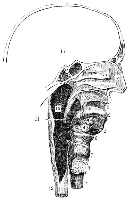
1. Mouth. 2. Tongue. 3. Section of the lower jaw. 4. Submaxillary gland. 5. Sublingual gland. 6. Hyoid bone. 7. Thyroid cartilage. 8. Thyroid gland. 9. Trachea. 10. Interior of the pharynx. 11. Section of the soft palate. 12. The esophagus. 13. The interior of the nose. 14. The two spongy bones dividing it into three chambers. 15. The posterior communication with the upper part of the pharynx.
Fig. CLIV.—Posterior view of the Nose, Mouth, Larynx, and Pharynx laid open.
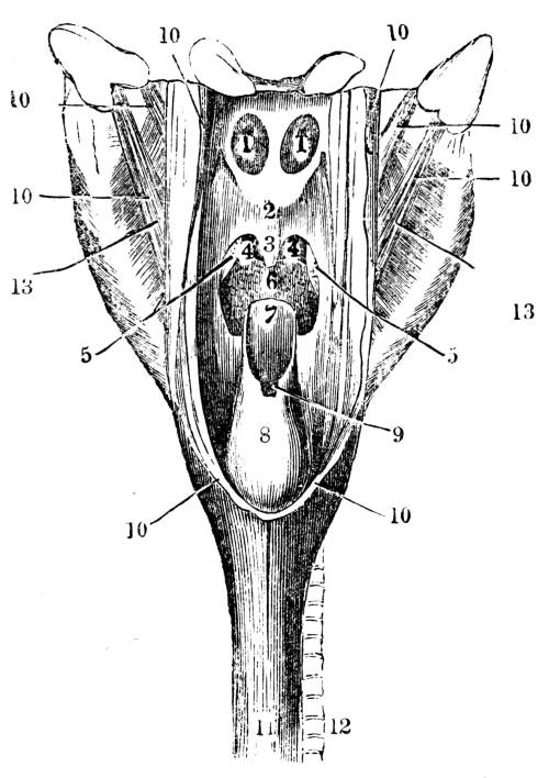
1. Posterior openings of the nose, communicating with the upper part of the pharynx. 2. Posterior surface of the soft palate. 3. The uvula. 4. Back part of the mouth communicating with the pharynx. 5. The tonsils. 6. Back part or root of the tongue. 7. Posterior surface of the epiglottis. 8. The larynx. 9. The opening of the larynx into the pharynx. 10. Cut edges of the pharynx. 11. Esophagus, the continuation of the pharynx. 12. The Trachea, continuation of the larynx. 13. Muscles acting on the pharynx.
568. From the posterior part of the bony arch of the palate is suspended, transversely, a moveable partition, called the soft palate (fig. CLII. 1 and 2), which is composed of muscular fibres enclosed in the mucous membranes of the mouth. No less than ten distinct muscles enter into the composition of the soft palate. These muscles are disposed in such a manner that they render the organ capable of descending and of applying itself against the tongue (fig. CLII. 6), so as completely to close the passage between the mouth and the pharynx (figs. CLII. 5, and CLIV. 1), and of ascending and carrying itself obliquely backwards towards the posterior wall of the pharynx, so as completely to close the passage between the pharynx and the nose (fig. CLIV. 2, 1); hence this moveable partition performs the office of a double valve, closing the passage from the mouth to the pharynx, and from the pharynx to the nose.
569. From the centre of the soft palate hangs pendulous the conical-shaped body called the uvula (fig. CLII. 4), which consists of a small muscle enveloped in the mucous membrane of the mouth. The uvula assists in completing the valve formed by the soft palate (fig. CLIV. 2, 3); it is also an important organ in the modulation of the voice. When destroyed by disease, both the deglutition of the food and the sound of the voice become imperfect.
570. The lateral edges of the soft palate separate into two layers, which enclose between them the bodies called the tonsils (fig. CLII. 3), two glands commonly about the size of an almond. These organs co-operate with other glands in secreting the fluids of the mouth.
571. The tongue (figs. CLII. 6, and CLIII. 2) is composed of six distinct muscles enveloped in the mucous membrane of the mouth. The fibres of these muscles are so interwoven with each other as to form an intricate net-work; and their number, arrangement, and exquisite organization render the organ capable of executing a surprising variety of movements with the most perfect precision, and with a velocity of which the mind can scarcely form a conception: some of these movements being requisite to bring the aliment under the operation of mastication, and others to produce articulate speech.
572. The tongue divided into base, apex, and dorsum, is supported by a bone called the hyoid bone (os hyoides) (figs. CXXXVI. 1, and CLIII. 6), which, unlike any other bone of the body, is placed at a distance from the general skeleton, and completely imbedded in muscles. This singularly posted and delicately constructed bone is not only connected with the tongue, but with many other highly important muscles, to which it affords a support and a lever.
573. Each jaw is provided with sixteen teeth (fig. CLV.), arranged with perfect uniformity, eight on each side of each jaw (fig. CLV.); those of the one side exactly corresponding with those of the other (fig. CLV.). The teeth, from the differences they present in their size, form, mode of connection with the jaw, and use, are divided into four classes, namely, on each side of each jaw, two incisors (figs. CLVI. and CLVII. 1, 2); one cuspid (figs. CLVI. and CLVII. 3); two bicuspid (figs. CLVI. and CLVII. 4, 5); and three molars (figs. CLVI. and CLVII. 6, 7, 8).
Fig. CLV.
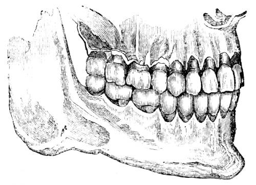
A lateral view of the whole series of the teeth, in situ, showing the relative situation of those of the upper with those of the lower jaw. This figure and the following figures to 159, are copied from Mr. T. Bell’s scientific and instructive work on the Anatomy, Physiology, and Diseases of the Teeth.
574. The incisor, or cutting teeth, are situated in the front of the jaw; that directly in the centre is called the central; and the next to it the lateral incisor (fig. CLV.). Their office, as their name imports, is to cut the food, which they do, on the principle of shears or scissors.
575. Standing next to the lateral incisor is the cuspid, canine, or eye-tooth (figs. CLV. , CLVI. , and CLVII. ). It is the longest of all the teeth. Its office is to tear such parts of the food as are too hard to be readily divided by the incisors.
576. Next the cuspid are the bicuspid, two on each side (fig. CLV., CLVII. ), so named from their being provided with two distinct prominences or points. Their office is to tear tough substances preparatory to their trituration by the next set.
Fig. CLVI.
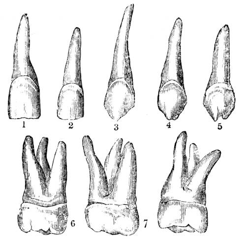
Front or external view of the upper teeth. 1. The central incisor. 2. The lateral incisor. 3. The cuspid. 4. The first bicuspid. 5. The second bicuspid. 6. The first molar. 7. The second molar. 8. The third molar, or dens sapientiæ.
577. The molars, or the grinders, three on each side (fig. CLVI. and CLVII. ), provided with four or five prominences on the grinding surface, with corresponding depressions, which are so arranged that the elevations of those of the upper are adapted to the concavities of those of the lower jaw, and the contrary.
Fig. CLVII.
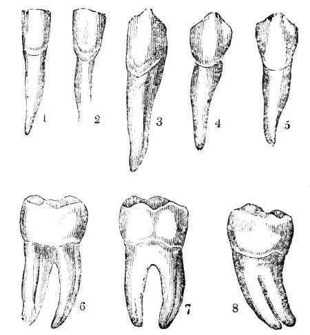
Front view of the lower teeth. 1. The central incisor. 2. The lateral incisor. 3. The cuspid. 4. The first bicuspid. 5. The second bicuspid. 6. The first molar. 7. The second molar. 8. The third molar, or dens sapientiæ.
578. From the incisor to the molar teeth there is a regular gradation in size, form, and use, the cuspid holding a middle place between the incisor and the bicuspid, and the bicuspid being in every respect intermediate between the cuspid and the molar. Thus the incisor are adapted only for cutting, the cuspid for tearing, the bicuspid partly for tearing and partly for grinding, and the molar solely for grinding. The incisor has only a single root, which is nearly round, and quite simple (fig. CLVII. 1, 2); the cuspid has only a single root, but this is flattened and partially grooved (fig. CLVII. 3); even the bicuspid has only a single root, but this is commonly divided at its extremity, and is always so much grooved as to have the appearance of two fangs partially united, the body having two points instead of one, thus approaching it to the form of the molar (fig. CLVII. 4, 5); and these last have always two, sometimes three, occasionally four roots, and their body is greatly increased in size, and has a complete grinding surface (fig. CLVII. 6, 7, 8).
579. In some animals whose food and habits require the utmost extension of the office of a particular class of teeth, a corresponding development of that class takes place. Thus in the carnivora, as is strikingly seen in the tiger and the polar bear, the cuspid or canine teeth are prodigiously elongated and strengthened, in order to enable them to seize their food, and to tear it in pieces. On the other hand, in the rodentia, or gnawing animals, as in the beaver, the incisors are exceedingly elongated; while in the graminivora, and especially in the ruminantia, the molar teeth are by far the most developed. In each case the other kinds of teeth are of little comparative importance; sometimes they are even altogether wanting. Thus the shark has only one kind of tooth, the incisor; but of these there are several rows, and all of them the creature has the power of erecting at will.
580. So intimately are these organs connected with the kind of food by which life is sustained, and the kind of food with the general habits of the animal, that an anatomist can tell the structure of the digestive organs, the kind of nervous system, the physical and even the mental endowments; that is, the exact point in the scale of organization to which the animal belongs, merely by the inspection of the teeth.
581. In man, the several classes of the teeth are so similarly developed, so perfectly equalized, and so identically constructed, that they may be considered as the true type from which all the other forms are deviations.
582. For the accomplishment of their office the teeth must be endowed with prodigious strength: for the fulfilment of purposes immediately connected with the apparatus of digestion, it is necessary that they should be placed in the neighbourhood of exceedingly soft, delicate, irritable, and sentient organs. That they may possess the requisite degree of strength, they are constructed chiefly of bone, the hardest organized substance. Bone, though not as sensible as some other parts of the body, is nevertheless sentient. The employment of a sensitive body in the office of breaking down the hard substances used as food would be to change the act of eating from a pleasurable into a painful operation. It has been shown (vol. i. p. 84) that provision is made for supplying to the animal a never-failing source of enjoyment in the annexation of pleasurable sensations with the act of eating, and that, taking the whole of life into account, the sum of enjoyment secured by this provision is incalculable. But all this enjoyment might have been lost, might even have been changed into positive pain, nay, must have been changed into pain, but for adjustments numerous, minute, delicate, and, at first view, incompatible.
583. Had a highly-organized and sensitive body been made the instrument of cutting, tearing, and breaking down the food, every tooth, every time it comes in contact with the food, would produce the exquisite pain now occasionally experienced when a tooth is inflamed. Yet a body wholly inorganic and therefore insensible, could not perform the office of the instrument; first, because a dead body cannot be placed in contact with living parts without producing irritation, disease, and consequently pain; and, secondly, because such a body being incapable of any process of nutrition, must speedily be worn away by friction, and there could be no possibility of repairing or of replacing it. The instrument in question, then, must possess hardness, durability, and, to a certain extent, insensibility; yet it must be capable of forming an intimate union with sentient and vital organs, must be capable of becoming a constituent part of the living system.
584. To communicate to it the requisite degree of hardness, the hard substance forming its basis is rendered so much harder than common bone that some physiologists have even doubted whether it be bone, whether it really possess a true organic structure. That there is no ground for such doubt the evidence is complete. For,
1. The tooth, like bone in general, is composed partly of an earthy and partly of an animal substance; the earthy part being completely removable by maceration in an acid, and the animal portion by incineration, the tooth under each process retaining exactly its original form.
2. The root of the tooth is covered externally by periosteum; its internal cavity is lined by a vascular and nervous membrane, and both structures are intimately connected with the substance of the tooth. If these membranes really distribute their blood-vessels and nerves to the substance of the tooth, which there is no reason to doubt, the analogy is identical between the structure of the teeth and that of bone.
3. Though the blood-vessels of the teeth are so minute that they do not, under ordinary circumstances, admit the red particles of the blood, and though no colouring matter hitherto employed in artificial injections has been able, on account of its grossness, to penetrate the dental vessels, yet disease sometimes accomplishes what art is incapable of effecting. In jaundice the bony substance of the teeth is occasionally tinged with a bright yellow colour; and in persons who have perished by a violent death, in whom the circulation has been suddenly arrested, it is of a deep red colour. Moreover, when the dentist files a tooth, no pain is produced until the file reaches the bony substance; but the instant it begins to act upon this part of the tooth, the sensation becomes sufficiently acute.
585. These facts demonstrate that the bony matter of the tooth, though modified to fit the instrument for its office, is still a true and proper organized substance.
586. Each tooth is divided into body, neck, and root (fig. CLVIII. 1, 2, 3). The body is that part of the tooth which is above the gum, the root that part which is below the gum, and the neck that part where the body and the root unite (fig. CLVIII. ). The body, the essential part, is the tooth properly so called, the part which performs the whole work for which the instrument is constructed, to the production and support of which all the other parts are subservient.
Fig. CLVIII.
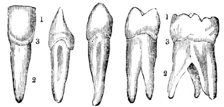
Views of different kinds of teeth, showing their anatomical division into, 1. The body or crown. 2. The fang or root. 3. The neck.
Fig. CLIX.—Sections of Teeth, exhibiting their Structure.
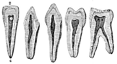
1. The bony substance. 2. The enamel. 3. The internal cavity. 4. The foramen, or hole at the extremity of the root.
587. When a vertical section is made in the tooth, it is found to contain a cavity of considerable size (fig. CLIX, 3), termed the dental cavity, which, large in the body of the tooth, gradually diminishes through the whole length of the root (fig. CLIX. 3). The dental cavity is lined throughout with a thin, delicate, and vascular membrane, continued from that which lines the jaw. It contains a pulpy substance. This pulp, highly vascular and exquisitely sensible, is composed almost entirely of blood-vessels and nerves, and is the source whence the bony part of the tooth derives its vitality, sensibility, and nutriment. The blood-vessels and nerves that compose the pulp enter the dental cavity through a minute hole at the extremity of the root (fig. CLIX. 4). The membrane which lines the dental cavity is likewise continued over the external surface of the root, so as to afford it a complete envelope.
588. Provision having been thus made for the organization of the tooth, for the support of its vitality, and for its connexion with the living system, over all that portion of it which is above the gum, and which constitutes the essential part of the instrument, there is poured a dense, hard, inorganic, insensible, all but indestructible substance, termed enamel (fig. CLIX. 2); a substance inorganic, composed of earthy salts, principally phosphate of lime with a slight trace of animal matter: a substance of exceeding density, of a milky-white colour, semi-transparent, and consisting of minute fibrous crystals. The manner in which this inorganic matter is arranged about the body of the tooth is worthy of notice. The crystals are disposed in radii springing from the centre of the tooth (fig. CLX. 3); so that the extremities of the crystals form the external surface of the tooth, while the internal extremities are in contact with the bony substance (fig. CLX. 3). By this arrangement a two-fold advantage is obtained; the enamel is less apt to be worn down by friction, and is less liable to accidental fracture.
Fig. CLX.
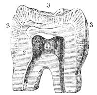
Magnified section of a tooth, to illustrate the arrangement of the fibrous crystals composing the enamel. 1. Cavity of the tooth. 2. Bony substance. 3. Enamel, showing the crystals disposed in radii.
589. In this manner an instrument is constructed possessing the requisite hardness, durability, and insensibility; yet organized, alive, as truly an integrant portion of the living system as the eye or the heart.
590. No less care is indicated in fixing than in constructing the instrument. It is held in its situation not by one expedient, but by many.
1. All along the margin of both jaws is placed a bony arch, pierced with holes, which constitute the sockets, called alveoli, for the teeth (fig. CLXI. ). Each socket or alveolus is distinct, there being one alveolus for each tooth (fig. CLXI. ). The adaptation of the root to the alveolus is so exact, and the adhesion so close, that each root is fixed in its alveolus just as a nail is fixed when driven into a board.
Fig. CLXI.
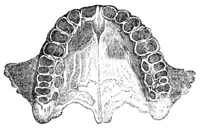
Upper jaw, showing the alveoli.
2. The roots of the tooth, when there are more than one, deviate from a straight line (fig. CLVI. 6, 7, 8); and this deviation from parallelism, on an obvious mechanical principle, adds to the firmness of the connexion.
3. Adherent by one edge to the bony arch of the jaw, and by the other to the neck of the tooth, is a peculiar substance, dense, firm, membranous, called the gum, less hard than cartilage, but much harder than skin, or common membrane; abounding with blood-vessels, yet but little sensible; constructed for the express purpose of assisting to fix the teeth in their situation.
4. The dense and firm membrane covering the bony arch of the jaw is continued into each alveolus which it lines; from the bottom of the alveolus this membrane is reflected over the root of the tooth, which it completely invests as far as the neck, where it terminates, and where the enamel begins: this membrane, like a tense and strong band, powerfully assists in fixing the tooth.
5. Lastly, the vessels and nerves which enter at the extremity of the root, like so many strings, assist in tying it down; hence, when in the progress of age, all the other fastenings are removed, these strings hold the teeth so firmly to the bottom of the socket, that their removal always requires considerable force.
591. But a dense substance like enamel, acting with force against so hard a substance as bone, would produce a jar which, propagated along the bones of the face and skull to the brain, would severely injure that tender organ, and effectually interfere with the comfort of eating.
592. This evil is guarded against,
1. By the structure of the alveoli (fig. CLXII.), which are composed not of dense and compact, but of loose and spongy bone (fig. CLXII.). This cancellated arrangement of the osseous fibres is admirably adapted for absorbing vibrations and preventing their propagation (90).
2. By the membrane which lines the socket.
3. By the membrane which covers the root of the tooth; and,
4. By the gum.
Fig. CLXII.
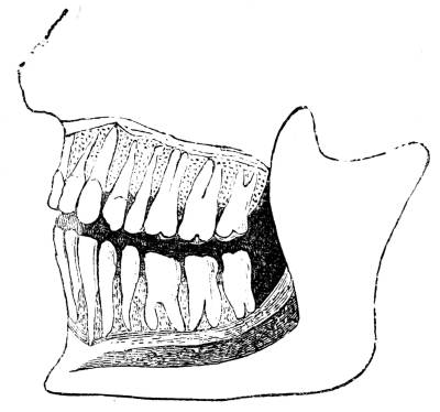
View of the upper and lower teeth in the alveoli; the external alveolar plate being cut away to show the cancellated structure of the alveoli, and the articulation of the teeth.
These membranous substances, even more than the cancellated structure of the alveoli, absorb vibrations and counteract the communication of a shock to the bones of the face and head when the teeth act forcibly on hard materials; so many and such nice adjustments go to secure enjoyment, nay to prevent exquisite pain, in the simple operation of bringing the teeth into contact in the act of eating.
Fig. CLXIII.
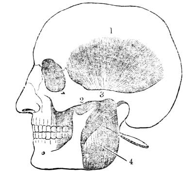
1. The temporal muscle. 2. Its insertion passing beneath. 3. The zygoma. 4. The masseter muscle, its anterior portion reflected to show the insertion of the temporal. The action of these powerful muscles is to pull the lower jaw upwards with great force against the upper jaw, and at the same time to draw it a little forwards or backwards, according to the direction of the fibres of the muscles.
593. The teeth in mastication are passive instruments put in motion by the jaws. The upper jaw is fixed, the lower only is movable. The lower jaw is capable of four different motions; depression, elevation, a motion forwards and backwards, and partial rotation. These simple motions are capable, by combination, of producing various compound motions. Numerous muscles, some of them endowed with prodigious power, are so disposed and combined as to be able, at the command of volition, to produce any of these motions that may be required, simple or compound.
Fig. CLXIV.—Muscles of the Jaw.
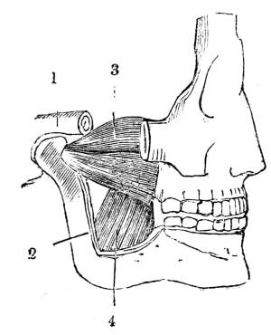
1. Portion of the zygomatic process of the temporal bone. 2. Ascending plate of the lower jaw removed to expose, 3. External pterygoid, and, 4. Internal pterygoid muscles. The action of these muscles is to raise the lower jaw, and to pull it obliquely towards the opposite side. When both muscles act together, they bring the lower jaw forwards, so as to make the fore-teeth project beyond those of the upper jaw.
594. By the combination, succession, alternation, and repetition of these motions, the lower is made to produce upon the upper jaw all the variety of pressure necessary for the mastication of the food. In this process the muscles of the tongue perform scarcely a less important part than the muscles of the lower jaw. Some of its muscular fibres shorten the tongue, some give it breadth, others render it concave, and others convex: so ample is the provision for moving this organ to different parts of the mouth and fauces, whether to bruise the softer parts of the aliment against the palate, to mix it with the saliva, or to place it under the pressure of the teeth.
595. By the combined action of the muscles of the lower jaw and tongue, and that of the teeth, the food is bruised, cut, torn, and divided into minute fragments. This operation is of so much importance that the whole process of digestion is imperfect without it. It is proved by direct experiment that the stomach acts upon the aliment with a facility in some degree proportionate to the perfection with which it is masticated. If an animal swallow morsels of food of different bulks, and the stomach be examined after a given time, digestion is found to be the most advanced in the smallest pieces, which are often completely softened, while the larger are scarcely acted upon at all.
596. At the same time that, by the operation of mastication, the aliment undergoes mechanical division, it imbibes a quantity of fluid derived from various sources.
1. From the membrane which lines the internal surface of the mouth, and which affords a covering to all the parts contained in it.
2. From numerous minute glands placed in clusters about the cheeks, gums, lips, palate, and tongue. Each of these glands is furnished with its own little duct, which, piercing the mucous membrane, opens into the cavity of the mouth. From these glands is derived the fluid with which the interior of the mouth is lubricated. It consists of a glutinous, adhesive, transparent fluid, of a light grey tint, salt taste, and slightly alkaline nature, termed mucus.
Fig. CLXV.—View of the Parotid Gland with the Muscles of the Face.
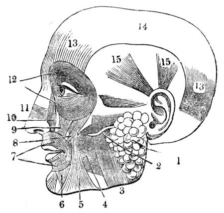
1. Parotid gland. 2. Parotid duct. 3. Masseter muscle. 4. Buccinator. 5. Triangularis, or depressor of the angle of the mouth. 6. Depressor of the lower lip. 7. Orbicularis, or circular muscle of the mouth. 8. Great zygomatic, or the distorter of the mouth, as in laughing. 9. Elevator of the angle of the mouth. 10. Elevator of the upper lip, and wing of the nose. 11. Compressor of the cartilage of the nose. 12. Orbicularis, or circular muscle of the eyelids. 13. Occipito frontalis; elevator of the eyelids; motor of the scalp, &c., an important muscle of expression. 14. Tendinous portion of the occipito frontalis. 15. Elevator of the ear.
3. From six large glands placed symmetrically, three on each side, termed the salivary glands, namely, the parotid (fig. CLXV. 1), situated before the ear; the submaxillary (fig. CLIII. 4), situated beneath the lower jaw; and the sublingual (fig. CLIII. 5), situated immediately under the tongue. Each of these glands is provided with a duct (figs. CLXV. 2, and cliii. 4, 5), by which it pours the fluid it elaborates, called saliva, into the mouth.
597. The other fluids of the mouth are always mixed with the saliva, and are all commonly included under that name. The secretion of these fluids is unceasing, and they pass into the stomach by successive acts of deglutition at nearly regular intervals; so that the stomach, after it has been some time without food, contains a considerable quantity of these fluids. But they are chiefly needed during the operation of mastication, and two provisions are made for securing their flow in the greatest abundance at that time.
598. First, the situation of the glands is such that they are all exposed to the action of the muscles of mastication (figs. CLXIII. and CLXIV. ), by which action the glands are excited, a large quantity of blood is determined to them, and the quantity of fluid they secrete is proportionate to the quantity of blood they receive. Secondly, the glands are placed under the influence of the mind, so that the very thought, and still more the taste, of grateful food, acting upon them as an additional stimulus, causes an additional secretion. The quantity of fluid formed from these different sources, and mixed with the food during the mastication of an ordinary meal, is estimated at half a pint. It must commonly be more than this, because, in a case described by Dr. Gairdner, of Edinburgh, in which the esophagus had been cut through, it was observed that from six to eight ounces of saliva were discharged during a meal, which consisted merely of broth injected through the divided esophagus into the stomach.
599. Saliva is a frothy, watery fluid, in its healthy state nearly insipid, and of a slightly alkaline nature. It is composed of water, a peculiar animal substance called salivary matter, mucus, osmazome, a little albumen, and several salts. It produces important changes on the food. By the water, and the salts contained in it, it softens and dissolves the food; and thus, while it renders it easier to be swallowed, it prepares it for the subsequent changes it is to undergo. To this latter object, the assimilation of the food, it seems to communicate the first tendency by the azotized substances, the salivary, and the albuminous matter which it adds to it. From this, the commencement of the assimilative process to its completion, animalized substances are successively added to the food which have the property of converting the food more and more into the nature of animal substance.
600. Comminuted by the teeth, and softened by the saliva, the food is reduced to a pulp. In this pulp there is a complete admixture of all the alimentary substances with the assimilative matter secreted from the blood, into the nature of which it is to be ultimately changed. The mass is at the same time brought to the temperature of the blood.
601. As long as the operations of mastication and insalivation go on, the mouth forms a closed cavity from which the food cannot escape; for the lips enclose it before, the cheeks at the sides, the tongue below, and the soft palate behind, the inferior edge of which being applied in close and firm contact with the base of the tongue, prevents all communication between the mouth and the pharynx.
602. When, by mastication, the food is sufficiently divided, and by insalivation softened and animalized to fit it for the future changes it is to undergo, it is collected by the tongue, and carried by that organ to the back part of the mouth. The soft palate (fig. CLII. 1), obedient to the stimulus of the duly prepared food, rises the instant it is touched by it, and affords it a free passage to the pharynx (figs. CLIII. . 10, and CLIV. . 10).
603. The pharynx (fig. CLIII. 10), a muscular bag, immediately continuous with the mouth (fig. CLIII. 1), is a vestibule into which open several highly important organs. Before is the entrance to the windpipe, termed the glottis (fig. CLIV. 9), leading directly to the larynx (fig. CLIV. 8); at the sides are the mouths of two ducts, termed the Eustachian tubes, which lead to the internal part of the organ of hearing; above are two entrances to the nose (fig. CLIV. 1); and below is the passage to the stomach (fig. CLIII. 12).
604. Were the food to enter the Eustachian tubes or the nose, it would occasion great inconvenience; were it to enter the glottis, it would cause death. It is prevented from entering the Eustachian tubes and the nose by the soft palate (fig. CLII. 1 and 2), which by the very act of rising to afford an opening from the mouth to the pharynx, is carried over the other apertures so as completely to close them. By the varied direction of the muscular fibres which enter into the composition of this organ, it is enabled to execute the different and even opposite motions required in the performance of its important office.
605. The food is prevented from entering the glottis partly by a cartilaginous valve (fig. CLIV. 7), termed the epiglottis, placed immediately above the glottis, and attached to the root of the tongue (fig. CLIV. 6). In delivering the food to the pharynx the tongue passes backwards (fig. CLIV. 6). In passing backwards it pushes in the same direction the epiglottis which is attached to it, and so necessarily carries it over the glottis, completely closing the aperture (fig. CLIV. 9). At the same time the opening is still more securely closed by the glottis itself, in consequence of the powerful and simultaneous contraction of the muscles that act upon it in the production of the voice. It is proved, by direct experiment, that the spontaneous closure of the glottis is a more powerful agent in excluding the food from the larynx even than the depression of the epiglottis; but both organs concur in producing the same result; and a double security is provided against an event which would be fatal.
606. It is deeply interesting to observe the part performed in these operations by sensation and volition, and the boundary at which their influence terminates and consciousness itself is lost. Mastication, a voluntary operation, carried on by voluntary muscles, at the command of the will, is attended with consciousness, always in the state of health of a pleasurable nature. To communicate this consciousness, the tongue, the palate, the lips, the cheeks, the soft palate, and even the pharynx, are supplied with a prodigious number of sentient nerves. The tongue especially, one of the most active agents in the operation, is supplied with no less than six nerves derived from three different sources. These nerves, spread out upon this organ, give to its upper surface a complete covering, and some of them terminate in sentient extremities visible to the naked eye. These sentient extremities, with which every point of the upper surface, but more especially the apex, is studded, constitute the bodies termed papillæ, the immediate and special seat of the sense of taste. This sense is also diffused, though in a less exquisite degree, over the whole internal surface of the mouth. Close to the sense of taste is placed the seat of the kindred sense of smell. The business of both these senses is with the qualities of the food. Mastication at once brings out the qualities of the food and puts the food in contact with the organs that are to take cognizance of it. Mastication, a rough operation, capable of being accomplished only by powerful instruments which act with force, is carried on in the very same spot with sensation, an exquisitely delicate operation, having its seat in soft and tender structures, with which the appropriate objects are brought into contact only with the gentlest impulse. The agents of the coarse and the delicate, the forcible and the gentle operations are in close contact, yet they work together not only without obstruction, but with the most perfect subserviency and co-operation.
607. The movements of mastication are produced, and, until they have accomplished the objects of the operation, are repeated by successive acts of volition. To induce these acts, grateful sensations are excited by the contact of the food with the sentient nerves so liberally distributed over almost the whole of the apparatus. To the provision thus made for the production of pleasurable sensation, is superadded the necessity of direct and constant attention to the pleasure included in the gratification of the taste. It is justly observed by Dr. A. Combe, that without some degree of attention to the process of eating, and some distinct perception of its gratefulness, the food cannot be duly digested. When the mind is so absorbed as to be wholly unconscious of it, or even indifferent to it, the food is swallowed without mastication; then it lies in the stomach for hours together without being acted upon by the gastric juice, and if this be done often, the stomach becomes so much disordered as to lose its power of digestion, and death is the inevitable result: so that not only is pleasurable sensation annexed to the reception of food, but the direct and continuous consciousness of that pleasurable sensation during the act of eating is made one of the conditions of the due performance of the digestive function.
608. With the operation of mastication and one part of the process of deglutition, immediately to be noticed, the agency of volition and sensation cease. Beyond this the function of digestion is wholly an organic process. In addition to the reasons assigned (vol. i. p. 55) why all the organic processes are placed alike beyond the cognizance of sense and the control of the will, there is this special reason why, in the function of digestion, they cease at the exact boundary assigned them.
609. Every time the act of deglutition is performed the openings to the windpipe and to the nose are closed, so that during this operation all access of air to the lungs is stopped, consequently it is necessary that the passage of the food through the pharynx should be rapid. Mastication, a voluntary process, may be performed slowly or rapidly, perfectly or imperfectly, without serious mischief; but life depends on the passage of the food through the pharynx with extreme rapidity and with the nicest precision. It is therefore taken out of the province of volition and entrusted to organs which belong to the organic life, organs which carry on their operations with the steadiness, constancy, and exactness of bodies whose motions are determined by a physical law.
610. No sooner does the duly-prepared food touch the soft palate than the whole apparatus of deglutition is instantly in motion. This movable partition suddenly rises to afford to the food a free passage to the pharynx. The pharynx itself, at the same instant, rises to receive the morsel thrust towards it by the pressure of the tongue; and one muscle, the stylo-pharyngeus, which concurs in producing this movement, seems specially intended, in addition, to expand the pharynx. Three muscles throw their fibres around the pharynx, termed its upper, middle, and lower constrictors, which, the moment the morsel reaches the pharynx, contract upon it, and embrace it firmly. At the same instant the larynx, closing its aperture, springs forward towards the base of the tongue, under which it is in a manner concealed, the additional shield of the epiglottis being simultaneously thrown over the glottis. By this movement of the larynx, upwards and forwards, the course of the morsel across the dangerous passage is shortened. All these motions take place with such rapidity that Boerhaave said the action is convulsive. And now the food, firmly pressed by the pharynx, cannot return to the mouth, for the root of the tongue is there stopping up the passage; it cannot enter the Eustachian tubes or the nose, for the soft palate is there closing the apertures; it cannot enter the larynx, for a double guard is placed upon the glottis securing its firm closure. The food can advance in one direction only, the direction required, that which leads to the esophagus. Well, therefore, on the contemplation of these complex structures and the consent and harmony with which they act, might Paley say, “In no apparatus put together by art do I know such multifarious uses so aptly contrived as in the natural organization of the human mouth and its appendages. In this small cavity we have teeth of different shape; first, for cutting; secondly, for grinding; muscles most artificially disposed for carrying on the compound motions of the lower jaw by which the mill is worked; fountains of saliva springing up in different parts of the cavity for the moistening of the food while the mastication is going on; glands to feed the fountains; a muscular contrivance in the back part of the cavity for the guiding of the prepared aliment into its passage towards the stomach, and for carrying it along that passage. In the mean time, and within the same cavity, is going on other business wholly different, that of respiration and of speech. In addition, therefore, to all that has been mentioned, we have a passage opened from this same cavity of the mouth into the lungs for the admission of air, for the admission of air exclusively of every other substance; we have muscles, some in the larynx, and, without number, in the tongue, for the purpose of modulating that air in its passage, with a variety, a compass, and a precision of which no other musical instrument is capable; and, lastly, we have a specific contrivance for dividing the pneumatic part from the mechanical, and for preventing one set of functions from interfering with the other. The mouth, with all these intentions to serve, is a single cavity; is one machine, with its parts neither crowded nor confined, and each unembarrassed by the rest.” It should be added, the mouth is also the immediate seat of one of the senses, and is in intimate communication with a second sense; both these senses are always excited while the principal business performed by the machine is carried on, and are necessarily excited by the very working of the machine, and the sensations induced in the natural and sound state of the apparatus are invariably pleasurable.
611. The food is delivered by the pharynx to the esophagus (fig. CLIII. 12), a tube composed partly of membrane and partly of muscle. Its muscular fibres consist of a double layer, an external and an internal layer; the external has a longitudinal direction; the internal describes portions of a circle around the tube. By the contraction of the longitudinal fibres the length, and by the contraction of the circular fibres, the diameter of the tube is diminished. Cellular membrane envelops these layers of fibres externally, and mucous membrane covers them internally. When the tube is contracted, the mucous membrane is disposed in folds, which disappear when it is dilated, and these folds allow of the expansion of the tube without injury to the delicate tissue that lines it. The food passes slowly along the esophagus urged towards the stomach, not by its own gravity, but by a force exerted upon it by the tube itself, chiefly by the contraction of its circular fibres. Delivered at length to the stomach, the food is incapable of returning into the esophagus in consequence of the oblique direction in which the esophagus enters the stomach, the obliquity of its entrance serving the office of a valve.
Fig. CLXVI.—View of the Stomach with its Muscular Coats displayed.
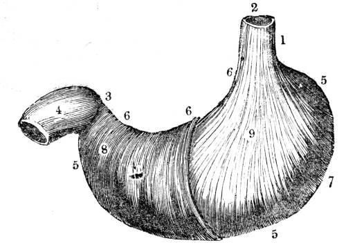
1. The esophagus terminating in the stomach. 2. The cardiac orifice. 3. The pylorus. 4. The commencement of the duodenum. 5. The large curvature of the stomach. 6. The small curvature. 7. The large extremity. 8. The small extremity. 9. The longitudinal muscular fibres. 10. The circular muscular fibres.
612. The stomach is a bag of an irregular oval shape (fig CLXVI.), capable, in the adult, of containing about three pints. It is placed transversely across the upper part of the abdomen (fig. LX. 7). It occupies the whole epigastric (fig. CV. 3), and the greater part of the left hypochondriac regions (fig. CVII. 3). Above, it is in contact with the diaphragm, the arch of which extends over it (fig. LX. 7, b); below with the intestines (fig. LX. 8, 9), on the right side with the liver (fig. LX. 6), and on the left side with the spleen (fig. CLXVIII. 5).
Fig. CLXVII. Internal View of the Stomach and Duodenum.
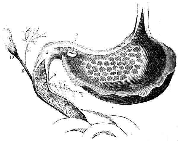
1. Mucous membrane, forming the rugæ. 2. Pyloric orifice opening into the duodenum. 3. Duodenum. 4. Interior of the duodenum, showing the valvulæ conniventes. 5. Termination of, 6. The biliary or choledoch duct. 7. Pancreatic duct, terminating at the same point as the choledoch duct. 8. Gall-bladder removed from the liver. 9. Hepatic duct proceeding from the liver. 10. Cystic duct proceeding from the gall-bladder, forming by its union with the hepatic, a common trunk, the choledoch.
613. Into the left extremity, which is much larger and considerably higher than the right (fig. CLXVI. 7), the esophagus opens by an aperture called the cardiac orifice (fig. CLXVI. 2). At the right extremity, a second aperture called the pyloric orifice (fig. CLXVII. 2), leads into the first intestine.
614. Between the cardiac and the pyloric orifices are two curvatures, one above, called the smaller (fig. CLXVI. 6), the other below, termed the larger curvature (fig. CLXVI. 5).
615. Like the esophagus, the stomach is composed of two layers of muscular fibres, the external longitudinal (fig. CLXVI. 9), the internal circular (fig. CLXVI. 10). By the contraction of the first the extent of the stomach, from extremity to extremity, is diminished, or the organ is shortened; by the contraction of the second the extent of the stomach, from curvature to curvature, is diminished, or the organ is narrowed. During digestion, by the contraction of these muscular fibres, the capacity of the stomach is changed alternately in both directions, whence a gentle motion is communicated to the aliment, which is thus brought in succession under the influence of the agent that acts upon it.
616. A thin but strong membrane, derived from the peritoneum, the membrane that lines the general cavity of the abdomen, forms the external tunic of the stomach; hence its outer covering is called the peritoneal coat.
617. The inner or mucous coat (fig. CLXVII. 1), a direct continuation of the lining membrane of the esophagus, is sometimes called also villous, on account of the minute bodies termed villi, with which every point of its internal surface is studded. It is these villi which give to this surface its pilous or velvety appearance,
Fig. CLXVIII.—View of the Vascular connexion between the Stomach, Liver, Spleen, and Pancreas.
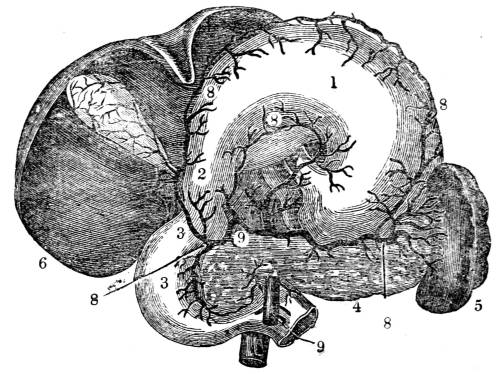
1. Stomach raised to exhibit its posterior surface. 2. Pylorus. 3. Duodenum. 4. Pancreas. 5. Spleen. 6. Undersurface of the liver. 7. Gall-bladder, in connexion with the liver. 8. Large vessels proceeding from. 9. A common trunk to supply the liver, gall-bladder, stomach, duodenum, pancreas, and spleen.
618. The mucous coat is far more extensive than the other two, in consequence of its being plaited into a number of folds (fig. CLXVII. 1), termed rugæ, which are so disposed as to present the appearance of a net-work. The object of the rugæ is to enlarge the space for the expansion of blood-vessels and nerves, and to admit of the occasional distension of the organ without injury to the delicate tissues of which it is composed.
619. Immediately beneath the mucous coat are the mucous follicles which secrete the mucous fluid by which the inner surface of the organ is defended. These glandular bodies are extremely numerous, and vary considerably in diameter. The largest are towards the great extremity, the smaller towards the pylorus.
620. Altogether different from the mucous secretion is another fluid, which also flows from the mucous surface, termed the gastric or the digestive juice, from its being the principal agent in the digestive process. By some anatomists the gastric juice is supposed to be secreted by minute glands placed between the mucous and the muscular coats, provided with ducts which pierce the mucous coat, and which bear their fluid into the stomach precisely as the salivary glands carry the saliva into the mouth. It is certain that this is the case with some animals, as in certain birds, the ostrich for example, in which glands of considerable magnitude, with ducts large enough to be visible, are seen to pour the digestive fluid into the stomach. But as no such glands have been discovered in the human stomach, it is generally conceived that in man the gastric juice is secreted by minute arteries expanded upon the villi.
621. All around the pyloric orifice (fig. CLXVII. 2) is placed a thick, strong, and circular muscle (fig. CLXVII. 2), termed, from its office, pylorus. It is about four times the thickness of the muscular coat of the stomach, and presents the appearance of a prominent and even projecting band (fig. CLXVII. 2). From the frequent action of its fibres, the pylorus often looks as if a piece of packthread had been tied around it (fig. CLXVI. 3). Its office is, by the contraction of its fibres, to guard and close the opening from the stomach until the aliment has been duly acted upon by the digestive fluid.
Fig. CLXIX.
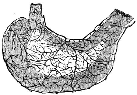
View of the stomach, showing the number and magnitude of its blood-vessels, and the mode of their distribution.
622. The quantity of blood sent to the stomach is greater than is spent upon any other organ except the brain. The vessels of the stomach (fig. CLXIX.) form two distinct layers, of which the external is distributed to the peritoneal and muscular coats, while the internal, after ramifying on the fine cellular tissue which unites the muscular and mucous tunics, penetrates the mucous coat, and is spent upon the villi, where it forms an exquisitely-delicate net-work. There is, moreover, an intimate vascular connexion between the spleen, pancreas and liver, and the stomach (fig. CLXVIII. 8, 9). The arteries which supply all these organs spring from a common trunk, and there is the freest communication between them by anastomosing branches.
Fig. CLXX.—View of the Organic Nerves of the Stomach.
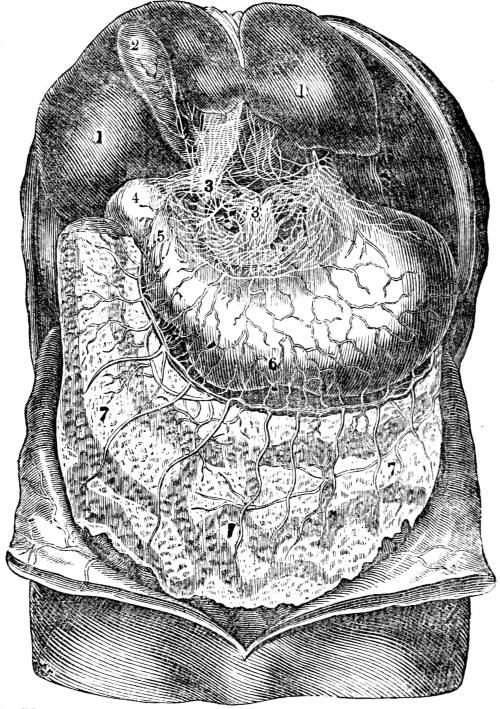
1. Under surface of the liver turned up, to bring into view the anterior surface of the stomach. 2. Gall bladder. 3. Organic nerves enveloping the trunks of the blood-vessels. 4. Pyloric extremity of the stomach and commencement of the duodenum. 5. Contracted portion of the pylorus. 6. Situation of the hour-glass contraction of the stomach, here imperfectly represented. 7. Omentum.
623. Equally abundant is its supply of nerves, some of which are derived from the organic or non-sentient system, and others from the animal or sentient system. The organic nerves are spread out in countless numbers upon the great trunks of the arteries, so as to give them a complete envelope (fig. CLXX. 3); these nerves, never quitting the arteries, accompany them in all their ramifications, and the fibril of the nerve is ultimately lost upon the capillary termination of the artery. It is by these organic nerves that the stomach is enabled to perform its organic functions, which, for the reason assigned (vol. i. p. 82), is placed beyond volition, and is without consciousness. By the nerves derived from the sentient system which mingle with the organic (fig. XVI.), the function of nutrition is brought into relation with the percipient mind, and is made part of our sentient nature. By the commixture of these two sets of nerves, derived from these two portions of the nervous system, though we have no direct consciousness of the digestive process—consciousness ceasing precisely at the point where the agency of volition stops (vol. i. p. 82, et seq.), yet pleasurable sensation results from the due performance of the function. Hence the feeling of buoyancy, exhilaration, and vigour, the pleasurable consciousness to which we give the name of health, when the action of the stomach is sound: hence the depression, listlessness, and debility, the painful consciousness which we call disease, when the action of the stomach is unsound: hence, too, the influence of the mental state over the organic process; the rapidity and perfection with which the stomach works when the mind is happy—when the repast is but the occasion and accompaniment of the feast of reason and the flow of soul; the slowness and imperfection with which the stomach works when the mind is harassed with care struggling against adverse events; or is in sorrow and without hope; when the friend that sat by our side, and with whom we were wont to take sweet counsel, is gone, and therefore gone that which made it life to live.
624. Renovation is the primary and essential office of the stomach, and its organic nerves enable it to supply the ever-recurring wants of the system. Gratification of appetite is a secondary and subordinate office of the stomach, and its sentient nerves enable it to produce the state of pleasurable consciousness when its organic function is duly performed. By the double office thus assigned it, the stomach is rendered what Mr. Hunter named it, the centre of sympathies.
625. From the whole length of the great arch of the stomach, and partly also from the commencement of the duodenum (fig. CLXX.), the peritoneal coat of the stomach is produced, forming a thin, delicate membranous bag, called the omentum, or cawl (fig. CLXX. 7). The omentum extends from the great arch of the stomach to below the umbilicus, and completely covers a large portion of the anterior surface of the abdominal viscera (fig. CLXX. 7). Between the two fine membranous layers of which it is composed is contained a quantity of fat, of which substance it serves as a reservoir, and by the transudation of which it appears to lubricate the intestines, and to assist in preventing their accretion.
626. The food, on reaching the stomach, does not occupy indifferently any portion of it, but is arranged in a peculiar manner always in one and the same part. If the stomach be observed in a living animal, or be inspected soon after death, it is seen that about a third of its length towards the pylorus is divided from the rest by the contraction of the circular fibre called the hour-glass contraction (fig. CLXX. 6). The stomach is thus divided into a cardiac and a pyloric portion (fig. CLXX. 6). The food, when first received by the stomach, is always deposited in the cardiac portion, and is there arranged in a definite manner. The food first taken is placed outermost, that is, nearest the surface of the stomach; the portion next taken is placed interior to the first, and so on in succession, until the food last taken occupies the centre of the mass. When new food is received before the old is completely digested, the two kinds are kept distinct, the new being always found in the centre of the old.
627. Soon after the food has been thus arranged, a remarkable change takes place in the mucous membrane of the stomach. The blood-vessels become loaded with blood; its villi enlarge, and its cryptæ, the minute cells between the rugæ, overflow with fluid. This fluid is the gastric juice, which is secreted by the arterial capillaries now turgid with blood. The abundance of the secretion, which progressively increases as the digestion advances, is in proportion to the indigestibility of the food, and the quietude of the body after the repast.
628. In the food itself no change is manifest for some time; but at length that portion of it which is in immediate contact with the surface of the stomach begins to be slightly softened. This softening slowly but progressively increases until the texture of the food, whatever it may have been, is gradually lost; and ultimately the most solid portions of it are completely dissolved.
629. When a portion of food thus acted on is examined, it presents the appearance of having been corroded by a chemical agent. The white of a hard-boiled egg looks exactly as if it had been plunged in vinegar or in a solution of potass. The softened layer, as soon as the softening is sufficiently advanced, is, by the action of the muscular coat of the stomach, detached, carried towards the pylorus, and ultimately transmitted to the duodenum; then another portion of the harder and undigested food is brought into immediate contact with the stomach, becomes softened in its turn, and is in like manner detached; and this process goes on until the whole is dissolved.
630. The solvent power exerted by the gastric juice is most apparent when the stomach of an animal is examined three or four hours after food has been freely taken. At this period the portion of the food first in contact with the stomach is wholly dissolved and detached; the portion subsequently brought into contact with the stomach is in the process of solution, while the central part remains very little changed.
631. The dissolved and detached portion of the food, from every part of the stomach flows slowly but steadily beyond the hour-glass contraction, or towards the pyloric extremity (626), in which not a particle of recent or undissolved food is ever allowed to remain. The fluid, which thus accumulates in this portion of the stomach, is a new product, in which the sensible properties of the food, whatever may have been the variety of substances taken at the meal, are lost. This new product, which is termed chyme, is an homogeneous fluid, pultaceous, greyish, insipid, of a faint sweetish taste, and slightly acid.
632. As soon as the chyme, by its gradual accumulation in the pyloric extremity amounts to about two or three ounces, the following phenomena take place.
633. First, the intestine called duodenum, the organ immediately continuous with the stomach, contracts. The contraction of the duodenum is propagated to the pyloric end of the stomach. By the contraction of this portion of the stomach, the chyme is carried backwards from the pyloric into the cardiac extremity, where it does not remain, but quickly flows back again into the pyloric extremity, which is now expanded to receive it. Soon the pyloric extremity begins again to contract; but now the contraction, the reverse of the former, is in the direction of the duodenum; in consequence of which, the chyme is propelled towards the pylorus. The pylorus, obedient to the demand of the chyme, relaxes, opens, and affords to the fluid a free passage into the duodenum. As soon as the whole of the duly prepared chyme has passed out of the stomach, the pylorus closes, and remains closed, until two or three ounces more are accumulated, when the same succession of motions are renewed with the same result; and again cease to be again renewed, as long as the process of chymification goes on.
634. When the stomach contains a large quantity of food, these motions are limited to the parts of the organ nearest the pylorus; as it becomes empty, they extend further along the stomach, until the great extremity itself is involved in them. These motions are always strongest towards the end of chymification.
635. The stomach during chymification is a closed chamber; its cardiac orifice is shut by the valved entrance of the esophagus, and its pyloric orifice by the contraction of the pylorus.
636. The rapidity with which the process of chymification is carried on is different according to the digestibility of the food, the bulk of the morsels swallowed, the quantity received by the stomach, the constitution of the individual, the state of the health, and above all, the class of the animal, for it is widely different in different classes. In the human stomach in about five hours after an ordinary meal the whole of the food is probably converted into chyme.
637. The great agent in performing the process of chymification is the gastric juice. The evidence of this is complete; for,
1. As soon as the food enters the stomach a large quantity of blood is determined to the arteries, which secrete the gastric juice (627); and this fluid continues to be poured into the stomach in great abundance during the whole time the process goes on.
2. The solvent power of this fluid is demonstrated by the fact that it sometimes dissolves the stomach itself, when death takes place suddenly during the act of digestion in a sound and vigorous state of the digestive organs.
3. On introducing into the stomach alimentary substances inclosed in metallic balls perforated with holes, or in pieces of porous cloth, it is found, on removing these bodies from the stomach, after a certain time, that the alimentary substances contained in them are as completely digested as if they had been in actual contact with the surface of the stomach; the metallic ball and the cloth remaining wholly unchanged. This experiment, which has been often performed with the same uniform result, was the first that led to the discovery of the true nature of the digestive process.
4. Though it be impossible to imitate out of the stomach all the circumstances under which the food is placed within it, yet, on procuring gastric juice from the stomachs of various animals, and mixing it with different alimentary substances, it is found not only to dissolve them, but to convert them into a pultaceous mass, closely resembling chyme. Gastric juice thus procured was put into a glass tube with boiled beef, which had been masticated; the tube was then hermetically sealed, and exposed near the fire to a uniform heat: by the side of this tube was placed another, containing the same quantity of flesh immersed in water. In twelve hours, the flesh in the tube containing the gastric juice began to lose its fibrous structure; in thirty-five hours it had nearly lost its consistence, being reduced to a soft homogeneous pultaceous mass. It experienced no further change during the two following days. On the other hand, the flesh that had been immersed in water was putrid in sixteen hours.
638. Since alimentary substances under the action of the stomach present precisely the appearance exhibited by bodies exposed to the influence of chemical agents, it appears that the gastric juice not only dissolves the food, but dissolves it by a chemical agency. Its action bears no proportion to the mechanical texture of bodies, nor to any of their physical properties. It acts upon the densest membrane, dissolves even bone itself; and yet produces no effect on other substances of the most tender and delicate texture. On the skin of fruit, on the finest fibre of flax and cotton, it is incapable of making the slightest impression. In this selection of substances it perfectly resembles a chemical agent acting by chemical affinity. On certain substances its action is unquestionably of a chemical nature. It occasions the coagulation of albuminous fluids; it prevents the accession of putrefaction; it stops the process after it has commenced. From the whole, it follows that the food in the stomach is converted into chyme by the agency of a fluid secreted by the inner surface of the stomach, and that this change is effected by a proper chemical action.
639. It had been long ascertained that the gastric juice contains an uncombined acid, and that if carbonate of lime be placed in a tube and introduced into the stomach, the carbonate is dissolved just as if it were put into weak vinegar. Several years ago, it was discovered by Dr. Prout that this free acid is muriatic acid. Some time after the publication of Dr. Prout’s experiments, Chevreul and Leuret and Lassaigne in France obtained different results; but Tiedemann and Gmelin, professors in the university of Heidelberg, in an extended series of experiments, arrived at precisely the same conclusion as the English physiologist, and apparently without any previous knowledge of the researches of the latter. Tiedemann and Gmelin state, as the result of their experiments, that the clear ropy fluid, or the gastric juice obtained from the stomach some time after it had been without food, is nearly or entirely destitute of acidity; that the presence of food, or indeed of any stimulus to the mucous membrane, causes the gastric juice to become distinctly acid; that this acidity increases according to the indigestibility of the food; that the quantity of acid poured out is very copious; that it consists partly of muriatic and partly of acetic acid; and that both these acids are efficient agents in the process of digestion. Dr. Prout, who had also recognised the presence of acetic acid, is of opinion that its formation is an accidental occurrence not necessary to digestion nor conducive to it; but is either derived from the aliment, or is the result of irritation or disease. He contends that the muriatic acid is the efficient digestive agent.
640. The still more recent experiments of Braconnot appear to have set this matter at rest, and to have proved, beyond all controversy, that the stomach, when stimulated by the presence of food or other foreign agents, possesses the power of secreting free muriatic acid in great quantity; and that it is by this acid that the solution of the food is effected. It is even found that muriatic acid is capable of digesting alimentary substances out of the body. It had been long known, that if meat and gastric juice be inclosed in a tube and kept at the temperature of the human body, a product is obtained closely resembling chyme (637.4). M. Blondelot, a physician at Nancy, has recently shown that the same result may be obtained by the digestion of the muscular fibre, in dilute muriatic acid. In both cases the muscular fibre retains its form and its original fibrous texture; but on the slightest motion it divides into an insoluble mass, perfectly homogeneous and similar to the chyme of the stomach;5 a very close approximation to the actual digestive process, more especially when it is considered that it is not possible to imitate out of the stomach several circumstances materially influencing chemical action under which the food is placed within the stomach.
641. Muriatic acid, the chemical agent by which the stomach dissolves the food, is probably obtained from the muriate of soda (common salt) contained in the blood. The soda, the basis of the salt, would appear to be retained in the blood, to preserve the alkaline condition essential to the maintenance of the sound constitution of the blood, while the muriatic acid, disengaged from the soda in the process of secretion, is poured into the stomach to act upon the food.
642. A remarkable confirmation of the correctness of the general conclusions to which observation and experiment had thus enabled physiologists to arrive, is afforded by the case of a young soldier in the American army, of the name of Alexis St. Martin, who received a wound on the left side by the accidental discharge of a musket. The charge, which consisted of duck shot, and which was received at the distance of one yard from the muzzle of the gun, entered the side posteriorly in an oblique direction, forward and inward; blew off the integument and muscles to the size of a man’s hand; fractured and carried away the anterior half of the sixth rib; fractured the fifth rib; lacerated the lower portion of the left lobe of the lungs; lacerated the diaphragm, and perforated the stomach.
643. Violent fever and extensive sloughing of the parts injured ensued, and the life of the young man was often in jeopardy, but he ultimately recovered. At the distance of about a year from the date of the accident, the injured parts had all become sound, with the exception of the perforation into the stomach, which never closed, but left an aperture permanently open, two inches and a half in circumference. This aperture was situated about three inches to the left of the cardia, near the left superior termination of the great curvature. For some time the food could be retained only by constantly wearing a compress and bandage; but at length a small fold of the mucous coat of the stomach appeared, which increased until it completely filled the aperture and acted as a valve, so as effectually to prevent any efflux from within, while it admitted of being easily pushed back by the finger from without: when the stomach was nearly empty, it was easy to examine its cavity to the depth of five or six inches by artificial distension; but, when entirely empty, the stomach was always contracted on itself, and the valve generally forced through the orifice, together with a portion of the mucous membrane equal in bulk to a hen’s egg.
644. It chanced that the admirable opportunity thus afforded of bringing the process of digestion, as far as it is carried on in the stomach, under the cognizance of sense, occurred to an observant and philosophical mind, and it was not lost.6 The following are some of the curious and instructive phenomena observed.
645. The inner coat of the stomach, in its natural and healthy state, is of a light or pale pink colour, varying in its hues according to its full, or empty state. It is of a soft or velvet-like appearance (617), and is constantly covered with a very thin transparent, viscid mucus, lining the whole interior of the organ (619).
646. Immediately beneath the mucous coat appear small spheroidal, or oval-shaped glandular bodies, from which the mucous fluid appears to be secreted (619).
647. By applying aliment or other irritants to the internal coat of the stomach, and observing the effect through a magnifying glass, innumerable minute lucid points, and very fine nervous or vascular papillæ are seen arising from the villous membrane, and protruding through the mucous coat, from which distils a pure, limpid, colourless, slightly viscid fluid (620). This fluid, thus excited, is invariably distinctly acid (639, et seq.). The mucus of the stomach is less fluid, more viscid or albuminous, semi-opaque, sometimes a little saltish, and does not possess the slightest character of acidity (619). On applying the tongue to the mucous coat of the stomach in its empty, un-irritated state, no acid taste can be perceived. When food or other irritants have been applied to the villous membrane and the gastric papillæ excited, the acid taste is immediately perceptible. The invariable effect of applying aliment to the internal, but exposed part of the gastric membrane, is the exudation of the solvent fluid from the papillæ. Though the aperture of these vessels cannot be seen even with the assistance of the best microscopes, yet the points from which the fluid issues are clearly indicated by the gradual appearance of innumerable very fine lucid specks rising through the transparent mucous coat, and seeming to burst and discharge themselves upon the very points of the papillæ, diffusing a limpid thin fluid over the whole interior gastric surface.
648. The fluid so discharged is absorbed by the aliment in contact; or collects in small drops, and trickles down the sides of the stomach to the more depending parts, and there mingles with the food, or whatever else may be contained in the gastric cavity. This fluid, the efficient cause of digestion, the true gastric juice is secreted only when it is needed; it is not accumulated in the intervals of digestion, to be ready for the next meal; it is seldom if ever discharged from its proper secreting vessels, except when excited by the natural stimulus of aliment, the mechanical irritation of tubes, or other excitants. When aliment is received, the juice is given out in exact proportion to its requirements for solution, except when more food has been taken than is necessary for the wants of the system.
649. On collecting this fluid, which it was easy to obtain, it was found to be transparent, inodorous, saltish, and acidulous to the taste; it consisted of water, containing free muriatic and acetic acids, phosphates and muriates, with bases of potass, soda, magnesia, and lime, together with an animal matter soluble in cold, but insoluble in hot water.
650. When a portion of liquid aliment, as a few spoonsful of soup, were introduced into the stomach at the external orifice, the rugæ (fig. CLXVII . 1) immediately closed gently upon it; gradually diffused it through the gastric cavity, and prevented the entrance of a second quantity till this diffusion was effected; then relaxation again took place, and admitted of a further supply. When solid food was introduced in the same manner, either in large pieces or finely divided, the same gentle contraction and grasping motions were excited, and continued from fifty to eighty seconds, so as to prevent more from being introduced, without considerable force till the contraction was at an end.
651. When the position of the body was such that the cardiac portion of the stomach was brought into view, and a morsel of food was swallowed in the natural mode, a similar contraction of the stomach, and closing of its fibres upon the bolus was invariably observed to take place; and till this was over, a second morsel could not be received without a considerable effort. Hence, in addition to the other purposes accomplished by mastication, insalivation, and deglutition, it is probable that these operations answer the further use of duly regulating the time for the admission of successive portions of the food into the stomach.7
652. On watching the phenomena that take place on the contact of a portion of food with the stomach, the circumstances described (627) are seen; the change in the mucous coat from a pale pink to a deep red colour, in consequence of the enlargement of the blood-vessels and their admission of a greatly increased number of red particles; the undulating motion of the stomach, in consequence of the contraction of its muscular fibres, excited by the stimulus of food; the distillation of the gastric juice from the enlarged and excited papillæ; the continuous flow of this fluid until the complete solution of the food, when food is present; and, on the contrary, the cessation of this discharge in a short time when it is produced by a mechanical irritant, as the bulb of a thermometer, although at first the gastric juice distil from the papillæ, from the contact of such an irritant, just as when excited by the contact of food.
653. On collecting the gastric juice and placing it in contact with an alimentary substance out of the stomach, its solution takes place more slowly, but not less completely, than when retained in the stomach. An ounce of this fluid was placed in a vial with a piece of boiled, recently salted beef, weighing three drachms; the vial was then tightly corked, and immersed in water, raised to the temperature of 100°, previously ascertained to be the ordinary heat of the stomach. In forty minutes the process of solution had commenced on the surface of the beef. In fifty minutes the texture of the beef began to loosen and separate. In sixty minutes an opaque and cloudy fluid was formed. In one hour and a half the muscular fibres hung loose and unconnected, and floated about in shreds in the more fluid matter. In three hours the muscular fibres had diminished about one half. In five hours only a few remained undissolved. In seven hours the muscular texture was no longer apparent; and in nine hours the solution was completed.
654. At the commencement of this experiment a piece of the same beef of equal weight and size was suspended within the stomach by means of a string. On examining this portion of beef at the end of half an hour, it was found to present precisely the same appearance as the piece in the vial; but on the removal of the string at the end of an hour and a half the beef had been completely dissolved, and had disappeared, making a difference of result in point of time of nearly seven hours. In both, the solution began on the surface, and agitation accelerated its progress by removing the external coating of chyme as fast as it was formed.
655. An ordinary dinner having been taken, consisting of boiled salted beef, bread, potatoes, and turnips, with a gill of pure water for drink, a portion of the contents of the stomach was drawn off into an open mouthed vial, twenty minutes after the meal. The vial was placed in a water-bath, maintained steadily at a temperature of 100°. It was continued in this temperature for five hours. At the end of that time the whole contents of the vial were dissolved. On comparing the solution with an equal quantity of chyme taken from the stomach, little difference could be distinguished between the two fluids, excepting that it was manifest that the digestive process had proceeded somewhat more rapidly in, than out of the stomach. The food, in this experiment, after having remained in contact with the stomach for the space of twenty minutes, had imbibed a sufficient quantity of gastric juice to complete its solution.
656. Fifteen minutes after half a pint of milk had been introduced into the stomach, it presented the appearance of a fine loosely-coagulated substance mixed with a semi-transparent whey-coloured fluid. A drachm of warm gastric juice poured into two drachms of milk at a temperature of 100°, produced a precisely similar appearance in twenty minutes. In another experiment, when four ounces of bread were given with a pint of milk, the milk was coagulated and the bread reduced to a soft pulp in thirty minutes, and the whole was completely digested in two hours.
657. When the albumen or white of two eggs was swallowed on an empty stomach, small white flakes began to be seen in about ten or fifteen minutes, and the mixture soon assumed an opaque whitish appearance. In an hour and a half the whole had disappeared. Two drachms of albumen mixed with two of gastric juice out of the stomach underwent precisely the same changes, but in a somewhat longer time.
658. Dr. Beaumont’s observations are adverse to the opinion, founded on numerous experiments, that the food is arranged in the stomach in a definite manner, and that a distinct line of separation exists between old and new food (626). In the human stomach, according to the subject of these experiments, the ordinary course and direction of the food are first from right to left along the small arch, and thence through the large curvature from left to right. The bolus as it enters the cardia turns to the left, passes the aperture, descends into the splenic extremity, and follows the great curvature towards the pyloric end. It then returns in the course of the smaller curvature, makes its appearance again at the aperture, in its descent into the great curvature, to perform similar revolutions. These revolutions are completed in from one to three minutes. They are probably induced in a great measure by the circular or transverse muscles of the stomach (615), as is indicated by the spiral motion of the stem of the thermometer, both in descending to the pyloric portion, and in ascending to the splenic. These motions are slower at first than after chymification has considerably advanced. The whole contents of the stomach, until chymification be nearly complete, exhibit a heterogeneous mass of solids and fluids, hard and soft, coarse and fine, crude and chymified; all intimately mixed, and circulating promiscuously through the gastric cavity like the mixed contents of a closed vessel, gently agitated or turned in the hand.
659. In attempting to pass a long glass thermometer through the aperture into the pyloric portion of the stomach, during the latter stages of digestion, a forcible contraction is perceived at the point of the hour-glass contraction of the stomach, and the bulb is stopped. In a short time there is a gentle relaxation, when the bulb passes without difficulty, and appears to be drawn quite forcibly, for three or four inches, towards the pyloric end. It is then released, and forced back, or suffered to rise again, at the same time giving to the tube a circular or rather a spiral motion, and frequently revolving it quite over. These motions are distinctly indicated and strongly felt in holding the end of the tube between the thumb and finger; and it requires a pretty forcible grasp to prevent it from slipping from the hand, and being drawn suddenly down to the pyloric extremity. When the tube is left to its own direction at these periods of contraction, it is drawn in, nearly its whole length, to the depth of ten inches; and when drawn back requires considerable force, and gives to the fingers the sensation of a strong suction power, like drawing the piston from an exhausted tube. This ceases as soon as the relaxation occurs, and the tube rises again, of its own accord, three or four inches, when the bulb seems to be obstructed from rising further; but if pulled up an inch or two through the stricture, it moves freely in all directions in the cardiac portions, and mostly inclines to the splenic extremity, though not disposed to make its exit at the aperture. These peculiar motions and contractions continue until the stomach is perfectly empty, and not a particle of food or chyme remains, when all becomes quiescent again.
660. The chambers in which the remaining part of the digestive process is carried on are much less accessible, and no such favourable opportunity as that enjoyed by Dr. Beaumont has occurred of rendering their operations manifest to the eye. Nevertheless, the researches of physiologists have succeeded in disclosing, with almost equal exactness and certainty, the successive changes which the food undergoes even in these more hidden organs, that admit of no exposure during life without extreme danger.