Volume 1 — CHAPTER V. (continued 2)
Reading edition
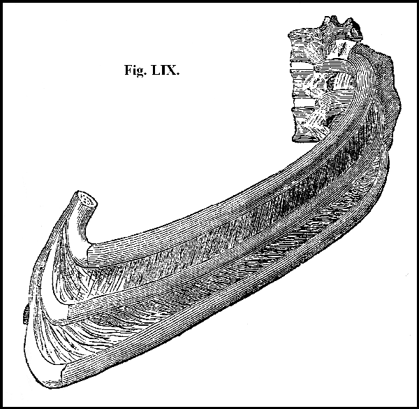
A view of the muscles called Intercostals, filling up the spaces between the ribs.
104. The inner surface of the walls of the thorax, in its whole extent, is lined by a serous membrane, exceedingly thin and delicate, but still firm, called the pleura. The same membrane is reflected over the organs of respiration contained in the cavity, so as to give them an external coat. The membrane itself is every where continuous, and every where the same, whether it line the containing or the contained parts; but it receives a different name as it covers the one or the other: that portion of it which lines the walls of the cavity being called the costal pleura (fig. LXI. a), while that which covers the organs contained in the cavity is termed the pulmonary pleura (fig. LX. 5, 1).
105. A fold of each pleura passes directly across the central part of the cavity of the thorax; extending from the spinal column to the sternum, and dividing the general cavity into two. This portion of the pleura is called the mediastinum, from its situation in the centre of the thorax, and it so completely divides the thoracic cavity into two, that the organs on one side of the chest have no communication with those of the other; so that there may be extensive disease in one cavity (for example, a large accumulation of water,) while the other may be perfectly sound.
106. The main organs contained in the cavity of the thorax are the lungs with their air tube; the heart with its great vessels; and the tube passing from the mouth to the stomach (fig. LX.).
107. The two lungs occupy the sides of the chest (fig. LX. 5). They are completely separated from each other by the membranous partition just described, the mediastinum. Between the two folds of the mediastinum, namely, in the middle of the chest, but inclining somewhat to the left side, is placed the heart, enveloped in another serous membrane, the pericardium (fig. LX. 2, 1).
108. The lungs are moulded to the cavities they fill; whence their figure is conical, the base of the cone being downwards, resting on the diaphragm (fig. LX. 5, b); and the apex upwards, towards the neck (fig. LX. 5).
109. That surface of each lung which corresponds to the walls of the chest is convex in its whole extent (fig. LX. 5); on the contrary, that surface which corresponds to the mediastinum is flattened (fig. LX. 5). The basis of the lung is concave, adapted to the convexity of the diaphragm on which it rests (fig. LX. 5).
110. The air-vessel of the lungs, termed the bronchus, together with the blood-vessels and nerves, enter the organ at its flattened side, not exactly in the middle, but rather towards the upper and back part. This portion is termed the root of the lung.
111. The lungs are attached to the neck by the trachea (fig. LX. 4), the continuation of which forms the bronchus; to the spinal column by the pleura, and to the heart by the pulmonary vessels (fig. LX. 3, d): their remaining portion is free and unattached.
112. In the living body, the lungs on each side completely fill the cavity of the chest, following passively the movements of its walls, and accurately adapting themselves to its size, whether its capacity enlarge in inspiration, or diminish in expiration, so that the external surface of the lung (the pulmonary pleura) is always in immediate contact with the lining membrane of the walls of the cavity (the costal pleura); consequently, during life, there is no cavity, the chest being always completely full.
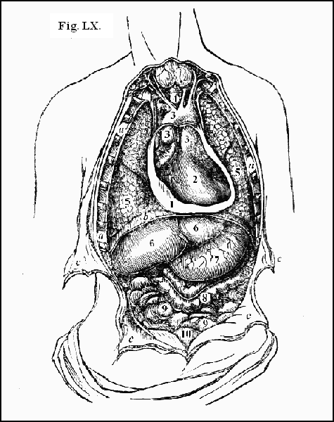
a. The cut edges of the ribs, forming the lateral boundaries of the cavity of the thorax. b. The diaphragm, forming the inferior boundary of the thorax, and the division between the thorax and the abdomen. c. The cut edges of the abdominal muscles, turned aside, exposing the general cavity of the abdomen. 1. The cut edge of the pericardium turned aside. 2. The heart. 3. The great vessels in immediate connexion with the heart. 4. The trachea, or wind-pipe. 5. The lungs. 6. The liver. 7. The stomach. 8. The large intestine. 9. The small intestines. 10. The urinary bladder.
113. The anterior surface of the pericardium, the bag which envelopes the heart, lies immediately behind the sternum, and the cartilages of the second, third, fourth, and fifth ribs, covered at its sides by the pleura, and firmly attached below to the diaphragm (fig. LX. 1).
114. Surrounded by its pericardium, within the mediastinum, the heart is placed nearly in the centre of the chest, but its direction is somewhat oblique, its apex being directly opposite to the interval between the fifth and sixth ribs on the left side (fig. LX. 2); while its basis is directed upwards, backwards, and towards the right (fig. LX. 2). That portion of its surface which is presented to view on opening the pericardium is convex (fig. LX. 2); but its opposite surface, namely, that which rests upon the part of the pericardium which is attached to the diaphragm, is flattened (fig. LX. 1). It is fixed in its situation partly by the pericardium and partly by the great vessels that go to and from it. But under the different states of expiration and inspiration, it accompanies, in some degree, the movements of the diaphragm; and in the varied postures of the body, the heart deviates to a certain extent from the exact position here described.
115. The second division of the trunk, the abdomen, is bounded above by the diaphragm (fig. LXI. 2), below by the pelvis (fig. LXI. 3), behind and at the sides by the vertebræ and muscles of the loins (fig. LXIII.), and before by the abdominal muscles (fig. LXIII. 9).
116. The organ which forms the superior boundary of the abdomen, the diaphragm (midriff), is a circular muscle, placed transversely across the trunk, nearly at its centre (fig. LXI. 2). It forms a vaulted partition between the thorax and the abdomen (fig. LXI. 2). All around its border it is fleshy (fig. LXI. 2); towards its centre it is tendinous (fig. LXI. 2); the surface towards the abdomen is concave (fig. LXI. 2); that towards the thorax convex (fig. LXI. 2); while its middle tendinous portion ascends into the thorax as high as the fourth rib (fig. LXI. 2).
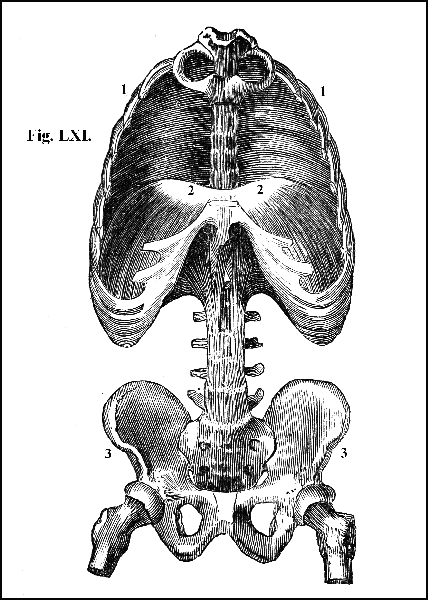
View of the diaphragm. 1. Cavity of the thorax; 2. diaphragm separating the cavity of the thorax from that of the abdomen; 3. cavity of the pelvis.
117. The diaphragm is perforated by several apertures, for the transmission of tubes and vessels, which pass reciprocally between the thorax and abdomen (fig. LXII.).
1. A separate aperture is formed to afford an exit from the thorax of the aorta, the common source of the arteries (fig. LXII. 2), and an entrance into the thorax of the thoracic duct, the tube that bears the digested aliment to the heart. 2. A little to the left of the former, there is another aperture, through which passes the esophagus or gullet (fig. LXII. 3), the tube that conveys the food from the mouth to the stomach. 3. On the right side, in the tendinous portion of the diaphragm, very carefully constructed, is a third aperture for the passage of the vena cava (fig. LXII. 4), the great vessel that returns the blood to the heart from the lower parts of the body.
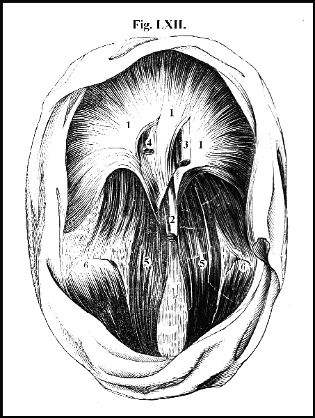
View of the diaphragm with the tubes that pass through it. 1. Arch of the diaphragm; 2. the trunk of the aorta passing from the chest into the abdomen; 3. the esophagus passing from the chest through the diaphragm to the stomach; 4. the vena cava, the great vein that returns the blood to the heart from the lower parts of the body, passing from the abdomen, into the chest, in its way to the right side of the heart; 5. 6. muscles that arise in the interior of the trunk and that act upon the thigh; 5. the muscle called psoas; 6. the muscle called iliacus.
118. The partition formed by the diaphragm between the thorax and abdomen, though complete, is moveable; for as the diaphragm descends in inspiration and ascends in expiration, it proportionally enlarges or diminishes the cavities between which it is placed; consequently, the actual magnitude of these cavities varies every moment, and the size of the one is always in the inverse ratio of that of the other.
119. Between the abdomen and the pelvis there is no separation; one cavity is directly continuous with the other (fig. LXI. 3); but along the inner surface of the expanded bones, which form a part of the lateral boundary of the abdomen, there is a prominent line, termed the brim of the pelvis (fig. XLV. 15), marking the point at which the abdomen is supposed to terminate and the pelvis to commence.
120. Behind and at the sides the walls of the abdomen are completed partly by the lumbar portion of the spinal column and partly by the lumbar muscles (fig. XLV. 4), and before by the abdominal muscles (fig. LXIII. 9).
121. The inner surface of the walls of the abdomen is lined throughout by a serous membrane, termed the peritoneum (fig. LXIII.). From the walls of the abdomen, the peritoneum is reflected upon the organs contained in the cavity, and is continued over them so as to form their external coat. The peritoneum also descends between the several organs, connecting them together, and holding them firmly in their situation; and it likewise forms numerous folds, in which are embedded the vessels and nerves that supply the organs. It secretes a serous fluid, by which its own surface and that of the organs it covers is rendered moist, polished, and glistening, and by means of which the organs glide smoothly over it, and over one another in the various movements of the body, and are in constant contact without growing together. In structure, distribution, and function, the peritoneum is thus perfectly analogous to the pleura.
122. Like the thorax, the abdomen is always completely full. When the diaphragm is in action, it contracts. When the diaphragm is in the state of contraction, the abdominal and lumbar muscles are in the state of relaxation. By the contraction of the diaphragm, the organs contained in the abdomen are pushed downwards, and the anterior and lateral walls of the cavity being at this moment in a state of relaxation, they readily yield, and, consequently, the viscera are protruded forwards and at the sides. But the abdominal and lumbar muscles in their turn contract, the diaphragm relaxing; and, consequently, the viscera, forced from the front and sides of the abdomen, are pushed upwards, together with the diaphragm, into the cavity of the thorax. A firm and uniform pressure is thus at all times maintained upon the whole contents of the abdomen: there is an exact adaptation of the containing to the contained parts, and of one organ to another. No space intervenes either between the walls of the abdomen and the organs they enclose, or between one organ and another: so that the term cavity does not denote a void or empty space, but merely the extent of the boundary within which the viscera are contained.
123. The contents of the abdomen consist of the organs which belong to the apparatus of digestion, and of those which belong to the apparatus of excretion.
124. The organs which belong to the apparatus of digestion are—1. The stomach (fig. LXIII. 2) 2. The duodenum (fig. LXIII. 4). 3. The jejunum (fig. LXIII. 5). 4. The ilium (fig. LXIII. 5). The three last organs are called the small intestines, and their office is partly to carry on the digestion of the aliment commenced in the stomach, and partly to afford an extended surface for the absorption of the nutriment as it is prepared from the aliment. 5. The pancreas (fig. LXIV. 5). 6. The liver (fig. LXIV. 2). 7. The spleen (fig. LXIV. 4). The three last organs truly belong to the apparatus of digestion, and their office is to co-operate with the stomach and the small intestines in the conversion of the aliment into nutriment.
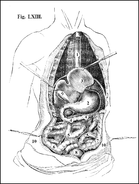
1. Esophagus; 2. stomach; 3. liver raised, showing its under surface; 4. duodenum; 5. small intestines; 6. cæcum; 7. colon; 8. urinary bladder; 9. gall bladder; 10. abdominal muscles divided and reflected.
125. The organs which belong to the apparatus of excretion are—1. The large intestines consisting of the cæcum (fig. LXIII. 6). 2. The colon (fig. LXIII. 7). 3. The rectum (fig. LXIV. 10). It is the office of these organs, which are called the large intestines, to carry out of the system that portion of the alimentary mass which is not converted into nourishment. 4. The kidneys (fig. LXIV. 6), the organs which separate in the form of the urine an excrementitious matter from the blood, in order that it may be conveyed out of the system.
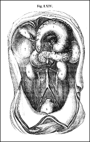
General view of the viscera of the abdomen. 1. Stomach raised; 2. under surface of liver; 3. gall bladder; 4. spleen; 5. pancreas; 6. kidneys; 7. ureters; 8. urinary bladder; 9. portion of the intestine called duodenum; 10. portion of the intestine called rectum; 11. the aorta.
126. The last division of the trunk, called the pelvis (fig. LXI. 3), consists of a circle of large and firm bones, interposed between the lower portion of the trunk and the inferior extremities (fig. XLV.). The bones that compose the circle, distinct in the child, are firmly united in the adult into a single piece; but as the original separation between each remains manifest, they are always described as separate bones. They are the sacrum (fig. XLV. 5), the coccyx (fig. XXXV.), the ilium (fig. XLV. 11), the ischium (fig. XLV. 12), and the pubis (fig. XLV. 13).
127. The sacrum, placed like a wedge between the moveable portion of the spinal column and the lower extremities, forms the posterior boundary of the pelvis. The figure of this bone is triangular (fig. XLV. 5); its anterior surface is concave and smooth, for enlarging the cavity of the pelvis and sustaining the organs contained in it (fig. XLV. 5); its posterior surface is convex, irregular, and rough (fig. XXXV.), giving origin to the great muscles that form the contour of the hip, and to the strong muscles of the back and loins that raise the spine and maintain the trunk of the body erect.
128. The base or upper part of the sacrum receives the last vertebra of the loins on a large and broad surface (fig. XLV. 4), forming a moveable joint; and the degree of motion at this point is greater than it is at the higher points of the spinal column. Firmly united at its sides with the haunch bones, it admits there of no degree of motion.
129. The coccyx, so named from its resemblance to the beak of the cuckoo, when elongated by a succession of additional bones, forms the tail in quadrupeds; but in man it is turned inwards to support the parts contained in the pelvis, and to contract the lower opening of the cavity. By means of a layer of cartilage, the medium by which this bone is connected with the sacrum, it forms a moveable articulation, continuing moveable in men until the age of twenty-five, and in women until the age of forty-five; continuing moveable in women thus long, in order that by yielding to the force which tends to push it backwards during the period of labour, it may enlarge the lower aperture of the pelvis, and so facilitate the process of parturition and diminish its suffering.
130. The lateral boundaries of the pelvis are formed by the ilium, the haunch bone (fig. XLV. 11), and by the ischium, the hip bone (fig. XLV. 12). The ilium forms the lower part of the abdomen and the upper part of the pelvis (fig. XLV. 11); its broad expanded wing supports the contents of the abdomen, and gives attachment to the muscles that form the anterior portion of its walls (figs. XLV. 11, and LXIII. 9); its external convex surface sustains the powerful muscles that extend the thigh; and along its internal surface is the prominent line which marks the brim of the pelvis (fig. XLV. 15), and which divides this cavity from that of the abdomen.
131. The ischium or hip bone is the lower part of the pelvis (fig. XLV. 12); at its undermost portion is a rounded prominence called the tuberosity (fig. XLV. 12), in its natural condition covered with cartilage, upon which is superimposed a cushion of fat. It is this part on which the body is supported in a sitting posture.
132. The pubis or share bone forms the upper and fore part of the pelvis (fig. XLV. 13), and together with the two former bones, completes the large and deep socket, termed the acetabulum (fig. XLV. 14), into which is received the head of the thigh-bone (fig. XXXIV. 4). The margin of the acetabulum and the greater part of its internal surface is lined with cartilage, so that in its natural condition it is much deeper than it appears to be when the bones alone remain.
133. The lower aperture of the pelvis, which appears large when all the soft parts are removed, is not really large, for in its natural state it is filled up partly by muscles and partly by ligaments, which sustain and protect the pelvic organs, leaving only just space enough for the passage to and from those which have their opening on the external surface.
134. The cavity of the pelvis, together with all the organs contained in it, are lined by a continuation of the membrane that invests the abdomen and its contents.
135. The organs contained in the pelvis are the rectum (fig. LXIV. 9), which is merely the termination of the large intestines, the urinary bladder (fig. LXIV. 8), and the internal part of the apparatus of reproduction.
136. The large and strong bones of the pelvis not only afford lodgment and protection to the tender organs contained in its cavity, but sustain the entire weight of the body, the trunk resting on the sacrum as on a solid basis (fig. XLV. 5), and the lower extremities being supported in the sockets in which the heads of the thigh-bones play, in the varied movements of locomotion (fig. XXXIV. 4).
137. The last division of the body comprehends the superior and the inferior extremities.
138. The superior extremities consist of the shoulder, arm, fore-arm, and hand.
139. The soft parts of the SHOULDER are composed chiefly of muscles; its bones are two, the scapula or the blade bone, and the clavicle or the collar bone (fig. LXV. 2, 4).
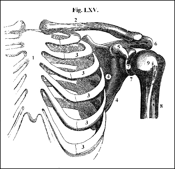
1. Sternum; 2. clavicle; 3. ribs; 4. anterior surface of scapula; 5. coracoid process of scapula; 6. acromion process of scapula; 7. margin of glenoid cavity of scapula; 8. body of the humerus or bone of the arm; 9. head of the humerus.
140. The SCAPULA is placed upon the upper and back part of the thorax, and occupies the space from the second to the seventh ribs (fig. LXV. 4)
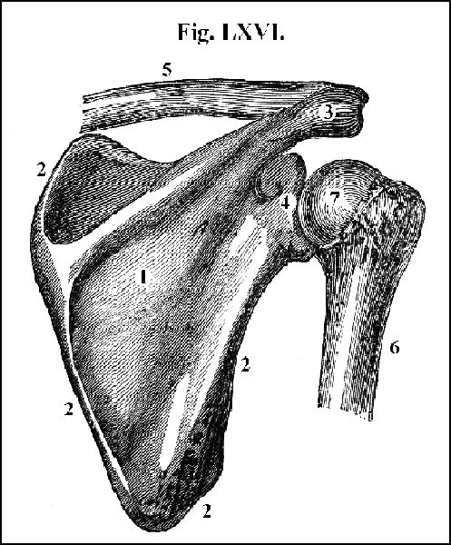
1. Posterior surface of scapula; 2. margin of scapula; 3. acromion process; 4. margin of glenoid cavity; 5. clavicle; 6. body of humerus; 7. head of humerus.
Unlike that of any other bone of the body, it is embedded in muscles, without being attached to any bone of the trunk, excepting at a single point. From the bones of the thorax it is separated by a double layer of muscles, on which it is placed as upon a cushion, and over the smooth surface of which it glides. Originally, like the bones of the skull, it consisted of two tables of compact bone, with an intermediate layer of spongy bony substance (diploë); but, by the pressure of the muscles that act upon it, it gradually grows thinner and thinner, until, as age advances, it becomes in some parts quite transparent and as thin as a sheet of paper.
141. The figure of the scapula is that of an irregular triangle (fig. LXVI.). Its anterior surface is concave (fig. LXV. 4), corresponding to the convexity of the ribs (fig. XLV. 7); its posterior surface is very irregular (fig. LXVI. 1), being in some parts concave and in others convex, giving origin especially to two large processes (figs. LXV. 5, and LXVI. 3); one of which is termed the acromion (fig. LXVI. 3), and the other the coracoid process of the scapula (fig. LXV. 5). The margins of the bone, whatever the thinness of some portions of it, are always comparatively thick and strong (fig. LXVI. 2), affording points of origin or of insertion to powerful muscles. At what is called the anterior angle of the bone there is a shallow oval depression covered with cartilage and deepened by a cartilaginous margin, called the glenoid cavity of the scapula (figs. LXV. 7, and LXVI. 4), which receives the head of the humerus or bone of the arm (figs. LXV. 9, and LXVI. 7, 6).
142. The clavicle, the second bone of the shoulder, is a long and slender bone, of the form of an italic , projecting a little forwards towards its middle, so as to give a slight convexity of outline to the top of the chest and the bottom of the neck (fig. LXV. 2). It is attached by one extremity to the sternum (fig. LXV. 2) and by the other to the scapula (fig. LXV. 2), by moveable joints. The nature of an immoveable joint has been explained (63). In the connexion of the bones of the trunk, while the main object is to secure firmness of attachment, some degree of motion is at the same time obtained (81 et seq.): but the mode in which the several bones of the extremities are connected with each other and with the trunk, admits of so great a degree of motion, that these articulations are pre-eminently entitled to the name of moveable joints. The component parts of all moveable joints are bone, cartilage, synovial membrane, and ligament. The great character of a moveable joint is the approximation of two or more bones; yet these bony surfaces are never in actual contact, but are invariably separated from each other by cartilage. The cartilage either covers the entire extent of the articulating surface of the bones, as in the shoulder-joint, where both the head of the humerus and the cavity of the scapula that receives it are enveloped in this substance (fig. LXV. 7. 9), or a portion of it is placed between the articulating surfaces of the bones, as in the joint between the clavicle and sternum (fig. LXVII. a); which, when so placed, is termed an interarticular cartilage (fig. LXVII. a). By its smooth surface cartilage lessens friction; while by its elasticity it facilitates motion and prevents concussion. Slightly organized cartilage is provided with comparatively few blood-vessels and nerves. Had it been vascular and sensible like the skin and the muscle, the force applied in the movements of the joint would have stimulated the blood-vessels to inordinate action, and the sensibility of the nerves would have been the source of constant pain: every motion of every joint would have been productive of suffering, and have laid the foundation of disease. The facility and ease of motion obtained by the smoothness, elasticity, and comparative insensibility of cartilage are still further promoted by the fluid which lubricates it, termed synovia, secreted by a membrane called synovial, which lines the internal surface of the joint, and which bears a close resemblance to the serous (30). Synovia is a viscid fluid of the consistence of albumen (5). It is to the joint what oil is to the wheel, preventing abrasion and facilitating motion; but it is formed by the joint itself, at the moment when needed, and in the quantity required. The motion of the joint stimulates the synovial membrane to secretion, and hence the greater the degree of motion, the larger the quantity of lubricating fluid that is supplied. The several parts of the apparatus of moveable joints are retained in their proper position by ligamentous substance, which, as has been shown (96 and 97), is oftentimes so strong that it is easier to fracture the bone than to tear the ligament, and in every case the kind and extent of motion possessed by the joint are dependent mainly on the form of the articulatory surfaces of the bones and on the disposition of the ligaments.

143. In the joint formed by the clavicle and the sternum (fig. LXVII. a) an interarticular cartilage is placed between the two bones which are united, first by a strong fibrous ligament, which envelops them as in a capsule (fig. LXVII. 1); by a second ligament, which extends from the cartilage of the first rib to the clavicle (fig. LXVII. 4), by which the attachment of the clavicle to the sternum is materially strengthened; and by a third ligament which passes transversely from the head of one clavicle to that of the other (fig. LXVII. 3). The joint thus formed, though so strong and firm that the dislocation of it is exceedingly rare, yet admits of some degree of motion in every direction, upwards, downwards, forwards, and backwards; and this articulation is the sole point by which the scapula is connected with the trunk, and consequently by which the upper extremity can act, or be acted upon, by the rest of the body.
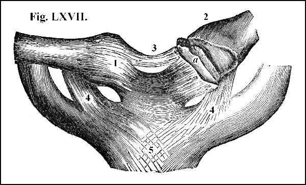
1. The fibrous capsule of the sternum and clavicle; 2. the same laid open, showing a, the interarticular cartilage; 3. the ligament connecting the two clavicles; 4. the ligament joining the clavicle to the first rib; 5. ligaments passing down in front of the sternum.
144. The scapular extremity of the clavicle (fig. LXVIII. 6) is attached to the processes of the scapula (fig. LXVIII. 4. 3) by several ligaments of great strength (fig. LXVIII. 7, 8, 9). First by very strong fasciculi which pass from the upper surface of the clavicle to the acromion of the scapula (fig. LXVIII. 6); and secondly by two ligaments which unite the clavicle with the coracoid process of the scapula (fig. LXVIII. 8, 9). These ligaments are so powerful that they resist a force capable of fracturing the clavicle; and they need to be thus strong, for the clavicle is a shaft which sustains the scapula, and through the scapula the whole of the upper extremity; and the main object of the joint by which these bones are united, is to afford a firm attachment of the scapula to its point of support.
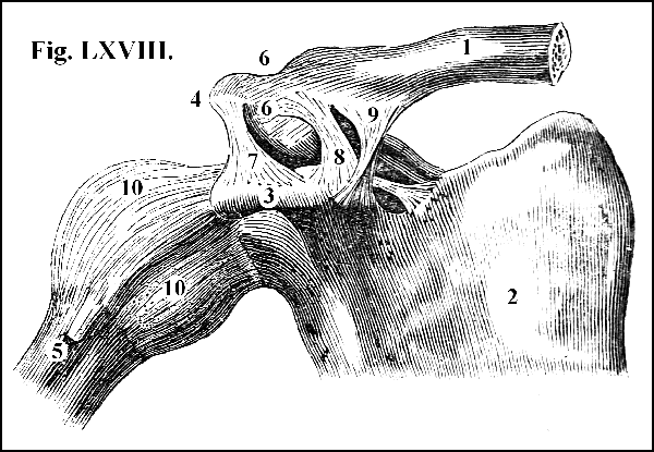
1. The clavicle; 2. the anterior part of the scapula; 3. the coracoid process; 4. the acromion process; 5. the humerus; 6. ligaments binding the scapular end of the clavicle to the acromion; 7. 8. 9. ligaments passing from one process of the scapula to the other; 10. the fibrous capsule of the shoulder-joint.
145. The clavicle serves the following uses: it sustains the upper extremity; it connects the upper extremity with the thorax; it prevents the upper extremity from falling forwards upon the thorax; and it affords a fixed point for steadying the extremity in the performance of its various actions.
146. The glenoid cavity of the scapula (fig. LXV. 7) receives the head of the humerus, the bone of the arm (fig. LXV. 9), and the two bones being united by ligament form the shoulder-joint (fig. LXVIII.). This joint is what is termed a ball and socket joint, the peculiarities of which are two: first, beyond all others this mode of articulation admits of free and extensive motion; in the present case, there is the utmost freedom of motion in every direction, upwards, downwards, backwards, and forwards. In the second place, this mode of articulation admits of the motion of the limb without that of the body, or of the motion of the body without that of the limb. When at rest, the arm may be moved in almost any direction without disturbing the position of any other part of the frame; the manifold advantages of which are obvious. On the other hand, by careful management, very considerable variations in the posture of the body may be effected without the communication of any degree of motion to the limb; an unspeakable advantage when the limb has sustained injury, or is suffering from disease.
147. It does not seem possible to construct a joint of great strength, capable, at the same time, of the degree of motion possessed by the joint of the shoulder. So shallow is the socket of the scapula, and so large the head of the humerus, that it seems as if the slightest movement must dislodge it from its cavity (fig. LXVI. 4. 7). For sustaining heavy weights or resisting a great amount of pressure, applied to it suddenly and in various directions, the arm is obviously unfitted. But this is not its office. The superior extremities are the organs of apprehension—the instruments by which the mind executes the commands of the will. They do not need the strength required by the organs that sustain the weight of the body and that perform the function of locomotion; but they do need freedom and extent of motion: to this strength may be sacrificed, and so it is; yet what can be done to combine strength with mobility is effected. Large and strong processes of bone, proceeding as has been shown (141), from the convex surface of the scapula (figs. LXV. and LXVI.), overhang, and to a considerable extent surround, the head of the arm-bone, especially resisting the force that would dislodge it from its socket and drive it upwards, inwards, and backwards (fig. LXV.), the directions in which force is most commonly applied to it. By these processes of bone the joint is greatly strengthened, especially in those directions. Moreover, a strong ligament, termed the fibrous capsule (fig. LXVIII. 10) envelops the joint. This ligament, arising from the neck of the scapula (fig. LXVIII. 10), expands itself in such a manner as completely to surround the head of the humerus (fig. LXVIII. 10); and then again contracts in order to be inserted into the neck of the bone (fig. LXVIII. 10). This ligament is strengthened by the tendons of no less than four muscles which are expanded over it, as well as by the powerful substance termed fascia which is reflected upon it from both the processes and ligaments of the scapula. In addition to all these expedients for fortifying the joint, it receives a further security in the position of the scapula, which is loose and unattached; which slides easily over the ribs upon its cushion of flesh; which thus obtains, by its facility of yielding, some compensation for its want of strength, eluding the force which it cannot resist.
148. The arm consists of numerous and powerful muscles, and of a single bone, the humerus, which belongs to the class of bones termed cylindrical (185).
149. The upper end of the humerus terminates in a circular head (fig. LXV. 9), which is received into the socket of the scapula (fig. LXV. 9. 7) termed, as has been stated (141), its glenoid cavity. The middle portion of the bone, or what is termed its shaft (fig. LXV. 8), diminishes considerably in magnitude, and becomes somewhat rounded (fig. LXV. 8), while its lower end again enlarges, and is spread out into a flattened surface of great extent (fig. LXIX. 1, 3, 2, 4). Of this broad flattened surface, the middle portion is grooved (fig. LXIX. 2): it is covered with cartilage; it forms the articulating surface by which the arm is connected with the fore-arm. On each side of this groove there is a projection of bone or tubercle, termed condyle (fig. LXIX. 3, 4), the inner (fig. LXIX. 3) being much larger than the outer (fig. LXIX. 4). The inner condyle gives origin to the muscles that bend, the outer to those that extend the fore-arm and the fingers (figs. LXXXIV. 1, 2, and LXXXV. 1).
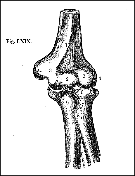
1. Lower extremity of the humerus; 2. grooved surface; 3. internal condyle; 4. external condyle; 5. the upper part of the ulna; 6. the head; 7. the neck; 8. the tubercle of the radius.
150. The muscles that act upon the arm arise from the back (fig. LXXII. 2), the chest (fig. LXXI. 1), the clavicle (fig. LXXI. 1), and the scapula (fig. LXXI. 3); and they move the arm with freedom and power upwards, downwards, forwards, backwards, inwards, and outwards. The chief muscle that raises the arm is the deltoid (fig. LXXI. 3), which arises partly from the clavicle and partly from the scapula (fig. LXXI. 3). It has the appearance of three muscles proceeding in different directions, the different portions being separated by slight fissures (figs. LXXI. 3, and LXXII. 3). The fibres converging unite and form a powerful muscle which covers the joint of the humerus (fig. LXXI. 3). It is implanted by a short and strong tendon into the middle of the humerus (fig. LXXI. 4). Its manifest action is to pull the arm directly upwards. Its action is assisted by the muscles that cover the back of the scapula, which are in like manner inserted into the humerus, and which, at the same time that they elevate the arm, support it when raised.
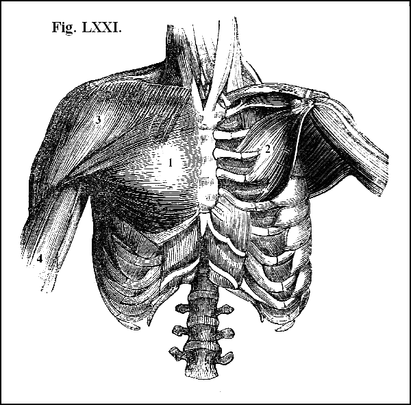
View of the muscles on the fore part of the chest that act upon the arm. 1. The muscle called the great pectoral; 2. the small pectoral; 3. the deltoid; 4. the humerus.
151. The principal muscle that carries the arm downwards is the latissimus dorsi (fig. LXXII. 2), the broadest muscle of the body, which, after having covered all the lower part of the back and loins, terminates in a thin but strong tendon which stretches to the arm, and is implanted into the humerus (fig. LXXII. 2), near the tendon of a muscle immediately to be described,—the great pectoral. When the arm is raised by the deltoid and its assistant muscles, the latissimus dorsi carries it downwards with force, and its powerful action is increased by that of other muscles which arise from the scapula and are inserted into the arm.
152. The principal muscle that carries the arm forwards towards the chest, is the great pectoral (fig. LXXI. 1), which, arising partly from the clavicle (fig. LXXI. 1), partly from the sternum (fig. LXXI. 1), and partly from the cartilages of the second, third, fourth, fifth, and sixth ribs (fig. LXXI. 1), covers the greater part of the breast (fig. LXXI. 1). Its fibres, converging, terminate in a strong tendon, which is inserted near the tendon of the longissimus dorsi into the humerus, about four inches below its head (fig. LXXI. 1). These two muscles form the axilla or armpit, the anterior border of the axilla consisting of the pectoral muscle. Though each of these muscles has its own peculiar office, yet they often act in concert, thereby greatly increasing their power, and the result of their combined action is to carry the arm either directly downwards or to the side of the chest.
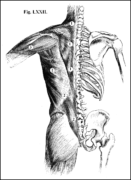
View of the muscles seated on the back part of the trunk that act upon the shoulder and arm. 1. The muscle called the trapezius; 2. the latissimus dorsi; 3. the deltoid.
153. Some of the muscles that elevate the arm carry it inwards, and others outwards; the muscles that carry it forwards likewise carry it inwards; while of the muscles that pull it downwards, some direct it forwards and inwards, and others backwards and outwards (151 and 152).
154. It has been already stated that the shoulder-joint is completely surrounded by the muscular fibres or the tendinous expansions of several of these powerful muscles, which have a far greater effect in maintaining the head of the humerus in its socket than the fibrous capsule of the joint; the latter being necessarily loose, in order to allow of the extended and varied motions of the arm, whereas the muscles that encompass the joint adhere closely and firmly to it. Moreover, by virtue of their vital power these muscles act with an efficiency which a mere ligamentous band is incapable of exerting; for they apportion the strength of resistance to the separating force, and react with an energy proportioned to the violence applied.
155. The bones of the fore-arm are two, the ulna and the radius (figs. LXIX. and LXXIII.). The ulna is essentially the bone of the elbow (figs. LXIX. 5, and LXXIII. 3); the radius that of the hand (fig. LXXV.). Supposing the arm to hang by the side of the body, and the palm of the hand to be turned forwards, the ulna, in apposition with the little finger, occupies the inner; and the radius, in apposition with the thumb, occupies the outer part of the fore-arm (fig. XXXIV. 3).
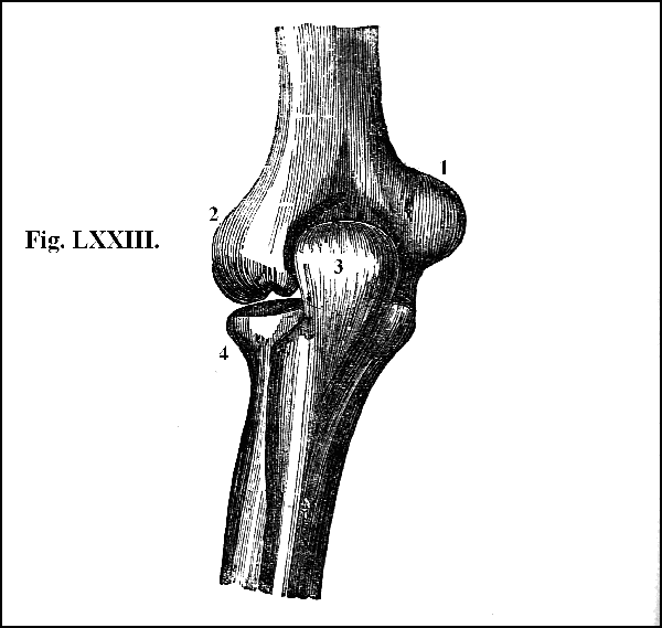
1. The internal condyle of the humerus; 2. the external condyle of the humerus; 3. the olecranon process of the ulna; 4. the head of the radius.
156. The upper end of the ulna belonging to the elbow is large (figs. LXIX. 5, and LXXIII. 3). It sends backwards the large projection commonly named the elbow or olecranon (fig. LXXII. 3), in the centre of which there is a smooth and somewhat triangular surface (fig. LXXIII. 3) which is always covered by skin of a coarse texture, like that placed over the lower part of the knee-pan, as if nature intended this for a part on which we may occasionally lean and rest. Large at the elbow, the ulna gradually grows smaller and smaller as it descends towards the wrist, where it ends in a small round head (fig. LXXXII. 2), beyond which, on the inner side, or that corresponding to the little finger, it projects downwards a small rounded point, termed the styloid process (fig. LXXXII. 3). As the styloid process and the olecranon, the two extremities of the ulna (figs. LXXIII. 3, and LXXII. 3), are easily and distinctly felt, the length of this bone was primitively used as a measure, called a cubit, which was the ancient name of the bone.
157. The radius, the second bone of the fore-arm, placed along its outer part next the thumb, is small at its upper end (figs. LXIX. 6, and LXXIII. 4); but its body is larger than that of the ulna; while its lower end, next the wrist to which it properly belongs, is very bulky (fig. LXXXII. 1). Its upper end is formed into a small circular head, which is united by distinct joints both to the humerus and to the ulna (fig. LXIX. 6). The top of its rounded head is excavated into a shallow cup (figs. LXIX. 6, and LXXIII. 4) which receives a corresponding convexity of the humerus (fig. LXIX. 2), and its lower extremity is excavated into an oblong cavity, which receives two of the bones of the wrist (fig. LXXXIII. 1. 4).
158. The joint of the elbow is composed above of the condyles of the humerus (fig. LXIX. 3. 2), and below by the heads of the ulna and radius (fig. LXIX. 5. 6).
159. The upper surface of the ulna is so accurately adapted to the lower surface of the humerus that the one seems to be moulded on the other (figs. LXIX. 5, and LXXIII. 3), and the form of these corresponding surfaces, which are everywhere covered with cartilage, is such as to admit of free motion backwards and forwards, that is, of extension and flexion; but to prevent any degree of motion in any other direction. The joint is therefore a hinge-joint, of which the two motions of flexion and extension are the proper motions. This hinge is formed on the part of the humerus by a grooved surface, with lateral projections (fig. LXIX. 2, 3, 4), and on the part of the ulna by a middle projection with lateral depressions (fig. LXIX. 5): the middle projection of the ulna turning readily on the grooved surface of the humerus (fig. LXIX. 2).
160. The bones are held in their proper situation, first, by a ligament on the fore part of the arm, called the anterior (fig. LXXIV. 6), which arises from the lower extremity of the humerus, and is inserted into the upper part of the ulna and the coronary ligament of the radius (fig. LXXIV. 6. 8); secondly, by another ligament on the back part of the arm, called the posterior ligament (fig.LXXV. 8), placed in the cavity of the humerus that receives the olecranon of the ulna (fig. LXXV. 8); and thirdly, by two other ligaments at the sides of the ulna (fig. LXXV. 6, 7). The ulna and radius are united, first, by a ligament called the coronary, which, arising from the ulna, passes completely around the head of the radius (fig. LXXVI. 3), and the attachment of which, while sufficiently close to prevent the separation of the two bones, is yet not adherent to the radius, for a reason immediately to be assigned; secondly, by another ligament which passes in an oblique direction from one bone to the other (fig. LXXVI. 4); and thirdly, by a dense and broad ligament, termed the interosseous (figs. LXXIV. 10, and LXXVI. 5), which fills up the space between the two bones nearly in their whole extent. This ligament serves other offices besides that of forming a bond of union, affording, more especially, a greater extent of surface for the attachment of muscles, and separating the muscles on the anterior from those on the posterior part of the limb.
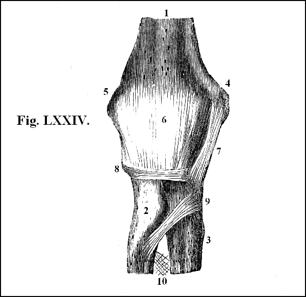
Anterior view of the ligaments of the elbow-joint. 1. The lower portion of the humerus; 2. the upper portion of the radius; 3. the upper portion of the ulna; 4. the internal condyle; 5. the external condyle; 6. the anterior ligament; 7. portion of the internal lateral ligament; 8. portion of the coronary ligament; 9. the oblique ligament; 10. upper portion of the interosseous ligament.
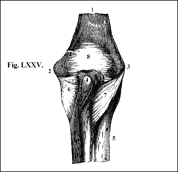
Posterior view of the ligaments of the elbow-joint. 1. Lower end of the humerus; 2. internal condyle; 3. external condyle; 4. the olecranon process of the ulna; 5. the upper portion of the radius; 6. the internal lateral ligament; 7. the external lateral ligament; 8. the posterior ligament.
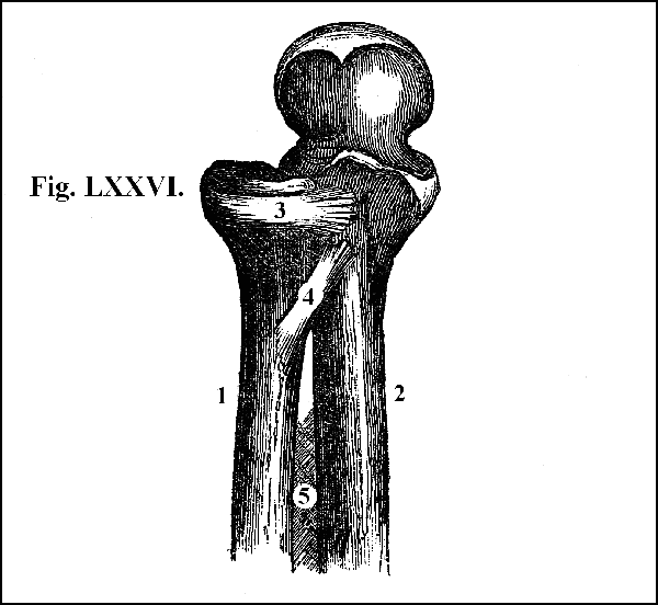
View of the ligaments connecting the ulna and radius at their upper part. 1. The radius; 2. the ulna; 3. the coronary ligament surrounding the head of the radius; 4. the oblique ligament passing from the ulna to the tubercle of the radius; 5 the upper portion of the interosseous ligament.
161. At their inferior extremities the ulna and radius are united partly by the interosseous ligament (fig. LXXVII. 1) and partly by ligamentous fibres which pass transversely from one bone to the other (fig. LXXVII. 2) on the anterior and the posterior surface of the fore-arm.
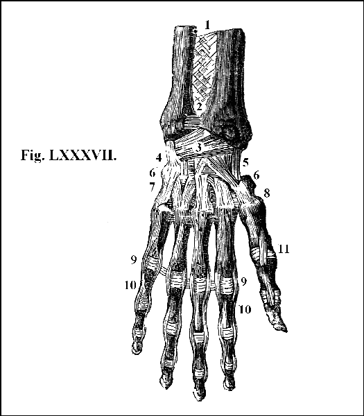
1. Interosseous ligament; 2. transverse fibres passing between the radius and ulna, and uniting the two bones; 3. 4. 5. posterior and lateral ligaments of the wrist joint; 6. ligaments uniting the bones of the wrist with one another; 7. 8. ligaments which attach the metacarpal to the carpal bones; 9. transverse ligaments for the attachment of the phalanges of the fingers; 10. lateral ligaments for the attachment of the phalanges of the fingers 11. ligaments of the thumb.
162. The lower extremity of the radius is also united to the wrist; and the hand being attached to the wrist, the junction of the hand and the fore-arm is effected by the articulation of the wrist with the radius (fig. LXXVII.). The ligaments which connect the bones of the wrist with the radius are bands of exceeding strength (fig. LXXVII. 3).
163. The muscles that act upon the fore-arm are placed upon the arm (fig. LXXVIII.). The joint of the elbow being a hinge-joint, the fore-arm can admit only of two motions, namely, flexion and extension. The muscles by which these motions are effected are four, two for each; the two flexors being placed on the fore part (fig. LXXVIII. 2. 4), and the two extensors on the back part of the arm (fig. LXXIX. 5).
164. The two flexor muscles of the fore-arm are termed the biceps and the brachialis (fig. LXXVIII. 2, 4). The biceps is so called because it has two distinct heads or points of origin (fig. LXXVIII. 2), both of which arise from the scapula (fig. LXXVIII. 2). About a third part down the humerus the two heads meet, unite and form a bulky muscle (fig. LXXVIII. 2), which, when it contracts, may be felt like a firm ball on the fore part of the arm, the upper part of the ball marking the point of union of the two heads (fig. LXXVIII. 2). The muscle gradually becoming smaller, at length terminates in a rounded tendon (fig. LXXVIII. 3), which is implanted into the tubercle of the radius a little below its neck (fig. LXXVIII. 3). It is an exceedingly thick and powerful muscle, and its manifest action is to bend the fore-arm with great strength. But since its tendon is inserted into the radius, besides bending the fore-arm, it assists other muscles that also act upon the radius in the performance of a function to be described immediately (168).
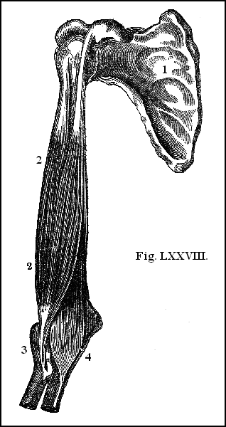
View of the flexor muscles of the fore-arm. 1. The anterior surface of the scapula; 2. the muscle called biceps; 3. tendon of the biceps passing to the tubercle of the radius; 4. the muscle called brachialis.
165. The second flexor of the fore-arm, termed the brachialis, is placed immediately under the biceps, and is concealed by it for a considerable part of its course (fig. LXXVIII. 4). Arising from the humerus, on each side of the insertion of the deltoid, it continues its attachment to the bone all the way down the fore part of the humerus, to within inch of the joint; it then passes over the joint, adhering firmly to the anterior ligament (fig. LXXVIII. 4), and is inserted by a strong tendon into the ulna (fig. LXXVIII. 4). It is a thick and fleshy muscle, powerfully assisting the action of the biceps.
166. The two extensor muscles are named the triceps and the anconeous (fig. LXXIX.). The triceps, seated on the back part of the arm, derives its name from having three distinct points of origin, or three separate heads (fig. LXXIX. 5); one of which arises from the scapula and two from the humerus (fig. LXXIX. 5). All these heads adhere firmly to the humerus, as the brachialis does on the fore part of the arm, down to within an inch of the joint (fig. LXXIX. 5), where they form a strong tendon, which is implanted into the olecranon of the ulna (fig. LXXIX. 3); the projection of which affords a lever for increasing the action of the muscle. In all animals that leap and bound, this process of the ulna is increased in length in proportion to their power of performing these movements. The triceps forms an exceedingly thick and strong muscle, which envelops the whole of the back part of the arm (fig. LXXIX.); its action is simple and obvious; it powerfully extends the fore-arm. The anconeous, a small muscle of a triangular form, arising from the external condyle of the humerus, and inserted into the ulna a little below the olecranon, assists the action of the triceps.
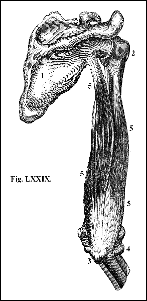
View of the extensor muscles of the fore-arm. 1. The scapula; 2. the upper part of the humerus; 3. upper end of the ulna; 4. upper end of the radius; 5. the muscle called triceps, the extensor of the fore-arm.
167. Such are the motive powers which act upon the fore-arm, and which produce all the motions of which the hinge-joint of the elbow renders it capable. But besides flexion and extension, the fore-arm is capable of the motion of rotation, which is accomplished by means of the radius. It has been shown (157) that the top of the rounded head of the radius is excavated into a shallow cup (figs. LXIX. 6, and LXXIII. 4) which receives a corresponding convexity of the humerus (figs. LXIX. 2, and LXXIII. 2). In consequence of this articulation with the humerus, the radius, like the ulna, can move backwards and forwards in flexion and extension, the proper movements of the hinge-joint; but that portion of the margin of the hinge of the radius which is in apposition with the ulna is convex (fig. LXIX. 6), and is received into a semilunar cavity hollowed out in the ulna (fig. LXIX. 5). In this cavity the rounded head of the radius revolves, the two bones being held together by the ligament already described (160), which surrounds the head of the radius (fig. LXXVI. 3), and which holds it firmly without being adherent to it, and without impeding in any degree the rotatory motion of the radius. Below, the surface of the radius next the ulna is hollowed out into a semilunar cavity (fig. LXXXII. 1), which receives a corresponding convex surface of the ulna (fig. LXXXII. 2), upon which convex surface the radius rolls (fig. LXXXII. 1). Thus, by the mode in which it is articulated with the ulna above, the radius turns upon its own axis. By the mode in which it is articulated with the ulna below, the radius revolves upon the head of the ulna; and, in consequence of both articulations, is capable of performing the motion of rotation. Moreover, the hand being attached to the radius through the medium of the wrist (figs. LXXXII. 1. 4. and LXXXIII. 1. 4) must necessarily follow every movement of the radius; the rotation of which brings the hand into two opposite positions. In the one, the palm of the hand is directed upwards (fig. LXXXII.); in the other, it is turned downwards (fig. LXXXIII.). When the hand is turned upwards, it is said to be in the state of supination (fig. LXXXII.); when downwards, in that of pronation (fig. LXXXIII.). A distinct apparatus of muscles is provided for effecting the rotation of the radius, in order to bring the hand into these opposite states: one set for producing its supination, and another its pronation.
168. The principal supinators arise from the external condyle of the humerus (fig. LXXX.), and are called long and short (fig. LXXX. 4, 5). The long supinator extends as far as the lower end of the radius, into which it is inserted (fig. LXXX. 4): the short supinator surrounds the upper part of the radius, and is attached to it in this situation (fig. LXXX. 5.). Moreover, the triceps, being inserted into the radius (164), often cooperates with the supinators and powerfully assists their action.
169. The principal pronators are also two, called the round and the square (figs. LXXXI. and LXXXVI. 1). The round pronator arises from the internal condyle, and passing downwards, is inserted into the middle of the radius (fig. LXXXI. 4); the square pronator is a small muscle between the radius and ulna, at their lower extremities being attached to each (fig. LXXXVI. 1).
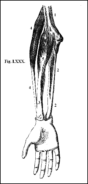
View of the supinators of the radius and hand. 1. The humerus; 2. the ulna; 3. the radius; 4. the muscle called the long supinator passing to be inserted into the lower portion of the radius; 5. the muscle, called the short supinator, surrounding the upper part of the radius.
170. The action of these muscles in producing the rotation of the radius, and so rendering the hand supine or prone, is sufficiently manifest from the mere inspection of the diagrams (fig. LXXXI. 4).
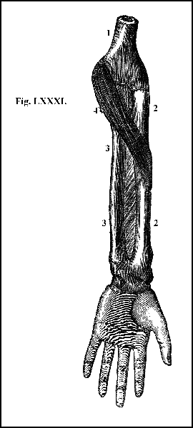
View of the pronators of the hand. 1. Lower end of the humerus; 2. the radius; 3. the ulna; 4. the muscle called the round pronator, one of the powerful pronators of the hand.
171. The hand is composed of the carpus, metacarpus, and fingers.
172. The carpus (fig. LXXXII. 4) consists of eight small wedge-shaped bones, placed in a double row, each row containing an equal number, and the whole disposed like stones in an arch (fig. LXXXII. 4). They do in fact form an arch, the convexity of which is upwards, on the dorsal surface (fig. LXXXIII. 4); and the concavity downwards, on the palmar surface (fig. LXXXII. 4). But they differ from the stones of an arch in this, that each bone is joined to its fellow by a distinct moveable joint, each being covered with a smooth articulating cartilage. At the same time all of them are tied together by ligaments of prodigious strength, which cross each other in every direction (fig. LXXVII. 6), so that the several separate joints are consolidated into one great joint. The consequence of this mechanism is that some degree of motion is capable of taking place between the several bones, which, when multiplied together, gives to the two rows of bones such an extent of motion, that when the wrist is bent the arch of the carpus forms a kind of knuckle. By this construction a facility and ease of motion, and a power of accommodation to motion and force, are obtained, such as belong to no arch contrived by human ingenuity.
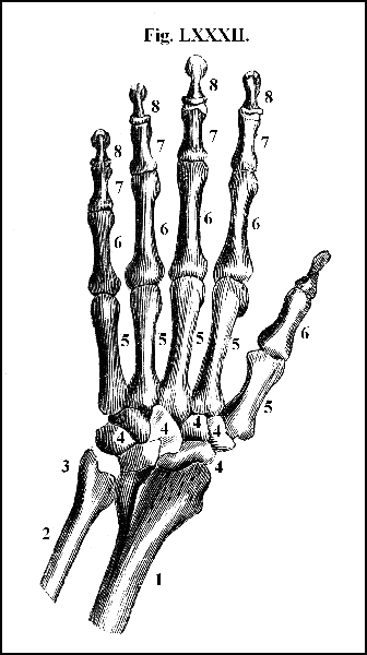
1. Lower extremity of the radius; 2. lower extremity of the ulna; 3. styloid process of the ulna; 4. bones of the carpus or wrist; 5. metacarpal bones; 6. first phalanges of the fingers; 7. second phalanges of the fingers; 8. third phalanges of the fingers.
173. The metacarpus (fig. LXXXII. 5), the middle portion of the hand, interposed between the wrist and the fingers, is composed of five bones, which are placed parallel to each other (fig. LXXXII. 5). They are convex outwardly, forming the back (fig. LXXXIII. 5), and concave inwardly, forming the hollow of the hand (fig. LXXXII. 5). They are large at each end, to form the joints by which they are connected with the wrist and fingers (figs. LXXXII. and LXXXIII.): they are small in the middle, in order to afford room for the lodgment and arrangement of the muscles, that move the fingers from side to side (fig. LXXXVI. 2). Their ends, which are joined to the carpus, are connected by nearly plane surfaces (figs. LXXXII. and LXXXIII.): their ends, which support the fingers, are formed into rounded heads, which are received into corresponding cup-shaped cavities, excavated in the top of the first bones of the fingers (fig. LXXXII. 5.). The powerful ligaments that unite these bones pass, both on the dorsal and the palmar surface, from the inferior extremity of the second row of the carpal to the bases of the metacarpal bones (fig. LXXVII, 7, 8). The ligaments are arranged in such a manner as to limit the motions of the joints chiefly to those of flexion and extension, allowing, however, a slight degree of motion from side to side.
174. Each of the fingers is composed of three separate pieces of bone, called phalanges; the thumb has only two (fig. LXXXII. 6, 7, 8): the phalanges are convex outwardly (fig. LXXXII. 6, 7, 8) for increasing their strength, and flattened inwardly (fig. LXXXIII. 6, 7, 8) for the convenience of grasping. The last bones of the fingers, which are small, terminate at their under ends, in a somewhat rounded and rough surface (fig. LXXXIII. 8), on which rests the vascular, pulpy, and nervous substance, constituting the special organ of touch, placed at the points of the fingers, and guarded on the upper surface by the nail (fig. LXXXII. 8).
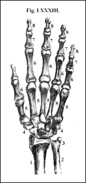
1. Lower extremity of the radius; 2. lower extremity of the ulna; 3. styloid process of the ulna; 4. bones of the carpus; 5. metacarpal bones; 6. 7. 8. first, second, and third phalanges of the fingers.
175. The round inferior extremity of the metacarpus is admitted into the cavity of the superior extremity of the first phalanx of the five fingers (figs. LXXXII. and LXXXIII.), and their joints are connected by lateral and transverse ligaments of great strength (fig. LXXVII. 9). The situation and direction of the ligaments which unite the several phalanges of the fingers (fig. LXXVII. 9) are precisely the same as those of the articulation of the phalanges with the metacarpus (fig. LXXVII. 7, 8); and the articulation of these bones with one another is such as to admit only of the motions of flexion and extension.
176. The muscles which perform these motions are seated for the most part on the fore-arm. Independently of the supinators and pronators which have been already described (167 et seq.), there are distinct sets of muscles for bending and extending the wrist and the fingers. The flexors arise from the internal, and the extensors from the external, condyle of the humerus (fig. LXIX. 3, 4). The internal condyle is larger and longer than the external (fig. LXIX. 3, 4); for the flexors require a larger point of origin and a longer fulcrum than the extensor muscles; because to the actions of flexion, such as grasping, bending, pulling, more power is necessary than to the action of extension, which consists merely in the unfolding or the opening of the hand previously to the renewal of the grasp.
177. For the same reason, two muscles are provided for flexing, while only one is provided for extending the fingers. The flexors, bulky, thick, and strong, are placed on the fore part of the fore-arm (fig. LXXXIV.). The first, named the superficial flexor (fig. LXXXIV. 1), about the middle of the arm, divides into four fleshy portions, each of which ends in a slender tendon (fig. LXXXIV. 1). As these tendons approach the fingers they expand (fig. LXXXIV. 1), and when in apposition with the first phalanx, split and form distinct sheaths for the reception of the tendons of the second flexor (fig. LXXXIV. 3). After completing the sheath, the tendons proceed forward along the second phalanx, into the fore part of which they are implanted, and the chief office of this powerful muscle is to bend the second joint of the fingers upon the first, and the first upon the metacarpal bone. Its action is assisted by a second muscle, called the deep or profound flexor (fig. LXXXIV. 2), because it lies beneath the former; or the perforans, because it pierces it. Bulky and fleshy, this second flexor, like the first, about the middle of the arm, divides into four tendons, which, entering the sheaths prepared for them in the former muscle (where the tendons are small and rounded for their easy transmission and play), pass to the root of the third phalanx of the fingers into which they are implanted (fig. LXXXIV. 3).
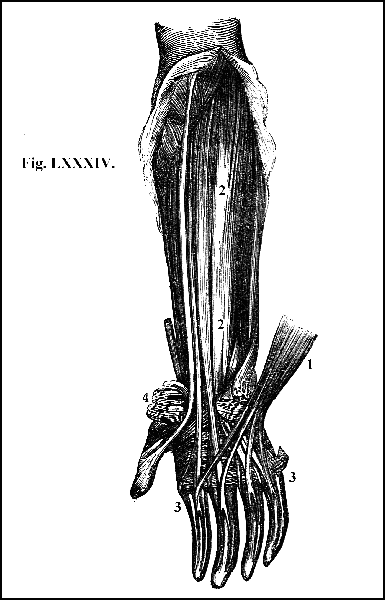
View of the flexor muscles of the fingers. 1. The superficial flexor, divided and turned aside, to show, 2. the deep flexor; 3. sheaths for the tendons of the deep flexor, formed by the splitting of the tendons of the superficial flexor; 4. the anterior annular ligament, divided and turned aside.
118. The muscle that extends the fingers, called the common extensor, is placed on the back part of the fore-arm (fig. LXXXV.), about the middle of which it divides into four portions which terminate in so many tendons (fig. LXXXV. 2). When they reach the back of the metacarpal bones, these tendons become broad and flat, and send tendinous expansions to each other, forming a strong tendinous sheath which surrounds the back of the fingers (fig. LXXXV. 2). These tendinous expansions are inserted into the posterior part of the bones of the four fingers (fig. LXXXV. 2); and their office is powerfully to extend all the joints of all the fingers (fig. LXXXV. 2).
179. On both the palmar and dorsal regions of the wrist are placed ligaments for tying down these tendons, and preventing them from starting from their situation during the action of the muscles (figs. LXXXIV. and LXXXV.). On the palmar region an exceedingly strong ligament passes anteriorly to the concave arch of the carpus (fig. LXXXIV. 4) for the purpose of tying down the tendons of the flexor muscles. On the dorsal surface (fig. LXXXV.), a similar ligament, passing in an oblique direction from the styloid process of the radius to the styloid process of the ulna (fig. LXXXV. 3), performs the same office in tying down the tendons of the extensor muscle. Both these ligaments are called annular.
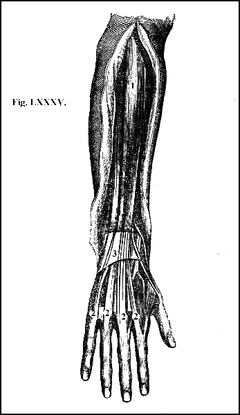
View of the extensor muscles of the fingers. 1. The common extensor, sending (2 2 2 2) tendons to each finger; 3. the posterior annular ligament.
180. In the palm of the hand are placed additional muscles which assist the flexors of the fingers (fig. LXXXVI. 2), being chiefly useful in enabling the fingers to perform with strength and precision short and quick motions. There are especially four small and rounded muscles (fig. LXXXVI. 2), resembling the earth worm in form and size, and hence called lumbricales; but as their chief use is to assist the fingers in executing short and rapid motions, they have also received the better name of the musculi fidicinales.
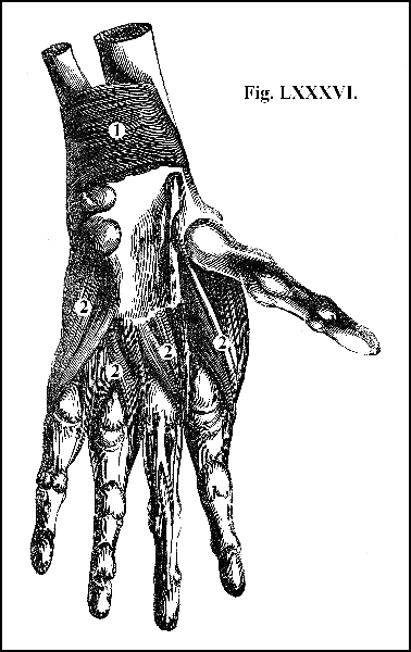
1. The muscle called the square pronator; 2. muscles seated in the palm of the hand, by which, chiefly, the fingers execute short and rapid motions.
181. The thumb, in consequence of the comparative looseness of its ligaments, is capable of a much greater extent of motion than the fingers, and can be applied to any part of each of the fingers, to different parts of the hand, and in direct opposition to the power exerted by the whole of the fingers and hand, in the act of grasping. The muscles which enable it to perform these varied motions, and which act powerfully in almost every thing we do with the hand, form a mass of flesh at the ball of the thumb (fig. LXXXVII. 1), almost entirely surrounding it. The little finger is also provided with a distinct apparatus of muscles (fig. LXXXVII. 2), which surrounds its root, just as those of the thumb surround its ball, in order to keep it firm in opposition to the power of the thumb in the act of grasping, and in various other motions.
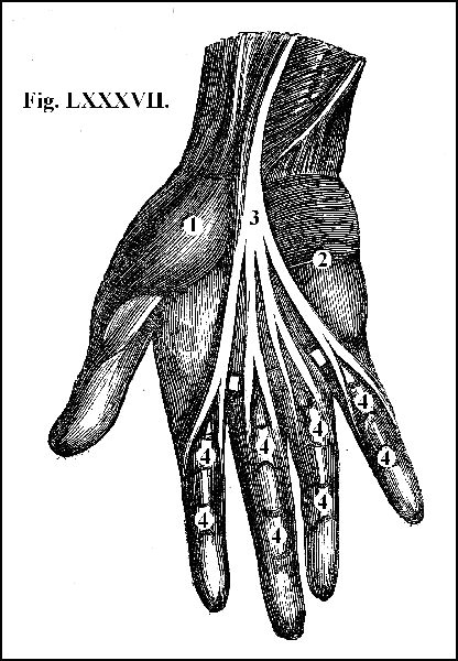
1. The mass of muscles forming the ball of the thumb; 2. the mass of muscles forming the ball of the little finger; 3. tendons of one of the flexor muscles of the fingers; 4. sheaths formed by the tendons of the superficial flexor for the reception of the tendons of the deep flexor.
182. The upper extremity is covered by a tendinous expansion or fascia which envelopes the whole arm, encloses its muscles as in a sheath, and affords them, in their strong actions, "that kind of support which workmen feel in binding their arms with thongs." This fascia likewise descends between many of the muscles, forming strong partitions between them, and affording points of origin to many of their fibres, scarcely less fixed than bone itself.
183. From the whole, it appears, that the first joint of the upper extremities, that of the shoulder, is a ball and socket joint, a joint admitting of motion in every direction; that the second joint, that of the elbow, is partly a hinge-joint, admitting of flexion and extension, and partly a rotation joint, admitting of a turning or rotatory motion; and that the joints of the wrist and of the fingers are likewise hinge-joints, admitting at the same time of some degree of lateral motion. When these various motions are combined, the result is that the hand can apply itself to bodies in almost every direction, in any part of the area described by the arm, when all the joints are moved to their utmost extent. There is thus formed an instrument of considerable strength, capable of a surprising variety and complexity of movements, capable of seizing, holding, pulling, pushing and striking with great power, yet at the same time capable of apprehending the minutest objects, and of guiding them with the utmost gentleness, precision, and accuracy, so that there are few conceptions of the designing mind which cannot be executed by the skilful hand.
184. The lower extremities consist of the thigh, leg, and foot.
185. The osseous part of the thigh consists of a single bone, called the femur (fig. XXXIV. 4), the longest, thickest, and strongest bone in the body. It sustains the entire weight of the trunk, and occasionally much heavier loads superimposed upon it. It is constructed in such a manner as to combine strength with lightness. This is effected by rendering the bone what is technically called cylindrical; that is, a bone in which the osseous fibres are arranged around a hollow cylinder. There are two varieties of osseous matter,—the compact, in which the fibres are dense and solid (fig. LXXXVIII. 1), and the spongy, in which the fibres are comparatively tender and delicate (fig. LXXXVIII. 2). Both varieties are, indeed, combined, more or less, in every bone, the compact substance being always external, and the spongy internal; but in the cylindrical bones the arrangement is peculiar. Every long or cylindrical bone consists of a body or shaft (fig. LXXXVIII. 4.), and of two extremities (fig. LXXXVIII. 5). The body is composed principally of compact substance, which on the external surface is so dense and solid, that scarcely any distinct arrangement is visible; but towards the interior this density diminishes; the fibres become distinct (fig. LXXXVIII. 5), and form an expanded tissue of a cellular appearance (fig. LXXXVIII. 5), the cells being called cancelli, and the structure cancellated. In the centre of the bone even the cancelli disappear; the osseous fibres terminate; and a hollow space is left filled up, in the natural state, by an infinite number of minute membranous bags which contain the marrow (fig. LXXXVIII. 3). In the body of the bone, to which strength is requisite, that part being the most exposed to external violence, the compact matter is arranged around a central cavity. By this means strength is secured without any addition of weight; for the resisting power of a cylindrical body increases in proportion to its diameter; consequently the same number of osseous fibres placed around the circumference of a circle produce a stronger bone than could have been constructed had the fibres been consolidated in the centre, and had the diameter been proportionally diminished. The hollow space thus gained in its centre, renders the bone lighter by the subtraction of the weight of as many fibres as would have gone to fill up that space; while its strength is not only not diminished by this arrangement, but positively increased. On the other hand, at the extremities of the bone, space, not strength, is required; required for the attachment and arrangement of the tendons of the muscles that act upon it, and for the formation of joints (fig. LXXXVIII. 5). Accordingly, at its extremities the bone swells out into bulky surfaces; but these surfaces are composed, not of dense and solid substance, but of spongy tissue, covered by an exceedingly thin crust of compact matter, and so, as by the former expedient strength is secured without increase of weight, by this, space is obtained without increase of weight.
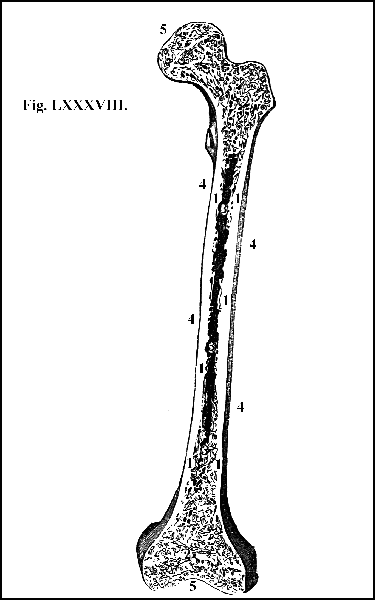
A section of the femur, showing, 1. the compact bony substance; 2. the spongy or cancellated structure; 3. the internal cavity containing the marrow; 4. body; 5, extremities of the bone.
186. The thigh-bone, placed at the under and outer part of the pelvis, has an oblique direction, the under being considerably nearer its fellow than the upper end (fig. XXXIV. 4), in order to afford space for the passages at the bottom of the pelvis, and also to favour the action of walking. The body of the bone, which is of a rounded form (fig. XXXIV. 4), is smooth on its anterior surface (fig. XXXIV. 4), where it is always slightly convex, the convexity being forwards (fig. XXXIV. 4), while its posterior surface is irregular and rough, and forms a sharp prominent line, termed the linea aspera (fig. XXXV. 4), giving attachment to numerous muscles.
187. The superior extremity of the femur terminates in a large ball or head, which forms nearly two-thirds of a sphere (fig. LXXXIX. 4.). It is smooth, covered with cartilage, and received into the socket of the ilium called the acetabulum, which, deep as it is, is still further deepened by the cartilage which borders the brim (fig. LXXXIX. 3). The brim is particularly high in the upper and outer part, because it is in this direction that the reaction of the ground against the descending weight of the trunk tends to dislodge the ball from its socket.
188. Passing obliquely downwards and outwards from the ball, is that part of the femur which is called the neck (fig. LXXXIX. 5). It spreads out archlike between the head and the body of the bone, and is more than an inch in length (fig. LXXXIX. 5). It is thus long in order that the head of the bone may be set deep in its socket, and that its motions may be wide, free, and unembarrassed.
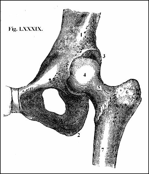
1. Lower portion of the ilium; 2. tuberosity of the ischium: 3. socket for the head of the femur, or thigh-bone; 4. head of the femur; 5. neck of the femur; 6. the great process of the femur called the trochanter major; 7. the body of the femur.
189. From the external surface of the femur, nearly in a line with its axis, proceeds the largest and strongest bony process of the body which gives insertion to its most powerful muscles, namely, those that extend the thigh and that turn it upon its axis (fig. LXXXIX. 6). Because, from its oblique direction, it rotates the thigh, this process is called the trochanter, and, from its size, the trochanter major. At the under and inner part of the neck on the posterior surface of the bone, is a similar process, but much smaller, called the trochanter minor (fig. XXXV. 4), into which are inserted the muscles that bend the thigh.
190. The inferior extremity of the femur, much broader and thicker than the superior (fig. XC. 1), is terminated by two eminences, with smooth surfaces, termed condyles (fig. XC. 2), which, articulated with the tibia, and the patella, form the joint of the knee (figs. XC. 2, 4, 5, and XCI. 1, 2, 3).
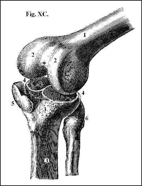
1. Lower end of the femur; 2. condyles of the femur; 3. upper end of the tibia; 4. articular surfaces on the head of the tibia on which the thigh-bone plays; 5. the patella, or knee-pan; 6. upper end of the fibula, not entering into the knee-joint.
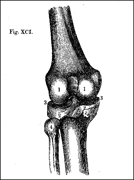
Posterior view of the bones forming the knee-joint. 1. Lower end of the femur; 2. upper end of the tibia; 3. articular surfaces on the head of the tibia, on which the thigh-bone plays; 4. upper end of the fibula, not entering into the knee joint.
191. The bones of the leg, two in number, consist of the tibia (fig. XC. 3) and fibula (fig. XC. 6). The tibia, next to the femur, the longest bone in the body, is situated at the inner side of the leg (fig. XC. 3). Its superior extremity is bulky and thick (fig. XC. 3). The top of it forms two smooth and slightly concave surfaces, adapted to the convex surfaces of the condyles of the femur (fig. XC. 4, 2). On its outer side there is a smooth surface, to which the head of the fibula is attached (fig. XC. 6). Its lower extremity, which is small, forms a concavity adapted to the convexity of the bone of the tarsus, called the astragalus, with which it is articulated (fig. XCII. 4.) Its inner part is produced so as to form the inner ankle (figs. XCII. 2, and XCIII. 3): its outer side is excavated into a semilunar cavity, for receiving the under end of the fibula, which forms the outer ankle (figs. XCII. 3, and XCIII. 4).
192. The fibula, in proportion to its length the most slender bone of the body, is situated at the outer side of the tibia (fig. XC. 6). Its upper end formed into a head, with a flat surface on its inner side (figs. XC. 6, and XCI. 4), is firmly united to the tibia (fig. XC. 4). Its lower end forms the outer ankle, which is lower and farther back than the inner (fig. XCII. 3, 2).
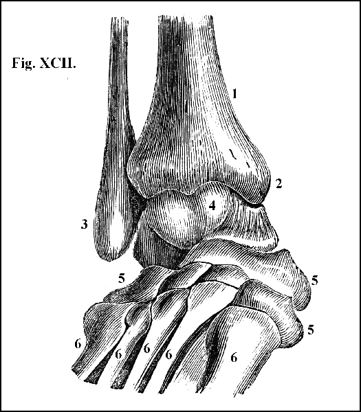
Anterior view of the bones forming the ankle-joint. 1. Lower end of the tibia; 2. production of the tibia, forming the inner ankle; 3. lower end of the fibula, forming the outer ankle; 4. upper part of the astragalus: these three bones form the ankle-joint; 5 5 5, other bones of the tarsus; 6 6 6 6 6 metatarsal bones.
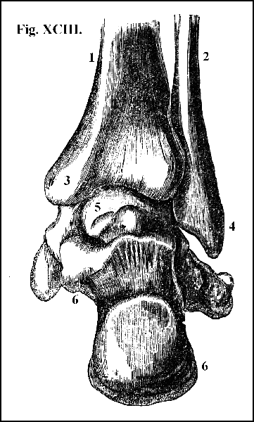
Posterior view of the bones forming the ankle-joint. 1. Lower end of the tibia; 2. lower end of the fibula; 3. internal malleolus or ankle; 4. external malleolus or ankle; 5. one of the tarsal bones, called the astragalus, with which the tibia and fibula are articulated; 6. the os calcis or heel.
193. The patella, or knee-pan (fig. XC. 5), is a light but strong bone, of the figure of the heart as painted on playing-cards, placed at the fore part of the joint of the knee, and attached by a strong ligament to the tibia, the motions of which it follows (fig. XC. 5). It is lodged, when the knee is extended, in a cavity formed for it in the femur (fig. XC.); when bent, in a cavity formed for it at the fore part of the knee (fig. XC. 5).
194. The foot consists of the tarsus, metatarsus, and toes.
195. The tarsus, or instep, is composed of seven strong, irregular-shaped bones, disposed like those of the carpus, in a double row (fig. XCII. 4, 5). The arrangement of the tarsal bones is such as to form an arch, the convexity of which above, constitutes the upper surface of the instep (fig. XCII. 4, 5): in the concavity below are lodged the muscles, vessels, and nerves that belong to the sole.
196. The metatarsus consists of five bones, which are placed parallel to each other (fig. XCII. 6), and which extend between the tarsus and the proper bones of the toes (fig. XCII. 6). Their extremities, especially next the tarsus, are large, in order that they may form secure articulations with the tarsal bones (fig. XCII. 6). Their bodies are arched upwards (fig. XCII. 6), slightly concave below, and terminate forwards in small, neat, round heads, which receive the first bones of the toes, and with which they form joints, admitting of a much greater degree of rotation than is ever actually exercised, in consequence of the practice of wearing shoes. The natural, free, wide-spreading form of the toes, and the consequent security with which they grasp the ground, is greatly impaired by this custom. Taken together, the bones of the metatarsus form a second arch corresponding to that of the tarsus (fig. XCVIII. 2).
197. Each toe consists of three distinct bones, called, like those of the fingers, phalanges (fig. XCVIII.), but the great toe, like the thumb, has only two (fig. XCVIII.). That extremity of the first phalanges which is next the metatarsal bones is hollowed into a socket for the head of the metatarsal bones.
198. Besides the bones already described, there are other small bones, of the size and figure of flattened peas, found in certain parts of the extremities, never in the trunk, called sesamoid, from their resemblance to the seed of the sesamum. They belong rather to the tendons of the muscles than to the bones of the skeleton. They are embedded within the substance of tendons, are found especially at the roots of the thumb and of the great toe, and are always placed in the direction of flexion. Their office, like that of the patella, which is, in truth, a bone of this class, is to increase the power of the flexor muscles by altering the line of their direction, that is, by removing them farther from the axis of the bone on which they are intended to act.
199. The ligaments which connect the bones of the lower extremities are the firmest and strongest in the body. Of these, the fibrous capsule of the hip-joint (fig. XCIV. 1), which secures the head of the femur in the cavity of the acetabulum (fig. XCIV.), is the thickest and strongest. It completely surrounds the joint (fig. XCIV. 1). It arises from the whole circumference of the acetabulum, and, proceeding in a direction outwards and backwards, is attached below to the neck of the femur (fig. XCIV. 1). It is thicker, stronger, and much more closely attached to the bones than the fibrous capsule of the shoulder-joint (144), because the hip-joint is formed, not like the shoulder-joint, for extent of motion, but for strength. Its internal surface is lined by synovial membrane, and its external surface is covered and strengthened by the insertion of muscles that move the thigh-bone. The joint is strengthened by another ligament, which passes from the inner and fore part of the cavity of the acetabulum (fig. XCV.) to be inserted into the head of the femur (fig. XCIV.), called the round ligament, the office of which obviously is to hold the head of the femur firmly in its socket.
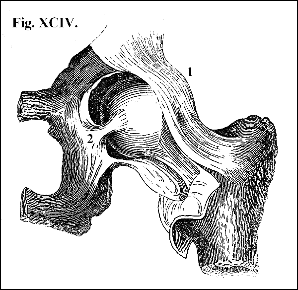
1. The fibrous capsule of the hip-joint, laid open and turned aside to show, 2. the round ligament in its natural position.
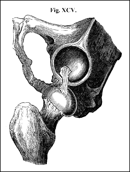
A view of the head of the femur drawn out of its socket, and suspended by the round ligament, to show more clearly the action of the ligament in retaining the head of the femur in its socket.
200. Numerous and complicated ligaments connect the bones that form the knee-joint (fig. XCVI.), and the strength of these powerful bands is greatly increased by the tendons that move the leg (fig. XCVI. 5), which pass over, and more or less surround, the joint.
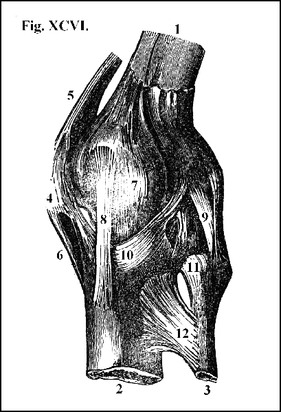
General view of the ligaments of the knee-joint. 1. Lower end of the femur; 2. upper end of the tibia; 3. upper end of the fibula; 4. the patella; 5. united tendons of the extensor muscles; 6. ligaments of the patella; 7. the capsular investment of the knee; 8. the internal lateral ligament; 9. the external lateral ligaments; 10. the posterior ligament; 11. the ligament connecting the tibia and fibula; 12. a portion of the interosseous ligament.
201. Strong ligaments maintain in their proper position the bones that form the ankle-joint (fig. XCVII.), connect the bones of the tarsus and metatarsus with one another (fig. XCVIII. 1), and articulate the several phalanges of the toes (fig. XCVIII. 2).
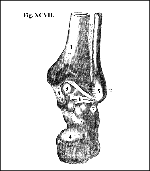
General view of the posterior ligaments of the ankle-joint. 1. Lower end of the tibia; 2. lower end of the fibula; 3. astragalus; 4. os calcis; 5. ligament between the tibia and fibula; 6. ligament passing from the fibula to the astragalus; 7. ligament passing from the fibula to the os calcis; 8. ligament passing from the tibia to the astragalus.
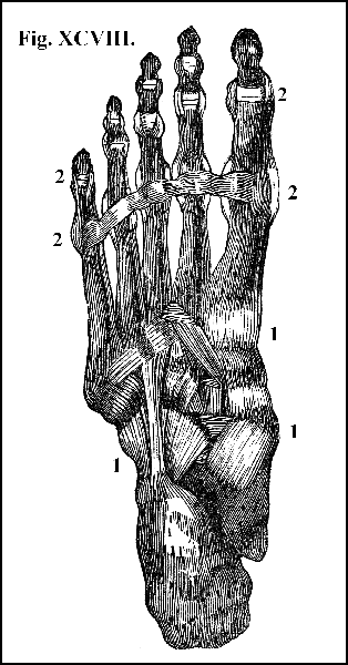
General view of the ligaments of the sole of the foot. 1. Ligaments connecting the bones of the tarsus; 2. ligaments connecting the bones of the toes.
202. The joint of the hip, like that of the shoulder, is capable of flexion, extension, and rotation; but its rotatory motions are to a much less extent, on account of the greater depth of the acetabulum and the stronger and shorter fibrous capsule. When the femur is flexed, the thigh is bent upon the pelvis, and its inferior extremity is carried forwards. When it is extended, the thigh is carried backwards. The two thighs may be separated from each other laterally (abduction), or brought near to each other (adduction), or the one may be made to cross the other, and they may be rotated outwards or inwards.
203. The apparatus of muscles that produces these varied motions is seated partly on the trunk and partly on the pelvis. Thus, the powerful muscle that flexes the thigh, or that carries it forwards, termed the psoas (fig. XCIX. 1), arises from the last vertebra of the back, and successively from each vertebra of the loins (fig. XCIX. 1), and is inserted into the lesser trochanter of the femur (fig. XCIX. 3). Its action is assisted first by a large and strong muscle named the iliacus (fig. XCIX. 2), which occupies the whole concavity of the ilium (fig. XCIX. 2), and which, like the psoas, is inserted into the lesser trochanter of the femur (fig. XCIX. 3).
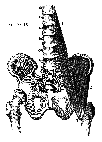
View of the muscles that bend the thigh. 1. The muscle called psoas; 2. the muscle called iliacus; 3. tendons of these muscles, going to be inserted into the trochanter minor of the femur.
204. The muscles that extend the thigh, or that carry it backwards, named the glutæi, the most powerful muscles of the body, are placed in successive layers, one upon the other, on the back part of the ilium (fig. C. 1, 2, 3), and are inserted into the linea aspera of the femur. They constitute the mass of flesh which forms the hip, and their powerful action in drawing the thigh backwards is assisted by several other muscles (fig. C. 4, 5, 6). Their action is never perfectly simple and direct; for those which move the thigh forwards sometimes carry it inwards, and sometimes outwards; and in like manner, those which move it backwards, at one time carry it inwards and at another outwards, according to the direction of the fibres of the muscle and the position of the limb when those fibres act; while some of them, and more especially those which carry it backwards, at the same time rotate it, or roll it upon its axis.
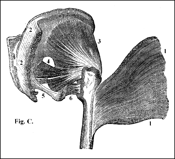
View of the muscles that extend the thigh. 1. The muscle called glutæus maximus, removed from its origin, 2, 2, to show the muscles which lie beneath it; 2. cut edge showing the origin of the same muscle; 3. the muscle called glutæus medius; 4, 5, 6. smaller muscles, assisting the action of the glutæi.
205. The knee is a hinge-joint, admitting only of flexion and extension, and is therefore provided only with two sets of muscles, one for bending and the other for extending the leg. The flexors of the leg arise from the under and back part of the pelvis, are seated on the back part of the thigh, and are inserted into the upper part either of the tibia or of the fibula (fig. CI). They consist for the most part of three muscles, named the semi-tendinosus, the semi-membranosus (fig. CI. 3), and the biceps of the leg (fig. CI. 1). The tendons of the two former muscles, in passing to be inserted into the leg, form the inner, and that of the latter the outer, hamstrings (fig. CI. 4, 5).
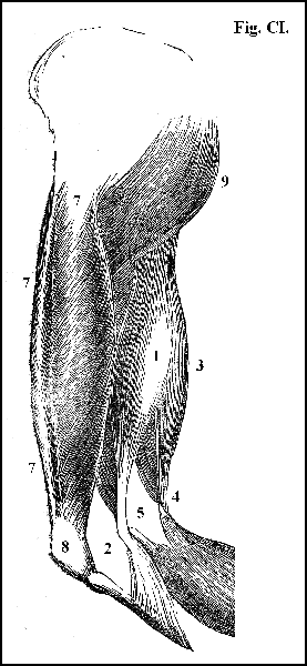
View of the flexor and extensor muscles of the leg. 1. The biceps of the leg; 2. tendon of the biceps, inserted into the head of the fibula; 3. the semi-membranosus, passing to be inserted into the head of the fibula; 4. tendon of the semi-membranosus forming the inner, and 5. tendon of the biceps forming the outer, hamstring; 6. upper part of the gastrocnemius muscle; 7. the four large muscles which unite to form the great extensor muscle of the leg, inserted into 8. the patella; 9. a portion of the glutæus maximus concealing the other muscles of the hip.
206. Four large muscles, blended together in such a manner as to form one muscle of prodigious size, termed the quadriceps cruris (fig. CI. 7), occupying nearly all the forepart and the sides, and a considerable portion of the back part of the thigh, constitute the great flexor of the thigh. This enormous mass of muscle arises partly from the ischium, and partly from the upper part of the femur (fig. CI. 7), and is all inserted into the patella (fig. CI. 8), which constitutes a pulley for the purpose of assisting the action of these powerful muscles.
207. The muscles which bend the toes and extend the foot, termed the gastrocnemii (fig. CII. 1, 2), are placed on the back part of the leg, and form the mass of muscle which constitutes the calf of the leg (fig. CII. 1, 2). They arise partly from the lower extremity of the femur (fig. CII.) and partly from the upper and back part of the fibula and tibia; and they form the largest and strongest tendon in the body, termed the tendo achillis (fig. CII. 3), which is implanted into the heel (fig. CII. 4).
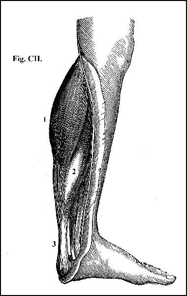
View of the muscles which bend the toes, and which, by lifting the heel, extend the foot. 1. The muscle called gastrocnemius externus, which, uniting with 2. the gastrocnemius internus, forms 3. the tendo achillis, which is inserted into 4. the heel.
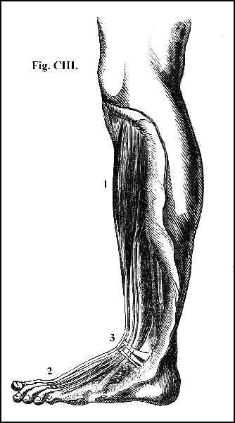
View of the muscles which extend the toes and bend the foot. 1. The common extensor; 2. the tendons of the same muscle inserted into the toes; 3. the anterior annular ligament of the foot.
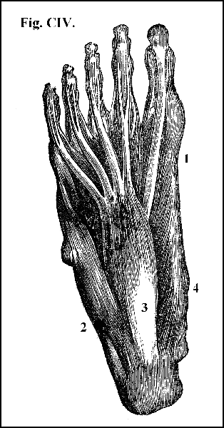
View of the muscles in the sole of the foot. 1 The muscle which draws the great toe from the other toes; 2. the muscle which draws the little toe from the other toes; 3. the muscle called the short flexor of the toes, which assists in bending the four smaller toes.
208. The muscles which extend the toes and bend the foot are seated on the fore part of the leg (fig. CIII.); split into tendons like the analogous muscles of the fingers (fig. CIII. 2); and are bound down by a ligament (fig. CIII. 3), exactly the same in name, disposition, and office, as that which belongs to the hand (fig. CIII. 3). Numerous minute muscles are placed in the sole of the foot (fig. CIV.), which act on the toes as the small muscles in the palm of the hand act on the fingers (fig. LXXXVI.).
209. Such are the moving powers which put in action the complicated mechanism provided for the function of locomotion. And these powers are adequate to their office; but they are what may be termed expensive powers; agents requiring a high degree, of organization and the utmost resources of the economy to support and maintain them. Hence in the construction of the framework of the machine which they have to move, whatever mechanical contrivance may economize their labour, is adopted. The construction, form, and disposition of the several parts of that framework have all reference to two objects: first, the combination of strength with lightness; and secondly, security to tender organs, with the power of executing rapid, energetic, and, sometimes, violent motions. The combination is effected and the object attained in a mode complicated in the detail, simple in the design, and perfect in the result. The weight of the body transmitted from the arch of the pelvis to a second arch, formed by the neck of the thigh-bone, and from this, in a perpendicular direction, to a third arch formed by the foot, is ultimately received by the heel behind, and by the metatarsal bones and the first phalanges of the toes before, and more especially by the metatarsal joints belonging to the great and little toe, which have a special apparatus of muscles, for the purpose of preserving steadily their relative situation to the heel. The weight of the body is thus sustained on a series of arches, from which it is, in succession, transmitted to the ground, where it ultimately rests upon a tripod: forms known and selected as the best adapted to afford support, and to give security of position. Columns of compact bone superimposed one upon another, and united at different points by bands of prodigious strength, form the pillars of support. But these bony columns never touch each other; are never in actual contact; are all separated by layers of elastic matter which, while they assist in binding the columns together, enable them to move one upon another, as upon so many pliant springs. The layers of cartilage interposed between the several vertebræ; the layer of cartilage interposed between the vertebral column and the pelvis; the layer of cartilage that lines the acetabulum and that covers the head of the femur; the layer of cartilage that covers the lower extremity of the femur and the upper extremity of the tibia and fibula and the tarsus; the successive layers of cartilage interposed between the several bones of the tarsus; and finally, the layer of cartilage that covers both the tarsal and the digital extremities of the metatarsal bones; are so many special provisions to prevent the weight of the body from being transmitted to the ground with a shock; and, at the same time, so many barriers established between the ground and the spinal cord, the brain and the soft and tender organs contained in the thoracic and abdominal cavities, to prevent these organs from being injured by the reaction of the ground upon the body. The excellence of this mechanism is seen in its results; in contemplating "from what heights we can leap—to what heights we can spring—to what distances we can bound—how swiftly we can run—how firmly we can stand—how nimbly we can dance—and yet how perfectly we can balance ourselves upon the smallest surfaces of support!"
210. It is necessary, in order to complete this general view of the structure of the human body, and of the combination and arrangement of its various parts, to denote the several regions into which, for the purpose of describing with accuracy the situation and relation of its more important organs, the body is divided. It is not needful to the present purpose to describe the regions of the head, because its internal cavity contains only one organ, the brain, and its external divisions do not differ materially from those which are common and familiar; but the chest, the abdomen, and the upper and lower extremities are mapped out into regions, of which it is very important to have an exact knowledge, which may be acquired by the study of the annexed diagrams.
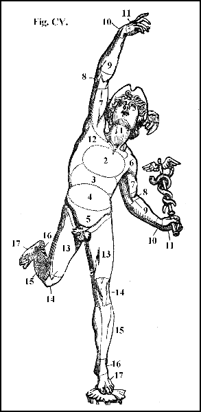
Anterior view of the regions of the body. 1. Region of the neck; 2. region of the chest or thorax. Abdominal regions: 3. epigastric; 4. umbilical; 5. hypogastric region. Regions of the upper extremities. 6. shoulder; 7. arm; 8. elbow; 9. fore-arm; 10. wrist; 11. ball of thumb; 12. the axilla or armpit. Regions of the lower extremities: 13. thigh; 14. knee; 15. leg; 16. ankle; 17. instep and foot.
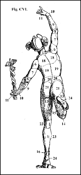
Posterior view of the regions of the body: 18. region to the scapula; 19. of the back; 20. of the loins; 21. of the hips; 22. of the ham; 23. of the calf of the leg; 24. of the heel and foot.
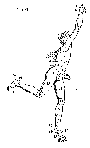
Lateral view of the regions of the body: 25. arch of the foot.
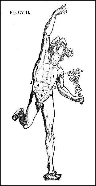
Anterior view of the situation of the more important internal organs: 1. lungs, right and left; 2. heart; 3. line representing the edge of the diaphragm; 4. liver; 5. stomach; 6. small intestines; 7. colon; 8. urinary bladder.
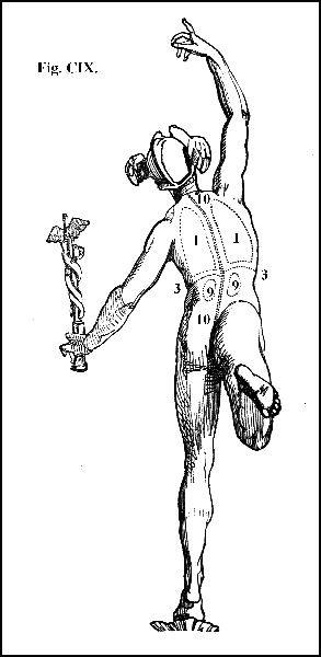
Posterior view of the situation of the more important internal organs: 9. kidnies, right and left; 10. the course of the spinal cord.
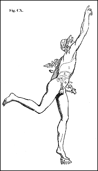
Lateral view of the situation of the more important internal organs.