THE SPERMATOCYTE DIVISIONS OF THE LOCUSTIDÆ. (continued 2)
Reading edition
5. The definitive chromosomes of the metaphase are produced by a concentration of the prophase elements, whereby they become shorter, heavier, and entirely homogeneous in structure. Distinct lines of division between the chromatids are not visible, but the tetrad character of the elements is readily established by observing the steps in their formation.
6. The accessory chromosome early becomes distinguishable because of its peripheral position and strong tendency to stain with safranin, while the remaining chromatin takes the gentian violet by Flemming’s three-color method. At first it appears as a homogeneous plate, but later this is seen to be a closely coiled thread. As the chromatin segments shorten and broaden to form the chromosomes of the mitotic figure, this thread also grows shorter and heavier until it forms an element of essentially the same character as that of the spermatogonial chromosome from which it was derived.
7. Upon the establishment of the mitotic figure, the chromosomes arrange themselves in the equatorial plate with their longer axis perpendicular to the spindle axis. Division of the elements is not synchronous, so that all stages of the chromatid movements may be observed in one nucleus. By this means it 228is possible to determine that separation of the chromosomes takes place along the plane which marked the longitudinal division of the prophase thread in such a way that the chromatids show no clear interspaces. The individual chromosome near the end of its division has the same form as that with which it started, except for the difference that the chromatids are now in contact for the greater part of their length along the plane of their cross-division. As the daughter chromosomes separate, this line of division comes into evidence through the springing apart of the two chromatids now composing each chromosome. The result is the formation of two V-shaped chromosomes with mantle fibers attached to their apices. The accessory chromosome does not participate in this division, but passes unchanged to one pole of the spindle.
8. By reason of the action of the accessory chromosome in the first spermatocyte mitosis, there are produced two numerically equal classes of second spermatocytes—(a) those containing sixteen dyad chromosomes and an undivided accessory chromosome, and (b) those with merely the sixteen dyad elements. In both cases the mitotic figure quickly reforms without an intervening rest stage in which the chromosomes lose their identity. There is a loosening up of the chromomeres in all the elements except the accessory chromosome, so that they have a structure and staining reaction similar to that of the first spermatocyte chromosomes just before they enter the metaphase. The dyads of the first spermatocyte telophase, and of the succeeding and greatly abbreviated second spermatocyte prophase, are quite as definite structures as are the chromosomes of the first spermatocyte prophase.
9. All the chromosomes of the second spermatocyte are paired structures and divide in a similar way. The spindle is small and weak as compared with that of the first spermatocyte, and the chromosomes arrange themselves radially on its periphery in such a way that the pairs lie in the plane of the spindle axis with their joined ends inward. The space between the chromatids represents the line of cross-division observable in the prophase segments of the first spermatocyte, and their separation accordingly represents a reduction division. The accessory chromosome, on the contrary, divides along the plane marking the longitudinal cleft of the spermatogonial spireme.
22910. From each first spermatocyte there are formed, by two divisions, four spermatids, of which two are distinguished from the remaining pair by the possession of an extra chromosome in addition to the number—sixteen—common to them all. Both classes undergo a like series of transformations by which they become mature spermatozoa. These are necessarily of two kinds; and it is believed that those containing the accessory chromosome, in the act of fertilizing the egg, determine that the germ-cells of the embryo shall be sexually male, or like themselves, while those from which it is absent are unable to impress their sex upon the egg and assist in producing female embryos.
VII. BIBLIOGRAPHY.
1. Atkinson, Geo. F., 1899. Studies on Reduction in Plants, Bot. Gaz., vol. 28.
2. Baumgartner, W. J., 1902. The Spermatid Transformations of Gryllus assimilis, etc., Kans. Univ. Sci. Bull., vol. 1, No. 1.
3. Brauer, A., 1893. Zur Kenntniss der Spermatogenese von Ascaris megalocephala, Arch. mikr. Anat., vol 42.
4. Carnoy, 1895. La Cytodiérese chez les Arthropodes, La Cellule, vol. 1.
5. Cholodkovsky, N., 1894. Zur Frage über die Anfangsstadien der Spermatogenese bei den Insecten, Zool. Anz., vol. 17.
6. Griffin, B. B., 1899. Studies on Maturation, Fertilization and Cleavage of Thalassema and Zirphæa, Jr. Morph., vol. 15.
7. Häcker, Valetin, 1897. Ueber weitere Uebereinstimmungen zwischen den Fortpflianzungsvorgängen der Tiere und Pflanzen, Biol. Cent., vol. 17.
8. Henking, H., 1890. Untersuchungen über die ersten Entwicklungsvorgänge in den Eiern der Insecten. 2. Ueber Spermatogenese und deren Beziehung zur Eientwicklung bei Pyrrhocoris apterus M., Z. wiss. Zool., vol. 51.
9. Henking, H., 1892, idem. 3. Specielles und Allgemeines, ibid., vol. 54.
10. Ischikawa, 1891. Studies of Reproductive Elements. Spermatogenesis, Ovogenesis and Fertilization in Diaptomus sp., J. Coll. Sc. Imp. Univ. Japan, vol. 5.
11. Kingsbury, B. F., 1902. The Spermatogenesis of Desmognathus fusca, Am. Jour. Anat., vol. 1, No. 2.
23012. Montgomery, T. H., jr., 1898. The Spermatogenesis in Pentatoma up to the Formation of the Spermatid, Zool. Jahrb., vol. 12.
13. Montgomery, T. H., jr., 1898. Chromatin Reduction in the Hemiptera: a Correction, Zool. Anz., vol. 22.
13a. Montgomery, T. H., jr., 1898. Observations on Various Nucleolar Structures of the Cell, Biol. Lect. of the Woods Holl Laboratory, 1898.
14. Montgomery, T. H., jr., 1900. The Spermatogenesis of Peripatus balfouri up to the Formation of the Spermatid, Zool. Jahrb., vol. 14.
15. Montgomery, T. H., jr., 1901. A Study of the Germ Cells of Metazoa, Trans. Amer. Phil. Soc., vol. 20.
16. McClung, C. E., 1899. A Peculiar Nuclear Element in the Male Reproductive Cells of Insects, Zool. Bull., vol. 2.
17. McClung, C. E., 1900. The Spermatocyte Divisions of the Acrididæ, Kans. Univ. Quart., vol. 9, No. 1.
18. McClung, C. E., 1901. Notes on the Accessory Chromosome, Anat. Anz., Bd. 20, Nos. 8, 9.
19. McClung, C. E., 1902. The Accessory Chromosome—Sex Determinant? Biol. Bull., vol. 3, Nos. 1, 2.
20. Moore, J. E. S., 1895. On the Structural Changes in the Reproductive Cells during the Spermatogenesis of Elasmobranchs, Quart. J. Micr. Sc., vol. 38.
21. Paulmier, F. C., 1898. Chromatin Reduction in the Hemiptera, Anat. Anz., vol. 14.
22. Paulmier, F. C., 1899. The Spermatogenesis of Anasa tristis, Jr. Morph., vol. 15.
23. Platner, G., 1886. Die Karyokinese bei den Lepidopteran als Grundlage für eine Theorie der Zelltheilung, Internat. Monatsschr. Anat. Histol., vol. 3.
24. Rath, O. vom, 1891. Ueber die Reduction der Chromatischen Elemente in der Samenbildung von Gryllotalpa, Ber. Naturf. Ges. Freiburg, vol. 6.
25. Rath, O. vom, 1892. Zur Kenntniss der Spermatogenese von Gryllotalpa vulgaris Latr., Arch. Mikr. Anat., vol. 40.
26. Rath, O. vom, 1895. Neue Beiträge zur Frage der Chromatinreduction in der Samen und Eireife, ibid., vol. 4.
27. Sabatier, A., 1890. De la Spermatogénese chez les Locustides, Comptes Rend., vol. 111.
23128. Sutton, W. S., 1900. The Spermatogonial Divisions of Brachystola magna, Kans. Univ. Quart., vol. 9, No. 2.
29. Toyama, K., 1894. On the Spermatogenesis of the Silkworm, Bull. Coll. Agric. Imp. Univ. Japan, vol. 2.
30. V. la Valette St. George, 1897. Zur Samen-und Eibildung beim Seidenspinner (Bombyx mori), Arch. mikr. Anat., vol. 50.
31. Wagner, J., 1896. Beiträge zur Kenntniss der Spermatogenese bei den Spinnen, Arb. Nat. Ges. St. Petersburg, vol. 26.
32. Wilcox, E. V., 1895. Spermatogenesis of Caloptenus femur-rubrum and Cicada tibicen, Bull. Mus. Comp. Zool. Harvard Univ., vol. 27.
33. Wilcox, E. V., 1896. Further Studies on the Spermatogenesis of Caloptenus femur-rubrum, ibid., vol. 29.
34. Wilcox, E. V., 1897. Chromatic Tetrads, Anat. Anz., vol. 14.
35. Wilcox, E. V., 1901. Longitudinal and Transverse Divisions of Chromosomes, Anat. Anz., Bd. 19, No. 13.
36. Wilson, E. B., 1900. The Cell in Development and Inheritance. New York; 2d ed.
37. de Sinéty, R., 1901. Recherches sur la Biologie et l’Anatomie des Phasmes, La Cellule, Tome 19, 1er fascicule.
DESCRIPTION OF FIGURES.
Drawings were made with a camera lucida, the optical combination being a 1–16 B. & L. objective and a Watson “Holoscopic” ocular No. 7. Details were studied with a Zeiss 2-mm. apochromat, N. A. 1.30. As reduced in reproduction, an enlargement of 1500 diameters exists. Photomicrographs, excepting those of figures 37 and 38, were made by the use of the arc light and horizontal camera. The exceptions represent illumination by ordinary diffuse daylight. In all cases the lenses used were the Zeiss 2 mm., N. A. 1.30 objective and projection oculars. A Watson “Parachromatic” oil-immersion condenser of 1.30 N. A. was employed to illuminate the objects. In use it was stopped down to between .75 N. A. and 1.0 N. A.
Explanation of Plate VII.
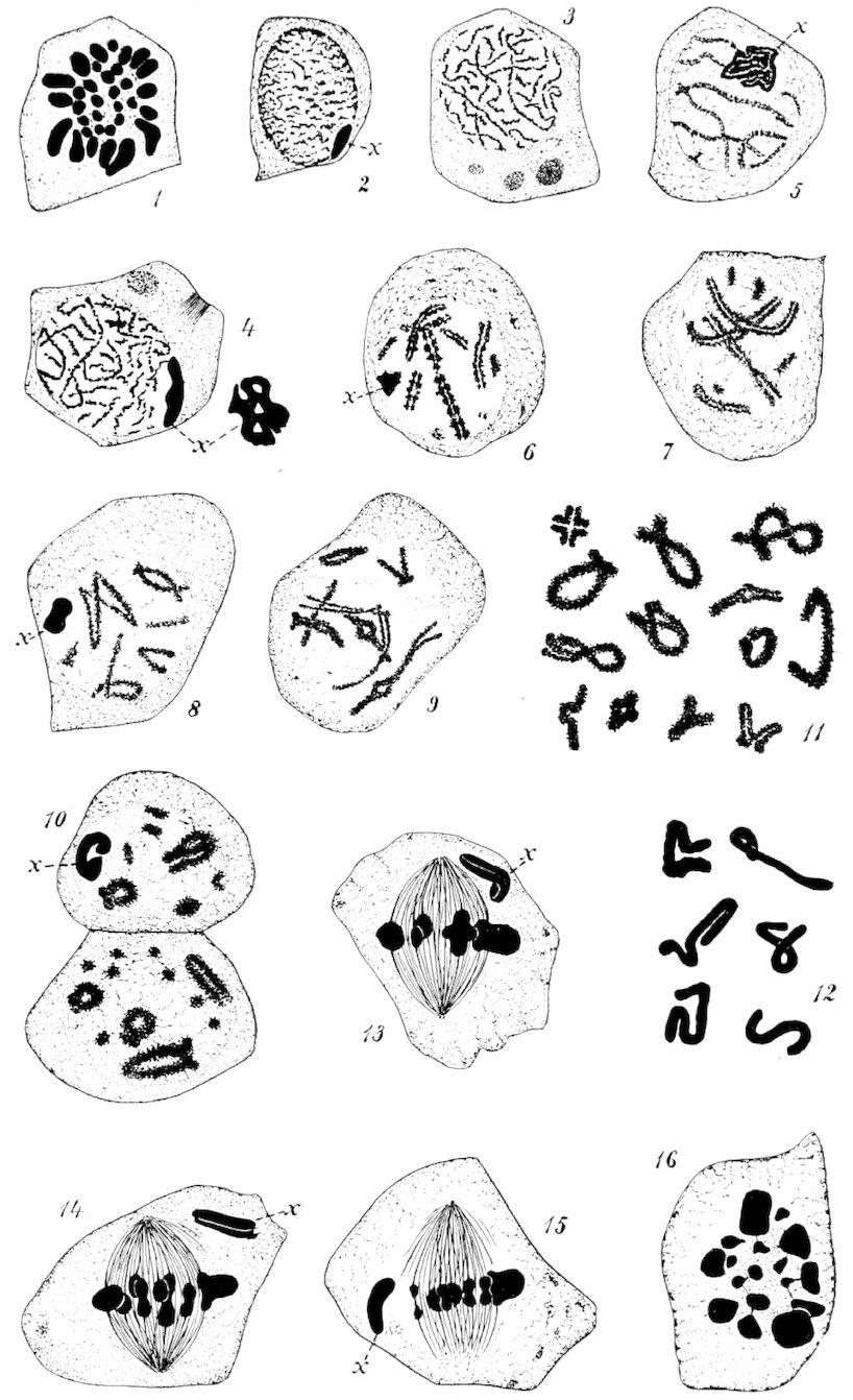
Fig. 1. Pole view of spermatogonial metaphase, showing the thirty-three chromosomes. It will be observed that the chromosomes are of unequal sizes, and that the large ones arrange themselves in a circle on the outside of the figure.
Fig. 2. Very young spermatocyte. The chromatin derived from the breaking down of the spermatogonial chromosomes in a diffuse condition, with no trace of a linear arrangement. The accessory chromosome x on the periphery of the nucleus, darkly staining and homogeneous.
Fig. 3. Early stage in the formation of the spireme. In the cytoplasm the remains of the spermatogonial spindle. The cell has entered upon the growth period.
Fig. 4. A later stage in the spireme formation. The accessory chromosome larger and more flattened. A surface view shows it as an apparently fenestrated plate. The remains of the two spermatogonial spindles still persisting.
Fig. 5. First appearance of definite chromosomes. One shown entire with longitudinal and cross-divisions marked. The accessory chromosome is here seen to be in a spireme condition.
Fig. 6. Condition of the chromosomes after further contraction of the early segments. As here shown, they are more granular than is usually the case.
Fig. 7. Common types of the prophase chromosomes.
Fig. 8. A cell in which one of the chromosomes has its halves widely separated along the longitudinal division, forming Paulmier’s double-V figure.
Fig. 9. In this cell may be seen the variation in form and size of the early spermatocyte chromosomes.
Fig. 10. Two cells of the late prophase, with the chromosomes at almost the extreme degree of concentration.
Fig. 11. Chromosomes of cells in the stage shown in figure 10. These represent the different types of rings, crosses, etc., commonly observed in first spermatocytes just before the formation of the mitotic figure.
Fig. 12. Different forms assumed by the accessory chromosome in the prophase of the first spermatocytes of Xiphidium.
Fig. 13. Metaphase of the first spermatocyte. The accessory chromosome is seen at one pole of the spindle, to which it has moved before the separation of the chromatids of the remaining chromosomes.
Fig. 14. Another cell in about the same stage as that represented in the preceding figure.
Fig. 15. A first spermatocyte metaphase in which the accessory chromosome has not as yet moved to the pole of the spindle. This is uncommon in Orchesticus, but frequent in Anabrus.
Fig. 16. Pole view of a first spermatocyte metaphase, showing seventeen chromosomes. The variation in size of the elements, so marked in the spermatogonia, is even more pronounced here. This is a cell similar to that of figure 15, in which the accessory chromosome lies in the equatorial plate.
Explanation of Plate VIII.
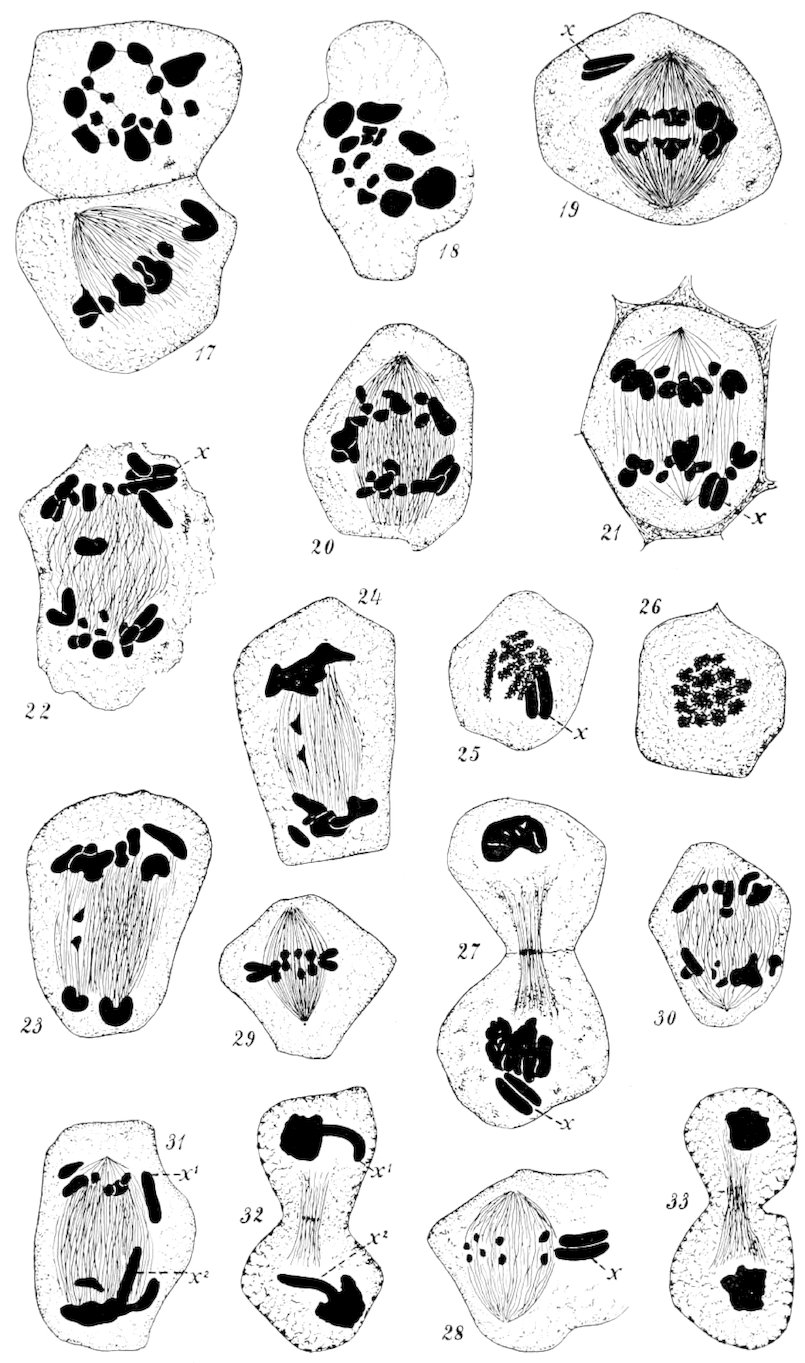
Fig. 17. Two cells in metaphase—a pole view of one and an oblique view of the other. The accessory chromosome does not show in the former, the cell being such a one as is represented in figures 14 and 15.
Fig. 18. Pole view of another cell, showing but sixteen chromosomes.
Fig. 19. Early anaphase of the first spermatocyte, with the accessory chromosome already at one pole.
Fig. 20. Mid-anaphase, with the giant chromosome still undivided.
Fig. 21. Later anaphase, in which the accessory chromosome is seen at the lower pole. This figure shows, also, the character and extent of the intercellular material.
Fig. 22. Later anaphase. The accessory chromosome at the upper pole. An undivided chromosome lying between the groups of daughter chromosomes.
Fig. 23. About the stage of figure 22, but the lagging chromosome has divided.
Fig. 24. Very late anaphase. Here, again, the lagging chromosome is divided.
Fig. 25. Pole view of first spermatocyte telophase, showing the accessory chromosome at one side of the daughter chromosomes.
Fig. 26. Pole view of a cell in the same stage as that represented in figure 25. Here, however, the accessory chromosome is not present.
Fig. 27. Lateral view of telophase, with the accessory chromosome in the lower daughter-cell.
Fig. 28. Fragment of second spermatocyte, showing the chromosomes in metaphase. The relative sizes of the accessory chromosome and the remaining chromosomes is well shown.
Fig. 29. Metaphase of a second spermatocyte, in which the accessory chromosome is not present.
Fig. 30. Anaphase of second spermatocyte, in which there is no accessory chromosome.
Fig. 31. Anaphase of second spermatocyte, where the accessory chromosome is present—x1 and x2.
Fig. 32. Telophase of the same class of second spermatocytes. The accessory chromosome extends out from the mass of chromosomes at each pole—x1 and x2.
Fig. 33. Telophase of the class of second spermatocytes from which the accessory chromosome is absent.
Explanation of Plate IX.
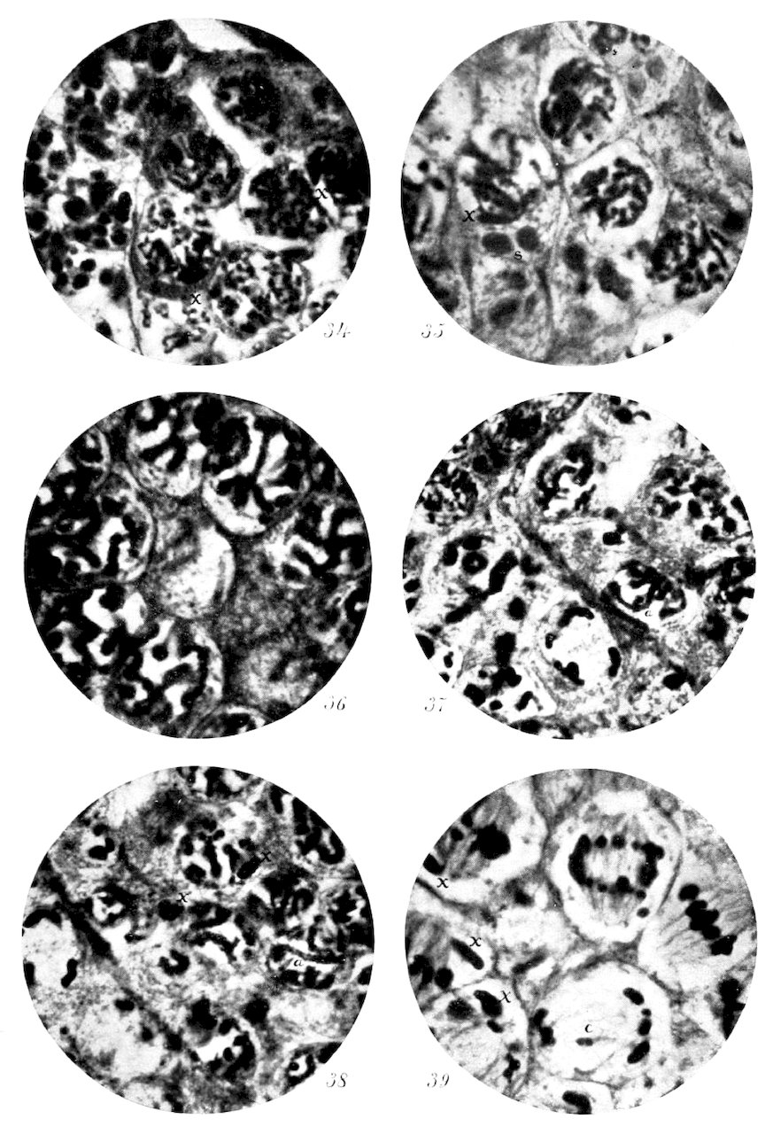
Fig. 34. Photomicrograph of early spireme stage of first spermatocyte, showing peripheral position of the accessory chromosome x. At the left, secondary spermatogonia, last generation. × 1300.
Fig. 35. A late prophase, showing accessory chromosome x, and spindle remains s (cf. figs. 3 and 4). × 1300.
Fig. 36. Coarse spireme of first spermatocyte. × 1300.
Fig. 37. Prophase, with chromosomes in the form of long segments. At a, the cell drawn in figure 9. In the cyst at the left are spermatocytes in a later stage, with the chromosomes homogeneous. × 1000.
Fig. 38. Prophase with segments divided longitudinally and across. At a is one shown en face. Accessory chromosome at x. × 1000.
Fig. 39. Metaphase and anaphase of first spermatocyte. The accessory chromosome x at one pole of the spindle. Lagging chromosome at c. × 1300.
Explanation of Plate X.
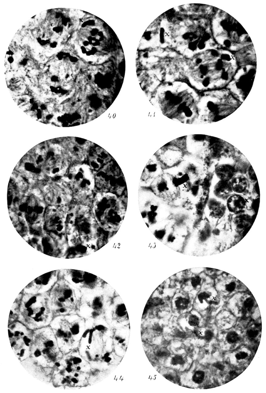
Fig. 40. Anaphase of first spermatocyte. Accessory chromosome x at one pole. The form of chromosome in the anaphase well shown. The lagging chromosome c seen in two cells. × 1300.
Fig. 41. Anaphase of the first spermatocyte, showing the longitudinally divided condition of the accessory chromosome x in the cell near the center. Compare with the accessory chromosome in the metaphase of second spermatocyte, figure 43. × 1300.
Fig. 42. Second spermatocyte in metaphase. In most of the cells the focus is upon the ends of the chromosomes, but in one a side view is obtainable. Compare with the chromosome of the upper cell in figure 40. No accessory chromosome in most of the cells in focus. × 1300.
Fig. 43. Second spermatocyte metaphase and spermatids. Note the relative sizes of the accessory chromosome and the other chromosomes. In the spermatids the accessory chromosome has taken its place on the periphery of the nucleus in the same way that it does in the prophase of the first spermatocyte. × 1300.
Fig. 44. Anaphase of the second spermatocyte, showing the accessory chromosome x separated. Other cells in metaphase. × 1300.
Fig. 45. Telophase of the second spermatocyte. Two daughter-cells with persisting spindle between, showing the accessory chromosome x in each. Other nuclei in focus show no accessory chromosomes. × 1300.
TRANSCRIBER’S NOTES
Silently corrected obvious typographical errors and variations in spelling.
Retained archaic, non-standard, and uncertain spellings as printed.