Front matter
Reading edition
SCLERO-CORNEAL TREPHINING IN THE OPERATIVE TREATMENT OF GLAUCOMA, 1913. Second Edition, 1914. George Pulman and Sons, London.
GLAUCOMA: DIGEST OF THE YEAR’S LITERATURE. The Ophthalmic Yearbook, 1913–1916. Herrick Book and Stationery Company, Denver, Colorado, U.S.A.
GLAUCOMA: A HANDBOOK FOR THE GENERAL PRACTITIONER, 1917. H. K. Lewis and Co. Ltd., London.
GLAUCOMA: A TEXTBOOK FOR THE STUDENT OF OPHTHALMOLOGY. (Now in the press.) H. K. Lewis and Co. Ltd., London.
PLATE I.
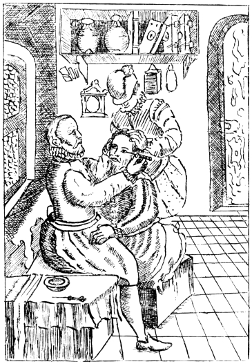
Copied by C. A. R. H. Fig. 1.—The positions of the patient, operator, and assistant, as depicted by Bartisch (Albertotti).
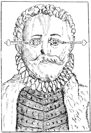
Copied by C. A. R. H. Fig. 2.—To illustrate the method of introducing the needle in Bartisch’s operation (Albertotti). The cataract is in situ in the right eye, and couched in the left eye.
THE INDIAN OPERATION OF COUCHING FOR CATARACT
INCORPORATING
THE HUNTERIAN LECTURES
DELIVERED BEFORE THE ROYAL COLLEGE OF SURGEONS OF ENGLAND ON FEBRUARY 19 AND 21, 1917
BY
ROBERT HENRY ELLIOT
M.D., B.S. Lond., Sc.D. Edin., F.R.C.S. Eng., etc. Lieut.-Colonel I.M.S. (retired)
LATE SUPERINTENDENT OF THE GOVERNMENT OPHTHALMIC HOSPITAL, MADRAS;
LATE PROFESSOR OF OPHTHALMOLOGY, MEDICAL COLLEGE, MADRAS; AND LATE FELLOW OF THE UNIVERSITY OF MADRAS;
HONORARY FELLOW OF THE AMERICAN ACADEMY OF OPHTHALMOLOGY AND OTOLARYNGOLOGY.
WITH 45 ILLUSTRATIONS

PAUL B. HOEBER 67 & 69 EAST 59TH STREET NEW YORK 1918
Printed in England
To E. C. I. E.
To
E. TREACHER COLLINS, Esq., f.r.c.s. eng.,
IN GRATEFUL ACKNOWLEDGMENT OF THE ASSISTANCE WHICH HIS WORK ON THE PATHOLOGY OF THE EYE HAS BEEN TO ALL WHO DESIRE TO ADVANCE THE SCIENCE OF OPHTHALMOLOGY
PREFACE
It is a quarter of a century since I first landed in India. In common with very many other surgeons, my attention was early attracted to the operation of couching as performed by its Indian exponents, and I was horrified to see how bad the majority of the results were. It appeared to me that the outstanding need was for carefully compiled statistics, in order that a fair judgment might be formed on the subject. I divested my mind of partisanship and bias, and sought every opportunity to discuss the method and its results with anyone and everyone whose knowledge was likely to be of use to me in my quest, whether they were laymen or surgeons, Europeans or Indians, officials or non-officials. The office of Superintendent of the Government Ophthalmic Hospital, Madras, afforded an unrivalled field for work, and the staff of the hospital co-operated with me in a manner which I find it impossible to acknowledge suitably. My thanks are especially due to Lieutenant H. C. Craggs, Assistant-Surgeon C. Taylor, and Dr. Ekambaram, for the valuable help they gave me. After I left India, Captain W. C. Gray acted for me as Superintendent, and later Major H. Kirkpatrick succeeded me permanently. Both of these officers most generously placed the material of the hospital still at my disposal, and rendered me very valuable service in the study of my subject. The great majority of the microscopic slides were very beautifully prepared for me by Mr. W. Chesterman. I am also indebted to Mr. S. Stephenson and Mr. A. C. Hudson for very kindly sectioning some of the globes for me. The photographs, both macroscopic and microscopic, were taken by Mrs. Elliot, without whose help I could not have written the book. Mr. E. Treacher Collins has generously given me advice and assistance of the greatest value in the study of a number of the preparations, and I have acted freely on the suggestions he has been good enough to make. I desire to express to the Council of the Royal College of Surgeons of England my acknowledgment of the honour they conferred on me by electing me to a Hunterian Professorship of the College. By the kindness of the authorities concerned, I have been enabled to include in this book articles which have appeared in the Lancet, in the Ophthalmic Record, and in the Proceedings of the Ophthalmological Society of the United Kingdom.
The collection of fifty-four eyeballs, on which the work for my Hunterian Lectures was founded, I have had the honour to present to the Museum of the Royal College of Surgeons. I think I am justified in claiming that it is unrivalled, and likely to remain so. At the suggestion of Sir John Bland-Sutton, I have endeavoured so to write the chapter on pathology that it may furnish a guide to any who care to study the subject within the walls of that museum. At the same time, I have striven to make it readable and of interest to those who have no such opportunity. In this the photographs have greatly helped me.
As I have already said, I have given nearly a quarter of a century’s intermittent work to elucidate these problems, which I felt were of wide interest, not merely to India or to the East alone, but to the whole civilized world. During the last two years I have devoted a very large proportion of my spare time continuously to the subject, but it is so immense and so far-reaching that I feel I have left much unfinished. On every hand fresh problems open up, till there seems no limit to what might be done, given time and opportunity. This operation of couching, so old that its origin is lost in the dim mists of antiquity, has still much that is new to be learnt for the seeking. There are many of the younger surgeons in the East who could carry the work much farther if they would, and who would be a hundred-fold repaid if they did. Will they?
ROBERT HENRY ELLIOT.
54, Welbeck Street, Cavendish Square, W., 1917.
CONTENTS
CHAPTER PAGE
I. THE HISTORY OF COUCHING 1
II. THE TECHNIQUE OF THE OPERATION 14
III. THE INDIAN COUCHER AND HIS HABITS 19
IV. STATISTICAL 25
V. THE PATHOLOGICAL ANATOMY OF COUCHED EYES 35
VI. DIAGNOSIS 77
VII. CLINICAL 85
INDEX 92
LIST OF ILLUSTRATIONS
FIGS. 1 AND 2. BARTISCH’S OPERATION (PLATE 1.) Frontispiece
FIG. PAGE
3. THE STAGES OF DEPRESSION 8
4. THE STAGES OF RECLINATION 8
5. SCARPA’S NEEDLE AS USED BY MACKENZIE 8
6. THE ANTERIOR METHOD OF COUCHING 14
7. THE POSTERIOR METHOD OF COUCHING 14
8. GROUP SHOWING THE INDIAN OPERATION OF COUCHING 15
9. THE INSTRUMENTS USED IN COUCHING IN THE SOUTH OF INDIA 17
PLATE II. FACING PAGE
10. LENS DISLOCATED BETWEEN CILIARY BODY AND SCLERA 38

11. NUCLEUS OF CATARACT FREELY MOVABLE BETWEEN THE AQUEOUS AND VITREOUS CHAMBERS
12. LENS IMPACTED IN ANGLE OF ANTERIOR CHAMBER
13. CAPSULE OF MORGAGNIAN CATARACT IMPACTED IN ANGLE OF ANTERIOR CHAMBER
14. LENS FLOATING FREE IN VITREOUS CHAMBER
15. LENS LIGHTLY IMPRISONED IN EXUDATE INTO VITREOUS CAVITY
PLATE III.
16. ABUNDANT EXUDATE INTO VITREOUS CAVITY 42

17. LENS FIRMLY FIXED BY ORGANIZED EXUDATE
18. LENS FIRMLY FIXED BY ORGANIZED EXUDATE, BUT IN UNUSUAL POSITION
19. TOTAL DETACHMENT OF RETINA, WITH CYST FORMATION
20. RECLINED LENS LYING IN FRONT OF THE HYALOID BODY
21. RECLINED LENS LYING IN FRONT OF THE HYALOID BODY
PLATE IV.
22. LENS DISLOCATED BEHIND RETINA 48

23. FISTULA OF THE CORNEA
24. CAPSULO-CORNEAL SYNECHIA
25. RETINO-CORNEAL SYNECHIA
26. SCLERAL FISTULA
27. " " (MAGNIFIED)
PLATE V.
28. INJURIES TO UVEAL TRACT 54
29. FOREIGN BODY (TIP OF COPPER PROBE) IMBEDDED IN THE EYE
30. TRAUMATIC DETACHMENT OF RETINA AND CHOROID
31. WHOLE-SECTION OF FIG. 19
32. PART OF THE ABOVE MAGNIFIED TO SHOW LENS IMBEDDED IN INFLAMMATORY EXUDATE
33. PHAGOCYTOSIS
PLATE VI.
34. L’IRIS BOMBÉ AND RETINAL CYST 58
35. MATTING OF STRUCTURES OF THE EYE DUE TO INFLAMMATION
36. " " " " " (HIGHER MAGNIFICATION)
37. UNUSUAL APPEARANCE OF EXUDATE INTO THE VITREOUS CAVITY
38. INFLAMED OPTIC NERVE HEAD
39. ADVANCED ORGANIZATION OF VITREOUS EXUDATE
PLATE VII.
40. HÆMORRHAGE INTO VITREOUS CAVITY 72
41. PROLIFERATIVE DOT IN RETINA
42. COLLECTION OF LEUCOCYTES ON SURFACE OF RETINA
43. SMALL CYSTS IN RETINA
44. WHOLE-SECTION OF FIG. 34, SHOWING L’IRIS BOMBÉ AND RETINAL CYST
45. ADHERENT LENS PRESSING ON IRIS BASE
COUCHING FOR CATARACT