IDIOPATHIC MULTIPLE PIGMENT SARCOMA (IDIOPATHIC MULTIPLE HÆMORRHAGIC SARCOMA) OF KAPOSI. By F. PARKES WEBER, M.D., F.R.C.P., AND PAUL DASER, M.D.
Reading edition
The patient, Jakob Z——, is a Polish Jew, aged 46 years, from Galicia, who has lived nine years in England, and says he has enjoyed good health. There is no evidence of previous alcoholism or syphilis. The patient was brought by Dr. Weber before the Dermatological Society of London on February 8th, 1905.
The present illness commenced about three years before, when he chanced to wound the sole of his right foot with a nail. A pedunculated growth, about the size of a cherry, arose from the wound. This description reminds one of the strawberry-like granulation tumours sometimes growing from small wounds or ulcers on the fingers, which have been termed “botryomycosis,” or rather “botryomycomata,” because at one time they were supposed to be caused by the “botryomyces.”[2] The growth was removed by a doctor. Afterwards small bluish nodules developed from time to time on the feet and legs. Some of these nodules apparently undergo the following series of changes: They slowly increase in size, and after about 136three months constitute little pendulous tumours, which ultimately fall off spontaneously or else are knocked off or pulled off by chance; in this way local healing occurs, but fresh nodules form elsewhere. Though, as stated, some of the nodules become pedunculated growths, others appear to atrophy and merely leave brownish pigment in the skin, whilst others seem to undergo very little change, and, at all events, persist for a long time in their primitive form, namely, as hard bluish lumps under the epidermis. Treatment has been as yet without result.
Present condition (January, 1905).—On the hands about the knuckles and backs of the fingers are a few minute slightly elevated bluish nodules. On the sole of the right foot, especially on the inner part, there are nodules in various stages of development. The minute ones, those in an early stage of development, are slightly elevated and of a bluish colour, similar to those on the hands. Others, further advanced in development, are small, sessile, or pedunculated hard outgrowths, without bluish colour and sometimes covered by thickened epidermis. There is a large patch of pigmented (brown) skin on the dorsal surface of the right foot, and in the neighbourhood of this pigmented area are some minute nodules of similar character to those already described. It is possible that the pigmented skin has been the site of minute growths which have undergone spontaneous involution, and have, as frequently happens in Lichen planus, left pigmentation of the skin behind them. The right thigh and leg are not affected.
The left lower extremity is the part most affected by the disease, and there is chronic, rather firm, œdema of the left foot and ankle. The nodules on the left foot are similar to those on the right, but are more widely distributed, and the left foot differs from the right in the absence of the pigmented area and in the presence of the œdema to which we have just alluded. Of the little sessile or pedunculated tumours on the back of the left foot and in the neighbourhood of the ankle-joint (see Fig. 1) the biggest are of the size of a large pea. There are likewise little tumours of various sizes—that is, in various stages of development—on the inner aspect of the left popliteal region (see Fig. 2) and in the neighbourhood of the patella, the smaller ones being bluish and slightly elevated, the larger ones being reddish, lentil-sized, sessile outgrowths.
There are no nodules or growths on other parts of the limbs, trunk, 137or head, with the exception of four or five minute purplish or bluish papules on the glans penis and neighbouring skin.
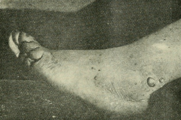
Figure 1
There is no evidence of any disease of the thoracic or abdominal viscera. The patient is not strongly built, but looks well nourished. He complains of various pains, but whether these are related to the disease or not is doubtful.
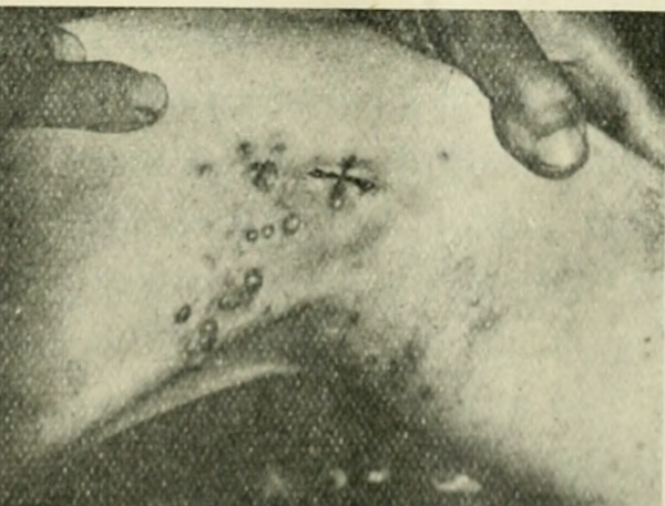
Figure 2
Microscopic examination.—One of the growths, of the size of a large pea, was removed from the left ankle, and a smaller one (lentil-sized) from the left knee. Sections of both were stained with hæmatoxylin and eosin-hæmatoxylin. The growths are seen to be situated in the corium, and are circumscribed. The epidermis over one of them is thickened. They consist of oval and spindle cells, 138which, in some parts, are more or less grouped into strands or bundles, and are cut in various directions. In some parts of the sections there are empty spaces (? lymph spaces) and in some parts there is homogeneous substance between the cells; in other parts there is extravasated blood. The cells in the larger and older of the two nodules are decidedly more elongated and fibrous-looking than in the smaller and more recently developed one. Dr. J. M. H. MacLeod is kindly going to make a further report of the microscopical features.
Remarks.—It seems to us clear that the case is a typical but relatively early one of the so-called idiopathic multiple pigment sarcoma (idiopathic multiple hæmorrhagic sarcoma) of Kaposi and later writers. For the literature on the subject we refer to the English summaries of Dr. J. H. Sequeira[3] and Dr. Radcliffe-Crocker.[4] The patient in our case is a male, as in nearly all other recorded cases of this disease; he is of the favourite age (between 40 and 50), and it may likewise be noted that he is a Galician Jew, as the patient of Sir Stephen Mackenzie[5] and Dr. J. J. Pringle was. The minute bluish nodules in the corium may be regarded as the primary feature of the disease. They either remain for a long time without undergoing much obvious change, or they undergo spontaneous involution and completely atrophy, leaving behind them only a little brownish cutaneous pigmentation; or else (thirdly) they may increase in size, become much elevated, and form definite sessile tumours, which, later on, become pedunculated, and finally drop off. This is the cycle in regard to the development of the little pendulous tumours which constitutes one of the most characteristic features of the disease. Another characteristic feature of the disease, probably sooner or later present in every case, is the œdema which occurs in the parts most affected, especially in the lower extremities. It is persistent œdema, and results in an elephantiasis-like condition. It is well marked in the left foot and ankle of our case. The striking blue or purple colour of the small nodules (which is absent in the pendulous tumours) seems, as in other cases, to be due to the venous blood contained in the blood-vessels or extravasated. What pigment there is 139in the growths is probably derived from blood. In our patient there is no evidence of gout as in certain cases described by Mr. Hutchinson.[6]
The prognosis in the present case is not altogether unfavourable, considering that the patient’s general health is good, that the disease is probably seldom of itself fatal, and that in some cases improvement or spontaneous care has, apparently, occurred after the disease has lasted twenty years or more.