THE LIFE HISTORY OF VAUCHERIA.1
Reading edition
THE LIFE HISTORY OF VAUCHERIA.1
By A.H. BRECKENFELD.
Nearly a century ago, Vaucher, the celebrated Genevan botanist, described a fresh water filamentous alga which he named Ectosperma geminata, with a correctness that appears truly remarkable when the imperfect means of observation at his command are taken into consideration. His pupil, De Candolle, who afterward became so eminent a worker in the same field, when preparing his "Flora of France," in 1805, proposed the name of Vaucheria for the genus, in commemoration of the meritorious work of its first investigator. On March 12, 1826, Unger made the first recorded observation of the formation and liberation of the terminal or non-sexual spores of this plant. Hassall, the able English botanist, made it the subject of extended study while preparing his fine work entitled "A History of the British Fresh Water Algæ," published in 1845. He has given us a very graphic description of the phenomenon first observed by Unger. In 1856 Pringsheim described the true sexual propagation by oospores, with such minuteness and accuracy that our knowledge of the plant can scarcely be said to have essentially increased since that time.
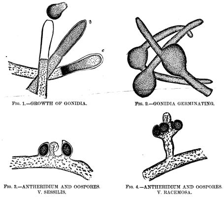
GROWTH OF THE ALGA, VAUCHERIA, UNDER THE MICROSCOPE.
Vaucheria has two or three rather doubtful marine species assigned to it by Harvey, but the fresh water forms are by far the more numerous, and it is to some of these I would call your attention for a few moments this evening. The plant grows in densely interwoven tufts, these being of a vivid green color, while the plant is in the actively vegetative condition, changing to a duller tint as it advances to maturity. Its habitat (with the exceptions above noted) is in freshwater—usually in ditches or slowly running streams. I have found it at pretty much all seasons of the year, in the stretch of boggy ground in the Presidio, bordering the road to Fort Point. The filaments attain a length of several inches when fully developed, and are of an average diameter of 1/250 (0.004) inch. They branch but sparingly, or not at all, and are characterized by consisting of a single long tube or cell, not divided by septa, as in the case of the great majority of the filamentous algæ. These tubular filaments are composed of a nearly transparent cellulose wall, including an inner layer thickly studded with bright green granules of chlorophyl. This inner layer is ordinarily not noticeable, but it retracts from the outer envelope when subjected to the action of certain reagents, or when immersed in a fluid differing in density from water, and it then becomes distinctly visible, as may be seen in the engraving (Fig. 1). The plant grows rapidly and is endowed with much vitality, for it resists changes of temperature to a remarkable degree. Vaucheria affords a choice hunting ground to the microscopist, for its tangled masses are the home of numberless infusoria, rotifers, and the minuter crustacea, while the filaments more advanced in age are usually thickly incrusted with diatoms. Here, too, is a favorite haunt of the beautiful zoophytes, Hydra vividis and H. vulgaris, whose delicate tentacles may be seen gracefully waving in nearly every gathering.
REPRODUCTION IN VAUCHERIA.
After the plant has attained a certain stage in its growth, if it be attentively watched, a marked change will be observed near the ends of the filaments. The chlorophyl appears to assume a darker hue, and the granules become more densely crowded. This appearance increases until the extremity of the tube appears almost swollen. Soon the densely congregated granules at the extreme end will be seen to separate from the endochrome of the filament, a clear space sometimes, but not always, marking the point of division. Here a septum or membrane appears, thus forming a cell whose length is about three or four times its width, and whose walls completely inclose the dark green mass of crowded granules (Fig. 1, b). These contents are now gradually forming themselves into the spore or "gonidium," as Carpenter calls it, in distinction from the true sexual spores, which he terms "oospores." At the extreme end of the filament (which is obtusely conical in shape) the chlorophyl grains retract from the old cellulose wall, leaving a very evident clear space. In a less noticeable degree, this is also the case in the other parts of the circumference of the cell, and, apparently, the granular contents have secreted a separate envelope entirely distinct from the parent filament. The grand climax is now rapidly approaching. The contents of the cell near its base are now so densely clustered as to appear nearly black (Fig. 1, c), while the upper half is of a much lighter hue and the separate granules are there easily distinguished, and, if very closely watched, show an almost imperceptible motion. The old cellulose wall shows signs of great tension, its conical extremity rounding out under the slowly increasing pressure from within. Suddenly it gives way at the apex. At the same instant, the inclosed gonidium (for it is now seen to be fully formed) acquires a rotary motion, at first slow, but gradually increasing until it has gained considerable velocity. Its upper portion is slowly twisted through the opening in the apex of the parent wall, the granular contents of the lower end flowing into the extruded portion in a manner reminding one of the flow of protoplasm in a living amoeba. The old cell wall seems to offer considerable resistance to the escape of the gonidium, for the latter, which displays remarkable elasticity, is pinched nearly in two while forcing its way through, assuming an hour glass shape when about half out. The rapid rotation of the spore continues during the process of emerging, and after about a minute it has fully freed itself (Fig 1, a). It immediately assumes the form of an ellipse or oval, and darts off with great speed, revolving on its major axis as it does so. Its contents are nearly all massed in the posterior half, the comparatively clear portion invariably pointing in advance. When it meets an obstacle, it partially flattens itself against it, then turns aside and spins off in a new direction. This erratic motion is continued for usually seven or eight minutes. The longest duration I have yet observed was a little over nine and one-half minutes. Hassall records a case where it continued for nineteen minutes. The time, however, varies greatly, as in some cases the motion ceases almost as soon as the spore is liberated, while in open water, unretarded by the cover glass or other obstacles, its movements have been seen to continue for over two hours.
The motile force is imparted to the gonidium by dense rows of waving cilia with which it is completely surrounded. Owing to their rapid vibration, it is almost impossible to distinguish them while the spore is in active motion, but their effect is very plainly seen on adding colored pigment particles to the water. By subjecting the cilia to the action of iodine, their motion is arrested, they are stained brown, and become very plainly visible.
After the gonidium comes gradually to a rest its cilia soon disappear, it becomes perfectly globular in shape, the inclosed granules distribute themselves evenly throughout its interior, and after a few hours it germinates by throwing out one, two, or sometimes three tubular prolongations, which become precisely like the parent filament (Fig 2).
Eminent English authorities have advanced the theory that the ciliated gonidium of Vaucheria is in reality a densely crowded aggregation of biciliated zoospores, similar to those found in many other confervoid algæ. Although this has by no means been proved, yet I cannot help calling the attention of the members of this society to a fact which I think strongly bears out the said theory: While watching a gathering of Vaucheria one morning when the plant was in the gonidia-forming condition (which is usually assumed a few hours after daybreak), I observed one filament, near the end of which a septum had formed precisely as in the case of ordinary filaments about to develop a spore. But, instead of the terminal cell being filled with the usual densely crowded cluster of dark green granules constituting the rapidly forming spore, it contained hundreds of actively moving, nearly transparent zoospores, and nothing else. Not a single chlorophyl granule was to be seen. It is also to be noted as a significant fact, that the cellulose wall was intact at the apex, instead of showing the opening through which in ordinary cases the gonidium escapes. It would seem to be a reasonable inference, I think, based upon the theory above stated, that in this case the newly formed gonidium, unable to escape from its prison by reason of the abnormal strength of the cell wall, became after a while resolved into its component zoospores.
WONDERS OF REPRODUCTION.
I very much regret that my descriptive powers are not equal to conveying a sufficient idea of the intensely absorbing interest possessed by this wonderful process of spore formation. I shall never forget the bright sunny morning when for the first time I witnessed the entire process under the microscope, and for over four hours scarcely moved my eyes from the tube. To a thoughtful observer I doubt if there is anything in the whole range of microscopy to exceed this phenomenon in point of startling interest. No wonder that its first observer published his researches under the caption of "The Plant at the Moment of becoming an Animal."
FORMATION OF OTHER SPORES.
The process of spore formation just described, it will be seen, is entirely non-sexual, being simply a vegetative process, analogous to the budding of higher plants, and the fission of some of the lower plants and animals. Vaucheria has, however, a second and far higher mode of reproduction, viz., by means of fertilized cells, the true oospores, which, lying dormant as resting spores during the winter, are endowed with new life by the rejuvenating influences of spring. Their formation may be briefly described as follows:
When Vaucheria has reached the proper stage in its life cycle, slight swellings appear here and there on the sides of the filament. Each of these slowly develops into a shape resembling a strongly curved horn. This becomes the organ termed the antheridium, from its analogy in function to the anther of flowering plants. While this is in process of growth, peculiar oval capsules or sporangia (usually 2 to 5 in number) are formed in close proximity to the antheridium. In some species both these organs are sessile on the main filament, in others they appear on a short pedicel (Figs. 3 and 4). The upper part of the antheridium becomes separated from the parent stem by a septum, and its contents are converted into ciliated motile antherozoids. The adjacent sporangia also become cut off by septa, and the investing membrane, when mature, opens: it a beak-like prolongation, thus permitting the inclosed densely congregated green granules to be penetrated by the antherozoids which swarm from the antheridium at the same time. After being thus fertilized the contents of the sporangium acquire a peculiar oily appearance, of a beautiful emerald color, an exceedingly tough but transparent envelope is secreted, and thus is constituted the fully developed oospore, the beginner of a new generation of the plant. After the production of this oospore the parent filament gradually loses its vitality and slowly decays.
The spore being thus liberated, sinks to the bottom. Its brilliant hue has faded and changed to a reddish brown, but after a rest of about three months (according to Pringsheim, who seems to be the only one who has ever followed the process of oospore formation entirely through), the spore suddenly assumes its original vivid hue and germinates into a young Vaucheria.
CHARM OF MICROSCOPICAL STUDY.
This concludes the account of my very imperfect attempt to trace the life history of a lowly plant. Its study has been to me a source of ever increasing pleasure, and has again demonstrated how our favorite instrument reveals phenomena of most absorbing interest in directions where the unaided eye finds but little promise. In walking along the banks of the little stream, where, half concealed by more pretentious plants, our humble Vaucheria grows, the average passer by, if he notices it at all, sees but a tangled tuft of dark green "scum." Yet, when this is examined under the magic tube, a crystal cylinder, closely set with sparkling emeralds, is revealed. And although so transparent, so apparently simple in structure that it does not seem possible for even the finest details to escape our search, yet almost as we watch it mystic changes appear. We see the bright green granules, impelled by an unseen force, separate and rearrange themselves in new formations. Strange outgrowths from the parent filament appear. The strange power we call "life," doubly mysterious when manifested in an organism so simple as this, so open to our search, seems to challenge us to discover its secret, and, armed with our glittering lenses and our flashing stands of exquisite workmanship, we search intently, but in vain. And yet not in vain, for we are more than recompensed by the wondrous revelations beheld and the unalloyed pleasures enjoyed, through the study of even the unpretentious Vaucheria.
The amplification of the objects in the engravings is about 80 diameters.
[1]
Read before the San Francisco Microscopical Society, August 13, and furnished for publication in the Press.