BOOK I. (continued 5)
Reading edition
Compressed into a small compass, I think the above is a fair statement of the leading facts and phenomena discovered by Lewis. The whole subject of hæmatozoology immediately received additional impulse, the consequences of which have not yet terminated. In this country Welch was stimulated to investigate the structure of Filaria immitis in the dog, whilst others sought diligently for nematoid hæmatozoa abroad.
On the 20th of April, 1874, Dr Prospero Sonsino communicated to the Neapolitan Royal Academy his memoir entitled “Researches concerning Bilharzia hæmatobia in relation to the endemic hæmaturia of Egypt, with a notice concerning a nematoid found in the human blood.” In this brochure he made known the fact of his having discovered microscopic Filariæ in a young Egyptian Jew, in the following words:—“On the 1st of February last, having well washed the finger of the boy, I placed one drop of blood under the microscope, when with astonishment I discovered a living organism of the form of a nematode, resembling Anguillula, in the midst of the hæmatic corpuscles. The worms glided amongst the globules, which were tossed about by their lively movements, showing various appearances according as they presented themselves either from the sides, the edges, or the front of the disk” (‘Ricerche,’ &c., pp. 11, 12). Dr Sonsino took every precaution to prevent error, subsequently verifying his “find” from the same patient. Dr Sonsino directs attention to two of his own characteristic figures of the worm, and subsequently states not only that he found examples of the Filariæ in the urine of this same youth, but also “in the urine of another patient.” The parasites from these two sources being figured side by side, it was clear, from their resemblance, that they referred to one and the same species of entozoon. Dr Sonsino having compared the facts supplied by these cases, was satisfied that the nematodes in question were specifically identical with those that I had previously obtained from my little African patient. However, Dr Sonsino was of opinion that his Filariæ were not precisely the same as those that had been described by Lewis.
On the 8th of April, 1876, I received from Dr William Roberts, of Manchester, some capillary tubes, charged with blood, obtained from a patient suffering from chyluria. The tubes had been transmitted by Dr Bancroft, of Brisbane, Queensland, Australia; and in fulfilment of the donor’s request, Dr Roberts afforded me an opportunity of examining their contents, he having himself verified Bancroft’s statement that they contained Filariæ. It was not until May 22nd that I found opportunity to confirm the observations of Drs Bancroft and Roberts. The contents of some of the tubes had by this time completely dried up; but in others, to which diluted glycerine had been added, the blood appeared tolerably fresh. In what might be reckoned as the sixth part of the contents of one of the tubes, spread on a glass slide, I detected about twenty Filariæ, three of which I sketched in sitû, in order to compare them with the figures of Lewis, and also with others that I had procured from my Bilharzia-patient in the year 1870. There could not, I thought, be any doubt as to the identity of all these sexually-immature nematoids. One novelty, however, presented itself in the presence of a solitary and empty egg envelope, measuring about 1/500 of an inch in its long diameter, and thus corresponding precisely with the ova that I obtained from the urine in my Bilharzia case.
According to Bancroft, chyluria is somewhat common in Brisbane; and the case here brought forward was not the only one of the kind which had already furnished Filariæ in the blood. The patient was a little girl ten years of age.
Thus stood the facts in the spring of 1876. Having informed Dr Bancroft that a nematoid egg had been detected in the Australian blood transmitted to England, he was induced to make further investigations. These happily resulted in the discovery of the adult worm; the circumstances attending the “find” being recorded by Dr Bancroft in a letter written to myself and dated from Brisbane, Queensland, April 20th, 1877. He wrote as follows:—“I have labored very hard to find the parental form of the parasite, and am glad to tell you that I have now obtained five specimens of the worm, which are waiting to be forwarded by a trustworthy messenger.
“I have on record about twenty cases of this parasitic disease, and believe it will be the solution of chyluria, one form of hæmaturia, one form of spontaneous lymphatic abscess, a peculiar soft varix of the groin, a hydrocele containing chylous fluid, together with some forms of varicocele and orchitis. These I have verified. In the colony there are no cases that I can find of elephantine leg, scrotal elephantiasis, or lymph scrotum; but from the description of these diseases in the volume on skin and other diseases of India by Fox, Farquhar, and Carter, and from Wm. Roberts’ article on the latter in his volume on urinary diseases, I am of opinion that the parasitic nature of the same will be established.
“The worm is about the thickness of a human hair, and is from three to four inches long. By two loops from the centre of its body it emits the Filariæ described by Carter in immense numbers.
“My first specimen I got on December 21st, 1876, in a lymphatic abscess of the arm; this was dead. Four others I obtained alive from a hydrocele of the spermatic cord, having caught them in the eye of a peculiar trochar I use for tapping. These I kept alive for a day and separated them from each other with great difficulty. The worm when immersed in pure water stretches itself out and lies quite passive. In this condition it could be easily washed out of hydroceles through a large-sized trochar from patients known to suffer from Filariæ.”
In July, 1877, I announced Bancroft’s discovery in the ‘Lancet,’ naming the parasite Filaria Bancrofti, and in the following September I sent the editor an account of the results of my study of the adult worms received from Brisbane in the interval. These examinations supplied me with the diagnosis already given (p. 181).
On the 29th of September, 1877, Dr Lewis published a paper in the ‘Lancet,’ wherein, after alluding to my previous announcement respecting the discovery of Filaria Bancrofti, he describes under the name of Filaria sanguinis hominis a mature worm, which was evidently the same parasite. Not unnaturally Dr Lewis put aside the nomenclature I had employed, on the ground that the name originally given by himself to the embryonal form ought to be retained, and that “a new name, if not necessary on anatomical grounds, would only lead to confusion.” Personally I have no objection to Lewis’s specific name, but if the question of priority is to determine the nomenclature, then I fear we ought to call the species Filaria Salisburyii. Obviously the retention of Dr Salisbury’s nomenclature (Trichina cystica) would be unsuitable and misleading.
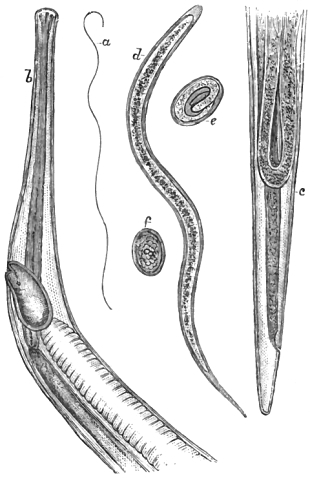
Fig. 39.—Filaria Bancrofti. a, Female (nat. size); b, head and neck (× 55 diam.); c, tail; d, free embryo (× 400 diam.); e, egg containing an embryo; f, egg, with mulberry cleavage of the yolk (× 360 diam.). Original.
When (prior to Lewis’s discovery of the hæmatozoa) I had myself encountered larval nematodes of the same character as those described by Salisbury, I, like Wucherer, was careful not to employ a special name for an immature form, which might or might not represent a worm hitherto known to science. The paper in which I described the adult worm from specimens supplied by Bancroft appeared in the ‘Lancet,’ Oct. 6th, 1877, the facts being stated as follows:—
On the 28th of August, 1877, I received a small collection of entozoa. The box contained the promised Filariæ, and also eight bottles filled with various intestinal worms taken from animals. The Filariæ were enclosed in four small tubes and preserved in glycerine. Three of the tubes (marked 1, 2, 3) contained sexually-mature worms, the fourth being labelled “Sediment from adult Fil. sang.—young and ova.” I described their contents in succession. Thus, on the 6th of September, 1877, I examined the Filaria in tube No. 3. The specimen was injured and in four portions, these collectively measuring three inches in length. Although, to the naked eye, the worm had appeared to Dr Bancroft to be of the thickness of an ordinary human hair, yet I found it about 1/90″ at the thickest part. It was a female. At the same time I examined the specimen in tube No. 1. This was also a female. Towards the centre of the body a hernial protrusion of the uterine horns and intestine had taken place. In a lithograph sent by Dr Bancroft this specimen was figured and described as the “parent worm of the Filaria sanguinis, emitting young Filaria from two loops.” Later on I examined the contents of tube No. 2. In it I found one tolerably perfect female Filaria, and also a delicate shred forming part of one of the uterine horns of another worm. This filament measured one inch and a half in length, and was coiled round the complete worm. On transferring it to a watch-glass containing water, hundreds of embryos made their escape. Owing to the transparency of the tissues I had much difficulty in finding the reproductive outlet, and the effort to find it was all the greater because Bancroft’s figure had misled me. At length I found the vagina and its orifice close to the head (about 1/20″ from it), the anal orifice being placed within the 1/90″ from the extremity of the tail. The vaginal pouch, 1/100″ long, was crowded with embryos, and a constriction marked its junction with the uterus proper, which appeared to divide lower down at a distance of 1/10″ from the head. Towards the tail a fold of the tuba Fallopii was seen to extend to within 1/20″ of the extremity. All sections of the uterine system were crowded with germs, eggs, and embryos in their usual relative situations.
My examinations of the ova and embryos were chiefly made from the “sediment” sent in a special glass tube. The fully formed embryos were 1/125″ in length by 1/2500″ in breadth. They each showed a double skin, the outer envelope in the more advanced specimens leaving clear spaces at either end of the body, resulting from commencing ecdysis. I saw no trace of intestinal tube, but a central line of condensation marked an early differentiation of the somatic granular contents. The less advanced embryos were mostly enclosed in a chorional envelope, the smallest free embryos measuring only 1/200″ in length by 1/3000″ in breadth. These had no double contour. The ova, whose yolk-contents were still in various stages of cleavage, gave an average long diameter of 1/900 to 1/1000 of an inch.
Such are the facts I made out, and they enabled me to amend the characters of the species.
As regards nomenclature, I associated Dr Bancroft’s name with the sexually-mature worm as being in harmony with the binomial method and little calculated to mislead; moreover, it helped to fix both the source and date of the discovery (Brisbane, Dec. 21st, 1876). The use of this nomenclature detracts nothing from the high merits of Lewis, who first named the immature worm Filaria sanguinis hominis. As it now turns out, both Dr Salisbury and myself had previously been made acquainted with the young of Filaria Bancrofti; but it was reserved for Lewis to discover the hæmatozoal character of the embryos of this worm, and actually to take them from the blood. It was a singular circumstance, that when I was engaged in treating my little African patient for trematode hæmatozoa, it never once occurred to me that the numerous nematoid embryos mixed with the Bilharzia ova were hæmatozoal. As before remarked, it was alleged that my patient had passed worms two or three inches long by the urethra. I therefore concluded that these were the parents of the eggs and embryos, and that all of them were urinary. The inference was wrong, but it has instructively shown how near one may go towards a great discovery without really making it. As regards the larvæ, notwithstanding some slight differences in regard to size and so forth, I have little hesitation in saying that all the embryo forms severally described by Salisbury, by myself, by Lewis, Sonsino, Wucherer, Crévaux and Corre, Silva Lima, Bancroft, Manson, and others, are referable to one and the same species.
Into the clinical bearings of this subject it is impossible for me to enter at any length, but I may remark that these parasites appear to be associated with, if not actually the cause of, several distinct morbid conditions. To one of these Bancroft has given a separate name (Helminthoma elastica). This is a highly elastic form of growth to which I have already alluded under the title of “lymphatic abscess of the arm.” In the first valuable report on Hæmatozoa, by Dr Patrick Manson, of Amoy, China, this careful observer gives interesting particulars of no less than fifteen cases in which hæmatozoa were found. Two of these patients had Elephantiasis scroti, two had lymph-scrotum, two were lepers (one having scrotal disease), two had enlarged inguinal glands, one had anasarca; and of the remaining six, spoken of as having no concomitant disease, one had enlarged glands and abscesses, and another suffered from marked debility. It would thus appear that what is ordinarily termed “good health” is rarely associated with a hæmatozoal condition of the blood in the human subject. The cases given by Lewis and Manson, where absolutely no recognisable disease existed, must be regarded as exceptional. Disease, moreover, may exist without any palpable symptoms being exhibited by the “bearer,” and thus perhaps it was with the hæmatozoal dogs of Gruby and Delafond to which I shall again have occasion to allude. Even those animals that carried upwards of two hundred thousand microscopic Filariæ in their blood appeared to suffer no inconvenience whatever.
In the autumn of 1877 Dr Da Silva Lima published an article in the ‘Gazeta Medica da Bahia,’ in which he dwelt upon the labors and merits of Wucherer, and, judging from an omission in one of my memoirs, he supposed that I had insufficiently acknowledged Wucherer’s claims. A translation of this article appeared in the ‘Archives de Médicine Navale,’ with an important appendix by Dr le Roy de Méricourt. In this addendum the French savant showed that the omission on my part was unintentional, and had been corrected by me in a later memoir. Not only had I been amongst the earliest in England to enforce Wucherer’s claims in respect of the micro-Filariæ, but I had first announced his discoveries in connection with Anchylostoma duodenale. In my translation of Wucherer’s memoir (‘Ueber die Anchylostomum Krankheit’) I spoke of the melancholy satisfaction I had in knowing that the memoir in question was “among the last that appeared from the pen of that gifted and amiable physician.” Some notice of Dr Lima’s paper and its appendix by Dr A. le Roy de Méricourt appeared in the ‘Lancet’ for Jan. 5th, 1878, and I also published a full translation of it, with explanatory notes, in the ‘Veterinarian’ for Feb., 1878. Later on, in the ‘Lancet’ (March 23rd, 1878), Dr Da Silva Lima published an interesting letter correcting a misconception that had incidentally arisen in the mind of a commentator (on the Helminthological work of 1877), and at the same time he pointed to the original facts connected with the discovery of Wucherer’s Filaria. As my views are in perfect accord with those of Dr Da Silva Lima, I can only regret that errors of interpretation should have crept into the discussion. Dr Lima honorably recognises the nomenclature (Filaria Bancrofti) which I proposed for the adult worm, and only claims for Wucherer that which is fairly due.
On the 4th of January, 1878, I received from Dr Patrick Manson a manuscript in which he announced the discovery of the larvæ of Filaria sanguinis hominis in the stomach of mosquitoes. Already, in April, 1877, Dr Bancroft had informed me of his expectation of finding that these insects sucked up the larvæ of the Filaria whilst engaged in their attacks on man. Dr Bancroft’s supposition was a very natural one, but it remained for Manson to make the actual discovery of the existence of human hæmatozoa, or parasites that had been such, within the stomach of Culex mosquito. I lost no time in making the principal facts public (‘Lancet,’ Jan. 12th, 1878). Dr Manson at the same time forwarded for publication a record of thirty-five additional cases of hæmatozoa occurring in Chinese subjects, together with additional particulars of one of the cases already published in the ‘Customs Gazette.’ These were afterwards published as separate contributions in the ‘Medical Times and Gazette.’ Dr Manson likewise forwarded materials for a paper entitled “Further Observations on Filaria sanguinis hominis.” In this communication he gave an analysis of the cases (sixty-two in all) in which he had observed the hæmatozoa, and he added valuable statistical evidence as to the prevalence of Filariæ in the Amoy district, dwelling especially on the influence of age, sex, and occupation in determining the presence of the parasite. He also described the morbid states with which these entozoa were commonly associated.
On the 7th of March, 1878, I formally communicated to the Linnean Society a detailed account of Manson’s investigations relating to the metamorphoses undergone by the Filariæ within the body of the mosquito. In this paper Manson pointed out that the female mosquito, after gorging itself with human blood, repairs to stagnant water for the purpose of digesting the blood, and also for the purpose of depositing its eggs. During this period, which lasts four or five days, the Filariæ undergo remarkable changes. Subsequently, in a more perfect state, they escape into the water, and in this advanced stage they are conveyed to the human body along with the water as drink. Dr Manson persuaded a Chinese, whose blood was previously ascertained to abound with Filariæ, to sleep in a “mosquito house.” In the morning the gorged insects were captured and examined under the microscope. A drop of blood from the mosquito was found to contain 120 Filariæ, but a drop taken from the man’s hand yielded only some thirty specimens. Further stages of development are accomplished within the human host, ending in the sexual maturity of the parasite. After fecundation successive swarms of embryos are discharged by the female worm, a part of whose progeny eventually gains access to the blood.
Before I proceed to summarise the whole body of facts I must in the next place state that Manson and myself contributed a joint communication to the Medical Society of London on the 25th of March, 1878. In this memoir I especially dealt with the question of priority in connection with the discovery of the adult worm. I then restated that the adult parasite was discovered by Dr Bancroft on December 21st, 1876. The discovery was verified by Dr Lewis on August 7th, 1877, by Dr Silva Araujo October 16th, 1877, and by Dr F. dos Santos November 12th, 1877. I gave these dates unhesitatingly, without, however, in any way prejudicing the question already raised in respect of the identity of the worms found in each case. My own mind was fully made up on that point, and affirmatively so. Dr dos Santos’ find was made in conjunction with Dr J. de Moura in a case of lymphatic abscess of the arm. Clinically viewed, the case published by Dr Araujo must be regarded as unique. Not only were adult and embryonic Filariæ found in the same patient, but, what was far more surprising and interesting, the patient displayed in his own person several of the disorders hitherto found apart; and he was more than once attacked by one or two of the diseases. He experienced a first attack of chyluria three years ago, then attacks of craw-craw commencing a year ago, the latter being attributed to bathing in a particular lagoon. He had a second attack of chyluria six months back, at which time lymph-scrotum appeared, and also scrotal elephantiasis. Dr Bourel-Roncière pronounced this case to be unique, and attributed nearly all the disorders to the presence of Wucherer’s embryonic Filariæ. In a very elaborate analysis of and commentary on Dr da Silva Lima’s second memoir, Dr Bourel-Roncière warmly claims for Wucherer the supreme honor in all these discoveries. A number of affections hitherto regarded as distinct, and all of which appear to be due to the action of Filariæ, are regarded by Dr Bourel-Roncière as mere phases of one and the same disorder. This affection he terms Wucherer’s helminthiasis. Dr Manson had indeed arrived independently at a similar conclusion, and I am confident that Wucherer, were he alive, would in this particular aspect of the question be the last to claim priority either to Lewis, to Bancroft, or to Manson.
In this place I may observe that Dr Pedro S. de Magalhães, of Rio de Janeiro, detected free microscopic nematodes in the potable waters of Rio (agua da Carioca), which from their similarity he supposes may have some genetic relation with Filaria Bancrofti. In this opinion I cannot share.
As regards the metamorphoses of the embryo, Manson states that for a little while after gaining access to the stomach of the mosquito the embryo undergoes no change (Fig. 40, a). In a very few hours changes commence, resulting in wider separation of the outer skin and an appearance of transverse markings on the body within (b). In the next stage oral movements occur; the striation becomes more marked, and the outer envelope is cast off (c). Then the striated lines disappear and a dotted appearance is substituted (d). From this condition the embryo passes to what Manson calls the chrysalis stage, in which nearly all movement is suspended and the large spots gradually disappear (e, f, g, h, i, j, k). The tail continues to be flexed and extended at intervals and the oral motions cease. By the close of the third day the embryo becomes much shorter and broader; but the finely pointed tail retains its original dimensions, projecting abruptly from the sausage-shaped body (m, n). Large cells next appear in the interior of the body, and by a little pressure one may detect indications of a mouth (o, p, q, r). At this period the embryo begins to elongate, and at the same time to diminish in width; but the growth takes place chiefly at the oral end of the body. The mouth becomes four-lipped, open, and funnel-shaped, and from it a delicate line can be distinctly traced passing to an opening near the caudal extremity, the tail itself gradually disappearing (s, t). Speaking of the most advanced stage Manson says:—“A vessel of some sort is seen in the centre running nearly the whole length of the body and opening close to one extremity. This end is slightly tapered down and is crowned with three or perhaps four papillæ, but whether this is the head or tail, and whether the vessel opening near it is the alimentary canal or the vagina, I cannot say.” Now it is quite evident, I think, from Manson’s figures that he has here faithfully represented the head and tail, the former (u) to the left, the latter (v) to the right. In his manuscript (from which I am now quoting) there is no special reference to these two figures; but it is easy to see that these terminal sections of the body of the advanced embryo closely correspond with the head and tail of the adult worm (Filaria Bancrofti). The curved line passing to the left (u) evidently indicates the commencement of the partially-formed vagina.
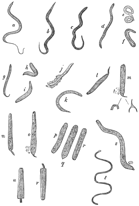
Fig. 40.—Larval Filariæ in various stage of growth from the mosquito; a to d, representing the first stage of metamorphosis during the first 36 hours, e to o, the changes occurring during the second stage, to the close of the third day; p to t, forms seen during the third stage of metamorphosis from the fourth day onwards. The figs. u, v, represent the head and tail only, whilst t shows the young Filaria in an advanced stage, and drawn to a much smaller scale, than the others which are here magnified about 125 diameters. Much reduced from Manson’s original figures.
How completely Manson took the initiative in this part of the work is evident even from Lewis’s own later observations. In a paper published in March, 1878, Dr Lewis, writing from Calcutta and speaking of the rôle of the mosquitoes, says:—“I had repeatedly examined, in a cursory fashion, these and other suctorial insects, but had not observed any parasites suggestive of these embryo-hæmatozoa, hence, when, on receipt of a communication from Dr Manson a couple of months ago, a renewed search was made, I was surprised to find that four out of eight mosquitoes, captured at random in one of the servants’ houses, harboured specimens of hæmatozoa to all appearances identical with those found in man in this country. After this, however, several days elapsed before any mosquitoes could be obtained which contained these embryo-nematoids, and the specimens obtained on the next occasion were devoid of the enveloping sheath, which appears to characterise the kind found in man out here, and apparently, according to Dr Manson, in China also.” Further on Lewis also remarks, “When the insect is caught shortly after feeding and the contents of its stomach examined microscopically, the hæmatozoa, if present, will be observed to manifest very active movements, which may possibly continue for several hours on the slide. If the insect be kept for twenty-four hours before examination it is probable that the movements of the parasites will be more sluggish, and their form probably altered owing to irregular contractions and dilatations of their substance—changes which may also occasionally be observed when embryo-hæmatozoa are preserved on a glass slide, and they may sometimes be kept alive thus, if in suitable media, for two or three days. When the insect is not examined till the third day, the contained parasites will probably manifest marked signs of disintegration—and possibly every indication of life will have disappeared from many of the specimens. After the third or fourth day I have not seen any active specimens of these entozoa in the stomach or in any part of the alimentary canal of the mosquito; those which remain have undergone more or less fatty degeneration, and are readily stained with eosin, which, as far as my experience goes, is not the case so long as they are alive and active. After the fourth or fifth day it is very rare that traces of any hæmatozoa-like objects can be detected at all, so that it must be inferred either that they have succumbed to the digestive action of the insect’s stomach or been disposed of along with the excreta.” An important addendum by Lewis records a fortunate incident as follows:—“It was observed that nearly all the mosquitoes captured in one of the servants’ houses contained hæmatozoa, so that the supply of suitable insects in all the stages of their growth became amply sufficient for all requirements. The result of the examinations under these favorable conditions has shown that although the stomach digests a great number of the ingested hæmatozoa, as mentioned above, nevertheless others actually perforate the walls of the insect’s stomach, pass out, and then undergo developmental stages in its thoracic and abdominal tissues.”
I may here observe that Sonsino has instituted a comparison between the embryos of this Filaria and those of Anchylostoma, by which it appears that the former measure 0·218 to 0·330 mm. in length, and those of Anchylostoma 0·430 mm. The hæmatozoa are about forty times longer than broad, and the larval anchylostomes only fourteen times longer. The tail of Filaria is conspicuously longer.
In the ‘Lancet’ for June 22nd, 1878, an announcement appeared from the pen of Mr D. H. Gabb, of Hastings, stating that a patient under his care formed the habitat of Filaria sanguinis hominis; and in the autumn of the same year a paper which I read to the Linnean Society in the spring was published. In that paper the following summary was offered:
1. Filaria Bancrofti is the sexually-mature state of certain microscopic worms hitherto obtained either directly or indirectly from human blood.
2. The minute hæmatozoa in question—hitherto described as Wucherer’s Filariæ, Filaria sanguinis hominis, Trichina cystica, Filariose dermathemaca, and so forth—are frequently associated with the presence of certain more or less well-marked diseases of warm climates.
3. The diseases referred to include chyluria, intertropical endemic hæmaturia, varix, elephantiasis, lymph scrotum, and lymphoid affections generally, a growth called helminthoma elastica, a cutaneous disorder called craw-craw, and also leprosy.
4. It is extremely probable that a large proportion, or at least that certain varieties of these affections are due to morbid changes exclusively resulting from the presence of Filaria Bancrofti or its progeny within the human body.
5. It is certain that the microscopic hæmatozoa may be readily transferred to the stomach of blood-sucking insects, and it has been further demonstrated that the digestive organs of the mosquito form a suitable territory for the further growth and metamorphosis of the larval Filariæ.
6. The character of the changes undergone by the microscopic Filariæ, and the ultimate form assumed by the larvæ whilst still within the body of the intermediate host (Culex mosquito), are amply sufficient to establish the genetic relationship as between the embryonal Filaria sanguinis hominis, the stomachal Filariæ of the mosquito, and the sexually-mature Filaria Bancrofti.
In the month of September, 1878, I received a letter from Dr da Silva Lima announcing the fact that Dr Araujo had verified the existence of the embryos of Filaria Bancrofti in mosquitoes, at Bahia. These mosquitoes had, I understood, attacked a French priest in whose blood Dr Araujo also detected Filariæ. Thus, it fell to the lot of Araujo, through his untiring zeal, to verify in Brazil all the separate discoveries of Bancroft, Manson, and Lewis.
In the October issue of the ‘Pathological Society’s Transactions’ for 1878 Dr Bancroft records numerous cases of filarious disease, and he gives a succinct account of the circumstances connected with his original discovery.
In a clinical lecture published October 12th, 1878, Dr Tilbury Fox seeks to diminish the value of these discoveries, characterising helminthological investigators as merely “recent writers.” Dr Fox denies that Filariæ are a cause of true elephantiasis, but admits the occurrence of “elephantoid inflammation and inflammations due to Filariæ.” Dr Fox’s statement that “Filariæ have not been found in uncomplicated elephantiasis, that is, in disease without chylous exudation,” seems to me to be directly at variance with Manson’s recorded experiences. I hold that Manson has confirmed the truth of Lewis’s views, and that he has thoroughly proved that (to use his own words) “varicose groin glands, lymph scrotum, elephantiasis, and chyluria are pathologically the same disease.” In the first instance I was myself led to conclude that some of the forms of elephantiasis might be due to other causes than obstruction of the lymphatics caused by the presence of Filariæ; but the explanations of Lewis, of Bancroft, and of Manson more especially, have almost entirely removed this doubt. Those who seek to explain away the connection between genuine elephantiasis and Filariæ will do well to study Manson’s last important memoir. He shows that “elephantiasis and allied diseases are much more frequently associated with the parasite than are other morbid conditions.” This fact is brought out very clearly in his table of 670 cases, from which it appears that 58 per cent. of cases of Filaria are associated with elephantoid disease.
When this opposition to Manson’s views is likely to cease (on the part of those who do not happen to have been in any way instrumental to the discoveries in question) it is not easy to say. In a brief communication which appeared in the last number of the ‘Medical Times and Gazette’ for 1878, Dr Manson successfully combats the doubts that have been entertained respecting the rôle of the mosquito. Because Lewis found that canine hæmatozoa were digested, and thus perished in the stomach of mosquitoes, it had been argued that human hæmatozoa must necessarily undergo similar processes, and consequently die. Those who oppose the views of helminthologists in respect of the intermediary host-function of insects on such grounds can have very little general, and still less special knowledge of the phenomena of parasitism. It is the old story. When any new discovery is made, it must always pass through the ordeals of denial and doubt before it can be generally accepted as true; and, as in the case of Jenner’s immortal discovery, there will always remain a certain number of peculiar people who show themselves hostile to every advance in science. Dr Manson may take comfort from this consideration, and rest assured that the value of his discovery is quite unaffected by the opposition referred to.
Since I communicated the results obtained by Manson, Lewis, myself, and others to the Linnean Society, an even more exhaustive summary of the facts has been published by Dr Bourel-Roncière, in the ‘Archives de Médecine Navale.’ The distinguished author does full justice to the writings of English helminthologists, and dwells, with emphasis, upon the finds and interpretations of Lewis, Manson, and Bancroft. Incidentally, also, he comments upon Sir Joseph Fayrer’s early recognition of the etiological identity of hæmato-chyluria and elephantiasis, on other than helminthic grounds. The frequent concurrence of the two affections had especially struck Sir J. Fayrer as pointing to a probable common origin. He had also surmised that the disorders might be due to parasites.
Dr Bourel-Roncière, alike with the caution, precision, and logical reasoning of a cultured savant, concludes his elaborate review in the following terms:—“There are the facts. Certainly, many points remain obscure, many problems await a solution, and the last word has not been said on the actual part which the parasite plays in the pathogenesis of the affections above enumerated—its mode of action, the importance of its rôle, the extent of its pathological domain, the habitat of its progenitors, their identity, and so forth. All these questions will only be elucidated by necroscopic researches, which at present remain absolutely wanting.”
“However, notwithstanding the doubts which hover over the future value of these curious discoveries, it is difficult not to recognise their importance in the study of certain tropical diseases—which up to the present time have been attributed to vague and undetermined causes—hæmato-chyluria and elephantoid affections principally. Apart from the interest which attaches to the natural history of the nematoids, they raise, in effect, etiological and prophylactic questions, the extreme importance of which we believe it would be needless to demonstrate. It is greatly to be desired that the researches should be taken up in other parts of the globe, where endemicity and perhaps greater facilities for necroscopic investigation would render them fruitful—Cochin-China, Tahiti, &c. Fresh observations are necessary to confirm the first and to fill up notable gaps. The way has been brilliantly opened by the English and Brazilian physicians. Let our colleagues in the French colonies put their shoulders to the wheel; they have before them a vast field of study to explore.”
Since the above remarks were written I have received several communications from Dr Bancroft, and also others from Drs da Silva Lima, Araujo, Assis Sousa, Paterson, Hall, of Bahia—the two last named being English physicians in practice there. I regret that I can do little more than refer to the writings of these authors in the Bibliography below; but I may observe that Drs Paterson and Hall have ascertained that the proportion of the population of Bahia affected by Filaria is 81/2 per cent. Out of 309 persons examined, 26 had hæmatozoa, which is, roughly, one in twelve, or more strictly, 8·666 per cent.
Amongst recent memoirs that by Sir J. Fayrer, read to the Epidemiological Society on the 5th of February, 1879, deserves especial attention. In regard to its significance, I have only space to remark that, much as we may regret the little interest shown by our hospital physicians and surgeons in this subject, it is particularly gratifying to see experienced Indian officers like Sir J. Fayrer, Mr Macnamara, and Dr John Murray, coming forward both to aid and render homage to their junior colleagues in Eastern parts, who are successfully labouring to advance the cause of helminthology and scientific medicine.
In concluding this subject I may observe, that one of the greatest hindrances to the due recognition of the remarkable part played by parasites in the production of human endemics and animal epizoötics arises from the circumstance that no inconsiderable number of minute worms may infest a host without obvious injury. This immunity proves nothing. If, for example, we take the case of Trichina we find that several millions of entozoa may exist in the human, or, at all events, in the animal bearer, without producing any symptom of discomfort. In such cases it is not possible to determine the strict limits of health and disease; nevertheless, were we to double the amount of infection, the imaginary line of demarcation is at once bridged over and the parasites become acknowledged as directly responsible for grave symptoms which may even prove fatal to the bearer. Again, the relative strength and size of the infected host constitute factors that materially limit the power of the parasite for injury. Where the entozoa are of minute size, and where their injurious action is primarily due to the mechanical obstructions they set up, it is clear that the virulence of the helminthiases, or resulting diseased conditions, will mainly depend upon the number of intruders.
Another consideration of the highest value in relation to epidemiology generally, and more especially in regard to the practical question as to the best methods of stamping out parasitic plagues, is that which refers to the life-history of the entozoon itself. It must be obvious that in all cases where the intermediate host can be captured and destroyed, the life-cycle of the parasite can be broken and interrupted, and if thus broken, there is an end to the further propagation of the species. The knowledge that we have acquired by experimental research in this connection has already enabled us to set a limit upon the prevalence of certain well-known disorders, such as Trichinosis, Cestode-tuberculosis, and so forth. In the case of epizoötics, however, which are indirectly due to the action of intermediary hosts that cannot be readily captured or destroyed, then our power of arresting the disease is comparatively limited. In the present case it is probably not necessary either that a dead or living mosquito should be swallowed to insure infection; but it is necessary that the parasitic larvæ should have dwelt within the mosquito in order to arrive at the highest stage of larval growth prior to their re-entrance within the human territory. Undoubtedly, the larvæ are swallowed with potable waters. Perfect filtration before use would certainly check, if in course of time it did not totally extinguish several of the many virulent diseases that now afflict the inhabitants of warm climates.
It is with reluctance that I terminate this article, but in the closing pages of this work (Book II, Section V) I hope to add a few more particulars in reference to Lewis’s latest researches.
Bibliography (No. 23).—Araujo, A. J. P. da Silva, “Memoria sobre a Filariose,” &c., Bahia, 1875; see also ‘Arch. de Méd. Nav.,’ 1875 and 1878.—Bancroft, J., “Cases of Filarious Disease,” in ‘Pathological Soc. Trans.’ for 1878, vol. xxix, p. 407.—Bourel-Roncière, “Résumé of and Commentary upon the writings of Silva Lima, Silva Araujo, and others,” in ‘Arch. de Méd. Nav.’ for March, 1878.—Idem, “Pathologie exotique. De l’hématozoaire nématoïde de l’homme et de son importance pathogénique, d’après les travaux Anglais et Bréziliens des dernières années;” ibid., for August and Sept., p. 113–134 and p. 192–214, 1878.—Cobbold, T. S., “Discovery of the Adult Representative of Microscopic Filariæ,” ‘Lancet,’ July, 1877, p. 70.—Idem, ‘On Filaria Bancrofti,’ ibid. Oct., 1877, p. 495.—Idem, “Verification of Hæmatozoal Discoveries in Australia and Egypt,” ‘Brit. Med. Journ.,’ June, 1876.—Idem, “Obs. on Hæmatozoa,” ‘Veterinarian,’ October, 1873.—Idem, “Remarks on the Ova of another Urinary Parasite (in the paper on ‘Bilharzia’) from Natal,” ‘Brit. Med. Journ.,’ July 27th, 1872, p. 89; see also Bibl. No. 12.—Idem, “Entozoa in Relation to the Public Health” (various papers), ‘Med. Times and Gaz.,’ Jan. and Feb., 1871.—Idem, ‘Worms’ (l. c., p. 151), 1872.—Idem, “Hæmatozoa; Fresh Discoveries by Lewis,” ‘Lancet’ for Feb. 6, 1875.—Idem (brief notice), the ‘Veterinarian,’ p. 209, March, 1875.—Idem, “On the Discovery of the Intermediary Host of Filaria sanguinis hominis,” ‘Lancet,’ Jan. 12, 1878, p. 69.—Idem, “On the question of Priority of Discovery,” Rep. of Med. Soc. of Lond., in ‘Lancet,’ March 30, 1878, p. 465.—Idem, ‘Mosquitoes and Filariæ’ (explanatory note), in ‘Brit. Med. Journ.,’ March 16, 1878, p. 366.—Idem, “On the Life-history of Filaria Bancrofti, as explained by the discoveries of Wucherer, Lewis, Bancroft, Manson, Sonsino, myself, and others,” “Report of the Proceed. of the Linnean Soc.” for March 7, 1878, in ‘Pop. Science Rev.,’ April, 1878; and afterwards published in extenso in ‘Journal Linn. Soc.,’ Oct. 31, 1878.—Idem, “On Filaria Bancrofti,” in Part iv of a series of papers on the Parasites of Man, in the ‘Midland Naturalist,’ August, 1878.—Idem, “On Filaria sanguinis hominis,” in a letter to the ‘Lancet,’ July 13, 1878, p. 64.—Idem, “Filariæ and Leprosy” (case from Bancroft); ‘Lancet,’ Feb. 1, 1879.—Corré, A., “Note sur l’helminthe rencontré dans les urines hémato-chyleuses,” ‘Rev. des Sci. Nat.,’ 1872.—Cossé, “Sur l’helminthe rencontré par Wucherer et Crevaux,” &c., ‘Rev. Montpellier,’ tom. i, p. 190.—Couto, A., “These de concourso,” Bahia, 1872.—Crevaux, J., “De l’hématurie chyleuse, &c.,” 1872; also in ‘L’Union Médicale,’ 1872 (abs. in ‘Brit. Med. Journ.,’ July, 1872, p. 100); also in ‘Arch. de Méd. Nav.,’ 1874; and in ‘Journ. de l’Anat. et de la Physiol.,’ 1875 (see also Silva Lima).—Davaine, C., ‘Traité,’ 2nd edit., p. 944; ‘Hæmatozoaires,’ supp., 1877.—Fayrer, Sir J., “Filaria sang. hom.,” ‘Lancet,’ March 16, 1878, p. 376.—Idem, “Elephantiasis Arabum,” ‘Med. Times and Gaz.,’ Dec. 1, 1877, p. 588; “On the Relation of Filaria sanguinis hominis to the Endemic Diseases of India,” in the ‘Lancet,’ Feb. 8 and 15, and reprinted from the ‘Med. Times and Gazette’ (same date), 1879.—Gabb, D. H., letter in ‘Lancet,’ June 22, 1878.—Leuckart, l. c., s. 638, 1876.—Lewis, T. K., “On a Hæmatozoon in Human Blood,” ‘San. Comm. 8th Rep.,’ Calcutta, 1872; ‘Med. Press,’ 1873, p. 234; ‘Indian Ann. Med. Sci.,’ 1874; ‘Lond. Med. Rec.’ (abs. by myself in vol. i, p. 5), 1873.—Idem, “Pathological Significance of Nematode Hæmatozoa,” ‘Tenth Ann. Rep.,’ 1873, Calcutta (reprint), 1874; ‘Ind. Ann.,’ 1875.—Idem, “Remarks regarding the Hæmatozoa found in the Stomach of Culex mosquito,” ‘Proc. Asiatic Soc. of Bengal,’ March, 1878, p. 89.—Idem, “Flagellated Organisms in the Blood of Rats” (being portion of a paper on “The Microscopic Organisms found in the Blood of Man and Animals,” in ‘14th Annual Report of the San. Comm. with the Govt. of India’), in the ‘Quart. Journ. of Micr. Science,’ Jan., 1879.—Idem (published since the present article was written), “The Nematoid Hæmatozoa of Man,” ibid., April, 1879.—Lima, J. F. da Silva (with Crevaux), ‘Memoria sobre hematuria chylosa ou gordurosa des paizes quentes;’ extrahida da ‘Gazeta Medica da Bahia,’ 1876; repr. in ‘Arch. de Méd. Nav.,’ Dec., 1878 (see also Le Roy de Méricourt).—Magalhães, Pedro S. de, “Filarias em estado Embryonario, encontradas n’agua tida como potavel (agua da Carioca),” ‘O Progresso Medico,’ Dezembro, 1877, p. 57.—Idem, “Nota sobre os nematoides encontrados no sedimento deposito pela agua (potavel) da Carioca,” ‘O Prog. Med.,’ 1 de Setemb., 1878, p. 577.—Idem, “Caso de filariose de Wucherer;” ibid., 15 de Setemb., 1878, p. 589.—Makina, M.D., “Filaria in Chyluria,” letter in ‘Lancet,’ Feb. 22, 1879, p. 286.—Manson, P., “Rep. on Hæmatozoa,” ‘Customs Gazette,’ No. 33, Jan.–March, 1877; see also ‘Med. Times and Gaz.’ for Nov. 10, p. 513, Nov. 17, p. 538, and Nov. 24, p. 563; Dec. 1, p. 589, 1877; also Jan., 1878.—Idem, “Additional Cases;” ibid., March 2, 9, 23, 1878.—Idem, “On Filaria sanguinis hominis, and on the Mosquito considered as a Nurse,” ‘Proc. Linn. Soc.,’ March 7, 1878; see also report in ‘Nature,’ March 28, 1878, p. 439.—Idem, “On Filaria sanguinis hominis, clinically considered in reference to Elephantiasis, Chyluria, and allied Diseases,” ‘Rep. of Med. Soc. of Lond.,’ in ‘Lancet,’ March 30, 1878.—Idem, “Further Observations on Filaria sanguinis hominis,” “Med. Rep.” for April–Sept., 1877, in ‘Customs Gazette,’ Shanghae, 1878.—Idem, “The Development of the Filaria sanguinis hominis,” ‘Med. Times and Gaz.’ for Dec. 28, 1878, p. 731.—Méricourt, A. Le Roy de, in Appendix to an art. entitled “Nouvelle phase de la question relative à la nature parasitaire de la chylurie. Découverte du représentant adulte de la ‘Filaire de Wucherer,’” par le Dr da Silva Lima, from the ‘Gaz. Med. da Bahia,’ Sept., 1877; see also the ‘Lancet,’ Jan., 1878, p. 22 (editorial notice).—Moura, J. de, ‘These de Concourso,’ 1877.—O’Neill, “On Craw-craw,” ‘Lancet,’ Feb., 1875.—Pareira, A. P., “On Bilharzia and Chyluria,” ‘Gazeta Med. da Bahia,’ No. 9, 1877 (noticed in ‘Lancet,’ Feb. 2, 1878).—Salisbury, J. H., “On the Parasitic forms developed in Parent Epithelial Cells of the Urinary and Genital Organs,” ‘Hay’s American Journ.,’ vol. iv, 1868, p. 376.—Santos, F. dos, in ‘Gaz. Med. da Bahia,’ March, 1877.—Sonsino, P., ‘Richerche,’ &c., 1874; ‘Della Bilharzia,’ &c., 1876; ‘Sugla Ematozoi,’ &c., 1876 (see Bibl. No. 12).—Idem, “On the Diagnosis of Embryos of Filaria,” in his paper ‘Sull’ Anchylostoma duodenale;’ ‘Estr. dall Imparziale,’ 1878.—Sousa, M. de A., ‘Memoria sobre a Elephantiasis do escroto,’ Bahia, 1878.—Wucherer, O., “Noticia Preliminar,” &c., ‘Gaz. Med. da Bahia,’ Dec., 1868.—Idem, ‘Sobre Hematuria no Brazil,’ ibid., Sept., 1869; see also “Méricourt’s trans. (De l’hématurie intertropicale observée au Brézil),” ‘Arch. de Méd. Nav.,’ p. 141, 1870, and the fuller references quoted in my memoir; ‘Linn. Soc. Journ., Zool.,’ vol. xiv, p. 368.
Filaria Loa, Guyot.—Although further examinations of this worm will probably result in placing it in some other genus than Filaria, yet it is by no means clear that Diesing was right in placing it with the genus Dracunculus. I therefore abandon the nomenclature adopted in my previous treatise. According to the surgeon, Guyot, who made seven separate voyages to the coast of Angola, these worms cannot be confounded with the Dracunculus. They are quite white, and relatively much thicker than guinea-worms. Under the title of Filaria oculi Moquin-Tandon has spoken of certain small nematodes as “not uncommon in the negroes of the Angola coast;” and he gives other localities where it occurs. The worms are identical with those described by Guyot as dwelling beneath the conjunctivæ of negroes at Congo and in the Gaboon region generally. The parasite is rather more than an inch and a quarter in length, being pointed at one end and blunt at the other. It is termed Loa by the natives, who state that after a period of several years the worm voluntarily quits the organ. The disease is thus naturally cured. This parasite enjoys a tolerably wide geographical distribution, as it has been observed by Clot Bey in a negress who had come from the town of Monpox, situated on the banks of the River Magdalena; by Sigaud, who saw one in the eye of a negress in Brazil; by Blot, at Martinique, who saw two in a negress originally from Guinea; by Bajon, who met with one in a little negro girl who had come from Guadeloupe; by Mongin, who found one in a negress who had been living in the Island of San Domingo; and by Lestrille, who removed one from beneath the conjunctiva of a negro who came from Gaboon.
Bibliography (No. 24). Davaine, l. c., p. 839.—Guyon, ‘Gaz. Méd. de Paris,’ p. 106, 1841, and in ‘Micr. Journ. and Struct. Record,’ p. 40, 1842, and in ‘Dublin Journ.,’ vol. xxv, p. 455, 1839.—Idem, ‘Compt. Rendus,’ tom. lix, p. 743, 1865.—Guyot, in ‘Mém. par Arrachait,’ p. 228, 1805.—Küchenmeister, l. c., s. 322.—Lestrille, in Gervais and Van Beneden’s ‘Zool. Med.,’ 1859, also quoted by Davaine, l. c., 2nd edit., p. 840.—Leuckart, l. c., s. 619.—Moquin-Tandon, A., ‘Zool. Med.,’ Hulme’s edit., p. 363, 1861.
Filaria lentis, Diesing.—This is a doubtful species. The worm was first discovered by Nordmann, in a case of lenticular cataract under the care of Von Gräfe, and it was afterwards found by Jüngken in a similar case, as recorded by Sichel. There is also the instance described by Gescheidt, in which Von Ammon operated, and from which brief descriptions of the worm have generally been taken. In this case there were three worms, two measuring about 1/6″ and the third 1/15″ in length. In Jüngken’s case (exhibited by Quadri, of Naples, at Brussels) the worm was more than 3/4″ long. In another case, reported by M. Fano, the worm was somewhat less than 1/4″ long. There is no certain evidence that any of these various worms had developed sexual organs in their interior. It is true that the reproductive organs were described in two of the worms observed by Gescheidt; but after a due consideration of all the facts I fear we must conclude that all the worms in question were sexually-immature and wandering nematodes, possibly referable to Gurlt’s Filaria lacrymalis, as Küchenmeister long ago suggested.
Bibliography (No. 25).—Cobbold, ‘Entozoa,’ p. 332.—Davaine, l. c., p. 821 et seq.—Diesing, ‘Syst. Helm.,’ p. 625.—Fano, ‘Traité des Malad. des Yeux,’ tom. ii, p. 498; and in ‘Rec. de Méd. Vét.,’ p. 140, 1869; quoted by Davaine, p. 831.—Gescheidt, Ammon’s ‘Zeitsch.,’ 1833, s. 435.—Leuckart, l. c., Bd. ii, s. 622.—Nordmann, l. c., Bibl. No. 2, s. 7, 1832.—Sichel, ‘Iconogr. Ophth.,’ p. 707, 1859.
Filaria labialis, Pane.—This is a filiform cylindrical worm measuring an inch and a quarter in length. The mouth is armed with four papillæ arranged in the form of a cross. The tail of the female is blunt, the vaginal outlet being placed at a very short distance from its extremity, and a little above or in front of the anus. This parasite was found by a medical student at Naples. It occupied the cavity of a pustule in the upper lip, giving rise to considerable irritation. Only the male worm is at present known.
Bibliography (No. 26).—Davaine, l. c., edit. ii, Synopsis, p. 107.—Leuckart, l. c. (with a fig.), Bd. ii, s. 616.—Pane, “Nota di un elminte nematoide,” in ‘Annali dell’ Acad. degli aspiranti Naturalisti,’ Napoli, ser. 3, vol. iv, 1864.
Filaria hominis oris, Leidy.—In the fifth volume of the ‘Proceedings of the Philadelphia Academy of Natural Sciences’ (1850, p. 117) Dr Leidy furnishes the following description of this worm as gathered from the examination of a simple specimen preserved in alcohol, and labelled as having been “obtained from the mouth of a child.” Body white, opaque, thread-like; mouth round, simple; posterior extremity obtuse, furnished with a short, curved, epidermal hooklet, 1/500″ in length, by 1/2000″ in diameter at base. Dr Leidy offers some speculations as to its origin, but from whatever source the worm was obtained by the bearer, it seems to be an immature form. Its length is five inches and seven lines.
Filaria (Nematoideum) trachealis, Bristowe and Rainey.—This is another very doubtful worm. It was originally described in the ‘Pathological Society’s Transactions’ for 1855. It evidently represents only a juvenile stage of growth of some species of round worm. Rainey discovered a considerable number of these worms in the trachea and larynx of a person who died from a disease affecting the lower extremities. Individually the parasites measured about the 1/50″ in length.
Strongylus (Filaria) bronchialis, Rudolphi.—This is a small nematode. The male measures rather more than half an inch, whilst the female is upwards of an inch in length. The caudal appendage of the male is furnished with a bilobed, membranous, half-bell-shaped bursa. This surrounds the cloacal outlet, the latter concealing a double spiculum. The tail of the female is sharply pointed, the anal orifice being placed a little in front or above. The body is filiform, of a pale yellow color. It is about 1/50″ broad in the male, and 1/35″ in the female. The mode of reproduction is viviparous.
The original specimens were discovered by Treutler in Germany, during the winter of 1791, in the bronchial glands of an emaciated subject, whilst those sent to Diesing for description were discovered by Dr Fortsitz at Klausenberg, in Transylvania, in the lungs of a boy six years old. Diesing and Weinland suggested the identity of Filaria bronchialis and Strongylus longevaginatus, whilst Küchenmeister went further, and pronounced them to be one and the same species.
Bibliography (No. 27).—Cobbold, ‘Entoz.,’ p. 357.—Davaine, ‘Synops.,’ l. c., ‘Synopsis’ cix.—Küchenmeister, l. c., Eng. edit., p. 381.—Leuckart, l. c., s. 618.—Treutler, F. A., “De vermibus filiformibus (Hamularia lymphatica) in glandulis conglobatis bronchiorum repertis,” in ‘Obs. Pathol. Anat.,’ 1793.—Wedl., ‘Die im Menschen vorkommenden Helminthen’ (quoted by Leuckart), Wien, 1862, s. 22.
Eustrongylus gigas, Diesing.—This is by far the largest nematode known to science, the male sometimes measuring a foot in length and the female more than three feet, whilst the breadth of the body reaches half an inch at the thickest part. Though fortunately very rare in man, this worm is known to occur in a great variety of animals, especially in weasels. According to Weinland and Jackson, it is particularly abundant in the kidney of the North American mink (Mustela vison), destroying the substance of the organ, the walls of which become the seat of calcareous deposit. It has been found in the dog, wolf, puma, glutton, raccoon, coati, otter, seal, ox, and horse.
The body of the adult worm is cylindrical, more or less red in color, and somewhat thicker behind than in front. The head is broadly obtuse, the mouth being supplied with six small, wart-like papillæ, two of which correspond with the commencement of the two lateral lines of the body. These lines are also distinguishable from other six longitudinal lines traversing the body from end to end by the presence of very minute papillæ which are less closely arranged towards the centre (Leuckart). The tail of the male shows a simple, thick, cup-shaped bursa, which is destitute of rays, and partly conceals the simple spiculum. The tail of the female is blunt and pierced by the centrally placed anal opening. The vulva is situated near the head in the ventral line. The eggs are stout and oval, measuring 1/300″ in length by about 1/550″ in breadth.
As regards development the recent researches of Schneider have shown that certain kinds of fish play the part of intermediary bearer. Balbiani preserved the ova in water for more than a year without their hatching, and all his attempts to rear the larvæ in the intestines of the dog by direct experiment failed. Similar feeding experiments upon fishes and reptiles also failed. The embryo, when removed from the egg, measures 1/104″ in length. It is vermiform, having a pointed head and simple mouth. Balbiani describes the buccal cavity as containing a protractile stylet. Notwithstanding the negative results obtained by Balbiani’s experiments on fishes, Schneider (from anatomical data, which Leuckart confirms) has placed it almost beyond question that the worm hitherto known as Filaria cystica is the sexually-immature Eustrongylus gigas. This worm is found encysted beneath the peritoneal membrane in Galaxias scriba and Synbranchus laticaudatus. It is worthy of remark that the genus Galaxias comes nearer to the Salmonidæ than to the pike family, whilst the Synbranchi are tropical oceanic fishes. Probably the sexually-immature worm occurs in other fishes, especially the Salmonidæ.
Remarkably fine examples of the adult worm may be seen in the Hunterian Collection, Lincoln’s Inn, and in the Museum of the Royal Veterinary College. The human example is undoubtedly genuine. The dissections in the Hunterian Collection of specimens were made by me in 1865. Objection has been taken to my description of the œsophagus as “spiral.” In Sheldon’s specimen it is certainly twisted upon itself, precisely in the manner in which Davaine has also figured it (‘Traité,’ fig. 68); but I cannot here give further anatomical particulars. Drelincourt found two worms sexually united in the kidney. When once the parasites have gained access to this organ, rapid destruction of the glandular substance follows. Ultimately the kidney is reduced to the condition of a mere cyst or bag, which, besides the worms, contains a quantity of sanguineo-purulent matter. Frequently only one worm is present, but oftener two or three. In the kidney of a puma D’Azara’s friend, Noseda, found no less than six worms, whilst Klein obtained eight from the kidney of a wolf.
Bibliography (No. 28).—Azara, F. de, ‘The Natural History of the Quadrupeds of Paraguay,’ trans. from the Spanish by W. P. Hunter; Valpy’s edit., p. 43, 1837; Black’s, 1838; French edit., p. 313, 1801.—Albers, ‘Beitr. z. Anat. &c.,’ Bd. i, s. 115.—Aubinais, ‘Revue Méd.,’ 1846, p. 284.—Balbiani, “Recherches,” &c., ‘Compt. Rend.,’ 1869, p. 1091; ‘Rec. de Méd Vét.,’ 1870, p. 5.—Bickford, “Spec. of Str. gigas found in the Kidney of a Dog,” the ‘Veterinarian,’ 1859, p. 312.—Blainville, ‘Dict. des Sci. Nat.,’ tab. 29.—Blanchard, ‘Ann. des Sci. Nat.,’ 1849, p. 186.—Idem, in ‘Cuvier’s Règne Animal’ (Masson’s edit.), ‘Les Intestinaux,’ p. 57, pl. 27.—Blasius, ‘Obs., &c.’ (with fig. of Lumbricus in renibus hominis), 1674, p. 125.—Bobe-Moreau, in ‘Journ. de Méd.,’ tom. xlvii.—Boerhaave, ‘Aphorism.,’ 1728.—Bremser (l. c., Bibl. 2), s. 223.—Chabert, ‘Traité des maladies verm. dans les Animaux,’ 1782.—Chiaje, ‘Comp. d. Elmintogr. umana,’ p. 106.—Clamorgan, J. de, ‘La Chasse de Loup,’ 1583 (quoted by Davaine, the worms being described as “serpents et bêtes fort venemeuses”).—Cobbold, ‘Entoz.,’ p. 358.—Idem, ‘Catalogue of Entozoa in the Museum of the Roy. Coll. of Surg.,’ “Descr. of preps. Nos. 19–25,” p. 3, 1866.—Idem, “Parasites of Man,” ‘Midland Naturalist,’ Dec., 1878.—Collet-Meygret, “Mém. sur un ver trouvé dans le rein d’un Chien,” in ‘Journ. de Physique,’ &c., 1802.—Cuvier, see Blanchard (supra).—Idem, ‘Voyage en Sicile,’ and in ‘Ann. des Sci. Nat.,’ tom. xi.—Davaine, C., ‘Traité,’ l. c., deuxième edit., p. 271 et seq. (with full lit. refs. at p. 290).—Diesing, l. c., vol. ii, p. 325.—Dujardin, l. c., p. 113.—Frank, F., “Ein Spulwürm in der Urinblase eines Hundes,” ‘Hufeland’s Journ.,’ Bd. xviii, s. 112.—Jackson, ‘Catalogue of the Boston Museum,’ 1847, p. 317.—Klein, T. K., “Anatomical Description of Worms found in the Kidneys of Wolves,” ‘Phil. Trans.,’ 1729–30, p. 269.—Küchenmeister, l. c., Eng. edit., p. 376.—Leblanc (rep. by Rayer and Bouley), in ‘Bull. de l’Acad. de Méd.,’ 1850, p. 640; in ‘Rec. de Méd. Vét.,’ 1862, p. 800; and quoted by Davaine.—Leuckart, l. c., Bd. ii, s. 353–401, 1876.—Moublet, “Mém. sur les vers sortis des reins et de l’urethre d’un enfant,” ‘Journ. de Méd-Chir. et Pharm.,’ 1758, pp. 244 and 337.—Otto (Anat.), in ‘Mag. d. Gesellsch. naturf.,’ 1814.—Owen, art. “Entozoa,” in Todd’s ‘Cyclop.’—Rayer, ‘Traité des maladies des reins,’ 1841.—Rayger, ‘Sur un serpent qui sortit du corps d’un homme après sa mort’ (quoted by Davaine, l. c., p. 272), 1675.—Schneider, ‘Monographie der Nematoden,’ 1866, s. 50.—Idem (mit Peters), quoted by Leuckart, l. c., s. 382.—Stratton, in ‘Edin. Med. and Surg. Journ.,’ p. 261, 1843.
Dochmius duodenalis, Leuckart.—Much time might be occupied and wasted over the nomenclature of this parasite. In my previous treatise, and for reasons there stated, I placed it under the genus Sclerostoma. On rather slender grounds Dubini formed the genus Anchylostoma for its reception, but Von Siebold thought that, on account of the absence of symmetry in the arrangement of the so-called dental organs, Dubini’s genus might very well be allowed to remain. Bilharz, Diesing, Küchenmeister, Wucherer, and others have retained the genus as either Anchylostoma or Anchylostomum. Schneider keeps it amongst the Strongyli; but after all that has been said and written there can, I think, be no doubt that if Dujardin’s genus Dochmius is to be retained at all, Dubini’s worm must be placed in it. The comparisons instituted by Leuckart afford sufficient proof of the intimate alliance as between Anchylostoma and Dochmius. Professor Molin thought to meet the difficulty by calling the worm Dochmius anchylostomum, but the specific term, duodenale, should certainly be retained.
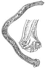
Fig. 41.—Male Dochmius duodenalis, with bursa separately enlarged. After Küchenmeister.
This worm was discovered by Dubini at Milan, and though at first thought rare, it is now known to be tolerably common throughout Northern Italy. The worm has also been recently found by Dr Kundrata at Vienna, in an Austrian subject. According to Pruner, Bilharz, and Griesinger, it is abundant in Egypt. Griesinger believed that about one fourth of the people of that country suffered from anæmic chlorosis, solely in consequence of the presence of this worm in the small intestines. From Wucherer’s observations especially, we know that Dubini’s worm is not limited to the localities above mentioned, for it occurs in the western tropics, in Brazil, and even in the Comoro Islands.
The worm may be described as a small nematode, the males measuring 3/8″ or rather more, whilst the females extend to very nearly 1/2″ (12 mm.). The head is pointed and tapering, and bent forward, having the mouth directed towards the ventral aspect. The oral opening is armed with four asymmetrically disposed, unequally-sized, horny, conical, converging teeth. The neck is continuous with the cylindrical body, which is 1/80″ in thickness. The body terminates in a straight cone-shaped, or rather sharply-pointed tail in the female, the caudal extremity of the male ending in a partially inflexed, blunt point. In the male there is a cup-shaped, bilobed bursa, the membranes of which are supported by eleven chitinous rays, ten being simple, whilst the median, or odd one, is bifurcated at the summit. The mode of reproduction is viviparous. Adult males and females occur in the proportion of one of the former to three of the latter.
As above mentioned, it was Griesinger who first pointed out the clinical importance of this entozoon. He first explained the manner in which the worm produces anæmia, the persons attacked losing blood as if they were being bitten by innumerable small leeches. Like the rest of their kindred, these worms are veritable blood-suckers. In the first instance the views of Griesinger met with opposition, but they have since received abundant confirmation. Whilst Küchenmeister’s ‘Manual’ furnishes an excellent account of the disorder as known in Europe, we are chiefly indebted to Wucherer for what is known of the disorder in Brazil. The experiences recorded in the ‘Deutsches Archiv für Klinische Medicin’ for Sept. 27th, 1872 (s. 379–400), were amongst the last that appeared from the pen of that gifted and amiable physician. As little or no notice of his writings appears to have been taken by professional men in this country, I depart somewhat from the design of this work when I venture to abstract a few of the clinical particulars which he has supplied. Their importance in relation to sanitary science is obvious, inasmuch as these parasites are introduced into the human body by drinking impure water, or, at least, water which either contains the free larvæ of the worm, or the intermediary bearers that harbor the larvæ.
It should be borne in mind that Dubini’s original discovery was made at Milan in 1838, whilst Griesinger’s recognition of the worm as a cause of the Egyptian chlorosis resulted from a post-mortem examination made on the 17th of April, 1851.
In the journal above mentioned, Wucherer records his own discoveries as follows (‘Ueber die Anchylostomunkrankheit,’ &c.):—“Although Griesinger with well-founded confidence gave an account of his ‘find’ and its significance, yet it remained for a long time unnoticed and unutilised, till at length a case led me to corroborate it. During my many years’ residence in Brazil, especially during the first year, I had very frequent opportunities for witnessing the tropical chlorosis, but seldom to treat it, as it is one of those diseases for which Brazilians seek no medical assistance. Its treatment falls to the lot of the curiosos, curadeiros (quacks), who employ the fresh pulp of a species of fig as a remedial agent with the best results. On the 13th of December, 1865, I was called to the Benedictine monastery in Bahia to see a slave of the order suffering from hypoæmia. The patient was about thirty years of age, married, a strongly built mulatto. He was a field laborer on the Ingua plantation of the order, who exhibited in a conspicuous degree all the symptoms that occur in hypoæmia except the diarrhœa. He was well nourished, but strikingly pale, his whole face, but especially the eyelids, being œdematously swollen, as also were the feet, legs, and hands. The hands and feet were very cold. His appearance betrayed the most horrible anguish or low despondency. With difficulty only could he raise himself, being obliged to lie down again immediately on account of his weakness. Auscultation revealed a diminished respiratory murmur, and bronchial expiration in both lungs. The pulse was very rapid and small, the patient complaining of pain in the region of the heart. He had frequent palpitation when he moved, and he complained of pain in other parts of the body. His abdomen was much distended by gases, but not sensitive to pressure from without, except in the region of the stomach. The urine was clear, its specific gravity 1007 to 10231/2°. Under great difficulties he resided for several months after his marriage at Inhatâ. Earlier he had been on the estates of the order at Rio de S. Francisco. He there suffered for a long time from intermittent fever, but at Inhatâ he entirely recovered. At Inhatâ the slaves frequently suffered from hypoæmia, but in S. Francisco not at all. He appears not to have made any misuse of brandy. The slaves of the order were well cared for, and supplied with good and wholesome nourishing food. The patient had already, for a long period, treated himself with steel wine, yet was continually getting worse and worse. He had not taken the pulp of the fig. As I was unaware he had suddenly become so ill, they hastily despatched a message to the town. There was no good to be expected from the further employment of iron, and the patient was in such a condition that from the very first I despaired of his recovery. I immediately prescribed the pulp of the Gammeleira (Ficus doliaria), but it could not be easily obtained. Considering that the Gammeleira would have a drastic effect, I therefore prescribed two grammes of elaterium, to be divided into eight doses, of which he should take one every three hours.” Dissatisfied with this advice, however, Dr Wucherer goes on to say that on reaching home he carefully looked up the literature of the subject. “In a ‘Geologico-Medical Report’ by Professor Hirch, recorded in the ninety-sixth volume of ‘Schmidt’s Jahrbucher,’ I found how Griesinger had recognised the Anchylostoma as the cause of the Egyptian chlorosis, which was clearly identical with our hypoæmia. He had employed this commended anthelmintic. I resolved the more to prescribe the pulp of the Gammeleira when I found it described as a worm-expelling remedy in Martin’s ‘Systema Materiæ Vegetabilis Braziliensis.’ The next morning, however, when I arrived at the monastery I learnt that my patient died about two hours after a slight evacuation. Only after much resistance would they permit the sectio cadaveris. I merely opened the abdomen, and was surprised to find everything as Griesinger had described. During the next season, through the courtesy of my colleagues attached to the General Infirmary at Bahia, especially of Drs Silva Lima, Faria, and Caldos, I was enabled to open more than twenty bodies of anæmically deceased individuals. All were selected as miserably poor in condition, but only five were bodies of persons in whom hypoæmia was diagnosed, and in these there were a great number of Anchylostomes in the small intestine. The intestines of the other bodies contained either none, one, or a few.” Dr Wucherer next states that he compared the characters presented by his entozoa with those given by Dubini, Diesing, and Von Siebold, and found a perfect agreement throughout. He sent several examples to Griesinger, who also established their identity, and communicated the results of his investigations accordingly (‘Archiv für Heilkunde,’ 1866, s. 387. See also Leuckart, ‘Die Mensch. Par.,’ Bd ii, s. 411). Dr Wucherer also forwarded a number of specimens to Dr Weber, who published a brief account of them with excellent figures (‘Path. Soc. Trans.,’ vol. xviii, 1867, p. 274). As mentioned in the text of his memoir (s. 394), Dr Wucherer also transmitted some strongyloids to myself. “The publication of my observations,” adds Dr Wucherer (‘Gazeta Medica da Bahia,’ 1866, p. 27 et seq.), “had a result in that Dr J. R. de Moura, of Thersepolis, in the province of Rio de Janeiro, sought for Anchylostomes in the bodies of tropical anæmics (Hypöæmikern). He at once found these parasites, as stated in the same journal (for 1866, p. 132). As occurred to myself, he saw no enduring results from the application of the remedies which appeared to be called for, whilst he well knew that unprofessional persons (Nichtärzte) succeeded in obtaining marked results by the exhibition of the pulp of the Gammeleira (Ficus doliaria). The anthelmintic action of this remedy was also unknown to him.” Dr Wucherer then records how his discovery of these entozoa was announced by Dr Jobini to the Rio academy, and how Dr Moura’s observations were subsequently communicated, adding remarks upon the interesting discussion that followed. The general opinion was that the Anchylostomata were not the primary and necessary cause of this tropical anæmia, but rather a co-operating agent in its production. Against this view Dr Wucherer afterwards very properly protested (‘Gazeta,’ Jan. 15th, 1868). In the mean time, says our author, “Dr le Roy de Méricourt, prompted by my first communication, had invited the physicians of the French colony to seek for Anchylostomes. Drs Monestier and Grenet, at Mayotta (one of the Comoro Isles, which lies about 12° S. lat. to the north-east of Madagascar), ascertained the presence of entozoa in hypoæmics. Dr Grenet sent the duodenum and a portion of the jejunum of an hypoæmic corpse to Le Roy de Méricourt, who compared the Anchylostomes with Davaine’s description, and recognised them as examples of A. duodenale.”
“In the year 1868 Dr Rion Kérangel found Anchylostomes in the bodies of hypoæmics in Cayenne. Thus, the occurrence of Anchylostomes in hypoæmics has been authenticated by Pruner, Bilharz, and Griesinger, in Egypt; by myself, Dr Moura, Dr Tourinho, and other physicians, in Brazil; by Monestier and Grenet, in the Comoros; and by Rion Kérangel in Cayenne. It thus also appears, from the wide separation of these several localities, that the Anchylostomes, if duly sought for, will be found in many other countries.”
These details given by Wucherer are so precise and instructive that I could not have further abridged them without injustice to his record. The bearing of the foregoing facts in relation to the question as to how we may hope to arrest the fatal action of many of these nematodes is sufficiently obvious. That strongyles and their allies prove highly destructive to man and beast is as well established as any other recognised conclusion in medical science; nevertheless, there are those who still doubt the power of these nematodes in relation to the production of fatal epidemics. I shall deal with the sanitary bearings of the subject hereafter. In conclusion, I may mention that Dr da Silva Lima has forwarded specimens of Anchylostomum to the Hunterian Museum, where they may be seen.
Bibliography (No. 29).—Bilharz, ‘Zeitschr. f. wiss. Zool.,’ Bd. iv, s. 55.—Cobbold, ‘Entozoa,’ p. 361.—Idem “Remarks on Recent Contributions to our Knowledge of the Parasitic Nematoids, especially in reference to the Wasting Diseases they produce in Man and Animals,” the ‘Veterinarian,’ Jan., 1876, p. 1.—Davaine, l. c., pp. 118 and 931.—Diesing “Revis. der Nematoden,” ‘Sitzb. d. m.-naturw. cl. d. k. Akad.,’ 1860, s. 716.—Dubini, ‘Entozoografia,’ &c., 1849.—Griesinger (quoted above), see also ‘Arch. f. Phys. Heilk.,’ 1854.—Küchenmeister, l. c., Eng. edit., p. 383.—Leuckart, l. c., ss. 410–455.—Molin, ‘Il sottordine degli Acroffali,’ p. 61 (quoted by Leuckart).—Siebold, ‘Zeitsch. f. wiss. Zool.,’ 1852, s. 55.—Sonsino, P., L’Anchilostoma duodenale in ‘relazione coll’ Anemia progressiva perniciosa,’ Egitto, 1877.—Idem, ‘Sull.’ Anch. duod., 1878 (see also Bibliog. No. 27, both reprinted from ‘Imparziale.’)—Weber, H., l. c., 1867.—Wucherer (quoted above), 1872.

Fig. 42.—Outline of a female Dracunculus medinensis. Nat. size. Original.
Dracunculus medinensis, Cobbold.—This parasite is popularly known as the guinea-worm, or Medina-worm. Probably Lister was the first writer who distinctly spoke of it as the Dracunculus, 1690, the same title being applied to it by Kaempfer, 1694. Be that as it may, Gmelin, long afterwards, placed the parasite in the genus Filaria, at the same time adopting the specific title medinensis. This had been previously employed by Linneus, who, however, regarded the worm as belonging to the genus Gordius. It being clear from the distinctive characters of the entozoon that it was desirable to separate it from the Filariæ, and that no better generic name could be devised than Dracunculus, I thought it right to combine Lister’s and Gmelin’s nomenclature as above, 1864. Leuckart pursued a similar course, crediting Linneus with the titles.
The guinea-worm having been known from the earliest times, it is not surprising that its true nature long remained a mystery. Any one who has read Küchenmeister’s elaborate narrative of the historical significance of the Dracunculus will hardly have failed to arrive at the conclusion that Moses was probably the earliest writer on the endemic disorder which is occasioned by this parasite. There can be no doubt that the “fiery serpents” which afflicted the children of Israel during their stay in the neighbourhood of the Red Sea were neither more nor less than examples of our Dracunculus. It is further evident that Plutarch spoke of Dracunculi, when in the eighth book of his ‘Symposiacon,’ he quotes Agatharchidas as stating that the people taken ill on the Red Sea suffered from many strange and unheard-of attacks, amongst other worms, from “little snakes, which came out upon them, gnawed away their legs and arms, and when touched retracted, coiled themselves up in the muscles, and there gave rise to the most insupportable pains.” In order to render the passage more readable, it will be seen that I have slightly altered the original version (‘Parasites,’ s. 305).
The guinea-worm may be described as a nematode measuring from one to six feet in length, having a thickness of 1/10th of an inch. The body is uniformly cylindrical, terminating below in a more or less curved and mucronately pointed tail. The head is flatly convex or truncate, having a central, simple mouth, which is surrounded by four equi-distantly and cruciately disposed papillæ. The mode of reproduction is viviparous, the body enclosing a prodigious number of hatched embryos, which, by distension of the uterine ducts, almost entirely obliterate the somatic cavity. Notwithstanding the statements of Owen to the contrary, the male Dracunculus is at present altogether unknown.
The guinea-worm possesses a comparatively limited geographical range, for not only is it proper to the tropical regions, but within intertropical limits it is almost exclusively confined to certain districts in Asia and Africa. Thus, according to Künsenmuller, as quoted by Busk, it occurs endemically in Arabia Petræa, on the borders of the Persian Gulf and Caspian Sea, on the banks of the Ganges, in Upper Egypt, Abyssinia, and the coast of Guinea. “In America the guinea-worm is unknown, except in persons who have had communication with Africa or other parts where it is indigenous. The island of Curaçoa is the only locality in the New World which offers an apparent exception to this fact, and it would be highly desirable to ascertain the real state of the case in this instance.” The observations of Chisholm showed that the Dracunculus is really prevalent in several of the West Indian islands, especially in Grenada, and the still later investigations of Dr Da Silva Lima point to its former prevalence in Brazil. Now, the worm is rarely seen at Bahia. Mr Busk said:—“Though endemic only in the above-mentioned parts of the world, it would yet appear that all races of mankind are obnoxious to the attacks of the Filaria when exposed to what may be called the contagion; that is, when placed in circumstances under which it might be supposed a contagious seminium could be conveyed to them.” Mr Busk also added:—“I have known many instances tending to prove that, in order that a European should become infected with the guinea-worm on the coast of Africa, it is not necessary that he should have been on shore at all. It has been quite sufficient for him to have exposed the bare surface of some parts of his person to the water in the native canoes alongside, or, it may be, to the discharge from the sores of those laboring under the disease. This mode of its introduction accounts for the frequency with which the legs and feet are attacked by the parasite, in preference to other parts of the body, as it will always, I believe, be found that the men who have become so affected have been in the habit of going about with bare feet, as is common among sailors in warm latitudes. That the contagious material is conveyed in water is also further indicated by the well-known fact that in India, where it is the custom of the natives to carry water in skins on their backs, the worm makes its appearance on the back and shoulders and upper part of the body.” These views were published by Busk in 1846, and I am free to confess that—confirmed as they appeared to be by subsequent and independent testimony—they completely dominated my conceptions as to the mode of ingress of the young parasites within the human bearer. Thus, those of our Indian troops which were most exposed during the rainy season, subsequently exhibited evidence of having been invaded by the Dracunculus. As, moreover, the period of incubation of the entozoon commonly extends from twelve to fifteen months, it necessarily happened that the disease often showed itself in localities far distant from the spot where the troops originally contracted the disorder. The statement that the period of incubation of the worm is not less than a year, is probably incorrect, since Carter mentions that in a school of fifty boys bathing in a certain pond at Bombay—the sediment of which swarmed with microscopic tank-worms (Urobales palustris, Carter)—twenty-one were attacked with Dracunculus during the year, whilst the boys of other schools, bathing elsewhere, remained, with few exceptions, uninfected. This is a remarkable occurrence, and it points to the possibility of the young Dracunculi being confined to particular pools. That they should, whether occupying the bodies of intermediary bearers or not, be more abundant in some waters than others, is just what might be expected, since such a distribution is in harmony with a recognised law affecting the abundance or limitation of species in particular localities. Much, indeed, has been written respecting the nature of the soil and geological formations occurring in the Indian worm-districts, but the speculative views enunciated on this point are little worthy of credit. Those who desire information on this head should at all events consult the valuable writings of Smyttan, Greenhow, Bird, Forbes, Chisholm, and Aitken, who, apart from the question at issue, supply abundance of practical information.
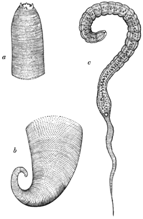
Fig. 43.—a, b, Head and tail of the adult guinea-worm (magnified 10 and 18 diameters respectively); c, embryo (magnified 500 diameters). Original.
Into the anatomy of the adult Dracunculus I do not enter, but I may remark in passing, that the structure of the worm has been exhaustively treated of by Busk and Bastian. A résumé of their views is given in my introductory treatise. Carter and Leuckart have also added important details. As regards the structure and development of the young worms, I have to observe that the discovery of the viviparous mode of reproduction in Dracunculus is due to Jacobson. Nearly a quarter of a century ago I recognised the fact that the uterine organs of the adult worm almost completely filled up the perivisceral cavity, and that they were crowded with microscopic worms. Referring to this “find,” the late Sir George Ballingall, of Edinburgh, in his well-known work on ‘Military Surgery,’ recorded the circumstance in the following terms:—“The Assistant Conservator of the Anatomical Museum in our University has detected in the oviduct of an adult specimen from my collection myriads of minute and perfectly-developed (embryonic) Dracunculi. They can be very well seen with an half-inch object-glass, but their structure is best exhibited if the magnifying power be increased to two hundred and fifty diameters linear.” As already stated in my introductory treatise, these observations were made during the winter of 1853–54. In July, 1854, M. Robin made a similar statement after examining a fresh Dracunculus which had been extracted from the leg of a man by M. Malgaigne. Robin, not unsuitably, compared the worm to a double tube, one tubular sheath, as it were, enclosing the other. “The second tube,” he distinctly affirms, “is the oviduct, or, rather, that part which represents the uterus. The young still remaining in the uterus were nearly all coiled, sometimes with the tail sallying outwards, at others rolled like the rest of the body.” I have thought it only due to Robin and myself to show that from the first we were perfectly well acquainted with the fact of the “great development of the genital tube and of its close adherence to the parietes of the body.” To be sure, many discrepancies occurred in our writings, and in those of Busk and Carter. It was Bastian’s skill and good fortune to correct these errors. Thus, most of us agreed in recognising a slightly trilobed or tripapillated mouth; but Carter failed to demonstrate the existence of these tubercles, and spoke of the oral aperture as being simple and “punctiform.” The body throughout its three upper fourths appeared to me to be cylindrical, but Robin found that it was flattened. It is finely striated transversely, except at the part where it contracts to form the slender, pointed tail. According to Carter, Robin, and Davaine, the young attain a length of about 1/33 of an inch, but Bastian gives it as about 1/42″. In thickness, Carter gives the approximative diameter as 1/633″, Robin makes it 1/990″ to 1/1320″, whilst Bastian gives their breadth at 1/1428″, and Davaine at 1/2500″. I estimated their greatest length and breadth to be 1/30″ by 1/1000″. Robin and myself thought we recognised a distinct, rounded, anal orifice; and whilst Busk, on the one hand, saw nothing which in the slightest degree indicated the presence of an anal opening, Carter, on the other hand, described the structure which we called the anus as a gland, at the same time placing the alimentary outlet on one side and a little above it. According to Bastian, “the intestinal tube is about 1/87″ in length, and appears to consist of a simple canal of varying calibre, pursuing a nearly straight course, and terminating exactly at about the middle, in length, of the worm.” Like Robin, Bastian recognised œsophageal and stomachal divisions, and in a few examples he observed the cæcal or terminal portion of the intestine to be partially reflected upon itself. In regard to the circular opening which Robin and myself described as the anus, Bastian says there is a rounded body, “about 1/2200″ in diameter, with a dark or light spot in the centre, according to the varying focal distance, and which seems to represent a central aperture. Sometimes, above this, traces of two or three large cells may be recognised, whilst behind nothing definite can be made out, save that the cavity of the body is visible for about 1/400″. In other specimens of the young worm the central body and spot are wanting, but, in its stead, two lateral sacculi are met with, about 1/3300″ in diameter, that communicate with the exterior by a minute channel through the integuments, which can sometimes be distinctly recognised. At other times the channel is obscured by protrusion, which appears to have taken place through it, of a minute bilobed papilla, projecting 1/10,000″ from the side of the body. When the projections are seen, the sacculi are indistinct.”
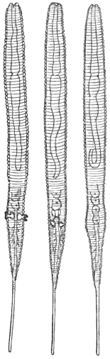
Fig. 44.—Embryos of Dracunculus. Magnified 500 diameters. After Bastian.
As Bastian found the young in all stages of development from the germ condition 1/5000″ in diameter up to the perfect embryo, and as, moreover, he, like the rest of us, could detect no sexual orifice in the adult Dracunculus, he was led to express his belief that the young were produced agamogenetically. He went so far as to call the germs pseudova. It was with great reluctance that I dissented from the views of so gifted an observer as Bastian; nevertheless, later researches have shown that I was justified in not hastily concurring in the theory of a non-sexual mode of reproduction for Dracunculus.
Among the many advances of modern helminthology, the discovery of the true source of the guinea-worm is not the least important. To the late M. Fedschenko (the lamented and accomplished Russian traveller, who lost his life in a snowstorm on the Alps), science stands indebted for this memorable advance. Fedschenko showed that the embryos of Dracunculi, after quitting the human host, succeed in effecting an entry into the bodies of entomostracous crustaceans belonging to the genus Cyclops. Within these intermediary bearers, after twelve hours’ sojourn, the embryos undergo a change of skin, attended with subsequent growth. Here they remain to complete their larval development, which takes place within a period of five weeks, or, as Fedschenko himself told me, one month and six days. At length, as perfected larvæ, they are, together with their crustacean hosts, transmitted to the stomach of the ultimate or human bearer. It is probable that sexual maturity is next acquired within the human stomach, copulation following. After this, the females migrate to the situations in which they are found beneath the skin of the human bearer, whilst the males perish and pass out with the fæces. Thus much I gathered from M. Fedschenko himself when he visited this country, and I possess a sketch of the larvæ made by him at the time (October 23rd, 1873). One of the figures represents a larva which has undergone ecdysis, the long and narrow embryonic tail being supplanted by one which is blunt and forked at the tip. The somatic contents of the embryo have at the same time differentiated into a complete intestinal tube, and a constriction marks the junction of the œsophagus with the stomach. There is also internally an oval-shaped mass of cells near the centre of the body. These represent the commencement of the reproductive organs.
What I had gathered from Fedschenko in conversation thus epitomises that which has since been much more fully stated by Leuckart; and it is only fair to add that the Russian traveller was led up to his discovery by the previous investigations of Leuckart respecting the young of Cucullanus. The Leipsic helminthologist had, indeed, specially instructed Fedschenko as to the probable source of Dracunculus.
It is often thus that science makes its clear advances, since a master-mind is needed to set others on the right track. The embryos of Cucullanus and Dracunculus bear a close resemblance to each other, and the similarity of the types is continued on, though not in the same degree, in the next stage of larval growth, after ecdysis. The higher larvæ of both have their tails trifurcate at the tip, the head of the Dracunculus-larva being distinguished by the presence of a pair of papillæ. In the case of Cucullanus the embryos are, according to Leuckart, passively transferred to the stomach of Cyclops by the mouth; but in the case of Dracunculus, Fedschenko saw the embryo in the act of perforating the bodies of the little crustacea at the ventral surface, where the segments are bound together by a thin and easily penetrated connecting membrane. The larvæ then proceed to coil themselves within the limbs, as many as six or even a dozen of the parasites being occasionally found within the body of a single crustacean host. When they have reached full larval growth they measure about 1/25″ in length. Of course, after attaining this stage, it is a matter of conjecture as to the precise way in which their final destiny is accomplished. Fedschenko fed dogs and cats with the infected crustacea, but failed to rear Dracunculi in these animals. Clearly, these carnivora were unsuitable hosts. Could Fedschenko have experimented on man the result would probably have been very different. Arguing from what happens in the case of Cucullanus amongst fishes, and Trichina in man, there can be little doubt that all the further and final changes undergone by the larvæ are accomplished within the human host. These changes are usually, if not invariably, consequent upon a direct transference of the infested entomostraca along with water used as drink. Thus, it must at once be evident that the simple sanitary precaution of filtering water before use is amply sufficient to ensure the prevention of attacks of dracontiasis or the guinea-worm disease. The theosophical remedy of Moses against this invasion by fiery serpents, as the worms were called in his time, and the modern prophylactic measures dictated alike by science and common sense, thus stand in striking contrast the one to the other. In the nature of things it must ever remain that unreason and reason will select diametrically opposite methods of action, equally, no doubt, with the good intention of bringing about beneficial results.
From what has now been advanced, it will be seen that as regards the mode of infection the views categorically expressed in my previous work (‘Entozoa,’ p. 387) cannot be maintained. What, however, is there stated in respect of treatment still holds good in the main, even as regards prophylaxis.
Bibliography (No. 30).—Adam, ‘Trans. Med. and Surg. Soc.,’ Calcutta, 1824.—Aitken, W., ‘The Science and Practice of Medicine,’ 6th edit., vol. i, 1872.—(Anonymous), “Review of the writings and opinions of Duncan, Johnson, Bird, Mylne, Kennedy, Chisholm, H. Scott, A. J. Robertson, Smyttan, Macgregor, Thomas, Mosely, Morehead, Twining, and others, on the Dracunculus or Guinea-worm,” in ‘Corbyn’s India Journ. of Med. and Phys. Sci.,’ vol. ii, p. 118, 1836.—(Anon.), “The Guinea-worm very Prevalent at Bokhara,” ‘Boston Med. and Surg. Journ.,’ 1843, p. 387.—Balfour, J., ‘Ind. Ann. Med. Sci.,’ 1859, p. 175.—Ballingall, G. (l. c., supra), 1854.—Bastian, H. C., “On the Structure and Nature of the Dracunculus or Guinea-worm,” ‘Linn. Soc. Trans.,’ vol. xxiv, p. 101, 1863.—Berncastle, J., in the ‘Lancet,’ 1851.—Bird, J., ‘Calcutta Med. and Phys. Trans.,’ 1825, p. 151.—Bremser (l. c., Bibl. No. 2), s. 194.—Brett, ‘Surgical Diseases of India,’ 1840; see also ‘Med.-Chir. Rev.,’ 1841.—Bruce, N., ‘Edin. Med. and Surg. Journ.,’ 1806, vol. ii, p. 145.—Busk, G., ‘Micr. Soc. Trans.’ (original series), 1846.—Carter, H. J., “Note on Dracunculus in the Island of Bombay,” ‘Bombay Med. and Phys. Soc. Trans.’ (new series), No. 2, p. 45, 1853–54; see also postscript, p. 252.—Idem, “Further Observ. on Dracunculus,” ‘Bomb. Med. and Phys. Soc. Trans.’ (new series), No. 4, p. 215, 1857–58.—Idem, “On Dracunculus and Microscopic Filaridæ,” ‘Ann. of Nat. Hist.,’ vol. iv (third series), 1859.—Idem, “Notes on Dracunculus,” &c., ‘Ann. of Nat. Hist.,’ vol. ix (third series), 1862.—Chapotin, ‘Bull. des Sci. Med.,’ 1810.—Charvet, ‘Ann. des Sci. Nat.,’ 1834.—Chiaje (l. c., Bibl. No. 2), p. 99.—Chisholm, C., “On the Malis Dracunculus or Guinea-worm (in Grenada),” ‘Edin. Med. and Surg. Journ.,’ vol. xi, 1815; see also the ‘Veterinarian,’ vol. ix, p. 508, 1836.—Clark, ‘Med.-Chir. Rev.,’ 1840.—Clarkson, N. F., “Alleged Case in the Horse,” the ‘Veterinary Record,’ 1845, p. 73.—Clot-Bey, ‘Aperçu sur le ver dragonneau observé en Egypte,’ 1830.—Cobbold, ‘Entozoa,’ p. 373.—Cuvier, ‘Règne animal,’ Orr’s Eng. edit., 1849, p. 644.—Davaine, ‘Traité,’ l. c., edit. ii, p. 783 (full lit. refs.), 1878.—Dickson, ‘Path. Soc. Trans.,’ 1851.—Drummond, ‘Med. Commentaries,’ 1793, p. 294.—Dubois, ‘Edin. Med. and Surg. Journ.,’ vol. ii, 1806.—Duncan, ‘Calcutta Med. and Phys. Soc. Trans.,’ 1835.—Ewart, J., “Questions relating to Dracunculus,” in a review of his memoir on the “Vital Statistics of the Meywar Bheel Corps,” in the ‘Madras Quart. Journ. of Med. Sci.,’ vol. i, 1860, p. 462.—Fedschenko, ‘Protocol of the Promoters (Freunde) of the Natural and Physical Sciences at Moscow’ (in the Russian language), 1869 and 1874 (quoted by Leuckart).— Forbes, D., “Observ. on Dracunculus” (extr. from the ‘Half-yearly Reports of the diseases prevailing at Dharwar in the 1st Grenadier Regiment, in the year 1836’), ‘Bombay Med. and Phys. Soc. Trans.,’ vol. i, 1838, p. 215.—Gibson, A., “Note on the Prevalence of Dracunculus,” in his remarks on the “Diseases of the Deckan,” in ‘Bomb. Med. and Phys. Soc. Trans.,’ vol. ii, 1839, p. 209.—Gramberg, ‘Geneeskundige tijdschrift voor nederl. Indie,’ 1861, p. 632 (quoted by Leuckart).—Greenhow, H. M., ‘Indian Ann. of Med. Sci.,’ vol. vii, 1861, p. 31.—Grierson, D., “Observ. on the Dracunculus, as it prevailed in the 22nd Regiment, N.I., from April till September, 1841,” ‘Bomb. Med. and Phys. Soc. Trans.,’ No. 4, 1841, p. 90.—Grundler, in ‘Commerc. Litt. Nov.,’ 1740, p. 239.—Henderson, J., “Note respecting Four Cases of Dracunculus in the 48th Regiment,” ‘Madras Quart. Journ.,’ vol. iii, 1841, p. 353.—Horton, J. A. B., ‘Army Med. Reports,’ 1868, p. 335.—Kennedy, R. H., ‘Calcutta Med. and Phys. Soc. Trans.,’ 1825, p. 165.—Küchenmeister (l. c., Eng. edit.), p. 389.—Leuckart (l. c., Bibl. No. 1), s. 644–725.— Lewis, T. R., in ‘On a Hæmatozoon,’ &c. (l. c., Bibl. No. 23), p. 30 et seq.—Lima, Da S., “Remarks on the Filaria medinensis, or Guinea-Worm; on the occurrence of this Parasite endemically in the Province of Bahia; on its entrance into the human body by drinking water,” in the ‘Veterinarian,’ Feb., March, et seq., 1879.—Lister, ‘Phil. Trans.,’ 1690, p. 417.—M’Clelland, J., ‘Calcutta Journ. of Nat. Hist.,’ vol. i, 1841, p. 366.—M’Grigor, J., “On the Guinea-worm” (in his “Account of the Diseases of the 88th Regiment in Bombay”), ‘Edin. Med. and Surg. Journ.,’ vol. i, 1805, p. 284.—Morehead, C., ‘Calcutta Med. and Phys. Soc. Trans.,’ vol. vi, 1833, p. 418; also noticed in ‘Edin. Med. and Surg. Journ.,’ vol. xliv, 1835.—Idem, part ii, ‘Calcutta Med. and Phys. Soc. Trans.,’ vol. viii, 1836–42.—Murray, J., “Guinea-worm a very Common Disease at Sattara” (in his Official Report on the Hospital, &c.), ‘Bombay Med. and Phys. Soc. Trans.,’ No. 9, art. vi, p. 198, 1847.—Oke, W. S., “Case of Guinea-worm,” ‘Prov. Med. and Surg. Journ.,’ vol. vi, 1843.—Oldfield, “Case of Dracunculus” (from Laird and Oldfield’s “Narrative of an Expedition into the Interior of Africa”), ‘Dublin Journ.,’ vol. xii, 1838.—Paton, “Cases of Guinea-worm,” ‘Edin. Med. and Surg. Journ.,’ vol. ii, 1806.—Raddock, “A Case of Guinea-worm,” ‘Indian Med. Gaz.,’ Oct., 1877, p. 265.—Scott, W., “Remarks on the Dracunculus,” in a letter to the Medical Board, Madras, ‘Edin. Med. and Surg. Journ.,’ vol. xvii, 1821.—Leverance, C. E., “History of a Case of Guinea-worm,” from ‘Amer. Med. Times,’ in the ‘Glasgow Med. Journ.,’ vol. ix, 1861–62, p. 377.—Smyttan, G., “On Dracunculus,” ‘Calcutta Med. and Phys. Soc. Trans.,’ vol. i, 1825, p. 179.—Stewart, L. W., ‘Indian Ann. of Med. Sci.,’ vol. vi, 1858, p. 88.—Twining, W., “Cases of Dracunculus,” ‘Calcutta Med. and Phys. Soc. Trans.,’ vol. vii, 1835.
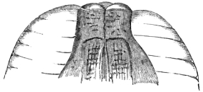
Fig. 45.—Head of Oxyuris vermicularis. Highly magnified. After Busk.
Oxyuris vermicularis, Bremser.—Of all the parasites infesting the human body this is the one concerning which the medical practitioner is most frequently consulted, partly on account of its remarkable frequency in children, and more particularly on account of the difficulty often experienced in getting permanently rid of it. The Oxyuris vermicularis is by no means confined to young persons, seeing that adults are infested even to old age. It is familiarly known as the threadworm or seatworm. The male measures about 1/6″, and the female from 1/3″ to 1/2″ in length. The female possesses a long capillary tail, which terminates in a three-pointed end. The extremity is said to act as a kind of holdfast. The tail of the male is obtusely pointed. In both sexes the body presents a more or less fusiform shape, the anterior end being narrowed to form a somewhat abruptly-truncated head, which is often rendered very conspicuous by a bulging of the transparent integument surrounding the mouth. This presents in profile the aspect of winged appendages (fig. 45). The oral opening is tripapillated, leading into a triangular œsophagus. The integument is transversely striated, and of a silvery-white appearance. The spicule is simple, single, and very minute. The eggs are oblong and unsymmetrical. They measure about 1/900″ from pole to pole, and 1/1400″ transversely.